Article

SELDI-TOF-MS Analysis of Transcriptional Activation Protein Binding to Response Elements Regulating Carcinogenesis Enzymes

by
Steve R. Bischoff
1,
Mahfuz B. Kahn
2,
Michael D. Powell
2 and
Ward G. Kirlin
1,*
1
Departments of Pharmacology/Toxicology, Morehouse School of Medicine, Atlanta GA 30310, USA
2
Microbiology, Biochemistry and Immunology Morehouse School of Medicine, Atlanta GA 30310, USA
*
Author to whom correspondence should be addressed.
Int. J. Mol. Sci. 2002, 3(10), 1027-1038; https://doi.org/10.3390/i3101027
Submission received: 7 June 2002 / Accepted: 31 October 2002 / Published: 31 October 2002

Abstract

:
The risk from chemical carcinogens and environmental toxins is dependent on the metabolic balance between bioactivation and detoxification enzymes. Therefore, agents that alter enzyme expression are critical factors in toxicity. Enhancement or suppression of enzyme activities through gene expression is in part regulated by interactions between specific DNA promoter response elements and specific transcription proteins. DNA-protein interactions are dependent upon translocation of proteins from the cytoplasm to the nucleus and the affinity of proteins for binding to transcription promoter sequences. A key factor in both processes is the intracellular redox state, which influences protein-protein interactions and protein-DNA binding and can be altered by exposure to electrophiles, antioxidants and oxidative stress. Oxidative stress levels can be readily detected by measurable effects on the intracellular glutathione (GSH):glutathione disulfide redox potential, the major intracellular redox buffer. Alterations in the GSH redox pool can directly affect enzyme activity by altering disulfide bonds in the transcription factors regulating enzyme expression. These may affect: 1) specific DNA-protein and protein-protein interactions, 2) cyst(e)ine redox state within transcriptional proteins and 3) translocation of transcription proteins from cytoplasmic to nuclear compartments within the cell. The studies reported here are designed to investigate the relative changes in enzyme expression in response to cellular redox potential changes using the new proteomics technology of surface enhanced laser desorption ionization time-of-flight mass spectrometry (SELDI). Treatment of HeLa and HT29 human cell lines to increase the expression of enzymes that are upregulated by oxidative stress was used as a model system to determine the efficacy of the SELDI technology in measuring changes in transcriptional protein binding to transcriptional response elements. An important goal is to determine whether the SELDI will allow simultaneous studies of multiple transcriptional protein-DNA interactions in response to controlled oxidative stress. This will provide a better understanding of the effect of electrophilic carcinogens and oxidants on the balance between activation and detoxification mechanisms in chemical carcinogenesis.

Introduction

Environmental chemicals and xenobiotics are generally metabolized to toxic compounds by mammalian enzymes either within the liver or in extrahepatic cells. In general, Phase I enzymes introduce a functional group onto xenobiotics which increase their hydrophilicity. Phase II enzymes then conjugate the xenobiotics, greatly increasing their hydrophilicity and promoting their excretion [1]. However, a Phase I oxidation reaction or a Phase II conjugation may actually increase the reactivity of a carcinogen to an electrophile capable of binding to cellular macromolecules such as DNA. The overall toxicity is often determined by the balance between the expression of these activating and detoxifying enzymes. This balance can be shifted by effects on the transcriptional regulation of these enzymes within a specific tissue. Biotransformation of xenobiotics may generate intracellular oxidative stress that can influence the expression of these metabolic enzymes through both up regulation and down regulation of their transcription.
Environmental carcinogens, such as the polycyclic aromatic hyrdrocarbon, benzo[a]pyrene (BP), are metabolized in a series of enzymatic steps to generate electrophililc compounds that can bind to DNA [1]. Catalysis by cytochrome P450 (CYP) enzymes, particularly CYP 1A1 and 1B1 in combination with epoxide hydrolase generates the ultimate DNA binding electrophile 7,8-dihydroxy-9,10-oxy-7,8,9,10-tetrahydrobenzo[a]pyrene (BPDE). BPDE, as well as precursor epoxides, can be inactivated by conjugation with glutathione, catalyzed by glutathione S-transferase (GST) [2]. Quinones that potentially give rise to redox cycling and reactive oxygen species are enzymatically inactivated by NADPH:quinone oxidoreductase (NQO1) [2]. Both of these inactivation enzymes are induced by oxidative stress, antioxidants and electrophilic metabolites [3,4,5]. Oxidative stress appears to also play a regulatory role in the expression of some of the activation enzymes [6,7,8] involved in the conversion of BP to BPDE.
There are at least two key elements of transcriptional activation that are responsive to oxidative stress and changes in intracellular redox potential: 1) translocation of transcription factors from the cytoplasm to the nucleus and 2) specific binding to DNA sequences. As most transcriptional proteins are either translated or sequestered in the cytoplasm, their effect on transcriptional regulation is dependent upon their movement into the nucleus of the cell [9]. Enzymatic metabolism of compounds may alter the expression of subsequent enzymes by generation of metabolites or reactive oxygen species that affect either of these elements of transcriptional regulation.
Xenobiotic interaction with the aryl hydrocarbon receptor (AhR) is the initiating step in the induction of CYP 1A1 expression. In the cytoplasm, the nuclear transcription factor AhR is in a complex with other cytoplasmic proteins [10]. Activation of the AhR by its association with a ligand (electrophile, antioxidant, xenobiotic) results in its translocation to the nucleus where it forms a transcriptionally active complex with the Ah receptor nuclear translocator (ARNT) protein [11] that binds to the xenobiotic response element (XRE) [12]. Both the AhR and the ARNT contain nuclear export signal (NES) sequences that limit their interaction with promotional DNA elements and thereby regulate transcription [13]. These NES and nuclear localization signals (NLS) which control translocation of transcription proteins may respond directly to oxidative stress. For example, NF6B in the cytoplasm exists as in a complex of Rel protein dimers with IκB inhibitor proteins [14]. This interaction apparently masks a nuclear localization signal. As NFκB is activated, IκB is phosphorylated, becoming a target for ubiquitination and degradation by proteasomes. This unmasks the nuclear localization signal and NFκB translocates to the nucleus [15]. NFκB activation can be inhibited by antioxidant treatments such as catalase or N-acetyl cysteine [16], which decreases the phosphorylation of IκBα.
Detoxification enzymes such as NQO1 and isozymes of GST are also regulated by transcription protein interactions with DNA response elements. In the absence of oxidative stress signals, the transcription factor NF-E2-related factor-2 (Nrf2) is sequestered in the cytoplasm by interaction with the Keap1 protein and the actin cytoskeleton of the cell. On exposure to electrophiles, Nrf2 is released and translocates into the nucleus [17]. The signal that releases Nrf2 from Keap is speculated to be an electrophile or an oxidative stress that may be directly sensed by Keap1, possibly due to oxidation of cysteine residues within the Keap1 protein that then undergoes conformational changes [17]. Once in the nucleus, Nrf2 protein and other transcription factors bind to an antioxidant response element (ARE) containing activator protein (AP-1) sites [18,19,20].
A recent review of the effect of ROS on AP-1 activation in yeast [21] points to the critical role of cysteine as an active site within transcription proteins. Under non-stress conditions yeast AP-1 (Yap1) proteins move into the nucleus but are quickly exported. This export appears to depend upon a carboxy terminal cysteine-rich domain within which are hydrophobic amino acid residues resembling a nuclear export sequence (NES). Treatment of cells with oxidants blocks the export, probably in response to oxidation of cysteines within the NES. Yap-1 proteins accumulate in the nucleus and through a bZIP DNA dimerization domain, similar to the mammalian c-Jun, bind to promoter regions of genes encoding thioredoxin (TRX). TRX, a small protein with redox active cysteines within its active site reduces the oxidative stress and Yap1 relocates back to cytoplasm diminishing Yap1-dependent gene expression [21].
Many of the mammalian transcription factors involved in the regulation of CYP1A1, NQO1 and the GST isoform, GST-P, bind DNA in a redox dependent manner. Protein-DNA binding appears to be dependent on the presence of critical cysteine residues [21,22,23]. Antioxidant treatments, such as catalase or N-acetyl cysteine, affect the binding of proteins in gel mobility shift assays [16]. AP-1 DNA binding is affected by reversible oxidation of a cysteine within the DNA binding domain of c-fos and c-jun proteins, with reduction of cysteine stimulating DNA binding and transactivation by AP-1 [21]. For CYP1A1 activation at the XRE site, the AhR-ARNT binding domain contains a critical cysteine in the XRE binding domain [24] that may be affected by the redox potential within the nuclear compartment.
Much of the information on nucleocytoplasmic shuttling proteins and their binding to transcriptional response elements has been derived from electrophoretic mobility shift analysis, DNA footprinting, and protein overexpression. These investigations have been extended with supershift assays using antibodies to identify specific proteins. However, this identification is limited to the known transcription factors, by the inability to resolve transcriptional isoforms, and due to degradation of complexes during electrophoresis. Additionally, the isolation of transcription factors is complicated by low concentrations of regulatory proteins and multiple purification steps that compromise final yield. The purpose of the studies described in this paper is to develop methods using the new proteomics technology of surface enhanced laser desorption ionization (SELDI) to more readily quantitate the amounts of binding protein and to more clearly identify whether there are multiple proteins involved in these protein-DNA interactions. SELDI utilizes surface-enhanced arrays and calculates the masses of matrix-assisted ionized particles from their time-of-flight values. In this study, we have utilized oligonucleotides containing the AP-1 consensus sequence for the binding of the transcription protein c-Jun. These have been attached via a 5' primary amine to the chip surface, prior to incubation with the purified c-Jun protein. This is similar to the method used by Forde et al [25] in their successful capture of the lac repressor protein.
Determination of variation in the amounts of transcription proteins in response to oxidative stress, alterations in redox potential or other stimuli that are present within cytoplasmic or nuclear compartments will increase our understanding of the role of translocation in this regulatory process. Identification of multiple transcriptional proteins involved in the regulation of enzyme expression can provide a better means of understanding the impact of xenobiotics and inducing agents on the balance between cellular metabolic activation and detoxification mechanisms. Modification in the expression of these enzymatic pathways may have a valuable role in inhibiting the activation of carcinogens, such as benzo[a]pyrene, to DNA binding species.

Methods

Cell Conditions. HeLa cells are from a human cervix adenocarcinoma epithelial cell line and have been used extensively as a source of transcriptional proteins. HT29 is a moderately well differentiated human colon adenocarcinoma epithelial cell line. Both cell lines were obtained from the American Type Culture Collection and their derivation is described in the ATCC manual. HeLa and HT29 cells were grown on 150 mm plates in Minimum Essential Medium (MEM) or McCoy’s 5A medium, respectively, in a humidified incubator at 37º C under 5% CO2/95% atmospheric air. MEM and McCoy’s were supplemented with fetal bovine serum 10% v/v, penicillin (100 IU/mL) and streptomycin (100 µg/mL). MEM also contained HEPES (20mM) and L-glutamine (2mM). McCoy’s includes 260 μM L-cysteine, 1.5 mM L-glutamine, 100 μM glycine and 1.6 μM reduced glutathione. For AP-1 stimulation experiments, confluent cultures were incubated 2-4 hrs with 162 nM 4ß-phorbol 12-myristate 13-acetate (PMA) and 2 μg/mL ionomycin from stock concentrations (500x) in DMSO. Control cells received 0.2% DMSO (v/v).
Isolation of nuclear proteins. Nuclear extracts were prepared from HeLa and HT29 cells following the Nuclear Extraction Kit protocol (Active Motif, Carlsbad, CA). Briefly, cells were rinsed twice with ice-cold PBS containing phosphatase inhibitors and centrifuged (500 rpm, 5 min, 4º C). Pellets were resuspended in 1 mL of hypotonic buffer, incubated (15 min, 4º C), then 25 µL of detergent was added and the suspension centrifuged (14,000 x g , 30 s at 4º C). To the nuclear pellet, 100 µL lysis buffer was added and proteins extracted on ice for 30 min on a rocking platform at 150 rpm, vortexed 30 s, and centrifuged (14,000 x g, 10 min, 4º C). Supernatant protein concentrations were quantified by the Bradford protein assay. Aliquots frozen in N2(l) were stored at -80º C.
Preparation of the AP-1 affinity chip surface. Oligonucleotides with the AP-1 consensus sequence (indicated in red) or a scrambled sequence were synthesized (Operon). Forward oligonucleotides included a 5’-amine and hydrocarbon linker.
AP-1consensus forward: 5’-[NH2-(CH2)12]-CGCTTGATGAGTCAGCCGGAA-3’
AP-1 consensus reverse:		      3’-GCGAACTACTCAGTCGGCCTT-5’
AP-1scrambled forward: 5’-[NH2-(CH2)12]-CAGGCGTGTCTGACTACAGAG-3’
AP-1 scrambled reverse:		      3’-GTCCGCACAGACTGATGTCTC-5’
	  
Complementary oligonucleotides were annealed at a hybridized concentration of 25 µM in 10 mM K2HPO4/KH2PO4, 10 mM MgCl2 buffer (pH 7.4) at 90o C, 5 min, cooled to ambient temperature for 30 min and stored at 4º C. Annealing buffer was adjusted to pH 9.0 for coupling of the hybridized DNA to the PS20 protein chip array epoxide surface (Ciphergen Biosystems, Fremont, CA) via reaction of the 5'- amine on the leading oligonucleotide to the activated epoxy groups on the chip surface. To each spot 250 pmol oligonucleotide was applied in 10 µL and incubated at 37o C for 1 h in a humid chamber. To block residual active sites on the chip surface, 4 µL 1 M ethanolamine, pH 8.0 in annealing buffer was added for 30 min at 37º C , then washed with buffer (100mM Tris, 250 mM NaCl, 1mM EDTA, pH 7.4).
Binding of transcription factors to the DNA substrate. Recombinant c-Jun protein or nuclear extract (20 µg) in protein chip binding buffer (10mM Tris-HCl [pH 7.9], 5 mM MgCl2, 50mM NaCl, 1mM EDTA, 5mM dithiothreitol, 5% sucrose, 10 µg/mL poly (dI:dC)) was added to the affinity surface using a bioprocessor (Ciphergen Biosystems) that allows larger samples (300 µL) to be applied to the protein array. Protein was incubated with the affinity surface (2h, 37º C) in a humid chamber with continuous shaking, then washed with buffer.
Detection of captured proteins. To facilitate ionization, sinapinic acid (20 mg in 50% acetonitrile, 0.5% trifluoroacetic acid) was added to each spot. Samples were analyzed in the PBS-II system from Ciphergen Biosystems using the supplied software (version 2.1c) by manual collection using a positive ion mode with a source and detector range of 2.0 and 2.2 kV, a digitizer rate of 250 MHz, time-lag focusing, pulse voltage and pulse lag time of 3000 V and 800 ns, nitrogen laser (337 nm) with 175 µJ maximum energy/4 ns pulse and 20 Hz maximum pulse rate.
Data and graphical analysis. As indicated in Figure 1, both a monomeric and dimeric protein peak were detected. Calculations for total protein bound to the oligonucleotide are given as total area of peak 1 (monomer) plus twice the area in peak 2 (dimer) in figures 2 – 5. This calculation may underestimate the amount of protein in the second peak relative to peak 1 in that less of the higher molecular weight dimer may be detected. However, within these experiments this potential error in the estimate should be relatively constant. Curve fitting to the data points were performed by SigmaPlot 2001 version 7 (SPSS, Inc., Chicago, IL) software. Equations are indicated in the figure legends.

Results and Discussion

Preliminary studies were done to determine optimum conditions for hybridization and attachment of double-stranded oligonucleotide to the PS20 epoxide surface, incubation of protein (c-Jun) for binding to the oligonucleotide, washing to increase specific binding and analysis on the SELDI instrument. These data are reported in the methods under appropriate headings.

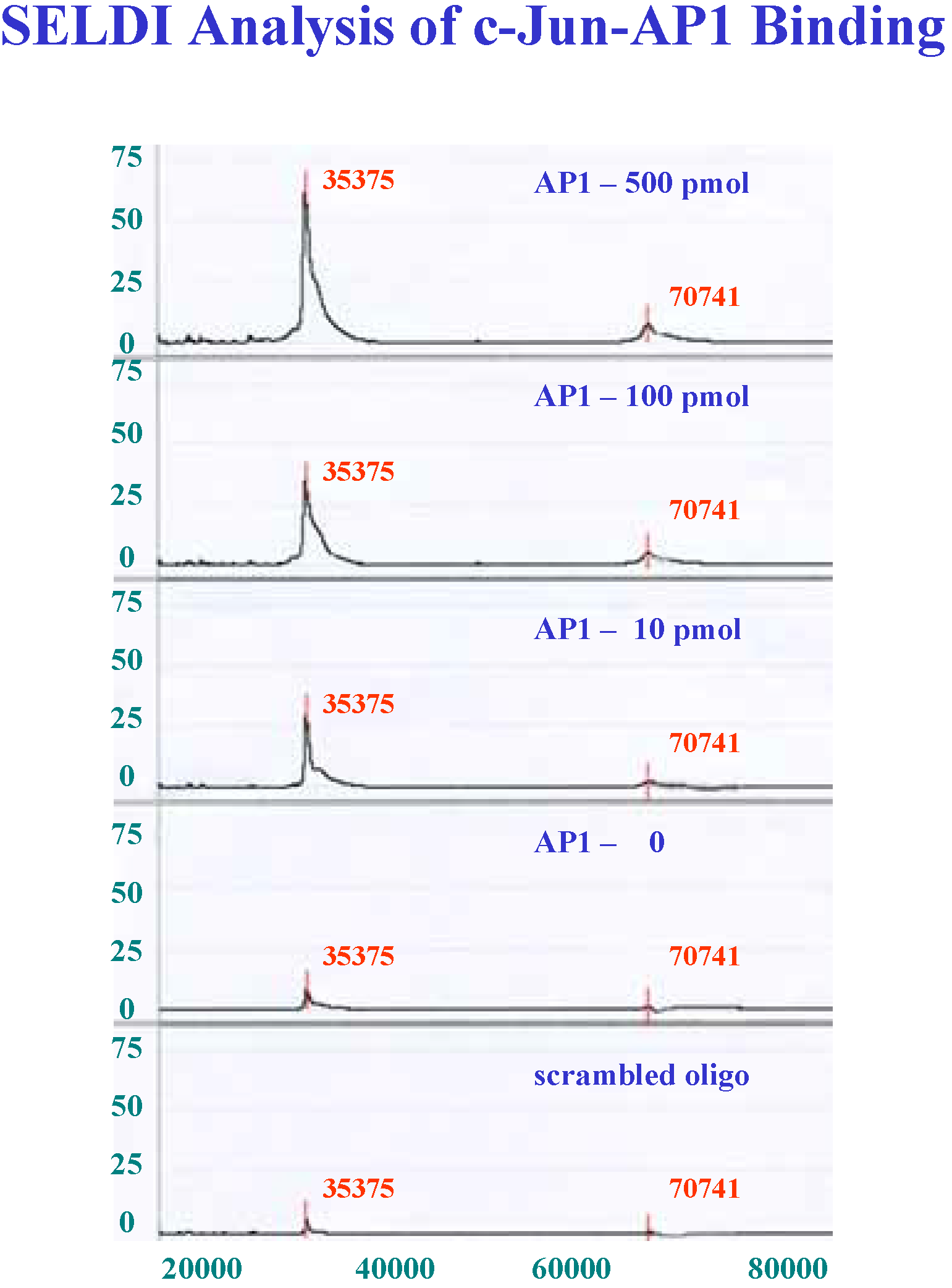
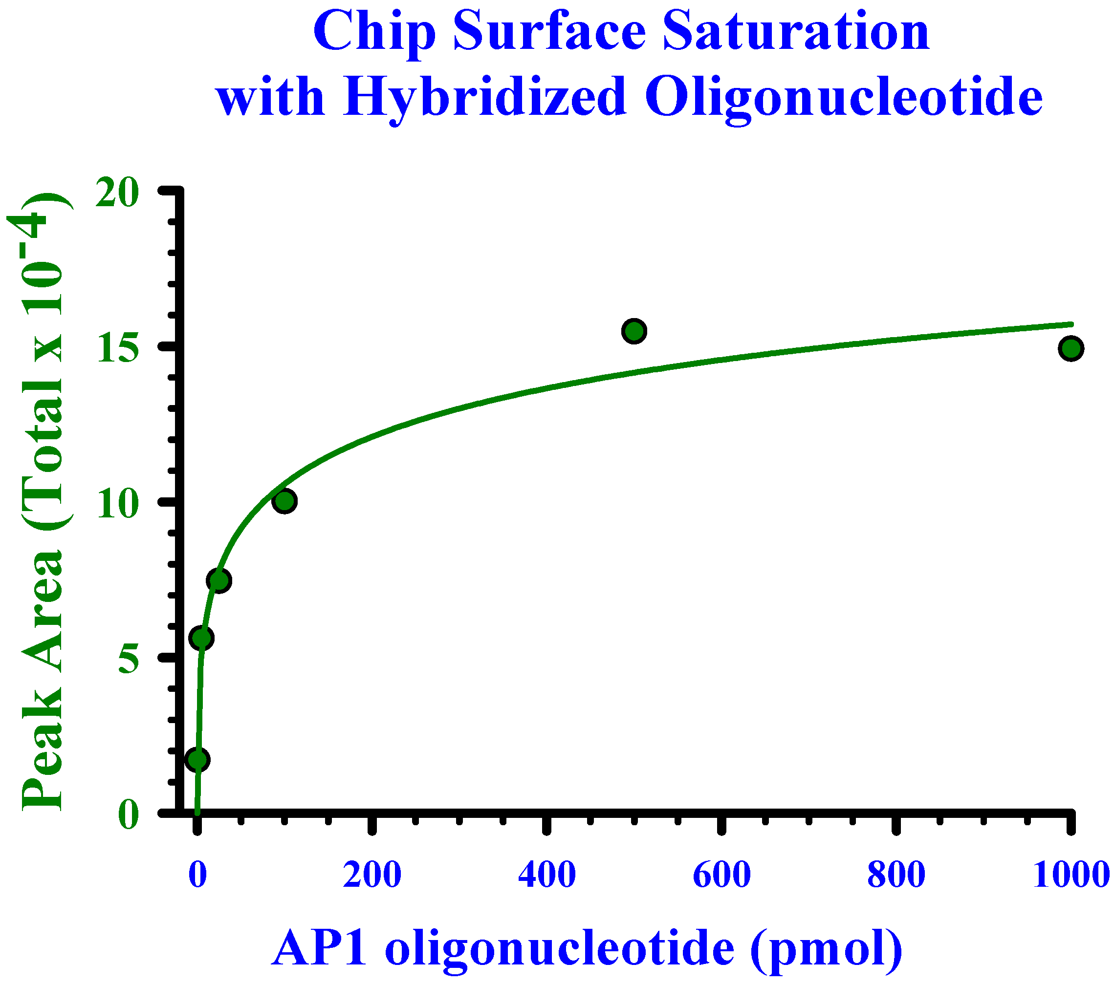
Determination of linearity and saturation of chip surface with oligonucleotide

To achieve saturation of the chip surface such that subsequent experiments with transcription proteins would be based on maximum target sequences, varying concentrations of hybridized oligonucleotide were prepared. Application of these concentrations to the chip surface was done in a constant 10μL volume such that 0 to1000 pmol were available for binding. Recombinant c-Jun protein at a fixed amount of 42 pmol was incubated with the oligonucleotide on the chip surface. As indicated in Figure 1, increasing amounts of c-Jun (indicated by MW 35375 and dimer at MW 70741 in chromatograms) were bound as the amount of oligonucleotide was increased to 500 pmol. The saturation curve in Figure 2 indicates an estimate of approximately 250 pmol of oligonucleotide for maximum binding of the 42 pmol of the c-Jun dimer. Subsequent experiments were therefore performed with 10 μL applications from hybridized oligonucleotides at 25 μM.
Figure 1. SELDI spectra of varying concentrations of duplexed AP-1 oligonucleotide and scrambled oligonucleotide sequence following incubation with 42 pmol of recombinant c-Jun protein. Protein signal height is represented on the y-axis and MW/z on the x-axis. Peak at 70,741 is a dimer of the 35,375 MW c-Jun protein.
Ijms 03 01027 g001
Figure 2. Saturation curve of increasing quantity of AP1 oligonucleotide applied to the epoxide surface of a PS-20 chip prior to incubation with 42 pmol of recombinant c-Jun. Saturation was estimated at approximately 250 pmol of hybridized oligonucleotide. The curve was fit to the data points using the function: y = axb/(cb+xb), with an r2 = 0.96.
Ijms 03 01027 g002
The apparent saturation of the PS20 chip surface is achieved by approximately 4 x 10¹² molecules of hybridized target sequence. To increase the sensitivity of this detection system for the capture of very low concentrations of transcription proteins may require the synthesis of tandem repeats of the binding motifs on the same oligonucleotide to increase the available target.

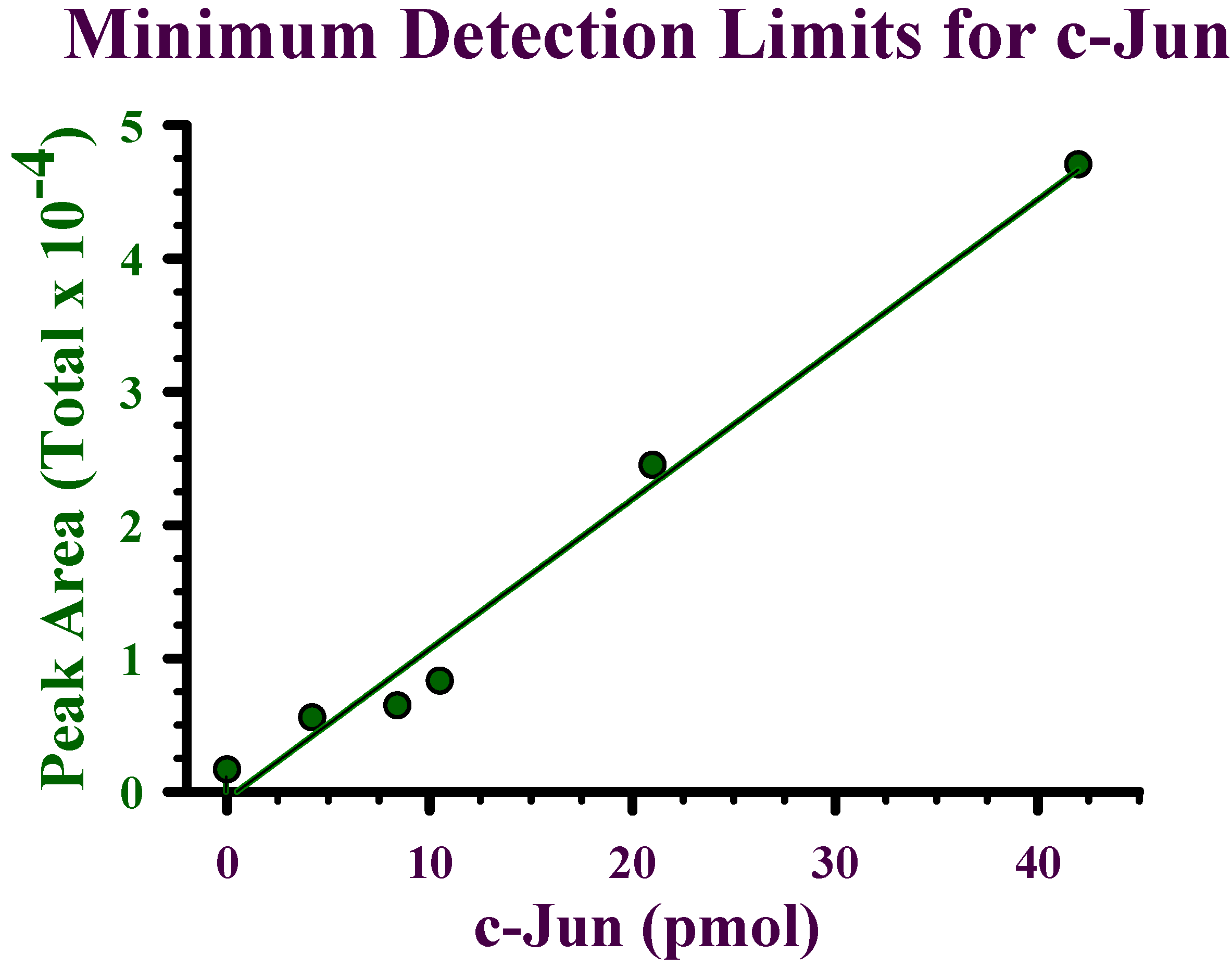
Detection limits for recombinant c-Jun

PS20 chip surfaces were incubated with 250 pmol of oligonucleotide and then with increasing amounts of c-Jun protein from 0 to 42 pmol. Under these conditions, saturation of the oligonucleotide was not achieved (Figure 3). Accurate and reliable signals were detectable to as little as 4.2 pmol. The lower limits of detection indicate that a maximum application of 1.5 mg of nuclear protein to the chip surface would allow detection of a transcription factor that was present at a concentration of approximately 0.01%. This maximum protein would require a nuclear extract with a concentration of 5 mg protein/ml incubated with the oligonucleotide using a bioprocessor to allow 300 μL application.

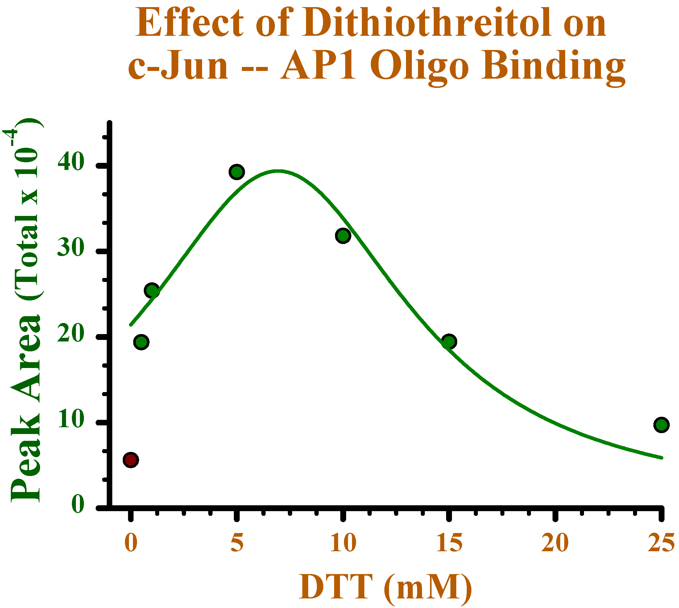
Effect of reducing conditions on binding of c-Jun to oligonucleotide

One of the applications of our subsequent studies will be to determine the effect of alterations in intracellular redox potential on the binding of transcription factors to oligonucleotide sequences. As an estimation of the effect of reducing conditions, dithiothreitol (DTT) at a concentration range of 0 to 25 mM was included in the incubation of the oligonucleotide with c-Jun protein under the binding conditions determined above. As indicated in Figure 4, DTT had a linear effect of increasing binding up to 5 mM, with greater concentrations resulting in diminished binding capacity. This latter effect may be due to disruption of the c-Jun dimer which may require less reducing conditions to maintain the homodimer conformation required for oligonucleotide binding. Although there is little information on the relative redox potential of the nucleus as compared to the cytoplasm, there are some indications of a nuclear compartmentalization of glutathione [26,27] and thioredoxin [28,29]. It is likely that reducing conditions are maintained within the cell nuclei which from our results would appear to favor the binding of transcription factors to the AP-1 oligonucleotide.
Figure 3. Linear increase in binding with increasing amounts of c-Jun incubated with 250 pmol AP-1 oligonucleotide. As little as 4.2 pmol c-Jun was detectable and oligonucleotide was not saturated by 42 pmol of c-Jun protein. Linear regression (y = ax+b, r2 = 0.98) was used to fit the data points.
Ijms 03 01027 g003
Figure 4. Dithiothreitol (0 - 25mM) was included in a 37º C, 15 min incubation with 21 pmol c-Jun prior to incubation with 250 pmol AP-1 oligonucleotide at 37º C, 2 h. Increased protein binding occurred at DTT concentrations from 0 to 5 mM, and then decreased at higher DTT concentrations. The curve was fit to the data points using the function: y = a/(1+((x-x0)/b)2), with an r2 = 0.93.
Ijms 03 01027 g004

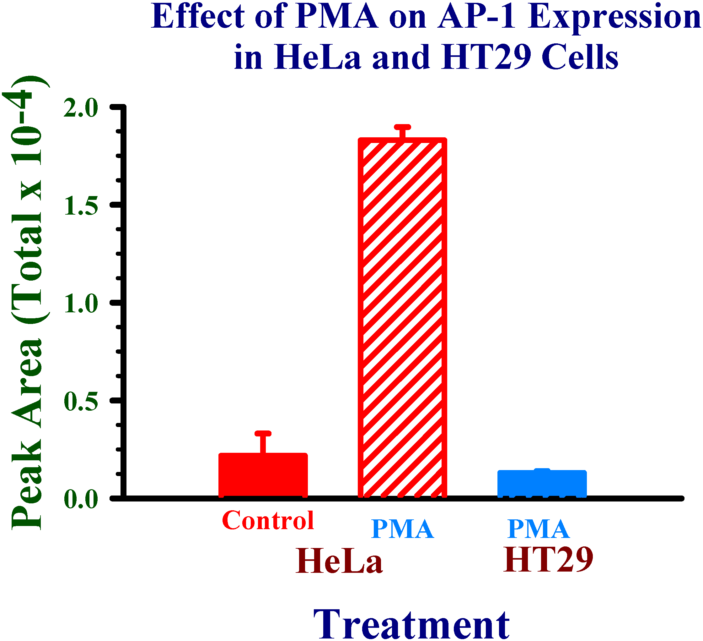
Application of experimental conditions to human cell nuclear protein extracts

The overall goal of these studies is to develop methods to investigate the effects of xenobiotics and their metabolites on transcriptional regulation of enzymes involved in the activation and detoxification of chemical carcinogens. Therefore, nuclear proteins were extracted from two human cell lines treated with 4β-phorbol 12-myristate 13-acetate (PMA) to stimulate the expression of c-Jun. As indicated in Figure 5, c-Jun was readily detectable in PMA-treated HeLa cell. However, neither control HeLa cells or HT29 PMA-treated or control (data not shown) indicated c-Jun levels significantly above background. In contrast to the multi-nucleated HeLa cells, HT29 cells under these conditions did not provide sufficient transcription protein for detection.
Results with the purified recombinant c-Jun protein indicate that the use of SELDI analysis for investigations of transcriptional protein interactions with specific transcriptional response element sequences is a feasible approach. Because of the specificity of detection of all of the proteins bound to the oligonucleotides, detection of candidate transcription factors is not limited by the availability of antibodies. Due to the complexity of the protein-protein interactions that are involved in transcriptional activation and suppression, this may provide a valuable new tool for understanding this feature of the regulatory process of enzyme expression.
Figure 5. HeLa and HT29 cells were treated with 162 nM 4β-phorbol 12-myristate 13-acetate (PMA) or DMSO (0.2%) for 4 h to stimulate expression of c-Jun. Nuclear protein extracts from treated and control (DMSO) cells were incubated with 250 pmol AP-1 oligonucleotides and analyzed by SELDI.
Ijms 03 01027 g005
As indicated from the results of this initial study from mammalian cell extracts, an improvement in the capture of specific proteins from the mixture of nuclear proteins must be achieved. We have estimated from these results that the transcription proteins must be present at approximately 0.01% of the total nuclear proteins. Increased expression of the nuclear proteins of interest will need to be achieved to readily isolate them from the nuclear and cytoplasmic proteins, as these may be present at only one-tenth of that amount in total cellular protein [30]. Alternatively, improvement in the capture of available protein may be accomplished by multiple copies of the oligonucleotide binding sites in the form of tandem repeats within the oligonucleotide sequence.

Acknowledgments

This research was supported by grants from the National Institutes of Health: NIH-RCMI RR03032 and NIH-MBRS GM-028248.

References

  1. Parkinson, A. Biotransformation of xenobiotics. In Casarett & Doull’s Toxicology The Basic Science of Poisons; Klaassen, C.D., Ed.; McGraw-Hill Inc.: New York, 2001; pp. 133–237. [Google Scholar]
  2. Hayes, J.D.; McLellan, L.I. Glutathione and glutathione-dependent enzymes represent a coordinately regulated defence against oxidative stress. Free Radic. Res. 1999, 31, 273–300. [Google Scholar] [CrossRef] [PubMed]
  3. Prochaska, H.J.; De Long, M.J.; Talalay, P. On the mechanisms of induction of cancer-protective enzymes: a unifying proposal. Proc. Natl. Acad. Sci. USA 1985, 82, 8232–8236. [Google Scholar] [CrossRef] [PubMed]
  4. Talalay, P.; De Long, M.J.; Prochaska, H.J. Identification of a common chemical signal regulating the induction of enzymes that protect against chemical carcinogenesis. Proc. Natl. Acad. Sci. USA 1988, 85, 8261–8265. [Google Scholar] [CrossRef] [PubMed]
  5. Rushmore, T.H.; Pickett, C.B. Glutathione-S-transferases, structure, regulation, and therapeutic implications. J. Biol. Chem. 1993, 268, 11475–11478. [Google Scholar] [PubMed]
  6. Baba, T.; Mimura, J.; Gradin, K.; Kuroiwa, A.; Watanabe, T.; Matsuda, Y.; Inazawa, J.; Sogawa, K.; Fujii-Kuriyama, Y. Structure and expression of the Ah receptor repressor gene. J. Biol. Chem. 2001, 276, 33101–33110. [Google Scholar] [CrossRef] [PubMed]
  7. Barouki, R.; Morel, Y. Repression of cytochrome P450 1A1 gene expression by oxidative stress: mechanisms and biological implications. Biochem. Pharmacol. 2001, 61, 511–516. [Google Scholar] [CrossRef] [PubMed]
  8. Kang, K.W.; Ryu, J.H.; Kim, S.G. Activation of phosphatidylinositol 3-kinase by oxidative stress leads to the induction of microsomal epoxide hydrolase in H4IIE cells. Toxicol. Lett. 2001, 121, 191–197. [Google Scholar] [CrossRef] [PubMed]
  9. Carmo-Fonseca, M. The contribution of nuclear compartmentalization to gene regulation. Cell 108, 513–52. [CrossRef]
  10. Kazlauskas, A.; Poellinger, L.; Pongratz, I. Two distinct regions of the immunophilin-like protein XAP2 regulate dioxin receptor function and interaction with hsp90. J. Biol Chem. 2002, 277, 11795–11801. [Google Scholar] [CrossRef] [PubMed]
  11. Whitelaw, M.; Pongratz, I.; Wilhelmsson, A.; Gustafsson, J.A.; Poellinger, L. Ligand-dependent recruitment of the Arnt coregulator determines DNA recognition by the dioxin receptor. Mol. Cell. Biol. 1993, 13, 2504–2514. [Google Scholar] [PubMed] [Green Version]
  12. Whitlock, J.P., Jr. Induction of Cytochrome P4501A1. Ann. Rev. Pharmacol. Toxicol. 1999, 39, 103–125. [Google Scholar] [CrossRef]
  13. Pollenz, R.S.; Barbour, E.R. Analysis of the complex relationship between nuclear export and aryl hydrocarbon receptor-mediated gene regulation. Mol. Cell. Biol. 2000, 20, 6095–6104. [Google Scholar] [CrossRef] [PubMed]
  14. Janssen-Heininger, Y.M.; Poynter, M.E.; Baeuerle, P.A. Recent advances towards understanding redox mechanisms in the activation of nuclear factor kappaB. Free Radic. Biol. Med. 2000, 28, 1317–1327. [Google Scholar] [CrossRef] [PubMed]
  15. Palombella, V.J.; Rando, O.J.; Goldberg, A.L.; Maniatis, T. The ubiquitin-proteasome pathway is required for processing the NF-kappa B1 precursor protein and the activation of NF-kappa B. Cell 1994, 78, 773–785. [Google Scholar] [CrossRef] [PubMed]
  16. Brar, S.S.; Kennedy, T.P.; Whorton, A.R.; Sturrock, A.B.; Huecksteadt, T.P.; Ghio, A.J.; Hoidal, J.R. Reactive oxygen species from NAD(P)H:quinone oxidoreductase constitutively activate NF-kappaB in malignant melanoma cells. Am. J. Physiol. Cell Physiol. 2001, 280, 659–676. [Google Scholar]
  17. Itoh, K.; Wakabayashi, N.; Katoh, Y.; Ishii, T.; Igarashi, K.; Engel, J.D.; Yamamoto, M. Keap1 represses nuclear activation of antioxidant responsive elements by Nrf2 through binding to the amino-terminal Neh2 domain. Genes Dev. 1999, 13, 76–86. [Google Scholar] [CrossRef] [PubMed]
  18. Wasserman, W.W.; Fahl, W.E. Functional antioxidant responsive elements. Proc. Natl. Acad. Sci. USA 1997, 94, 5361–5366. [Google Scholar] [CrossRef] [PubMed]
  19. Moffat, G.J.; McLaren, A.W.; Wolf, C.R. Transcriptional and post-transcriptional mechanisms can regulate cell-specific expression of the human Pi-class glutathione S-transferase gene. Biochem. J. 1997, 15, 91–95. [Google Scholar]
  20. Jaiswal, A.K. Regulation of genes encoding NAD(P)H:quinone oxidoreductases. Free Radic. Biol. Med. 2000, 29, 254–262. [Google Scholar] [CrossRef] [PubMed]
  21. Toone, W.M.; Morgan, B.A.; Jones, N. Redox control of AP-1-like factors in yeast and beyond. Oncogene 2001, 20, 2336–2346. [Google Scholar] [CrossRef] [PubMed]
  22. Sun, Y.; Oberley, L.W. Redox regulation of transcriptional activators. Free Radic. Biol. Med. 1996, 21, 335–348. [Google Scholar] [CrossRef] [PubMed]
  23. Ikuta, T.; Watanabe, J.; Kawajiri, K. Characterization of the LxxLL motif in the aryl hydrocarbon receptor: effects on subcellular localization and transcriptional activity. J. Biochem. (Tokyo) 2002, 131, 79–85. [Google Scholar] [CrossRef]
  24. Sun, W.; Zhang, J.; Hankinson, O. A mutation in the aryl hydrocarbon receptor (AHR) in a cultured mammalian cell line identifies a novel region of AHR that affects DNA binding. J. Biol. Chem. 1997, 272, 31845–31854. [Google Scholar] [CrossRef] [PubMed]
  25. Forde, C.E.; Gonzales, A.D.; Smessaert, J.M.; Murphy, G.A.; Shields, S.J.; Fitch, J.P.; McCutchen-Maloney, S.L. A rapid method to capture and screen for transcription factors by SELDI mass spectrometry. Biochem. Biophys. Res. Commun. 2002, 290, 1328–1335. [Google Scholar] [CrossRef] [PubMed]
  26. Bellomo, G.; Vairetti, M.; Stivala, L.; Mirabelli, F.; Richelmi, P.; Orrenius, S. Demon-stration of nuclear compartmentalization of glutathione in hepatocytes. Proc. Natl. Acad. Sci. USA 1992, 89, 4412–4416. [Google Scholar]
  27. Voehringer, D.W.; McConkey, D.J.; McDonnell, T.J.; Brisbay, S.; Meyn, R.E. Bcl-2 expression causes redistribution of glutathione to the nucleus. Proc. Natl. Acad. Sci. USA 1998, 95, 2956–2960. [Google Scholar] [CrossRef] [PubMed]
  28. Hirota, K.; Murata, M.; Sachi, Y.; Nakamura, H.; Takeuchi, J.; Mori, K.; Yodoi, J. Distinct roles of thioredoxin in the cytoplasm and in the nucleus. A two-step mechanism of redox regulation of transcription factor NF-kappaB. J. Biol. Chem. 1999, 274, 27891–27897. [Google Scholar] [CrossRef] [PubMed]
  29. Tanaka, T.; Nishiyama, Y.; Okada, K.; Hirota, K.; Matsui, M.; Yodoi, J.; Hiai, H.; Toyokuni, S. Induction and nuclear translocation of thioredoxin by oxidative damage in the mouse kidney: independence of tubular necrosis and sulfhydryl depletion. Lab. Invest. 1997, 77, 145–155. [Google Scholar] [PubMed]
  30. Kadonaga, J.T.; Tjian, R. Affinity purification of sequence-specific DNA binding proteins. Proc. Natl. Acad. Sci. USA 1986, 83, 5889–5893. [Google Scholar] [CrossRef] [PubMed] [Green Version]

