Review

Phototoxic or Photoprotective?—Advances and Limitations of Titanium (IV) Oxide in Dermal Formulations—A Review

1
Chair and Department of Pharmaceutical Technology, Poznan University of Medical Sciences, 6 Grunwaldzka Street, 60-780 Poznań, Poland
2
CNRS, LRGP, Université de Lorraine, F-54000 Nancy, France
*
Author to whom correspondence should be addressed.
Int. J. Mol. Sci. 2023, 24(9), 8159; https://doi.org/10.3390/ijms24098159
Submission received: 31 March 2023 / Revised: 26 April 2023 / Accepted: 28 April 2023 / Published: 2 May 2023

Abstract

:
The widespread role of titanium (IV) oxide (TiO2) in many industries makes this substance of broad scientific interest. TiO2 can act as both a photoprotector and photocatalyst, and the potential for its role in both applications increases when present in nanometer-sized crystals. Its sunlight-scattering properties are used extensively in sunscreens. Furthermore, attempts have been made to incorporate TiO2 into dermal formulations of photolabile drugs. However, the propensity to generate reactive oxygen species (ROS) rendering this material potentially cytotoxic limits its role. Therefore, modifications of TiO2 nanoparticles (e.g., its polymorphic form, size, shape, and surface modifications) are used in an effort to reduce its photocatalytic effects. This review provides an overview of the potential risks arising from and opportunities presented by the use of TiO2 in skin care formulations.

Graphical Abstract

1. Introduction

The beneficial effect of sunlight on the functioning of our bodies was already known and described thousands of years ago. Photoexposure in treating rickets and neurological or skin diseases was recommended, for example, in ancient Egypt [1]. Unfortunately, solar radiation also has another, less favorable side. As a form of energy, it can negatively interact with drugs, leading to their decomposition or even their transformation into harmful photoproducts [2]. Photosensitization of active compounds, especially after topical administration, is considered a real-world problem, while their interaction with UV light often leads to serious phototoxic or photoallergic reactions [3]. Literature data evidence the scale of the problem, according to which almost 400 active substances are considered as photolabile [4]. There is a strong need to carefully analyze the photochemical properties of drugs since there is a clear correlation between phototoxicity and skin cancer that has been confirmed [4]. Moreover, treating patients with photodamaged skin is long-term and requires multiple medications, such as antihistamines, steroids, and nonsteroidal anti-inflammatory drugs [5], generating additional costs. One of the possibilities to reduce the effect of light on drugs applied to the skin is the use of excipients that are able to absorb or scatter the radiation, and therefore produce a photoprotective function. To date, one of the most frequently used was titanium (IV) oxide (TiO2)—a substance that has received significant press coverage recently [6]. On the basis of scientific analysis, concerning the inclusion of TiO2 in various medical products, on 8 September 2021 the European Medicines Agency (EMA) released a document which indicated the need for further research aimed at finding safer alternatives [7]. In short, the main purpose of this report was to exert pressure on the pharmaceutical industry to develop substitutes for TiO2 (to date, most commonly used as color and opacifier) within the time-frame of 3 years. This recommendation was adopted in January 2022 and the main focus was on the genotoxic potential of TiO2. Although this detrimental effect has been proven for inhaled particles, the current knowledge regarding the use of TiO2 on skin surfaces still remains to be clarified and analyzed in detail [8]. The main assumption of the presented review is to assess the current state of knowledge about the advantages (photoprotection) and disadvantages (phototoxicity) of the use of TiO2, mainly in dermal formulations. The goal is to answer whether and to what extent a risk may accompany its application to the skin and what are the possibilities of modifying its properties to maintain the photoprotective function while minimizing potentially harmful effects.

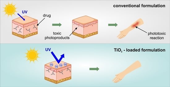
2. Photolability of Drugs Applied to the Skin

In general, photosensitization can be defined as a process involving the excitation of a given chemical compound by light. Under the right conditions, with a sufficiently high wavelength of light, radiation can lead to photochemical degradation or photochemical transformation into pharmacologically inactive products, which are more likely to cause skin allergies or burns by a phototoxic or photoallergic mechanism [3]. In the case of drugs applied to the skin, such as analgesics [9], antibiotics [10], antiallergics [11], or anti-acne agents [12], UVA radiations are the most harmful. Direct and uncontrolled exposure of the treated skin to sunlight is likely to result in serious side effects and damage that is difficult to recover from [13].
Acute phototoxic reactions often result in processes occurring at the cellular level, such as DNA or cell membrane damage. Typical phototoxicity is accompanied by sudden changes, which, after proper treatment, disappear upon complete recovery. Photoallergic reactions can last significantly longer, since the absorbed energy induces formation of stable haptens with affinity to the cell proteins, followed by production of antigens [3]. It should also be kept in mind that it is not only topically applied drugs that are responsible for photochemical reactions. In the case of substances administered orally, used chronically in long-term therapies, there is a risk of their accumulation in the skin, which can also result in unwanted effects.
The differences between the mechanism of phototoxic and photoallergic reactions are shown in Figure 1, while the characteristics of both types of photosensitization are presented in Table 1. However, reactions caused by topically applied photosensitizers (e.g., in dermatology or ophthalmology) tend to be more severe than those caused by drugs applied systemically [14].
Photoallergy is more relevant to photosensitization than to topically applied agents. Nevertheless, some compounds, such as psoralen and its derivatives, retinoids, and 5-fluorouracil can also cause photosensitization through a phototoxic mechanism [3,14,15]. The severity of the reaction depends on the drug absorption depth, as well as its metabolism in the skin [16]. Drugs in the form of creams or ointments tend to accumulate in the upper layers of the epidermis and primarily cause damage to keratinocytes [17]. However, an acute reaction may be followed not only by necrosis of keratinocytes, but also by epidermal spongiosis, edema, vasodilation, and an infiltration of neutrophils, lymphocytes, and macrophages [14]. An example of an acute photosensitization reaction following the topical application of ketoprofen to sun-exposed skin is shown in Figure 2.
Figure 2. Acute phototoxic reaction following topical application of ketoprofen. Published with the permission of Elsevier [18] published by Elsevier Spain, S.L.U. All rights reserved.
Ijms 24 08159 g002
Table 1. Characteristics of phototoxicity and photoallergy.
PhototoxicityPhotoallergyReferences
Clinical symptomserythema, vesicles, and bullae, burning, stinging, hyperpigmentationeczematous, pruritic rash[3]
Histological effectsnecrotic keratinocytes, epidermal degeneration, sparse dermal infiltrate of lymphocytes, macrophages, and neutrophilsspongiotic
dermatitis, dermal
lymphohistiocytic
infiltrate
[3,17]
Pathophysiologydirect tissue injurytype iv delayed hypersensitivity response[2]
Occurrence after first exposureyesno[2,3]
Onset after exposureminutes to hours24 to 48 h[2,14]
Dose of agent needed for the reactionlargesmall[14]
Cross-reactivity with other agentsnonecommon[2,17]
Diagnosisclinicalphotopatch tests[3,19]
The common active pharmaceutical ingredients recognized as photolabile are presented in Table 2.

3. Titanium Dioxide

3.1. General Information

Crystalline TiO2 may exist in three phases, namely rutile, anatase, and brookite (Figure 3) [28].
Variations between these polymorphs involve octahedral units arranged in a three-dimensional space. Importantly, each of the crystal structures exhibits different properties—they can differ in shape, structure, refractive index, and photocatalytic activity [30]. Furthermore, only rutile is stable, while anatase and brookite are metastable and convert to rutile upon heating. The differences between the polymorphs are particularly apparent when comparing their photocatalytic activity. The higher energy gap of anatase and brookite and their superior electron mobility make these polymorphs more reactive than rutile. Moreover, there is a significant difference in the refractive index of rutile and anatase (2.7 and 2.5, respectively). While there are many arguments in favor of the safety of TiO2, potential risk factors also need to be considered. On the one hand, excellent photoprotective properties are reported, and on the other hand, the risk of inducing photochemical reactions, as shown in the following sections.

3.2. Titanium Dioxide Nanoparticles (NPs) Photoprotection vs. Phototoxicity

TiO2 nanoparticles (TiO2-NPs) have attracted a considerable attention due to their unique properties, which make them desirable for a wide range of applications. Among industrial applications, they have found a role in cosmetic and pharmaceutical technology. Properties that make it an excellent candidate for the use as a sunscreen include its high photoprotective capacity (strong light scattering) associated with a large photoactive area, insolubility in aqueous media, and transparency. It has been proven that TiO2-NPs can act as a shield for both the skin and drugs against UVA and UVB radiations. Moreover, the argument for using TiO2-NPs as a sunscreen to date is that they are less photoreactive than other chemical ingredients (especially organic sunscreens), and thus less likely irritating to people with sensitive skin. The EU Scientific Committee on Consumer Safety (SCCS) has approved crystalline TiO2-NPs as safe for use in cosmetic products intended for use on healthy, undamaged, or sunburnt skin [31]. As a UV filter, TiO2 can be incorporated into cosmetic formulations at a maximum concentration of 25%. However, it has to be considered that the high surface-to-volume ratio makes it legitimate to raise the issue of the increased bio-reactivity of nanomaterials (NMs) and their potential toxicity. Moreover, recent studies and reports on TiO2-induced phototoxicity have brought the topic increasingly into the headlines. Therefore, the question of safety must be asked to consider whether chronic exposure to this material is dangerous or not. Although the phototoxic effect of TiO2-NPs has already been demonstrated for all polymorphs, it is important to note that different phases can induce a reaction of different severities. However, the main problem with the use of TiO2 in products intended for use on the skin surface is its potential to generate ROS [32,33,34,35,36,37,38].

3.3. Illuminated TiO2-NPs Generate Reactive Oxygen Species (ROS)

Reactive oxygen species are a group of chemically reactive molecules/radicals derived from molecular oxygen. Examples of ROS include superoxide (O2●−), hydroxyl radical (HO) hydrogen peroxide (H2O2), and singlet oxygen (1O2), each of which causes a variety of biological damage and has been implicated in a multitude of diseases, including cancer and ageing [39,40]. As already proven, ROS are deleterious to biological structures (e.g., membranes, lipids, proteins, and nucleic acids) and may be generated by TiO2-NPs under certain conditions, e.g., when illuminated with UV light and in an aqueous environment [41].
TiO2 is a wide bandgap semiconductor (3.2 eV for anatase) [42] and can only be photo-activated under UV illumination [43]. Only light with a wavelength lower than 387 nm allows for the excitement of an electron from the valence band (VB) to the conduction band (CB). As a result, holes in the VB (h+VB) and electrons in the CB (eCB) are generated and subsequently migrate to the NPs surface to react with water and adsorbed O2 molecules, respectively [44]. These reactions are responsible for the production of ROS in the aquatic and aerobic environment. The reactions mentioned are presented as chemical Equations (1)–(6) [44,45].
H2O + h+ → OH + H+
O2 + 2e + 2H+ → H2O2
O2●− + O2●− +2H+ → H2O2 + O2
H2O2 + e → OH + OH
H2O2 + O2●− → OH + OH + O2
H2O2 + hv → OH + OH
The correlation between TiO2-NPs and hydroxyl radical concentrations, UVA light intensity, and concentration of dissolved organic carbon (DOC) with toxicity was assessed by the team of Coral et al. Their study demonstrated that the radical production rate was positively correlated with an increase in TiO2-NPs concentration and UVA intensity, and negatively correlated with increased DOC concentrations. Moreover, the authors showed that the concentration of TiO2-NPs is a poor predictor of toxicity, when considered alone, and that the concentration of generated hydroxyl radical is a better predictor in assessing the overall risk [45].
An alternative of TiO2-NPs phototoxicity was also described. Taking into account the energy transfer of O2●− radicals to the holes in the VB, singlet oxygen (1O2) may also be generated according to Equation (7) [46].
h+ + O2●− → 1O2
1O2 can oxidize fatty acids or damage nucleotides leading to several oxidative products [46]. The presence of 1O2 photo-produced by TiO2-NPs was confirmed by Fenoglio et al. By modification of the NPs surface, the ROS-generating potential can be decreased. For example, by the introduction to the surface carbonaceous or carbonate/carboxylate-like products, that generation of ROS can be decreased, although the presence of singlet oxygen was confirmed [44].

3.4. Factors Influencing the Phototoxic Potential of TiO2-NPs

3.4.1. Polymorphic Form

As it was previously mentioned, the polymorphic form of TiO2-NPs influences both the material stability as well as the potential of ROS generation. Usually, the rutile phase is less photoreactive than anatase or brookite. This phenomenon is related to a different lifetime of holes (that is longer for rutile) and the electron-trap depth that is shallow for anatase (<0.1 eV), moderate for brookite (~0.4 eV), and high for rutile (~0.9 eV). The deeper the electron trap is located, the less photoreactivity is observed due to lower electron mobility [47]. Another difference between polymorphs is the interaction of these materials with oxygen. Molecular oxygen shows a high affinity to oxygen vacancies. In anatase, the interaction of molecular oxygen with the surface is weaker than for rutile. It explains the difference in the concentration of absorbed oxygen in both forms of TiO2-NPs. As shown previously, adsorption of O2 relates to an electron transfer from the surface defects, forming superoxide radical anions, which are well known as ROS with a weaker oxidation ability than hydroxyl radicals [48]. The different photoreactivities of anatase and rutile were confirmed in cytotoxicity studies [49].
Tests on immortalized human keratinocytes (HaCaT), both with and without UV light were conducted. In the dark, TiO2-NPs (rutile or anatase) did not affect the mitochondrial activity or cell membrane. However, the exposition to rutile NPs may inhibit cell growth and induce OH generation, which seems to be dependent on the surface properties of nanomaterial, and was confirmed for the hydrophobic surface of rutile NPs. In this study, the cytotoxicity under UVA irradiation was tested and a strong polymorphic-dependent effect was seen. The rutile NPs did not show any cellular effect while the anatase showed strong phototoxicity and induced a marked decrease in mitochondrial activity, cell membrane damage, and the induction of oxidative stress [50]. Another research that settled the higher cytotoxic potential of anatase was conducted by Hering et al. By using both the monolayer cell culture and UV light, the authors confirmed the influence of the polymorphic of TiO2-NPs and the material concentration on the cell viability. The results showed that anatase led to a lower effective dose (ED50) for all UV doses, the lowest being when irradiated with UVB light. However, rutile showed higher cell viability compared to anatase at the highest intensity of UVB light [51]. The polymorphic-dependent toxicity is mainly connected with the potential to generate ROS, which can damage the cell membrane. This conclusion was also reached by Yin et al. Their work showed that the anatase TiO2-NPs are more photoreactive than rutile, being more phototoxic to HaCaT cells [52]. However, some studies confirmed that the mixture of rutile/anatase may be more toxic than smaller pure anatase particles [53,54].
The cytotoxic effect of various polymorphs is also connected with the cellular uptake of NPs. Although the absorption of particles into the cells depends mostly on their diameter, the hydrophilic and hydrophobic surface properties should also be taken under consideration.

3.4.2. Material Size, Surface, and Morphology

In physics—scale matters. The size of the material determines its properties, especially when nanomaterials are considered. The reason that properties of nanomaterials are quite different from bulk concerns both the surface effects and quantum size effects.
The atoms in the surface of the material have fewer neighbors than atoms in bulk, and thus generate a lower coordination and dangling bonds. This makes those atoms less stable compared to bulk atoms. The smaller the particle, the greater the number of surface atoms; therefore, the surface-to-volume ratio scales with the inverse of size. Moreover, in semiconductors (e.g., TiO2), the electronic wave functions of conductive electrons are delocalized over the entire particle. Electrons are described as “particles in the box” and the density of state and the energy of particles depend crucially on the size of the box, which leads to a smooth-size dependence. Absorption wavelengths, ionization potentials, and electron affinities are tuned between the atomic values and the work function of the bulk material by varying the size of the NPs. These properties are connected to the availability of electrons for redox reactions. Therefore, the photoreactivity depends on the NPs size [55].
Considering the influence of the size on the cytotoxic effect, tests using the Sinorhizobium meliloti bacteria exposed to aqueous dispersions of micrometer-sized TiO2 (44 μm) and TiO2-NPs (21 nm) under various concentrations were conducted. A hundred percent mortality of the bacteria was reached using TiO2-NPs at a concentration of 100 mg/L (under UVA irradiation) and 900 mg/L (under dark conditions). However, the exposure to micrometer-sized TiO2 showed that there is no effect on the bacteria in a concentration of less than 300 mg/L (under dark or UVA light irradiation). One hundred percent mortality was only achieved with a nominal concentration of 600 mg/L or more (UVA light irradiated) [56]. This result clearly shows that there is a direct correlation between the effect of material size and the toxic effect.
Sanders et al., tested the uptake and accumulation of TiO2-NPs in ARPE-19 cells and showed that after a short-term or long-term exposure, the NPs were observed inside the cells and remained there for an extended period. Moreover, the amount of NPs inside the cells was dose-dependent. However, no clear effects on viability or morphology were observed. The cells that were treated with any of the TiO2-NPs and remained in the dark were 100% viable up to the concentration of 30 μg/mL, except for the sample treated with 31 nm-sized anatase/rutile, which showed a dose-related decrease in viability beginning at 10 μg/mL. Moreover, the tests conducted under visible light illumination showed no difference in viability compared to the dark conditions, which is not surprising as TiO2 cannot be photo-activated under these irradiation conditions. The differences between samples were observed when cells were exposed to UVA radiation. The outcomes showed clearly that the LC50 decreased with the particles size [57]. However, the outcomes of Wyrwoll et al., suggested that these results are only valid on a limited number of particle sizes. The authors showed that NPs of 25 nm were more toxic than those of 10 nm. Although the smaller NPs are able to generate a higher amount of OH radicals, the sum of all measured ROS (free and surface attached OH and O2●− radicals) was the highest for 25 nm-sized NPs [58]. These phenomena may be explained by the different mechanisms that remain behind the photoactivity. For the particles smaller than 25 nm, the mechanism depends more on optical and electronic properties, including light absorption, scattering efficiencies, and charge-carrier dynamics. In contrast, for particles larger than 25 nm, the photoactivity depends more on the surface area available for redox reactions [59]. Moreover, Wyrwoll et al., confirm that the ionic strength (IS) of the medium highly influences the material toxicity as a high IS promotes material agglomeration and thus sedimentation; therefore, lowering its phototoxicity [58].
Another important factor influencing the toxicity of TiO2-NPs is their specific surface area (SSA). As already mentioned, the smaller the particles, the higher the surface/volume ratio. The large surface area of the material allows it to come into close contact with the cells, which is essential for maximizing the damaging effects of ROS, whose lifetime is extremely short. However, particles with the same size may have a different SSA, and therefore produce various photo- and cytotoxicity effects. This effect was tested on RAW264 cells in the absence and presence of light. It was shown that a larger SSA induced higher cyto- and phototoxicity (UV activated) in cells. The increased interactions between the NPs and biomolecules and the greater number of ROS generated contribute to these effects [60].
When considering the physical properties of a material that affect its phototoxic potential, the shape of the particles, which influences their interaction with the cell, should be mentioned. TiO2-NPs are usually produced in spherical shapes. However, TiO2 may also exhibit low-dimensional nanostructures, such as one-dimensional nanotubes, nanorods, nanobelts, and nanowires or two-dimensional nanosheets. By measuring the membrane integrity of bacteria, the evaluation of how different morphologies influence the cytotoxicity of TiO2-NPs was assessed. The outcome showed that the toxic effect strongly depends on the shape with the following order: nanospheres > nanorods > nanosheets ≈ nanorods, and thus indicating that low-dimensional materials are less toxic. Using scanning electron microscopy, it was suggested that the material morphology influences TiO2-NPs phototoxicity by governing the alignment of TiO2-NPs at the bacterial cell surface. The different shapes that TiO2-NPs can take are shown in Figure 4 and the influence of the particles’ shape on bacterial viability is presented in Figure 4 and Figure 5 [61].
The determination of the cytotoxic and phototoxic effects of TiO2 cannot be described as a simple function of the photocatalytic reactivity or ROS production. Rather, the evaluation of TiO2-NPs phototoxicity must encompass a three-pronged approach, involving the intrinsic photoactivity (influenced by, e.g., polymorphic form, particles size, or specific surface area), aggregation of TiO2-NPs (influenced by, e.g., particles size, ionic strength of the medium, or the materials surface charge), and the TiO2-NPs/cell interactions (influenced by, e.g., material morphology, specific surface area, or ability to penetrate the cell).

3.5. Cytotoxicity as a Response to Photocatalytic Properties of TiO2-NPs

The photocatalytic properties of TiO2-NPs lead to cytotoxicity, thus inducing apoptosis or, in some cases, necrosis of the cell [62,63]. Necrosis, as premature cell death, can be caused directly by ROS generated by NPs exposed to UV light, which causes damage to the cell membrane. In contrast, apoptosis is a controlled and programmed cell death. In response to cytotoxicity, cells may undergo apoptosis by activating cell signals that cause the cell to break down and die. The mechanism of apoptosis involves a sequence of biochemical events that results in the activation of proteases, such as caspases, that cleave the cell proteins and DNA, leading to cell death. The cell may be induced to initiate apoptosis by an external signal, e.g., in the form of free-oxygen radicals present in its environment [64].
As shown, the type of cell death (i.e., by apoptosis or necrosis) depends on the cell line. Among the mechanisms responsible for cell necrosis, the main one seems to be oxidative stress. By reducing the mitochondrial membrane potential and damaging those structures, the cell becomes devoid of its “engine”. An essential role in the regulation of the mitochondrial membrane potential is played by the mitochondrial permeability transition pore (mPTP)—a non-specific channel between the inner and outer membrane of mitochondria [65]. The disorder in the form of abnormal opening of the mPTP in response to TiO2-NPs under UVA irradiation was proven for the HeLa cell line (immortalized epithelial cells), confirming that necrosis plays a key role in cell death. Moreover, the researchers suggest that by regulating the ROS-mPTP pathway, it is possible to influence the cell response to TiO2-NPs [66].
However, caspase-dependent apoptosis seems to play a major role in the death of HaCaT cells caused by ROS generated by UV-illuminated TiO2-NPs. Exposure of HaCaT cells to those NPs resulted in accumulation in lysosomes via endocytosis or autophagy. Under UVA light, TiO2-NPs damage the lysosomal membrane and subsequently stimulate ROS production. Therefore, it was suggested that lysosomal membrane permeabilization (LMP)-dependent oxidative stress plays a critical role in the UVA phototoxicity of TiO2-NPs in HaCaT cells [67]. Nonetheless, not only ROS have been proven to induce NPs toxicity. Ren et al., point out that reactive nitrogen species (RNS) also induce apoptotic signaling and pathologies in intracellular redox homeostasis. The authors observed the effects of TiO2-NPs on cell glycans—structures that play a regulatory role in various physiological processes, such as sialylation. Sialylation is the glycosylation modification playing a key role in cell adhesion, signaling, and recognition, as well as aging and senescence [68]. Using the HaCaT cells, they have proven the changes in sialic acid concentration in the response of ROS generated by UV-illuminated TiO2-NPs, the changes that overall have been reported to cause cancer [69]. Among the biochemical mechanisms that can be influenced by TiO2-NPs, Janus kinase/signal transducer and activator of transcription (JAK/STAT) pathway may be disrupted by these NPs. JAK/STAT pathway is known to regulate the development and reproduction in many organisms. In the example of Caenorhabditis elegans, the alteration of this pathway was observed, as a response to oxidative stress. Moreover, down-regulation in glutathione activity also appeared. As the JAK/STAT pathway is cross-talked with transforming growth factor beta (TGF-β; the protein that plays an important role in regulating cell growth and extracellular matrix synthesis), the pathway gene expression of this protein was also tested. As with JAK/STAT, significant changes in this gene expression under the influence of TiO2-NPs (under UV) were observed, indicating a significant reproductive toxicity [70].
Although the cytotoxicity was proven in cell tests, there is a significant discrepancy between 3D and 2D cultures. In 2D cell culture, NPs are suspended in the cell media. Due to their physicochemical properties, a sedimentation of NPs was observed, as the result of agglomeration, thus increasing the amount of TiO2 at the bottom of culture wells. This increases the cellular uptake resulting in an overestimated toxic effect [71]. Moreover, the 2D models do not consider the natural barrier of the skin. Due to the above, many studies have proven that the toxic effects on 2D cell cultures do not reflect in 3D models [50,51].
A review that brought together 50 papers focusing on in vitro and in vivo toxicity testing of TiO2 suggests that the genotoxic effect of TiO2 cannot be directly confirmed or ignored. The authors point out that more in vitro than in vivo studies should be performed, leading to more positive results of genotoxicity in vitro models than in vivo models. Moreover, they conclude that more well-designed in vivo studies must be performed to assess TiO2 genotoxicity [72].
Furthermore, a very recent review including 192 articles with 34 robust datasets does not support a direct DNA damaging mechanism for TiO2 in nano- or micro-form [49].

3.6. Might Modifications of TiO2-NPs Surface Decrease Phototoxicity?

The widespread potential use of TiO2 in medicine and cosmetics has led researchers to try to reduce phototoxicity by modifying the particles surface. Most of the modifications involve the introduction of an inert shell on the surface of the NPs. Among the materials used for coating, the most common are silica (SiO2), alumina (Al2O3), zirconium dioxide (ZrO2), or siloxanes [36]. However, the main challenge in synthesizing these types of particles with decreased phototoxicity is to suppress the photoactivity with preserved UV-shielding ability. Moreover, due to the potential application of these particles, the shell must be built of inert materials, the role of which is to inhibit the formation of ROS or to scavenge ROS. However, to date, there is no evidence that this method leads to a complete deactivation of the catalytic activity of TiO2-NPs [36,73].
One of the most promising materials for shelling TiO2-NPs is SiO2, mainly due to its wide bandgap of ca. 9 eV. To date, several trials have been reported in which the properties of TiO2-NPs have been successfully modified [74,75,76,77]. Notable is the decrease in photocatalytic activity by 85% relative to the TiO2 particles while preserving 76% of the original UV-scattering ability [75]. El-Toni et al., decreased the photocatalytic activity of TiO2-NPs and the UV-shielding ability was not significantly reduced after coating [78].
Nevertheless, the nanoparticle coating procedure has led to a change in the mechanism of the potential generation of toxic effects by the newly formed material. Based on the model of fish gill cells, Martin et al., point out that the coating influences NPs ability to be taken up by cells. They examined three different materials: TiO2/SiO2 (core/shell; hydrophilic), TiO2/Al(OH)3/PDMS (core/shell/shell; hydrophobic), and TiO2/Al2O3/stearic acid (core/shell/shell; hydrophobic). The results showed that particles with hydrophilic surfaces were present inside the cells, whereas those with hydrophobic surfaces were not [79]. Moreover, considering that NPs can be incorporated into the cells raises questions about their safety [80]. Furthermore, Tang et al., showed that NPs coated with silica or alumina exhibited high phototoxic potency that might originate from the interaction of harmful metal ions released from the oxide coating (e.g., Al3+) [81]. However, this effect was not confirmed considering the 3D skin model, as the particles could not penetrate the skin barrier.
Reducing the toxicity of TiO2-NPs by coating with a chemically inert material carries the risk of other toxicity mechanisms of the resulting core/shell NPs. At the same time, the lack of comprehensive in vitro and in vivo studies of the newly engineered materials suggests that further research is needed to confirm the safety of coated TiO2-NPs for medicinal applications.

4. Titanium Dioxide Nanoparticles as Photostabilizing Agents in Topical Drug Formulations

The use of TiO2 as a photoprotector for photolabile active pharmaceutical ingredients (APIs) in dermal drug formulations is very limited, and to date, was tested only for retinoic acid and ketoprofen. However, all the papers confirm the increased photostability due to the use of this inorganic sunscreen, compared to formulations without TiO2 [13,82,83]. Asfour et al., combined all-trans retinoic acid (ATRA) and TiO2 for the alleviation of photo-induced sensitivity. The authors confirmed the positive impact of the prepared formulation on product safety in vitro and in vivo studies. They have proven that the addition of 6% (w/w) TiO2 resulted in the enhanced photostability of ATRA by about 2 times, compared to its methanolic solution. Moreover, in vivo studies on mice exposed to direct sunlight for 4 days, confirmed that the prepared formulation alleviated the photosensitization in comparison to the marketed product [82]. The positive effect of TiO2 on ketoprofen (KP) photostability has been proven for transdermal patches [13] and gels [83]. By incorporating TiO2 into the fabric backing of the patch, the UV transmission through the product was effectively decreased in the UVA region, exhibiting the same increased photostability of KP. Moreover, in vivo studies demonstrated lower photosensitization of the patch with TiO2, compared to the KP patch without TiO2. In the latter study, the lowered decomposition of KP in the gel was confirmed. The three materials (crystalline TiO2, pharmaceutical grade TiO2, and TiO2 coated with alumina and silica) were tested and the latter-one showed the best effect. The 2 h exposure of the gels with and without coated TiO2 to ordinary sunlight caused a decrease in KP concentration from 2.5% to 0.05% (gel without TiO2) and from 2.5% to 0.71% (gel with TiO2).
Both presented studies conclude the high potential of TiO2 in the protection of photolabile API applied to the skin. However, given the scarcity of work on the subject, more in-depth studies and a comprehensive evaluation of the effect of TiO2 addition on the formulation stability are required.

5. The Use of TiO2 in Sunscreens and Cosmetics

The common link between photocarcinogenesis, photoaging, and photosensitivity is their induction by exposure to sunlight. Although sunlight is essential for certain metabolic pathways, such as vitamin D3 synthesis, overexposure to UV radiation leads to more harm than benefit [84]. The World Health Organization (WHO) indicates that, as of 2018, there is a reported incidence of approximately 3 million keratinocyte skin cancers and 132,000 melanoma skin cancers, stating that one of every three cancers diagnosed is some form of skin cancer [85]. Given these statistics, it is necessary to provide some protection from UV radiations.
Among the various sun protection strategies, the use of sunscreens is a common process. In general, we can distinguish between organic and inorganic sunscreens. Organic agents (such as, e.g., benzophenone, para-aminobenzoic acid, or menthyl anthranilate) act by absorbing UV radiation, which then leads to the activation of electrons to an excited state. When returning to a stable state, energy is emitted in the form of fluorescent radiation. In contrast, the mechanism of action of inorganic sunscreens (e.g., TiO2 and ZnO) exploits the ability of these particles to reflect and scatter UV and visible radiations from a layer of particles [86]. Furthermore, considering the mechanism of action of both types of sunscreens, it is worth noting that inorganic agents show less variability in protective action than organic ones (which absorb UVA and UVB light of narrow wavelength ranges). Furthermore, the structural stability of inorganic sunscreens accounts for their photoprotection even after prolonged UV exposure [87].
As described in the previous paragraph, TiO2 exists in different polymorphic forms. For the use as a sunscreen, rutile TiO2 is preferred since its absorption starts around 380–400 nm, while for the anatase type, the onset of absorption is located at 360–380 nm. The effectiveness of the photoprotective potential also depends on the shape and size of the particles [88]. Although TiO2 can be effective in scattering UV and visible radiations, it does not meet the requirements set by the cosmetic industry when it is micrometer-sized. Its high refractive index (of ca. 2.6) results in a white appearance, which reduces its cosmetic acceptability [89]. For this reason, it is mandatory to reduce the particle size, making the particles transparent, but also shifting the protective spectrum toward shorter wavelengths [90]. The highest visible light scattering efficiency has been proven for particles of around 150–250 nm. Particles with a size of about 10–20 nm are completely lacking in visible light scattering. Therefore, the decrease in particle size to 50–100 nm results in excellent UV absorption and low visible light scattering [88]. However, given the size of these NPs, favored for their aesthetic and physical characteristics, the safety of TiO2-NPs has been continuously questioned in many studies. As indicated previously, the cytotoxicity and phototoxicity of NPs have been proven for various cells. Nevertheless, to damage a cell, these particles would have to penetrate the skin, reaching the living cells. However, as some reviews show, inorganic nanoparticles used as sunscreens are safe. Moreover, they are preferable to organic UV filters [91,92]. A comprehensive review presented by Nohynek et al. [93] shows that nanoparticles of TiO2 are mostly non-toxic, as they do not penetrate through the stratum corneum and do not affect living cells. Moreover, the authors highlight that in vitro assessment of NPs toxicity does not translate into in vivo toxicity.
For safety reasons, the concentration of TiO2 in a cosmetic formulation must not exceed 25%. However, when safety plays a key role, the properties of the prepared formulation, such as rheological profile or API release, are also crucial for the product. Moreover, sunscreens are evaluated in terms of the effectiveness of their protective action expressed as sun protection factor (SPF). The SPF is the ratio of UV energy required to produce minimal erythema on sunscreen-protected skin to the energy required to produce the same erythema on unprotected skin. The higher the SPF value, the more effective the product. Usually, in cosmetics, SPF is between 2 and 50 [94]. Another important factor characterizing sun filters is the critical wavelength (CW). It is the wavelength below which 90% of the area under the absorbance curve resides. The higher the CW, the better sun protection against UVA displaying higher wavelength values. For broad spectrum coverage, the CW must be above 370 nm [95].
Kamel et al., examined how different TiO2 concentrations (1%, 2%, 5%, and 7.5%) affected the SPF of the product loaded with rutin. Using in vitro tests, the authors showed that sun protection was 2.04, 2.51, 4.9, and 3.25 times higher (for the TiO2 concentrations tested, respectively) than the reference product. The 5% addition of an inorganic filter ensures an SPF of 5.33, which means that more than 80% of UV radiation will be absorbed by the formula. Moreover, the same formula reached the CW of 390.4 nm [94].
Kubác et al., studied three types of TiO2-NPs. Using rutile particles with a primary size below 100 nm, they obtained agglomerates with a diameter of 120–150 nm. The authors tested the photoactivity and UV-protective efficacy of rutile TiO2, PretioxTM UVS30W (commercial product; Precheza; rutile deactivated with silica and alumina surface), and SoltexTM TIO/H2O (product deactivated with colloidal nanosilica and dialkylsulfosuccinate). The CW of all tested products was about 378 ± 1 nm and decreased by ~1 nm after irradiation. PretioxTM UVS30W had the lowest photo-acquired potential, showing a non-significant effect on dye stability (test based on Orange I decomposition in the presence of TiO2 and UV/visible irradiation), with decomposition rates similar to pure dye. On the other hand, rutile TiO2 showed the highest decomposition rate of Orange I, resulting in an undesirable effect on the stability of the other UV filters used in the formulation. The SPF of the tested samples was around 20 for all products and increased slightly after irradiation. Furthermore, none of the TiO2 particles tested penetrated the pig skin. More than 70% of TiO2 remained on the skin surface, about 20–30% was detected in the stratum corneum/epidermis, and less than 3% in the dermis. As previously reported in other studies, the authors observed no phototoxic potential against the skin. This study showed that, despite the lack of induction of a phototoxic reaction against the skin, the reactivity of NPs may play an important role on the stability of other compounds present in the formulation, and thus the API [88].
Incorporating coated TiO2-NPs into sunscreen formulations could be a way to engineer products with better aesthetics and greater safety due to lower ROS production. These conclusions were reached by researchers who coated TiO2-NPs with SiO2 Al2O3, ZrO2, and sodium polyacrylate (PAANa) using a low-cost and ultrafast sono-chemical strategy. Importantly, all the particles obtained have SPFs similar to those for the original, pure TiO2, while having lower photoactivity. Furthermore, TiO2/PAANa showed an increase in colloidal stability [96]. The potential of the coating procedure to increase the efficacy of the filters was demonstrated by Bartoszewska et al. [97]. They compared the level of agglomeration and sun protection efficacy of three prepared materials: Pure TiO2, TiO2/SiO2, and TiO2/Ag. The best effect was observed for TiO2/Ag, as this composite showed the lowest degree of aggregation. Furthermore, TiO2/Ag NPs were found to provide the longest sun protection and water resistance, indicating that Ag significantly increased SPF, especially when compared to pristine samples. Furthermore, the SiO2 coating was found to be able to dissolve in aqueous environments, leading to an increase in the phototoxicity of the material, which is in line with current knowledge [97,98,99]. Although the coating of TiO2-NPs with inorganic materials has shown promising results, attempts have also been made to reduce the photoactivity of NPs with an increase in the sun protection spectrum through the addition of organic particles. Loto et al., coated TiO2 with an aqueous extract of native trees from the Chaco region of Argentina, providing the same almost completely reduced photocatalytic activity compared to an aqueous suspension of bare TiO2 and high stability in synthetic fresh and seawater. Moreover, the material prepared in this way showed higher broadband photoprotection [100]. Enhanced UV-blocking efficiency of TiO2-NPs was also achieved by embedding the particles in cellulose nanofibres (TiO2@CNF). The optimized material showed significantly higher UV absorption than pure TiO2. Furthermore, TiO2@CNF has proven to be considerably less photoactive, as the hybrid degraded only 14.8% of Rhodamine B in a photodegradation test. In comparison, pure TiO2 degraded 95% of the dye [101].

6. Conclusions

Over the past few years, interest and public awareness of the use of nano-sized TiO2 has increased rapidly. This interest is mainly due to the effective sun protection provided by this material [102]. Given the potential risks associated with the photocatalytic activity of TiO2-NPs, many studies have been conducted to reduce ROS production. As researches point out, coating the NPs with organic or inorganic compounds seems to be an effective way to achieve this goal. To increase the safety of inorganic sunscreens, particles with reduced photoactivity could be engineered, and thus achieve a higher safety profile due to the minimized interactions between formulation components and skin sensitizers, as well as greater aesthetic value. The addition of TiO2 to sunscreens or cosmetics is common. However, the use of this ingredient in topical preparations is underutilized. When case reports confirm the phototoxic or photoallergic potential of drugs applied to the skin, there is a great need to reduce the photolability of photosensitive APIs. Although the number of papers confirming the positive effect of TiO2 added to drugs applied to the skin is still small, all confirm a positive effect on formulation stability. Combining the use of an inorganic sunscreen with high photoprotective and low photocatalytic activity with the drug delivery technology seems to be the right way to increase the safety of drug therapy delivered via this route.
There is no doubt that TiO2, despite its photoprotective effect, is also phototoxic. Many in vitro studies indicate that, when exposed to UV photons, it induces cell necrosis or apoptosis. The toxic effect of TiO2-NPs is confirmed in almost all 2D cytotoxic studies. However, the same effect is not commonly confirmed in 3D studies using real skin models. Extensive review studies on this topic indicate a great need for more toxicity tests that reflect real-world conditions, including naturally occurring skin barriers.
Therefore, when considering the dilemma: TiO2 phototoxic or photoprotective, the following should be pointed out:
  • The phototoxic potential of TiO2-NPs must always be taken into account when designing cosmetic and drug formulations;
  • Optimization of physical characteristics, such as polymorphic form, size, shape, active surface area, and hydrophilic/hydrophobic profile of the particles makes it possible to reduce the toxicity of the material and to increase the UV/visible light-scattering capacity depending on the intended use of the product;
  • Coating NPs with inert inorganic or organic shells, as well as incorporating them into organic structures, can effectively reduce their toxicity and increase their protective properties;
  • TiO2 can be effectively used not only as a sunscreen, but also as a substance enhancing the stability of formulations containing photolabile ingredients;
  • Given the inorganic sunscreen materials available on the market, TiO2 appears to be invaluable for effective protection against harmful UV radiations as it has no other equivalent, showing high protection against UVB radiation and moderately high protection against UVA radiation.
Given the studies proving the effectiveness of TiO2 in scattering sunlight, the prospect of its use in drug formulations applied to the skin is high, provided that it will not affect the stability of the drug.

Author Contributions

Conceptualization, M.G., T.O. and J.L.; writing—original draft preparation, M.G., T.O., A.F., F.O. and J.L.; writing—review and editing, M.G., T.O., R.S. and J.L.; validation, visualization, supervision, T.O., R.S. and J.L. All authors have read and agreed to the published version of the manuscript.

Funding

This research received no external funding.

Institutional Review Board Statement

Not applicable.

Informed Consent Statement

Not applicable.

Data Availability Statement

Not applicable.

Conflicts of Interest

The authors declare no conflict of interest.

References

  1. McDonagh, A.F. Phototherapy: From Ancient Egypt to the New Millennium. J. Perinatol. 2001, 21, S7–S12. [Google Scholar] [CrossRef] [PubMed]
  2. Kowalska, J.; Rok, J.; Rzepka, Z.; Wrześniok, D. Drug-Induced Photosensitivity—From Light and Chemistry to Biological Reactions and Clinical Symptoms. Pharmaceuticals 2021, 14, 723. [Google Scholar] [CrossRef] [PubMed]
  3. Montgomery, S.; Worswick, S. Photosensitizing Drug Reactions. Clin. Dermatol. 2022, 40, 57–63. [Google Scholar] [CrossRef]
  4. Hofmann, G.A.; Weber, B. Drug-Induced Photosensitivity: Culprit Drugs, Potential Mechanisms and Clinical Consequences. J. Dtsch. Dermatol. Ges. J. Ger. Soc. Dermatol. JDDG 2021, 19, 19–29. [Google Scholar] [CrossRef] [PubMed]
  5. Lugović-Mihić, L.; Duvančić, T.; Ferček, I.; Vuković, P.; Japundzic, I.; Ćesić, D. Drug-Induced Photosensitivity—A Continuing Diagnostic Challenge. Acta Clin. Croat. 2017, 56, 277–283. [Google Scholar] [CrossRef] [PubMed]
  6. EFSA (European Food Safety Authority). EFSA Statement on the Review of the Risks Related to the Exposure to the Food Additive Titanium Dioxide (E 171) Performed by the French Agency for Food, Environmental and Occupational Health and Safety (ANSES). EFSA J. 2019, 17, e05714. [Google Scholar] [CrossRef]
  7. New Questions and Answers from the EMA Regarding Titanium Dioxide in Medicines—ECA Academy. Available online: https://www.gmp-compliance.org/gmp-news/new-questions-and-answers-from-the-ema-regarding-titanium-dioxide-in-medicines (accessed on 27 March 2023).
  8. EUR-Lex—32022R0063—EN—EUR-Lex. Available online: https://eur-lex.europa.eu/eli/reg/2022/63/oj (accessed on 15 March 2023).
  9. Araujo, M.A.R. Ketoprofen Hypersensibility and Idiosyncratic Response—A Case Report. Immunopharmacol. Immunotoxicol. 2020, 42, 174–177. [Google Scholar] [CrossRef]
  10. Niu, S.; Zhou, Y.; Yu, H.; Lu, C.; Han, K. Investigation on Thermal Degradation Properties of Oleic Acid and Its Methyl and Ethyl Esters through TG-FTIR. Energy Convers. Manag. 2017, 149, 495–504. [Google Scholar] [CrossRef]
  11. Ambrogi, V.; Nocchetti, M.; Latterini, L. Promethazine-Montmorillonite Inclusion Complex to Enhance Drug Photostability. Langmuir ACS J. Surf. Colloids 2014, 30, 14612–14620. [Google Scholar] [CrossRef]
  12. Kryczyk-Poprawa, A.; Zupkó, I.; Bérdi, P.; Żmudzki, P.; Popiół, J.; Muszyńska, B.; Opoka, W. Photostability Testing of a Third-Generation Retinoid-Tazarotene in the Presence of UV Absorbers. Pharmaceutics 2020, 12, 899. [Google Scholar] [CrossRef]
  13. Choi, Y.-G.; Lee, J.H.; Bae, I.-H.; Ah, Y.-C.; Ki, H.-M.; Bae, J.-H.; Park, Y.-H.; Lee, K.C.; Lim, K.-M. Titanium Dioxide Inclusion in Backing Reduce the Photoallergenicity of Ketoprofen Transdermal Patch. Arch. Toxicol. 2011, 85, 219–226. [Google Scholar] [CrossRef] [PubMed]
  14. Lankerani, L.; Baron, E.D. Photosensitivity to Exogenous Agents. J. Cutan. Med. Surg. 2004, 8, 424–431. [Google Scholar] [CrossRef] [PubMed]
  15. Dawe, R.S.; Ibbotson, S.H. Drug-Induced Photosensitivity. Dermatol. Clin. 2014, 32, 363–368. [Google Scholar] [CrossRef]
  16. Epstein, J.H.; Wintroub, B.U. Photosensitivity Due to Drugs. Drugs 1985, 30, 42–57. [Google Scholar] [CrossRef]
  17. Glatz, M.; Hofbauer, G.F.L. Phototoxic and Photoallergic Cutaneous Drug Reactions. Adverse Cutan. Drug Erupt. 2012, 97, 167–179. [Google Scholar] [CrossRef]
  18. Gómez Moyano, E.; Martín Carmona, J.; Gómez Huelgas, R.; Martínez Pilar, L. Reacción fototóxica causada por contacto con ketoprofeno tópico. Med. Clin. 2021, 156, 153. [Google Scholar] [CrossRef]
  19. Zuba, E.B.; Koronowska, S.; Osmola-Mańkowska, A.; Jenerowicz, D. Drug-Induced Photosensitivity. Acta Dermatovenerol. Croat. 2016, 24, 55. [Google Scholar]
  20. Gumieniczek, A.; Berecka-Rycerz, A.; Hubicka, U.; Żmudzki, P.; Lejwoda, K.; Kozyra, P. Photodegradation of the H1 Antihistaminic Topical Drugs Emedastine, Epinastine, and Ketotifen and ROS Tests for Estimations of Their Potent Phototoxicity. Pharmaceutics 2020, 12, 560. [Google Scholar] [CrossRef]
  21. Wang, L.; Zheng, Y.; Zhou, Y.; Lu, J.; Chovelon, J.-M.; Ji, Y. Aquatic Photolysis of Ketoprofen Generates Products with Photosensitizing Activity and Toxicity. Water Res. 2022, 210, 117982. [Google Scholar] [CrossRef]
  22. Ioele, G.; Tavano, L.; De Luca, M.; Ragno, G.; Picci, N.; Muzzalupo, R. Photostability and Ex-Vivo Permeation Studies on Diclofenac in Topical Niosomal Formulations. Int. J. Pharm. 2015, 494, 490–497. [Google Scholar] [CrossRef]
  23. Sammartino, M.P.; Castrucci, M.; Ruiu, D.; Visco, G.; Campanella, L. Photostability and Toxicity of Finasteride, Diclofenac and Naproxen under Simulating Sunlight Exposure: Evaluation of the Toxicity Trend and of the Packaging Photoprotection. Chem. Cent. J. 2013, 7, 181. [Google Scholar] [CrossRef]
  24. Patel, M.R.; Patel, R.B.; Parikh, J.R.; Patel, B.G. Improving the Isotretinoin Photostability by Incorporating in Microemulsion Matrix. ISRN Pharm. 2011, 2011, 838016. [Google Scholar] [CrossRef] [PubMed]
  25. Kryczyk, A.; Żmudzki, P.; Koczurkiewicz, P.; Piotrowska, J.; Pękala, E.; Hubicka, U. The Impact of ZnO and TiO2 on the Stability of Clotrimazole under UVA Irradiation: Identification of Photocatalytic Degradation Products and in vitro Cytotoxicity Assessment. J. Pharm. Biomed. Anal. 2017, 145, 283–292. [Google Scholar] [CrossRef] [PubMed]
  26. Mohebali, A.; Abdouss, M.; Afshar Taromi, F. Fabrication of Biocompatible Antibacterial Nanowafers Based on HNT/PVA Nanocomposites Loaded with Minocycline for Burn Wound Dressing. Mater. Sci. Eng. C Mater. Biol. Appl. 2020, 110, 110685. [Google Scholar] [CrossRef]
  27. Niu, X.-Z.; Glady-Croué, J.; Croué, J.-P. Photodegradation of Sulfathiazole under Simulated Sunlight: Kinetics, Photo-Induced Structural Rearrangement, and Antimicrobial Activities of Photoproducts. Water Res. 2017, 124, 576–583. [Google Scholar] [CrossRef]
  28. Eddy, D.R.; Permana, M.D.; Sakti, L.K.; Sheha, G.A.N.; Solihudin; Hidayat, S.; Takei, T.; Kumada, N.; Rahayu, I. Heterophase Polymorph of TiO2 (Anatase, Rutile, Brookite, TiO2 (B)) for Efficient Photocatalyst: Fabrication and Activity. Nanomaterials 2023, 13, 704. [Google Scholar] [CrossRef] [PubMed]
  29. Samat, M.H.; Ali, A.M.M.; Taib, M.F.M.; Hassan, O.H.; Yahya, M.Z.A. Hubbard U Calculations on Optical Properties of 3d Transition Metal Oxide TiO2. Results Phys. 2016, 6, 891–896. [Google Scholar] [CrossRef]
  30. Hiroi, Z. Inorganic Structural Chemistry of Titanium Dioxide Polymorphs. Inorg. Chem. 2022, 61, 8393–8401. [Google Scholar] [CrossRef]
  31. Revision of the Opinion on Titanium Dioxide (Nano Form). Available online: https://health.ec.europa.eu/publications/revision-opinion-titanium-dioxide-nano-form_en (accessed on 27 March 2023).
  32. Gupta, S.; Tripathi, M. A Review on the Synthesis of TiO2 Nanoparticles by Solution Route. Open Chem. 2012, 10, 279–294. [Google Scholar] [CrossRef]
  33. Baldassari, S.; Komarneni, S.; Mariani, E.; Villa, C. Microwave-Hydrothermal Process for the Synthesis of Rutile. Mater. Res. Bull. 2005, 40, 2014–2020. [Google Scholar] [CrossRef]
  34. Hu, Y.; Tsai, H.-L.; Huang, C.-L. Phase Transformation of Precipitated TiO2 Nanoparticles. Mater. Sci. Eng. A 2003, 344, 209–214. [Google Scholar] [CrossRef]
  35. Dubey, R.S. Temperature-Dependent Phase Transformation of TiO2 Nanoparticles Synthesized by Sol-Gel Method. Mater. Lett. 2018, 215, 312–317. [Google Scholar] [CrossRef]
  36. Racovita, A.D. Titanium Dioxide: Structure, Impact, and Toxicity. Int. J. Environ. Res. Public Health 2022, 19, 5681. [Google Scholar] [CrossRef]
  37. Egerton, T. UV-Absorption—The Primary Process in Photocatalysis and Some Practical Consequences. Molecules 2014, 19, 18192–18214. [Google Scholar] [CrossRef]
  38. Egerton, T.A.; Tooley, I.R. UV Absorption and Scattering Properties of Inorganic-Based Sunscreens: Absorption and Scattering by TiO2. Int. J. Cosmet. Sci. 2012, 34, 117–122. [Google Scholar] [CrossRef]
  39. Meo, S.D.; Venditti, P.; Victor, V.M.; Napolitano, G. Harmful and Beneficial Role of ROS 2020. Oxid. Med. Cell. Longev. 2022, 2022, 9873652. [Google Scholar] [CrossRef]
  40. Martínez de Toda, I.; Vida, C.; Sanz San Miguel, L.; De la Fuente, M. Function, Oxidative, and Inflammatory Stress Parameters in Immune Cells as Predictive Markers of Lifespan throughout Aging. Oxid. Med. Cell. Longev. 2019, 2019, e4574276. [Google Scholar] [CrossRef]
  41. Gojznikar, J.; Zdravković, B.; Vidak, M.; Leskošek, B.; Ferk, P. TiO2 Nanoparticles and Their Effects on Eukaryotic Cells: A Double-Edged Sword. Int. J. Mol. Sci. 2022, 23, 12353. [Google Scholar] [CrossRef]
  42. Dette, C.; Pérez-Osorio, M.A.; Kley, C.S.; Punke, P.; Patrick, C.E.; Jacobson, P.; Giustino, F.; Jung, S.J.; Kern, K. TiO2 Anatase with a Bandgap in the Visible Region. Nano Lett. 2014, 14, 6533–6538. [Google Scholar] [CrossRef] [PubMed]
  43. Bhuvaneshwari, M.; Sagar, B.; Doshi, S.; Chandrasekaran, N.; Mukherjee, A. Comparative Study on Toxicity of ZnO and TiO2 Nanoparticles on Artemia Salina: Effect of Pre-UV-A and Visible Light Irradiation. Environ. Sci. Pollut. Res. 2017, 24, 5633–5646. [Google Scholar] [CrossRef]
  44. Fenoglio, I.; Ponti, J.; Alloa, E.; Ghiazza, M.; Corazzari, I.; Capomaccio, R.; Rembges, D.; Oliaro-Bosso, S.; Rossi, F. Singlet Oxygen Plays a Key Role in the Toxicity and DNA Damage Caused by Nanometric TiO2 in Human Keratinocytes. Nanoscale 2013, 5, 6567–6576. [Google Scholar] [CrossRef]
  45. Coral, J.A.; Kitchens, C.L.; Brumaghim, J.L.; Klaine, S.J. Correlating Quantitative Measurements of Radical Production by Photocatalytic TiO2 with Daphnia Magna Toxicity. Environ. Toxicol. Chem. 2021, 40, 1322–1334. [Google Scholar] [CrossRef] [PubMed]
  46. Cadet, J.; Douki, T.; Ravanat, J.-L. Oxidatively Generated Damage to the Guanine Moiety of DNA: Mechanistic Aspects and Formation in Cells. Acc. Chem. Res. 2008, 41, 1075–1083. [Google Scholar] [CrossRef]
  47. Vequizo, J.J.M.; Matsunaga, H.; Ishiku, T.; Kamimura, S.; Ohno, T.; Yamakata, A. Trapping-Induced Enhancement of Photocatalytic Activity on Brookite TiO2 Powders: Comparison with Anatase and Rutile TiO2 Powders. ACS Catal. 2017, 7, 2644–2651. [Google Scholar] [CrossRef]
  48. Buchalska, M.; Kobielusz, M.; Matuszek, A.; Pacia, M.; Wojtyła, S.; Macyk, W. On Oxygen Activation at Rutile- and Anatase-TiO2. ACS Catal. 2015, 5, 7424–7431. [Google Scholar] [CrossRef]
  49. Kirkland, D.; Aardema, M.J.; Battersby, R.V.; Beevers, C.; Burnett, K.; Burzlaff, A.; Czich, A.; Donner, E.M.; Fowler, P.; Johnston, H.J.; et al. A Weight of Evidence Review of the Genotoxicity of Titanium Dioxide (TiO2). Regul. Toxicol. Pharmacol. 2022, 136, 105263. [Google Scholar] [CrossRef] [PubMed]
  50. Horie, M.; Sugino, S.; Kato, H.; Tabei, Y.; Nakamura, A.; Yoshida, Y. Does Photocatalytic Activity of TiO2 Nanoparticles Correspond to Photo-Cytotoxicity? Cellular Uptake of TiO2 Nanoparticles Is Important in Their Photo-Cytotoxicity. Toxicol. Mech. Methods 2016, 26, 284–294. [Google Scholar] [CrossRef] [PubMed]
  51. Hering, H.; Zoschke, C.; König, F.; Kühn, M.; Luch, A.; Schreiver, I. Phototoxic versus Photoprotective Effects of Tattoo Pigments in Reconstructed Human Skin Models: In vitro Phototoxicity Testing of Tattoo Pigments: 3D versus 2D. Toxicology 2021, 460, 152872. [Google Scholar] [CrossRef]
  52. Yin, J.-J.; Liu, J.; Ehrenshaft, M.; Roberts, J.E.; Fu, P.P.; Mason, R.P.; Zhao, B. Phototoxicity of Nano Titanium Dioxides in HaCaT Keratinocytes—Generation of Reactive Oxygen Species and Cell Damage. Toxicol. Appl. Pharmacol. 2012, 263, 81–88. [Google Scholar] [CrossRef]
  53. Coleman, H.M.; Marquis, C.P.; Scott, J.A.; Chin, S.-S.; Amal, R. Bactericidal Effects of Titanium Dioxide-Based Photocatalysts. Chem. Eng. J. 2005, 113, 55–63. [Google Scholar] [CrossRef]
  54. Uchino, T.; Tokunaga, H.; Ando, M.; Utsumi, H. Quantitative Determination of OH Radical Generation and Its Cytotoxicity Induced by TiO2-UVA Treatment. Toxicol. Vitro Int. J. Publ. Assoc. BIBRA 2002, 16, 629–635. [Google Scholar] [CrossRef] [PubMed]
  55. Roduner, E. Size Matters: Why Nanomaterials Are Different. Chem. Soc. Rev. 2006, 35, 583–592. [Google Scholar] [CrossRef]
  56. Wang, J.; Jia, Y.; Whalen, J.K.; McShane, H.; Driscoll, B.T.; Sunahara, G.I. Evidence That Nano-TiO2 Induces Acute Cytotoxicity to the Agronomically Beneficial Nitrogen-Fixing Bacteria Sinorhizobium meliloti. Can. J. Microbiol. 2021, 68, 67–72. [Google Scholar] [CrossRef]
  57. Sanders, K.; Degn, L.L.; Mundy, W.R.; Zucker, R.M.; Dreher, K.; Zhao, B.; Roberts, J.E.; Boyes, W.K. In vitro Phototoxicity and Hazard Identification of Nano-Scale Titanium Dioxide. Toxicol. Appl. Pharmacol. 2012, 258, 226–236. [Google Scholar] [CrossRef] [PubMed]
  58. Wyrwoll, A.J.; Lautenschläger, P.; Bach, A.; Hellack, B.; Dybowska, A.; Kuhlbusch, T.A.J.; Hollert, H.; Schäffer, A.; Maes, H.M. Size Matters—The Phototoxicity of TiO2 Nanomaterials. Environ. Pollut. 2016, 208, 859–867. [Google Scholar] [CrossRef] [PubMed]
  59. Almquist, C.B.; Biswas, P. Role of Synthesis Method and Particle Size of Nanostructured TiO2 on Its Photoactivity. J. Catal. 2002, 212, 145–156. [Google Scholar] [CrossRef]
  60. Specific Surface Area of Titanium Dioxide (TiO2) Particles Influences Cyto- and Photo-Toxicity—PubMed. Available online: https://pubmed.ncbi.nlm.nih.gov/23295712/ (accessed on 11 February 2023).
  61. Tong, T.; Shereef, A.; Wu, J.; Binh, C.T.T.; Kelly, J.J.; Gaillard, J.-F.; Gray, K.A. Effects of Material Morphology on the Phototoxicity of Nano-TiO2 to Bacteria. Environ. Sci. Technol. 2013, 47, 12486–12495. [Google Scholar] [CrossRef]
  62. Wang, J.; Zhou, G.; Chen, C.; Yu, H.; Wang, T.; Ma, Y.; Jia, G.; Gao, Y.; Li, B.; Sun, J.; et al. Acute Toxicity and Biodistribution of Different Sized Titanium Dioxide Particles in Mice after Oral Administration. Toxicol. Lett. 2007, 168, 176–185. [Google Scholar] [CrossRef]
  63. Abbasi-Oshaghi, E.; Mirzaei, F.; Pourjafar, M. NLRP3 Inflammasome, Oxidative Stress, and Apoptosis Induced in the Intestine and Liver of Rats Treated with Titanium Dioxide Nanoparticles: In vivo and in vitro Study. Int. J. Nanomed. 2019, 14, 1919–1936. [Google Scholar] [CrossRef]
  64. Xue, C.; Luo, W.; Yang, X.L. A Mechanism for Nano-Titanium Dioxide-Induced Cytotoxicity in HaCaT Cells under UVA Irradiation. Biosci. Biotechnol. Biochem. 2015, 79, 1384–1390. [Google Scholar] [CrossRef]
  65. Karch, J.; Molkentin, J.D. Identifying the Components of the Elusive Mitochondrial Permeability Transition Pore. Proc. Natl. Acad. Sci. USA 2014, 111, 10396–10397. [Google Scholar] [CrossRef]
  66. Geng, R.; Ren, Y.; Rao, R.; Tan, X.; Zhou, H.; Yang, X.; Liu, W.; Lu, Q. Titanium Dioxide Nanoparticles Induced HeLa Cell Necrosis under UVA Radiation through the ROS-MPTP Pathway. Nanomaterials 2020, 10, 2029. [Google Scholar] [CrossRef] [PubMed]
  67. Kim, I.Y.; Lee, T.G.; Reipa, V.; Heo, M.B. Titanium Dioxide Induces Apoptosis under UVA Irradiation via the Generation of Lysosomal Membrane Permeabilization-Dependent Reactive Oxygen Species in HaCat Cells. Nanomaterials 2021, 11, 1943. [Google Scholar] [CrossRef]
  68. Pinho, S.S.; Reis, C.A. Glycosylation in Cancer: Mechanisms and Clinical Implications. Nat. Rev. Cancer 2015, 15, 540–555. [Google Scholar] [CrossRef] [PubMed]
  69. Ren, Y.; Liu, X.; Geng, R.; Lu, Q.; Rao, R.; Tan, X.; Yang, X.; Liu, W. Increased Level of A2,6-Sialylated Glycans on HaCaT Cells Induced by Titanium Dioxide Nanoparticles under UV Radiation. Nanomaterials 2018, 8, 253. [Google Scholar] [CrossRef]
  70. Kim, H.; Jeong, J.; Chatterjee, N.; Roca, C.P.; Yoon, D.; Kim, S.; Kim, Y.; Choi, J. JAK/STAT and TGF-ß Activation as Potential Adverse Outcome Pathway of TiO2NPs Phototoxicity in Caenorhabditis Elegans. Sci. Rep. 2017, 7, 17833. [Google Scholar] [CrossRef] [PubMed]
  71. Hering, H.; Zoschke, C.; Kühn, M.; Gadicherla, A.K.; Weindl, G.; Luch, A.; Schreiver, I. TatS: A Novel in vitro Tattooed Human Skin Model for Improved Pigment Toxicology Research. Arch. Toxicol. 2020, 94, 2423–2434. [Google Scholar] [CrossRef]
  72. Wani, M.R.; Shadab, G. Titanium Dioxide Nanoparticle Genotoxicity: A Review of Recent in vivo and in vitro Studies. Toxicol. Ind. Health 2020, 36, 514–530. [Google Scholar] [CrossRef]
  73. Smijs, T.G.; Pavel, S. Titanium Dioxide and Zinc Oxide Nanoparticles in Sunscreens: Focus on Their Safety and Effectiveness. Nanotechnol. Sci. Appl. 2011, 4, 95–112. [Google Scholar] [CrossRef]
  74. Choi, J.-W.; Kim, J.-Y.; Lee, W.; Lee, W.H.; Jang, H.-D.; Lee, S.B. UV Protection Characteristics and Fabrication of Modified TiO2 Nanoparticles. J. Ind. Eng. Chem. 2004, 10, 428–434. [Google Scholar]
  75. Furusawa, T.; Honda, K.; Ukaji, E.; Sato, M.; Suzuki, N. The Microwave Effect on the Properties of Silica-Coated TiO2 Fine Particles Prepared Using Sol–Gel Method. Mater. Res. Bull. 2008, 43, 946–957. [Google Scholar] [CrossRef]
  76. Park, O.K.; Kang, Y.S. Preparation and Characterization of Silica-Coated TiO2 Nanoparticle. Colloids Surf. Physicochem. Eng. Asp. 2005, 257–258, 261–265. [Google Scholar] [CrossRef]
  77. Siddiquey, I.A.; Furusawa, T.; Sato, M.; Honda, K.; Suzuki, N. Control of the Photocatalytic Activity of TiO2 Nanoparticles by Silica Coating with Polydiethoxysiloxane. Dye. Pigment. 2008, 76, 754–759. [Google Scholar] [CrossRef]
  78. El-Toni, A.M.; Yin, S.; Sato, T.; Ghannam, T.; Al-Hoshan, M.; Al-Salhi, M. Investigation of Photocatalytic Activity and UV-Shielding Properties for Silica Coated Titania Nanoparticles by Solvothermal Coating. J. Alloy. Compd. 2010, 508, L1–L4. [Google Scholar] [CrossRef]
  79. Martin, N.; Wassmur, B.; Slomberg, D.; Labille, J.; Lammel, T. Influence of TiO2 Nanocomposite UV Filter Surface Chemistry and Their Interactions with Organic UV Filters on Uptake and Toxicity toward Cultured Fish Gill Cells. Ecotoxicol. Environ. Saf. 2022, 243, 113984. [Google Scholar] [CrossRef]
  80. Camaioni, A.; Massimiani, M.; Lacconi, V.; Magrini, A.; Salustri, A.; Sotiriou, G.A.; Singh, D.; Bitounis, D.; Bocca, B.; Pino, A.; et al. Silica Encapsulation of ZnO Nanoparticles Reduces Their Toxicity for Cumulus Cell-Oocyte-Complex Expansion. Part. Fibre Toxicol. 2021, 18, 33. [Google Scholar] [CrossRef]
  81. Tang, Y.; Cai, R.; Cao, D.; Kong, X.; Lu, Y. Photocatalytic Production of Hydroxyl Radicals by Commercial TiO2 Nanoparticles and Phototoxic Hazard Identification. Toxicology 2018, 406–407, 1–8. [Google Scholar] [CrossRef]
  82. Asfour, M.H.; Kassem, A.A.; Salama, A. Topical Nanostructured Lipid Carriers/Inorganic Sunscreen Combination for Alleviation of All-Trans Retinoic Acid-Induced Photosensitivity: Box-Behnken Design Optimization, in vitro and in vivo Evaluation. Eur. J. Pharm. Sci. Off. J. Eur. Fed. Pharm. Sci. 2019, 134, 219–232. [Google Scholar] [CrossRef]
  83. Lodén, M.; Akerström, U.; Lindahl, K.; Berne, B. Novel Method for Studying Photolability of Topical Formulations: A Case Study of Titanium Dioxide Stabilization of Ketoprofen. J. Pharm. Sci. 2005, 94, 781–787. [Google Scholar] [CrossRef]
  84. Wright, F.; Weller, R.B. Risks and Benefits of UV Radiation in Older People: More of a Friend than a Foe? Maturitas 2015, 81, 425–431. [Google Scholar] [CrossRef]
  85. Radiation: Ultraviolet (UV) Radiation and Skin Cancer. Available online: https://www.who.int/news-room/questions-and-answers/item/radiation-ultraviolet-(uv)-radiation-and-skin-cancer (accessed on 23 February 2023).
  86. Lautenschlager, S.; Wulf, H.C.; Pittelkow, M.R. Photoprotection. Lancet 2007, 370, 528–537. [Google Scholar] [CrossRef]
  87. Kullavanijaya, P.; Lim, H.W. Photoprotection. J. Am. Acad. Dermatol. 2005, 52, 937–958. [Google Scholar] [CrossRef]
  88. Kubáč, L.; Akrman, J.; Kejlová, K.; Bendová, H.; Klánová, K.; Hladíková, Z.; Pikal, P.; Kovaříková, L.; Kašparová, L.; Jírová, D. Characteristics of Titanium Dioxide Microdispersions with Different Photo-Activity Suitable for Sunscreen Formulations. Int. J. Pharm. 2015, 481, 91–96. [Google Scholar] [CrossRef]
  89. Baumann, L.; Baumann, L. Cosmetic Dermatology and Medicine: Principles and Practice, 2nd ed.; McGraw-Hill: New York, NY, USA, 2009; ISBN 978-0-07-149062-7. [Google Scholar]
  90. Shedding Light on Sunscreens|Clinical and Experimental Dermatology|Oxford Academic. Available online: https://academic.oup.com/ced/article/23/4/147/6631068 (accessed on 21 February 2023).
  91. Fajzulin, I.; Zhu, X.; Möller, M. Nanoparticulate Inorganic UV Absorbers: A Review. J. Coat. Technol. Res. 2015, 12, 617–632. [Google Scholar] [CrossRef]
  92. Gilbert, E.; Pirot, F.; Bertholle, V.; Roussel, L.; Falson, F.; Padois, K. Commonly Used UV Filter Toxicity on Biological Functions: Review of Last Decade Studies. Int. J. Cosmet. Sci. 2013, 35, 208–219. [Google Scholar] [CrossRef]
  93. Nohynek, G.J.; Dufour, E.K.; Roberts, M.S. Nanotechnology, Cosmetics and the Skin: Is There a Health Risk? Skin Pharmacol. Physiol. 2008, 21, 136–149. [Google Scholar] [CrossRef]
  94. Kamel, R.; Mostafa, D.M. Rutin Nanostructured Lipid Cosmeceutical Preparation with Sun Protective Potential. J. Photochem. Photobiol. B Biol. 2015, 153, 59–66. [Google Scholar] [CrossRef]
  95. Diffey, B.L.; Tanner, P.R.; Matts, P.J.; Nash, J.F. In vitro Assessment of the Broad-Spectrum Ultraviolet Protection of Sunscreen Products. J. Am. Acad. Dermatol. 2000, 43, 1024–1035. [Google Scholar] [CrossRef]
  96. Barbosa, J.S.; Neto, D.M.A.; Freire, R.M.; Rocha, J.S.; Fechine, L.M.U.D.; Denardin, J.C.; Valentini, A.; de Araújo, T.G.; Mazzetto, S.E.; Fechine, P.B.A. Ultrafast Sonochemistry-Based Approach to Coat TiO2 Commercial Particles for Sunscreen Formulation. Ultrason. Sonochem. 2018, 48, 340–348. [Google Scholar] [CrossRef]
  97. Bartoszewska, M.; Adamska, E.; Kowalska, A.; Grobelna, B. Novelty Cosmetic Filters Based on Nanomaterials Composed of Titanium Dioxide Nanoparticles. Molecules 2023, 28, 645. [Google Scholar] [CrossRef]
  98. Slomberg, D.L.; Catalano, R.; Bartolomei, V.; Labille, J. Release and Fate of Nanoparticulate TiO2 UV Filters from Sunscreen: Effects of Particle Coating and Formulation Type. Environ. Pollut. 2021, 271, 116263. [Google Scholar] [CrossRef]
  99. Nicoara, A.I.; Ene, V.L.; Voicu, B.B.; Bucur, M.A.; Neacsu, I.A.; Vasile, B.S.; Iordache, F. Biocompatible Ag/Fe-Enhanced TiO2 Nanoparticles as an Effective Compound in Sunscreens. Nanomaterials 2020, 10, 570. [Google Scholar] [CrossRef]
  100. Loto, A.M.; Morales, J.M.N.; Cisneros, A.B.; Coria, M.S.; Tulli, F.; Morán Vieyra, F.E.; Borsarelli, C.D. Simple Preparation of Broadband UV Filters Based on TiO2 Coated with Aqueous Extracts of Native Trees from the Chaco Region of Argentina. Photochem. Photobiol. Sci. 2023, 22, 319–331. [Google Scholar] [CrossRef]
  101. Rabani, I.; Jang, H.-N.; Park, Y.-J.; Tahir, M.S.; Lee, Y.-B.; Moon, E.-Y.; Song, J.W.; Seo, Y.-S. Titanium Dioxide Incorporated in Cellulose Nanofibers with Enhanced UV Blocking Performance by Eliminating ROS Generation. RSC Adv. 2022, 12, 33653–33665. [Google Scholar] [CrossRef]
  102. Wang, S.Q.; Tooley, I.R. Photoprotection in the Era of Nanotechnology. Semin. Cutan. Med. Surg. 2011, 30, 210–213. [Google Scholar] [CrossRef]
Figure 1. The major pathways of phototoxic and photoallergic tissue damage [4].
Ijms 24 08159 g001
Figure 3. Unit cell structure of TiO2 polymorphs rutile (a), anatase (b), and brookite (c). Based on [29].
Ijms 24 08159 g003
Figure 4. TEM micrographs of TiO2 nanotubes (A), nanorods (B), nanosheets (C), nanospheres (D), and AEROXIDE® P25 (E); (F) XRD patterns of different TiO2 nanostructures. Reprinted with permission from [61] Copyright 2013 American Chemical Society.
Ijms 24 08159 g004
Figure 5. Bacterial viability of E. coli and A. hydrophila exposed to different TiO2-NPs. (A,B) E. coli (A) and A. hydrophila (B) under simulated solar irradiation. The exposure durations for E. coli and A. hydrophila were 1 h and 20 min, respectively. (C,D) E. coli (C) and A. hydrophila (D) in the dark for 1 h. One asterisk (*) indicates a statistically significant decrease in bacterial viability (p < 0.05) compared to the TiO2-free control, whereas two asterisks (**) indicate an increase in bacterial viability (p < 0.05). Reprinted with permission from [61] Copyright 2013 American Chemical Society.
Ijms 24 08159 g005
Table 2. Photosensitivity or photostability assessment for selected pharmacological groups of drugs with the topical route of administration.
Active CompoundPhotosensitivity or Photostability AssessmentConclusionsReferences
H1-receptor antagonists (antihistamines)
Emedastine1. pH-dependent photodegradation in the presence of UV/VIS light
2. HPLC-UV analysis—a quantifying percentage of photodegradation
3. UPLC-MS/MS analysis—identifying photodegradation products, their chemical structure, and possible degradation pathways
photolability in the whole range of pH values[20]
Epinastinephotostability at pH 7.0 and 10.0, decreased at 3.0
Ketotifenmoderately photolabile at pH 3.0 and 7.0, completely decreased at 10.0
Promethazine1. intercalation promethazine into the montmorillonite (mont) matrix (promethazine salt complex)
2. XRD, DSC, and FT-IR—the behavior of complexes during different light exposure times
3. data analysis—obtaining kinetic of photodegradation and drug photostability information
promethazine-mont salt complex demonstrates a higher value of photostability; this compound can develop topical formulation without photosensitization and adverse reactions in the skin[11]
Non-steroidal anti-inflammatory drugs (NSAIDs)
Ketoprofen1. irradiation aqueous ketoprofen solutions
2. LC-MS/MS, HR-MS analysis—identifying photodegradation products
3. spectroscopic analysis—characterization of photophysical properties of photolysis products
ketoprofen is a strongly photolabile drug; it is necessary to further study and determine the behavior of ketoprofen under the influence of sunlight[21]
Diclofenac1. compounding niosomal gels based on diclofenac and ascorbic acid with antioxidant properties
2. irradiation commercial formulations based on diclofenac and niosomal gels
3. spectroscopic analysis—obtaining kinetic of photodegradation and drug photostability information
photodegradation of diclofenac is oxygen-concentration dependent. Niosomal formulations enhanced diclofenac permeation and strongly increased photostability[22]
Naproxen1. irradiation aqueous naproxen solutions
2. HPLC-UV analysis—a quantifying percentage of photodegradation
3. toxicity tests—amperometric biosensor based on suspended yeast cell
naproxen and its photodegradation products exhibit toxic properties which lead to yeast cell culture death[23]
Retinoids
Isotretinoin1. compounding microemulsion based on isotretinoin
2. irradiation isotretinoin-methanol solution and isotretinoin microemulsion
3. spectroscopic analysis—obtaining kinetic of photodegradation and drug photostability information
the inclusion of isotretinoin in the microemulsion matrix increases photostability[24]
Tazarotene1. irradiation ethanolic solutions containing tazarotene without/in the presence of ZnO, TiO2, and benzophenone-derivative UV-filters
2. UPLC-MS/MS analysis—identifying photodegradation products, their chemical structure, and possible degradation pathways
3. MTT—analyzing cytotoxic properties
UV irradiation favors retinoids photodegradation. Photodegradation is UV-filter-dependent that exhibits photoprotective properties[25]
Antibiotics and antifungals
Minocycline1. development of nanocomposite film based on polyvinyl alcohol and halloysite nanotubes for minocycline delivery
2. XRD, FT-IR, Zeta potential, TG analysis
minocycline is a slightly photolabile drug. polymeric formulations increased photostability[26]
Sulfathiazole1. irradiation aqueous sulfathiazole solutions
2. LC-MS/MS analysis—identifying photodegradation products
3. antimicrobial assays
irradiated sulfathiazole indicated less antibacterial potency against Escherichia coli[27]
Clotrimazole1. irradiation clotrimazole-methanol solution with ZnO/TiO2 powder-mixture
2. pH-dependent photodegradation in the presence of UV/VIS light
3. UPLC-MS/MS analysis—identifying photodegradation products, their chemical structure, and possible degradation pathways, obtaining kinetic of photodegradation
photodegradation of clotrimazole is strongly pH dependent. Instability is marked at acidic pH[25]
Disclaimer/Publisher’s Note: The statements, opinions and data contained in all publications are solely those of the individual author(s) and contributor(s) and not of MDPI and/or the editor(s). MDPI and/or the editor(s) disclaim responsibility for any injury to people or property resulting from any ideas, methods, instructions or products referred to in the content.

