Article

PLA2G12A as a Novel Biomarker for Colorectal Cancer with Prognostic Relevance

1
Research Group of Cancer Biomarkers, Biomedical Research Institute of Lleida (IRBLleida), 25198 Lleida, Spain
2
Department of Experimental Medicine, University of Lleida, 25198 Lleida, Spain
3
Department of Medical Oncology, Arnau de Vilanova University Hospital (HUAV), 25198 Lleida, Spain
4
Department of Basic Medical Sciences, University of Lleida and IRBLleida, 25198 Lleida, Spain
5
Department of Pathology and Molecular Genetics/Oncologic Pathology Group, Biomedical Research Institute of Lleida (IRBLleida), University of Lleida, CIBERONC, 25198 Lleida, Spain
6
School of Biosciences, The University of Sheffield, Sheffield S10 2TN, UK
7
Department of Medicine, University of Lleida, 25198 Lleida, Spain
*
Author to whom correspondence should be addressed.
These authors contributed equally to this work.
Int. J. Mol. Sci. 2023, 24(13), 10889; https://doi.org/10.3390/ijms241310889
Submission received: 28 April 2023 / Revised: 13 June 2023 / Accepted: 23 June 2023 / Published: 30 June 2023
(This article belongs to the Special Issue Molecular Advances in Colorectal Cancer)

Abstract

:
Metastasis is the leading cause of colorectal cancer (CRC)-related deaths. Therefore, the identification of accurate biomarkers predictive of metastasis is needed to better stratify high-risk patients to provide preferred management and reduce mortality. In this study, we identified 13 new genes that modified circulating tumor cell numbers using a genome-wide genetic screen in a whole animal CRC model. Candidate genes were subsequently evaluated at the gene expression level in both an internal human CRC cohort of 153 patients and an independent cohort from the TCGA including 592 patients. Interestingly, the expression of one candidate, PLA2G12A, significantly correlated with both the time to recurrence and overall survival in our CRC cohort, with its low expression being an indicator of a poor clinical outcome. By examining the TCGA cohort, we also found that low expression of PLA2G12A was significantly enriched in epithelial–mesenchymal transition signatures. Finally, the candidate functionality was validated in vitro using three different colon cancer cell lines, revealing that PLA2G12A deficiency increases cell proliferation, migration, and invasion. Overall, our study identifies PLA2G12A as a prognostic biomarker of early-stage CRC, providing evidence that its deficiency promotes tumor growth and dissemination.

1. Introduction

Colorectal cancer (CRC) is one of the most frequently diagnosed cancers worldwide [1]. However, its incidence has slightly decreased from 1976 to 2005 in the US [2] and the same happened with mortality, now being more than 50% lower than the maximum mortality rates [3]. These improvements are due to the implementation of cancer prevention and screening programs, as well as better treatment modalities. Conversely, the CRC incidence and mortality shows age-dependent trends, being specially increased in people aged <50 [4].
It is estimated that 40–50% of CRC patients will present with metastases throughout the disease [5]. Indeed, metastasis is the main cause of cancer-related deaths; thus, a better understanding of this multistep process is urgently needed. In particular, additional tools that could help to better stratify patients based on the recurrence risk would help clinicians in decision making and thus optimize current treatments.
Regarding CRC treatment, adjuvant chemotherapy with fluoropyrimidine and oxaliplatin is recommended after a surgical excision for stages III and II at a high risk [6]. A risk assessment is based on clinic-pathological factors such as the number of lymph nodes analyzed after surgery (less than 12), poorly differentiated histology, lymphatic/vascular invasion, perineural invasion, tumor budding, bowel obstruction, localized perforation, positive margins, and stable microsatellite status (MSS) [7]. Among these factors, lymph node sampling (less than 12) and pT4 are currently recognized as the major prognostic parameters associated with worse survival, regardless of MS [8]. According to the National Comprehensive Cancer Network guidelines, stage II MSS patients categorized as low-risk are recommended to be only under observation or submitted to not oxaliplatin adjuvant chemotherapy (MOSAIC trial [9]). Nevertheless, the question is whether or not the treatment has to be given. In addition, in high-risk MSS stage II and all stage III patients, the recommendations are the administration of fluoropyrimidines plus oxaliplatin (FOLFOX or CAPEOX regimens) for a variable time [10,11]. However, there are no data correlating risk characteristics and a chemotherapy regimen selection.
Several biomarkers have also been proposed to improve recurrence prediction, such as CDX2, a nuclear protein essential for the proliferation and development of intestinal epithelial cells and that is frequently downregulated during tumorigenesis [12]. Dalerba et al. indicated that a lack of CDX2 could identify a subgroup of high-risk stage II CRC patients who could benefit from adjuvant chemotherapy [13]. Another study showed that the presence of circulating tumor DNA (ctDNA) after chemotherapy is associated with a lower recurrence-free survival (RFS) in stage II patients compared with those not having detectable ctDNA [14]. Furthermore, gene expression profiles have been analyzed to identify correlations with the clinical course of CRC [15]. Oncotype DX colon cancer [16], ColoPrint [17], and ColDx [18] assays performed well in predicting recurrence in stage II/III patients independently of other classical risk factors. Nevertheless, the international guidelines did not recommend the use of these panels, as they failed to predict the benefit of adjuvant chemotherapy; therefore, further validation is required.
During cancer progression, carcinoma cells often undergo epithelial–mesenchymal transition (EMT), where they acquire enhanced migratory abilities that enable them to disseminate and form secondary metastases at distant sites [19]. Preclinical in vivo models of metastasis are needed to obtain novel insights with potential clinical implications. Several organisms have been used to model metastasis in vivo, mainly mice and rats [20]. Other inferior organisms have also been employed to model metastasis or at least, to decipher the events preceding it in zebrafish [21], in worms [22], and also in yeast [23]. In addition, in vivo fly models have been useful in addressing the roles of several signaling pathways implicated in mammalian tumorigenesis and metastasis [24,25]. Specifically, we used a model of intestinal tumors in Drosophila melanogaster based on the activation of the Wnt and EFGR/Ras pathways, which reproduced many features of human CRC [26,27].
In this study, we used this innovative Drosophila melanogaster CRC model to screen several genes, searching for candidates that modify the migration capabilities of tumor cells. These results were evaluated at the gene expression level in human CRC cohorts and were functionally validated in vitro. Interestingly, our results reveal that PLA2G12A can act as a tumor suppressor gene being involved in dissemination and associated with a good prognosis of CRC.

2. Results

2.1. Screening of Candidate Genes Involved in Tumor Cell Dissemination Using an In Vivo CRC Model

We used a non-metastatic CRC Drosophila model to search for candidate genes with a functional role in primary tumor cell dissemination. We selected 97 genes downregulated in a Drosophila metastatic CRC model from previous experiments [27] that may play a role in inhibiting cell dissemination in vivo. To test this hypothesis, we expressed an RNAi transgene for each of these genes in primary non-metastatic tumor cells that also expressed luciferase, the activity of which correlated linearly with the number of tumor cells present [27]. Through a highly sensitive luciferase assay, we quantified the number of circulating tumor cells (CTCs) in the hemolymph (analogous to the blood in vertebrates) and the primary tumor burden (Supplementary Table S1). We identified 13 genes that showed a statistically significant increase in the number of CTCs, tumor cells that had broken through the basement surrounding the primary tumor and disseminated into the hemolymph, compared to control flies bearing non-metastatic tumors (Figure 1A) (Supplementary Table S2). We conclude that these 13 genes may act as potential tumor suppressor genes because their inhibition in the fly intestine increases the number of CTCs. For technical reasons, we continued our study with 10 of these genes.

2.2. Identification of PLA2G12A as a Potential Tumor Suppressor Gene Associated with Prognosis in Human CRC Cohorts

To evaluate the clinical relevance of the Drosophila functional screening, we analyzed the impact of candidate genes on TTR and OS in our internal CRC human HUAV cohort (n = 153). First, we determined the human orthologues of the Drosophila genes and analyzed RNA gene expression for the 10 candidates. Among all genes (Table 1), the expression of PLA2G12A was the only one with a significant association with both the time to recurrence (TTR) (HR = 0.590, 95% CI: 0.375–0.928, p = 0.022) (Figure 2A) and overall survival (OS) (HR = 0.433, 95% CI: 0.190–0.989, p = 0.047) (Figure 2B), where its low expression indicated a poor clinical outcome in terms of a higher risk of recurrence and subsequent death.
Interestingly, this higher risk of recurrence determined by PLA2G12A expression was maintained in stage II tumors (HR = 0.546, 95% CI: 0.308–0.967, p = 0.038) (Figure 2C), the ones with less evidence of a benefit from adjuvant treatments. Notably, no other clinico-pathological features were associated with PLA2G12A expression (Table 2), indicating that its biological relevance is independent of other classic prognostic factors. PLA2G12A codify for a member of the family of secreted phospholipases A2 (PLA2s), enzymes that hydrolyze phospholipids and are involved in several processes related to inflammation and cancer [28,29].
To further validate the prognostic properties of the candidate genes, we used the external CRC from The Cancer Genome Atlas (TCGA) cohort (n = 592) with available transcriptome data (Supplementary Table S3). Again, a low expression of PLA2G12A was significantly associated with a higher risk of recurrence or death in terms of the composite endpoint recurrence-free survival (RFS) (HR = 0.580, 95% CI: 0.414–0.813, p = 0.002) (Figure 2D). Finally, to explore the biological underpinnings of PLA2G12A in CRC, we analyzed 50 hallmark gene expression signatures with a gene set enrichment analysis (GSEA) in the TCGA cohort (Supplementary Table S4, Figure 2E). The analysis revealed that a low PLA2G12A expression was significantly enriched in EMT signatures (ES = 0.64, p = 0.012) (Figure 2F), reinforcing its potential role in the first steps of the metastatic process.

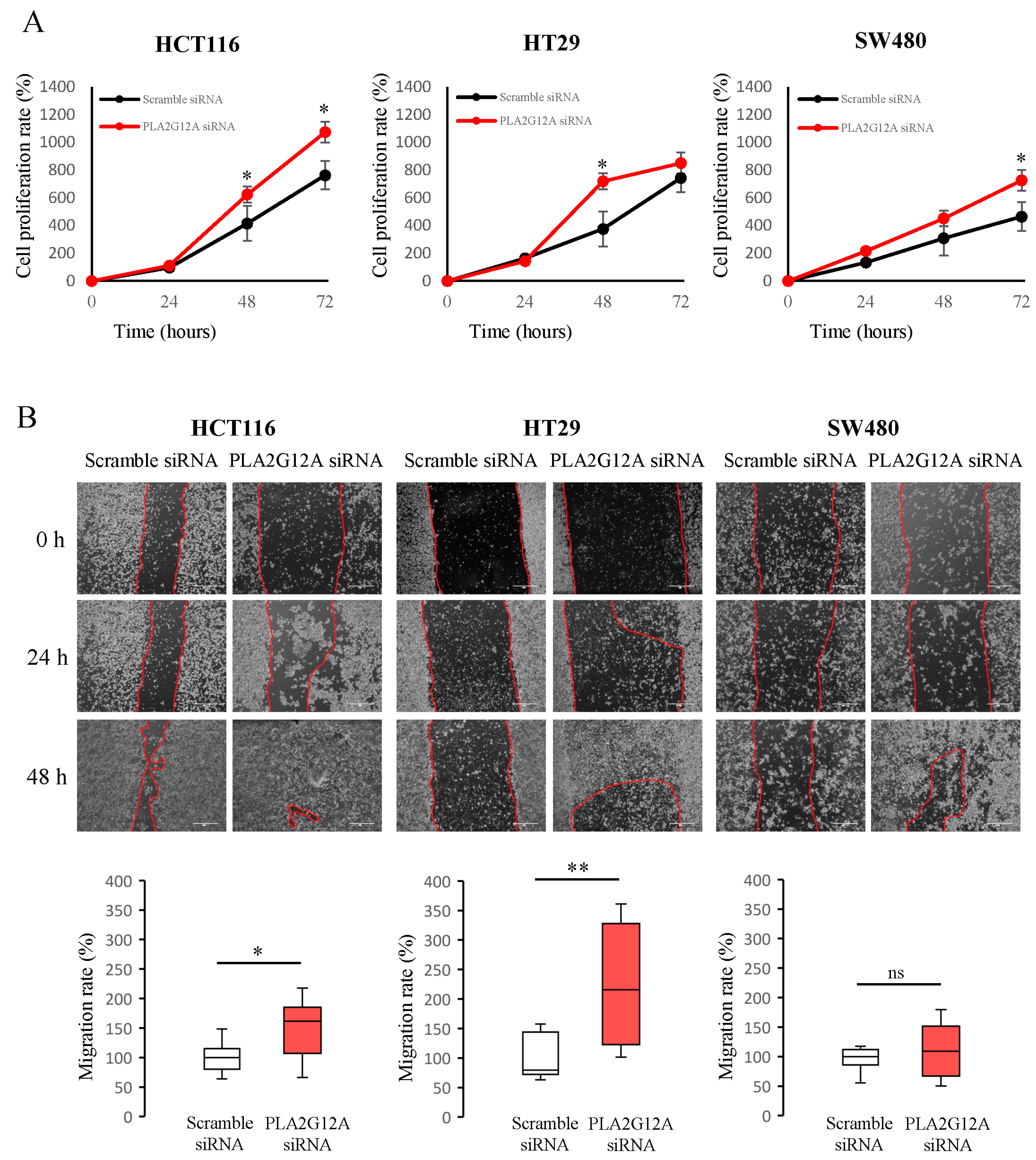
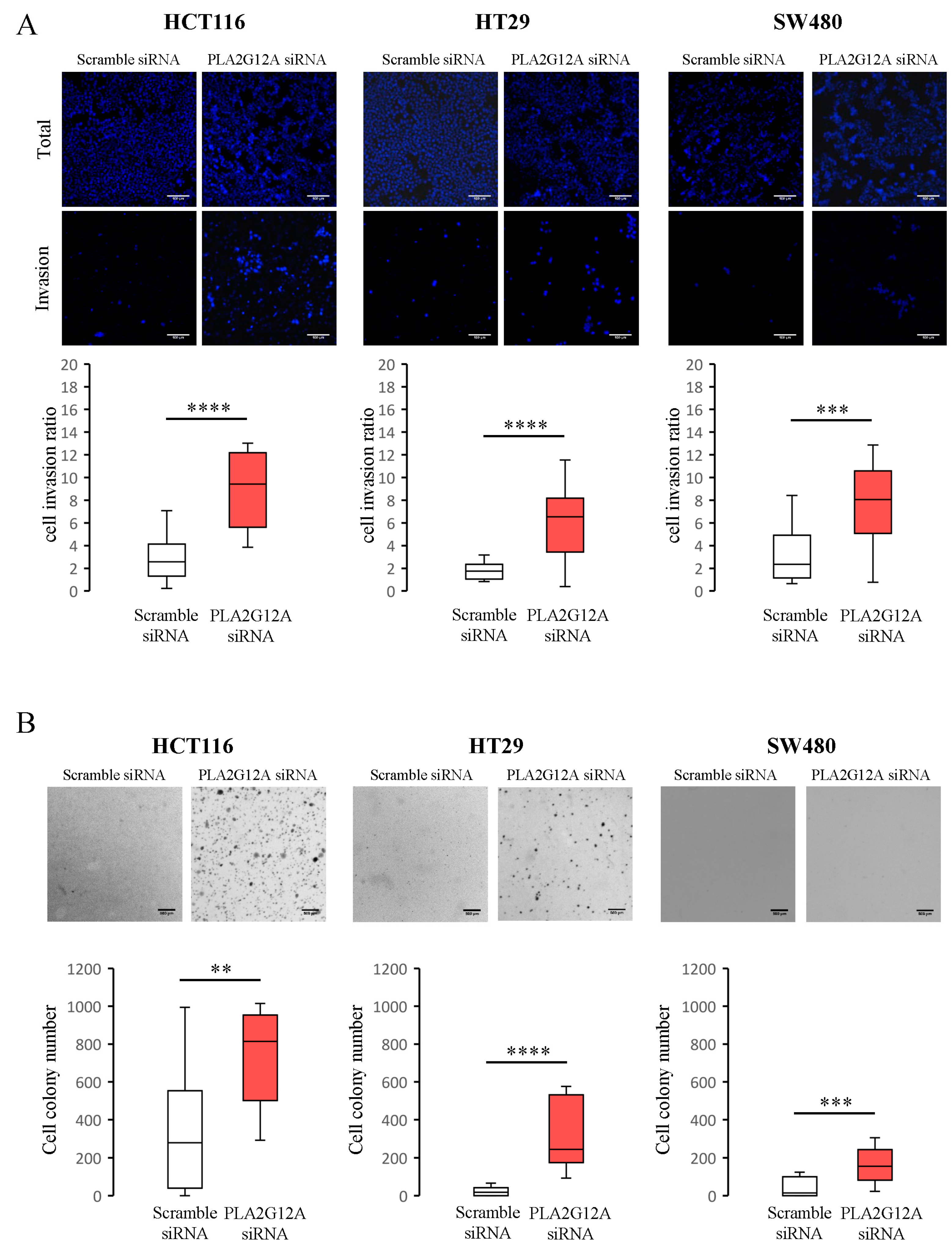
2.3. Functional Characterization of PLA2G12A in CRC Preclinical Models

To further investigate the potential of PLA2G12A as a clinical prognostic marker, we assessed its role in tumor growth and development. In the Drosophila CRC model previously described, the inhibition of GXIVsPLA2 (the fly orthologue of PLA2G12A) not only increased the number of CTCs but also increased the intestinal primary tumor burden (Supplementary Table S1, Figure 1D). As these primary tumor cells also express GFP, we dissected several midguts and measured the total tumor area, as described previously by Adams et al. [30]. Our results showed that the inhibition of GXIVsPLA2 induced the spread of primary tumors along the midgut compared to midguts bearing control Apc-Ras tumors (Figure 1B,C). Together, our results suggest a role for GXIVsPLA2 in inhibiting both intestinal primary tumor growth and the invasion of tumor cells into the hemolymph.
We also performed in vitro experiments with CC cell lines to investigate possible changes in cell behavior induced by PLA2G12A expression. To this end, we used three CC cell lines (HCT116, HT29, and SW480) with distinct proliferation and migration capacities. As shown in Figure 3A, PLA2G12A inhibition induces an increase in proliferation rates compared with the control in all three cell lines. HCT116 and HT29 cells showed proliferation increments 48 h after transfection, whereas for SW480 cells, the effect appeared to be significant 72 h post-transfection. We next analyzed migration rate changes caused by PLA2G12A inhibition using a scratch wound healing assay, where transfected cells were seeded and the migratory ability of the cells at covering the scratch was monitored. Figure 3B shows three different time points in the experiment and representative images of the migration rate measurement. According to our observations, PLA2G12A-downregulated cells showed increased migration rates compared to control cells, as significant changes were seen in both HCT116 and HT29 cell lines.
Lastly, we analyzed whether PLA2G12A expression could alter other cell aspects related to the metastatic process, such as cell invasion and anchorage-independent cell growth abilities. To study these processes, we transfected and seeded cells in a transwell chamber treated with matrigel and incubated them for 48 h. We noticed that PLA2G12A-inhibited cells appeared in a greater number in the bottom layer of the chamber than in the control, indicating an increased invasion capacity when PLA2G12A levels were low in the three CC cell lines used (Figure 4A). By analyzing the growth of transfected cells in an agar layer, we found that PLA2G12A-inhibited cells formed a more significant number of colonies than the control cells (Figure 4B) in all cell lines studied. Together, these results provide evidence that PLA2G12A inhibition promotes the cell proliferation, migration, invasion, and anchorage-independent growth of CC cells.

3. Discussion

In this study, we performed wide molecular screening of genes potentially involved in tumor progression in an in vivo Drosophila CRC model and a subsequent validation of its prognostic power in two independent human cohorts, and discovered a novel putative tumor suppressor gene for CRC, PLA2G12A. This gene, a phospholipase A2 family member, can modify tumor growth and dissemination in Drosophila, suggesting that it could be a potential driving factor for CRC metastatic tumorigenicity.
Despite numerous efforts, it has been difficult to find a reliable recurrence risk biomarker for CRC that could select patients requiring adjuvant therapies. In this regard, some gene expression panels have been used to add value in predicting a prognosis in stage II/III patients [16,17,18]. In fact, several ongoing clinical trials have aimed to validate these gene expression signatures for a recurrence risk assessment in these patients. For instance, the PARSC trial (NCT00903565) focused on assessing the utility of the ColoPrint assay to estimate the 3-year relapse rate in stage II CRC patients.
In this work, we focused our research on searching for candidates that directly modify tumor behavior in an in vivo Drosophila model. The fruit fly Drosophila melanogaster has become an important model system for cancer studies because of its potential for conducting large-scale genetic screens [31]. Moreover, research on the signaling pathways involved in tumor formation, such as Hippo, Notch, Dpp, and JAK-STAT, has been enriched with investigations performed in Drosophila [32,33,34]. Our model is based on the induction of mutant clones in the Wnt pathway (Apc and Apc2) in the adult fly intestine, and the overexpression of the oncogenic form of Ras, RasV12, facilitating the generation of tumor-like overgrowths similar to those found in human CRC tumors. These are characterized by an increased proliferation, blockage of cell differentiation, alterations in cell polarity, and disruption of the organ architecture [26]. No sign of cell dissemination was found in these Apc-Ras flies, whose clones were confined to the gut and surrounded by a thick layer of the basement membrane [26]. For these reasons, the model is an excellent tool to easily detect molecules able to promote tumor cell migration and propagation outside the gut. The molecular screening of 97 different candidates identified 13 genes that increased in vivo tumor cell dissemination. Each of these genes is capable of changing the number of CTCs that can escape from the primary tumor and be detected in the hemolymph, which is analogous to human blood.
To determine whether the candidates obtained in the Drosophila model maintain invasive capabilities in more complex organisms, we analyzed their orthologous genetic expression in two independent cohorts of CRC patients. We found one gene, PLA2G12A, whose low expression was correlated with tumor recurrence and a poor clinical outcome. PLA2G12A is a member of the family of phospholipases A2 (PLA2s), a large superfamily of enzymes that hydrolyzes phospholipids and releases fatty acids and lysophospholipids [35].
Phospholipases can be divided into three major classes—PLA (consisting of A1 and A2), PLC, and PLD, which are differentiated by the type of reaction that they catalyze [36]. Moreover, each class of phospholipase is composed of many isotypes with distinct functions, domains, and regulatory mechanisms [37,38,39]. Within the phospholipase A2 (PLA2) superfamily, secreted PLA2 (sPLA2) enzymes comprise the largest family containing 12 mammalian isoforms with a conserved catalytic site. Every group of sPLA2s exhibit unique tissue and cellular localizations with specific enzymatic properties, suggesting distinct biological roles [40]. In humans, two PLA2G12, PLA2G12A, and a catalytically inactive PLA2G12A-like protein (PLA2G12B) were cloned [41,42]. The functional characterization of PLA2G12A revealed that its catalytic activity is relatively low in comparison with sPLA2s. While the enzymatically inactive PLA2G12B is mainly expressed in the liver, small intestine, and kidney [42], PLA2G12A is strongly expressed in the human heart and skeletal muscle, kidney, pancreas [41], and intestinal tissues [43]. PLA2G12 are suggested to mediate their physiological roles in part via alternative mechanisms independent of their catalytic activity [41,42,44], which at present are poorly characterized.
Many studies have suggested different roles for sPLA2s in relation to inflammation and cancer. Their functional roles are incompletely understood and seem to be dependent on the enzyme studied, the tissue, and the cancer type involved [17]. sPLA2s play a pro-tumorigenic role in breast, lung, prostate, ovarian, and esophageal cancers, and on the contrary, play an anti-tumorigenic role in gastric and intestinal cancers [18]. Alterations in the expression of diverse sPLA2s, such as groups II, III, and X, are well documented in CRC [19]. However, sPLA2s-associated signaling pathways are yet to be elucidated. Schewe et al. provided a potential mechanism by which secreted PLA2G2A, another family member of sPLAs, suppresses colon cancer by inhibiting Wnt signaling through the intracellular activation of Yap1 [45]. On this note, Ganesan et al. also reported an association between PLA2G2A expression and components of the Wnt signaling pathway, including β-catenin and the Wnt target gene EphB2 in gastric cancer [46].
Remarkably, using a gene set enrichment analysis, we found that PLA2G12A expression correlated with EMT signaling pathways, indicating a potential role for PLA2G12A in the initiation of metastasis. In fact, the in vitro data generated in this study indicate that the downregulation of PLA2G12A increases malignant behavior in intestinal cells, altering tumor cell growth, migration, and invasion. EMT induction could explain this malignant behavior found in our cell lines when PLA2G12A is inhibited. In this regard, different works demonstrate an association between phospholipases and transforming growth factor β (TGF-β)-induced EMT in hepatic cells [47], as well as in breast cancer [48]. In other studies on breast cancer, it has also been found that arachidonic acid, the main product of phospholipase activity, promotes migration and invasion through a PI3K/Akt-dependent pathway [49,50]. However, all these associations still need to be demonstrated and validated in colon cancer cells.

4. Materials and Methods

4.1. Fly Strains

Drosophila melanogaster strains were raised at 25 °C on standard cornmeal media. We used the GAL4/Upstream Activating Sequence (UAS) system [51] for the tissue-specific expression of transgenes. yw hsp70-flp; esg Gal4 UAS-GFP UAS-RasV12/CyO; and UAS-Luciferase FRT82B Gal80/TM6b flies were crossed with yw hsp70-flp; Sp/CyO; and FRT82B Apc2N175KApcQ8/TM6b flies to generate non-metastatic Apc-Ras control clones as described in [26] or to different yw hsp70-flp; UAS-RNAi line/CyO; and FRT82B Apc2N175KApcQ8/TM6b for each gene tested. RNAi lines were obtained from the Bloomington Drosophila Stock Center (Indiana, IN, USA) and The Vienna Drosophila Resource Center (Vienna, Austria). Apc2N175K is a loss-of-function allele, ApcQ8 is a null allele, UAS-RasV12 is a gain of function transgene, and each gene analyzed had its specific UAS-RNAi line. A mosaic analysis with repressible cell marker (MARCM) [52] clones were generated with a 1 h heat shock at 37 °C of 2–7-day-old females and were marked by the progenitor cell marker escargot (esg) Gal4 line, driving the expression of UAS green fluorescent protein (GFP).

4.2. Luciferase Assay

Luciferase assays were performed using the dual-luciferase(R) reporter assay system [27] 2 weeks after MARCM generation. The UAS-luciferase transgene introduced into fly intestine mutant clones confered a linear correlation between the number of mutant cells and the amount of luciferase activity detected. For a hemolymph analysis, hemolymph was extracted from whole flies according to the instructional video published by Laura Musselman (“Drosophila hemolymph collection procedure”. Youtube, uploaded by Laura Musselman, www.YOUTUBE.com/watch?v=im78OIBKlPA 4 November 2013). For whole-fly lysates, flies were squashed using a pipette tip into a luciferase buffer (E1500, Promega, Madison, WI, USA). The samples were loaded into 96-well plates and read on a Tecan Infinite® 200 plate reader (Tecan, Männedorf, Switzerland).

4.3. Human Samples

The internal CRC Hospital Universitari Arnau de Vilanova (HUAV) human cohort was composed of 741 patients treated with a surgical resection at the HUAV from January 2010 to December 2020 with pathological stages I–II–III and no residual disease. Patients who received neoadjuvant treatment or had synchronous tumors were excluded from the study. Tumor tissue were collected during surgery and stored with support from the Xarxa de Bancs de Tumors de Catalunya sponsored by the Pla Director d’Oncología de Catalunya (XBTC), IRBLleida Biobank (B.0000682), and Plataforma Biobancos PT20/00021.
Clinical and pathological information of the patients was collected, including age, sex, anatomical location, pathological stage, histology, differentiation degree, venous/lymphatic/perineural invasion, microsatellite status, RAS mutations, adjuvant chemotherapy, RFS, and OS. The OS and TTR durations were defined as the intervals from initial surgery to death and from initial surgery to clinically proven recurrence or metastasis, respectively.
To reduce the size of the cohort and maintain the whole spectrum of clinical outcomes, paired matching was conducted between patients with recurrence (n = 90) and without recurrence after more than 5 years of follow up (n = 239), accounting for a balanced pathological stage, differentiation degree, and anatomical location. The final cohort was composed of 180 patients (Supplementary Table S5).

4.4. Real-Time PCR (RT-PCR)

Total RNA was successfully extracted from 153 patients from the internal CRC human HUAV cohort using the Maxwell® RSC simplyRNA Tissue Kit (Promega), and cDNA was synthesized from mRNA using the Reverse Transcriptase assay iScript™ cDNA Synthesis Kit (BioRad, Hercules, CA, USA). RT-PCR was performed using predesigned hydrolysis fluorescent probes for the genes of interest (Integrated DNA Technologies, Coralville, IA, USA) with 40 cycles at 95 °C for 15 s and 60 °C for 1 min using the QuantStudio 7 (ThermoFisher Scientific, Waltham, MA, USA). Relative mRNA expression levels were calculated using the 2−ΔΔCt method, with β-actin as a housekeeping gene control.

4.5. TCGA Data Analysis

RNA sequencing data and clinico-pathological information of 592 patients were obtained from Colorectal Adenocarcinoma of The Cancer Genome Atlas (TCGA) and PanCancer Atlas [53] databases. Gene expression data were explored through the Broad Institute Memorial Sloan Kettering Cancer Center cBioPortal and downloaded for an analysis [54,55]. For a gene set enrichment analysis, we used the GenePattern platform from the Gene Set Enrichment Analysis (GSEA) software (UC San Diego and Broad Institute) [56].

4.6. Cell Culture Assays

Human CC cell lines used in our experiments, HCT116, HT29, and SW480, were obtained from the American Type Culture Collection (Manassas, VA, USA). Cell lines were cultured in a DMEM (Dulbecco’s modified Eagle’s medium) supplemented with 10% fetal bovine serum, 100 μg/mL of penicillin, and 100 U/mL of streptomycin (all from ThermoFisher Scientific) at 37 °C in a 5% CO2 incubator.

4.7. Proliferative Assays

Cells were transfected with interference RNA (iRNA) (Integrated DNA Technologies) with Lipofectamine RNAiMAX (ThermoFisher Scientific) against the specific gene or with scramble iRNA for the controls. Gene-specific inhibition was assessed with a RT-PCR and Western blot (Supplementary Figure S1). The cell growth ability was monitored by seeding 6000–10,000 cells/well of transfected cells in 24-well dishes and counting the cell number every 24 h for 3 days in a Neubauer chamber.

4.8. Cell Migration Assays

Migration rates were measured with a scratch wound healing assay where 500,000 transfected cells/well were seeded onto 6-well plates. After 6 h, a wound was scratched using a sterile pipette tip and the cells were washed with PBS (phosphate-buffered saline) until there were no floating cells. Photographs were taken using an optical microscope from 0 to 72 h. Migration rates (mm2/hour) were calculated by measuring the decrease in the wound area.

4.9. Invasion Assays

In total, 50,000 transfected cells were seeded into the upper chamber of a matrigel-treated transwell (ThermoFisher Scientific), while the bottom well contained a medium supplemented with glucose as an induction stimulus. After 48 h of incubation, cells in the upper chamber that had not migrated through the filter were wiped off with a cotton swab, whereas those that had migrated to the bottom surface were fixed with 4% paraformaldehyde (Sigma-Aldrich, St. Louis, MO, USA) for 15 min, stained with Hoechst (1:400) (ThermoFisher Scientific), and counted under a fluorescence microscope.

4.10. Anchorage-Independent Cell Growth Assays

Six-well plates were coated with a 0.50% agar (ThermoFisher Scientific) layer and after solidifying, 6000 cells/well of transfected cells were seeded in a second superimposed layer at 0.35% agar. After incubation for 15 days, when colonies were visible, clones were stained with crystal violet (0.005%) (ThermoFisher Scientific) and the number of colonies was counted under an optical microscope.

4.11. Statistical Analyses

Results are presented as the mean ± standard error of the mean (SEM) and statistical differences were determined with a Student’s t-test, Fisher’s test, or Mann–Whitney U test as appropriate and corrected with the Benjamini–Hochberg procedure if needed. All statistical tests were two-tailed and p values < 0.05 were considered statistically significant. For survival analyses, patients were included in low or high groups if gene levels were respectively below or above the cutoff, defined as the median for each independent marker. A Kaplan–Meier survival analysis was used for time-to-event variables, and the log-rank test was used to determine significance. A Cox proportional hazards model was used to calculate the univariate hazard ratios (HR) ±95% confidence intervals (CI) for OS, TTR, and RFS. Statistical analyses were performed using the Statistical Package for the Social Sciences for Windows (SPSS v.25, Inc., Chicago, IL, USA).

5. Conclusions

We propose PLA2G12A as a prognostic biomarker in early-stage CRC, providing evidence that its deficiency promotes tumor growth and dissemination, unveiling novel functions for this poorly characterized phospholipase.

Supplementary Materials

The following supporting information can be downloaded at: https://www.mdpi.com/article/10.3390/ijms241310889/s1.

Author Contributions

Conceptualization, E.P., A.C., M.A.S. and A.S. (Antonieta Salud) methodology, E.P., I.H., J.T., A.N. and M.A.S.; software, R.M.; validation, E.P., R.M. and M.A.S.; formal analysis, E.P. and R.M.; investigation, E.P. and I.H.; resources, M.A.S. and A.S. (Antonieta Salud); data curation, E.P., R.M. and O.P.; writing—original draft preparation, E.P.; writing—review and editing, E.P., R.M., A.S. (Anabel Sorolla), K.C., A.C., M.A.S. and A.S. (Antonieta Salud); supervision, A.C., M.A.S. and A.S. (Antonieta Salud); project administration, M.A.S.; funding acquisition, A.S. (Antonieta Salud). All authors have read and agreed to the published version of the manuscript.

Funding

This research was funded by the Merck Salud Foundation Project Award (2019), the University and Research Grants Management Agency (AGAUR) of the Catalonia Government grants 2017SRG1620 and 2021FIB00025, Instituto de Salud Carlos III grants PI21/01619 and JR21/00039, Spanish Ministry of Science and Innovation grant PID2019-1946GB-100, The National Centre for the Replacement, Refinement and Reduction of Animals in Research grant NC/W001136/1 and Wellcome Trust grant 204615/Z/16/Z. This research was also supported by the CERCA Program of the Generalitat de Catalunya and the IRBLleida (Fundació Pifarré).

Institutional Review Board Statement

This study was approved by the Committee on Biomedical Research Ethics (CEIC-2047) of our hospital and was performed in accordance with the Declaration of Helsinki.

Informed Consent Statement

All the participants provided written informed consent.

Data Availability Statement

The data generated in this study are available within the article and its supplementary data files. Other expression profile data analyzed in this study were obtained from the Broad Institute Memorial Sloan Kettering Cancer Center cBioPortal and Colorectal Adenocarcinoma TCGA PanCancer Atlas (https://www.cancer.gov/tcga, id=coadread_tcga_pan_can_atlas_2018, accessed on 17 March 2022).

Acknowledgments

The authors would like to thank Alba Vicario, Rita González-Resina, and Judith Casals for their help with the Drosophila management. This work was supported by the Xarxa de Bancs de Tumors de Catalunya sponsored by the Pla Director d’Oncología de Catalunya (XBTC), IRBLleida Biobank (B.0000682), and PLATAFORMA BIOBANCOS PT20/00021.

Conflicts of Interest

The authors declare no conflict of interest.

References

  1. Ferlay, J.; Colombet, M.; Soerjomataram, I.; Parkin, D.M.; Piñeros, M.; Znaor, A.; Bray, F. Cancer statistics for the year 2020: An overview. Int. J. Cancer 2021, 149, 778–789. [Google Scholar] [CrossRef] [PubMed]
  2. Cheng, L.; Eng, C.; Nieman, L.Z. Trends in Colorectal Cancer Incidence by Anatomic Site and Disease Stage in the United States. Am. J. Clin. Oncol. 2011, 34, 573–580. [Google Scholar] [CrossRef] [PubMed]
  3. Siegel, R.; Miller, K.; Sandeep Wagle, N.; Jemal DVM, A. Cancer statistics, 2023. CA Cancer J. Clin. 2023, 73, 17–48. [Google Scholar] [CrossRef] [PubMed]
  4. Bailey, C.; Hu, C.; You, N.; Bednarski, B.; Rodriguez-Bigas, M.; Skibber, J.; Cantor, S.; Chang, G. Increasing Disparities in Age-Related Incidence of Colon and Rectal Cancer in the United States, 1975-2010. JAMA Surg 2015, 150, 17–22. [Google Scholar] [CrossRef] [Green Version]
  5. Brenner, H.; Kloor, M.; Pox, C.P. Colorectal cancer. Lancet 2014, 383, 1490–1502. [Google Scholar] [CrossRef]
  6. Cunningham, D.; Atkin, W.; Lenz, H.J.; Lynch, H.T.; Minsky, B.; Nordlinger, B.; Starling, N. Colorectal cancer. Lancet 2010, 375, 1030–1047. [Google Scholar] [CrossRef]
  7. Benson, A.B.; Schrag, D.; Somerfield, M.R.; Cohen, A.M.; Figueredo, A.T.; Flynn, P.J.; Krzyzanowska, M.K.; Maroun, J.; McAllister, P.; Van Cutsem, E.; et al. American Society of Clinical Oncology recommendations on adjuvant chemotherapy for stage II colon cancer. J. Clin. Oncol. 2004, 22, 3408–3419. [Google Scholar] [CrossRef]
  8. Argilés, G.; Tabernero, J.; Labianca, R.; Hochhauser, D.; Salazar, R.; Iveson, T.; Laurent-Puig, P.; Quirke, P.; Yoshino, T.; Taieb, J.; et al. Localised colon cancer: ESMO Clinical Practice Guidelines for diagnosis, treatment and follow-up†. Ann. Oncol. 2020, 31, 1291–1305. [Google Scholar] [CrossRef]
  9. André, T.; De Gramont, A.; Vernerey, D.; Chibaudel, B.; Bonnetain, F.; Tijeras-raballand, A.; Scriva, A.; Hickish, T.; Tabernero, J.; Van Laethem, J.L.; et al. Adjuvant Fluorouracil, Leucovorin, and Oxaliplatin in Stage II to III Colon Cancer: Updated 10-Year Survival and Outcomes According to BRAF Mutation and Mismatch Repair Status of the MOSAIC Study. J. Clin. Oncol. 2023, 33, 4176–4187. [Google Scholar] [CrossRef]
  10. Baxter, N.N.; Kennedy, E.B.; Bergsland, E.; Berlin, J.; George, T.J. Adjuvant Therapy for Stage II Colon Cancer: ASCO Guideline Update. J. Clin. Oncol. 2023, 40, 892–910. [Google Scholar] [CrossRef]
  11. Grothey, A.; Sobrero, A.F.; Shields, A.F.; Yoshino, T.; Paul, J.; Taieb, J.; Souglakos, J.; Shi, Q.; Labianca, R.; Meyerhardt, J.A.; et al. Duration of Adjuvant Chemotherapy for Stage III Colon Cancer. N. Engl. J. Med. 2019, 378, 1177–1188. [Google Scholar] [CrossRef]
  12. Wang, Y.S.; Kou, Y.; Zhu, R.T.; Han, B.W.; Li, C.H.; Wang, H.J.; Wu, H.B.; Xia, T.M.; Che, X.M. CDX2 as a Predictive Biomarker Involved in Immunotherapy Response Suppresses Metastasis through EMT in Colorectal Cancer. Dis. Markers 2022, 2022. [Google Scholar] [CrossRef]
  13. Dalerba, P.; Sahoo, D.; Soonmyung, P.; Guo, X.; Yothers, G.; Song, N.; Wilcox-Fogel, N.; Forgó, E.; Rajendran, P.S.; Miranda, S.P.; et al. CDX2 as a Prognostic Biomarker in Stage II and Stage III Colon Cancer. N. Engl. J. Med. 2016, 374, 211–222. [Google Scholar] [CrossRef] [PubMed]
  14. Tie, J.; Wang, Y.; Tomasetti, C.; Li, L.; Springer, S.; Silliman, N.; Tacey, M.; Wong, H.; Christie, M.; Skinner, I.; et al. Circulating Tumor DNA Analysis. Sci. Transl. Med. 2017, 8. [Google Scholar]
  15. Cavalieri, D.; Dolara, P.; Mini, E.; Luceri, C.; Castagnini, C.; Toti, S.; Maciag, K.; De Filippo, C.; Nobili, S.; Morganti, M.; et al. Analysis of gene expression profiles reveals novel correlations with the clinical course of colorectal cancer. Oncol. Res. 2007, 16, 535–548. [Google Scholar] [CrossRef] [PubMed]
  16. O’Connell, M.J.; Lavery, I.; Yothers, G.; Paik, S.; Clark-Langone, K.M.; Lopatin, M.; Watson, D.; Baehner, F.L.; Shak, S.; Baker, J.; et al. Relationship between tumor gene expression and recurrence in four independent studies of patients with stage II/III colon cancer treated with surgery alone or surgery plus adjuvant fluorouracil plus leucovorin. J. Clin. Oncol. 2010, 28, 3937–3944. [Google Scholar] [CrossRef] [PubMed] [Green Version]
  17. Salazar, R.; Roepman, P.; Capella, G.; Moreno, V.; Simon, I.; Dreezen, C.; Lopez-Doriga, A.; Santos, C.; Marijnen, C.; Westerga, J.; et al. Gene expression signature to improve prognosis prediction of stage II and III colorectal cancer. J. Clin. Oncol. 2011, 29, 17–24. [Google Scholar] [CrossRef] [Green Version]
  18. Kennedy, R.D.; Bylesjo, M.; Kerr, P.; Davison, T.; Black, J.M.; Kay, E.W.; Holt, R.J.; Proutski, V.; Ahdesmaki, M.; Farztdinov, V.; et al. Development and independent validation of a prognostic assay for stage ii colon cancer using formalin-fixed paraffin-embedded tissue. J. Clin. Oncol. 2011, 29, 4620–4626. [Google Scholar] [CrossRef] [Green Version]
  19. Cao, H.; Xu, E.; Liu, H.; Wan, L.; Lai, M. Epithelial-mesenchymal transition in colorectal cancer metastasis: A system review. Pathol. Res. Pract. 2015, 211, 557–569. [Google Scholar] [CrossRef]
  20. Khanna, C.; Hunter, K. Modeling metastasis in vivo. Carcinogenesis 2005, 26, 513–523. [Google Scholar] [CrossRef] [Green Version]
  21. Astell, K.R.; Sieger, D. Zebrafish In Vivo Models of Cancer and Metastasis. Cold Spring Harb Perspect Med 2020, 10, 1–17. [Google Scholar] [CrossRef] [PubMed]
  22. Kirienko, N.; Mani, K.; Fay, D. Cancer models in C. elegans. Dev. Dyn. 2010, 239, 1413–1448. [Google Scholar] [PubMed] [Green Version]
  23. Chakraborty, U.; Dinh, T.; Alani, E. Genomic Instability Promoted by Overexpression of Mismatch Repair Factors in Yeast: A Model for Understanding Cancer Progression. Genetics 2018, 209, 439–456. [Google Scholar] [CrossRef] [PubMed] [Green Version]
  24. Brumby, A.M.; Richardson, H.E. Using Drosophila melanogaster to map human cancer pathways. Nat. Rev. Cancer 2005, 5, 626–639. [Google Scholar] [CrossRef]
  25. Gonzalez, C. Drosophila melanogaster: A model and a tool to investigate malignancy and identify new therapeutics. Nat. Rev. Cancer 2013, 13, 172–183. [Google Scholar] [CrossRef]
  26. Martorell, Ò.; Merlos-Suárez, A.; Campbell, K.; Barriga, F.M.; Christov, C.P.; Miguel-Aliaga, I.; Batlle, E.; Casanova, J.; Casali, A. Conserved mechanisms of tumorigenesis in the Drosophila adult midgut. PLoS ONE 2014, 9, e88413. [Google Scholar] [CrossRef] [PubMed] [Green Version]
  27. Campbell, K.; Rossi, F.; Adams, J.; Pitsidianaki, I.; Barriga, F.M.; Garcia-Gerique, L.; Batlle, E.; Casanova, J.; Casali, A. Collective cell migration and metastases induced by an epithelial-to-mesenchymal transition in Drosophila intestinal tumors. Nat. Commun. 2019, 10, 2311. [Google Scholar] [CrossRef] [Green Version]
  28. Peng, Z.; Chang, Y.; Fan, J.; Ji, W.; Su, C. Phospholipase A2 superfamily in cancer. Cancer Lett. 2021, 497, 165–177. [Google Scholar] [CrossRef]
  29. Brglez, V.; Lambeau, G.; Petan, T. Secreted phospholipases A2 in cancer: Diverse mechanisms of action. Biochimie 2014, 107, 114–123. [Google Scholar] [CrossRef]
  30. Adams, J.; Casali, A.; Campbell, K. Sensitive high-throughput assays for tumour burden reveal the response of a drosophila melanogaster model of colorectal cancer to standard chemotherapies. Int. J. Mol. Sci. 2021, 22, 5101. [Google Scholar] [CrossRef]
  31. Miles, W.O.; Dyson, N.J.; Walker, J.A. Modeling tumor invasion and metastasis in Drosophila. DMM Dis. Model. Mech. 2011, 4, 753–761. [Google Scholar] [CrossRef] [PubMed] [Green Version]
  32. Brumby, A.M.; Richardson, H.E. scribble mutants cooperate with oncogenic Ras or Notch to cause neoplastic overgrowth in Drosophila. EMBO J. 2003, 22, 5769–5779. [Google Scholar] [CrossRef] [PubMed] [Green Version]
  33. Vidal, M.; Cagan, R.L. Drosophila models for cancer research. Curr. Opin. Genet. Dev. 2006, 16, 10–16. [Google Scholar] [CrossRef] [PubMed]
  34. Januschke, J.; Gonzalez, C. Drosophila asymmetric division, polarity and cancer. Oncogene 2008, 27, 6994–7002. [Google Scholar] [CrossRef] [PubMed] [Green Version]
  35. Nevalainen, T.J.; Cardoso, J.C.R.; Riikonen, P.T. Conserved domains and evolution of secreted phospholipases A 2. FEBS J. 2012, 279, 636–649. [Google Scholar] [CrossRef] [Green Version]
  36. Park, J.B.; Lee, C.S.; Jang, J.H.; Ghim, J.; Kim, Y.J.; You, S.; Hwang, D.; Suh, P.G.; Ryu, S.H. Phospholipase signalling networks in cancer. Nat. Rev. Cancer 2012, 12, 782–792. [Google Scholar] [CrossRef]
  37. Murakami, M.; Taketomi, Y.; Miki, Y.; Sato, H.; Hirabayashi, T.; Yamamoto, K. Recent progress in phospholipase A2 research: From cells to animals to humans. Prog. Lipid Res. 2011, 50, 152–192. [Google Scholar] [CrossRef]
  38. Suh, P.G.; Park, J.I.; Manzoli, L.; Cocco, L.; Peak, J.C.; Katan, M.; Fukami, K.; Kataoka, T.; Yun, S.; Sung, H.R. Multiple roles of phosphoinositide-specific phospholipase C isozymes. J. Biochem. Mol. Biol. 2008, 41, 415–434. [Google Scholar] [CrossRef] [Green Version]
  39. Aoki, J.; Inoue, A.; Makide, K.; Saiki, N.; Arai, H. Structure and function of extracellular phospholipase A1 belonging to the pancreatic lipase gene family. Biochimie 2007, 89, 197–204. [Google Scholar] [CrossRef]
  40. Murakami, M.; Taketomi, Y.; Miki, Y.; Sato, H.; Yamamoto, K.; Lambeau, G. Emerging roles of secreted phospholipase A2 enzymes: The 3rd edition. Biochimie 2014, 107, 105–113. [Google Scholar] [CrossRef]
  41. Gelb, M.H.; Valentin, E.; Ghomashchi, F.; Lazdunski, M.; Pharmacologie, I. De Cloning and Recombinant Expression of a Structurally Novel Human Secreted Phospholipase A2. J Biol Chem 2000, 275, 39823–39826. [Google Scholar] [CrossRef] [PubMed] [Green Version]
  42. Rouault, M.; Bollinger, J.G.; Lazdunski, M.; Gelb, M.H.; Lambeau, G. Novel mammalian group XII secreted phospholipase A2 lacking enzymatic activity. Biochemistry 2003, 42, 11494–11503. [Google Scholar] [CrossRef] [PubMed]
  43. Peuravuori, H.; Kollanus, S.; Nevalainen, T.J. Expression of group XIIA phospholipase A2 in human digestive organs. Apmis 2014, 122, 1171–1177. [Google Scholar] [CrossRef] [PubMed]
  44. Ho, I.; Arm, J.P.; Iii, C.O.B.; Choi, A.; Austen, K.F.; Glimcher, L.H. A Novel Group of Phospholipase A 2 s Preferentially Expressed in Type 2 Helper T Cells *. J. Biol. Chem. 2001, 276, 18321–18326. [Google Scholar] [CrossRef] [PubMed] [Green Version]
  45. Schewe, M.; Franken, P.F.; Sacchetti, A.; Schmitt, M.; Joosten, R.; Böttcher, R.; van Royen, M.E.; Jeammet, L.; Payré, C.; Scott, P.M.; et al. Secreted Phospholipases A2 Are Intestinal Stem Cell Niche Factors with Distinct Roles in Homeostasis, Inflammation, and Cancer. Cell Stem Cell 2016, 19, 38–51. [Google Scholar] [CrossRef] [PubMed]
  46. Ganesan, K.; Ivanova, T.; Wu, Y.; Rajasegaran, V.; Wu, J.; Ming, H.L.; Yu, K.; Sun, Y.R.; Hyun, C.C.; Ylstra, B.; et al. Inhibition of gastric cancer invasion and metastasis by PLA2G2A, a novel β-catenin/TCF target gene. Cancer Res. 2008, 68, 4277–4286. [Google Scholar] [CrossRef] [Green Version]
  47. Zhao, L.; Gandhi, C.R.; Gao, Z.H. Involvement of cytosolic phospholipase A 2 alpha signalling pathway in spontaneous and transforming growth factor-beta-induced activation of rat hepatic stellate cells. Liver Int. 2011, 31, 1565–1573. [Google Scholar] [CrossRef]
  48. Jiang, J.; Wang, K.; Chen, Y.; Chen, H.; Nice, E.C.; Huang, C. Redox regulation in tumor cell epithelial–mesenchymal transition: Molecular basis and therapeutic strategy. Signal Transduct. Target. Ther. 2017, 2, 1–12. [Google Scholar] [CrossRef] [Green Version]
  49. Martinez-Orozco, R.; Navarro-Tito, N.; Soto-Guzman, A.; Castro-Sanchez, L.; Perez Salazar, E. Arachidonic acid promotes epithelial-to-mesenchymal-like transition in mammary epithelial cells MCF10A. Eur. J. Cell Biol. 2010, 89, 476–488. [Google Scholar] [CrossRef]
  50. Villegas-Comonfort, S.; Castillo-Sanchez, R.; Serna-Marquez, N.; Cortes-Reynosa, P.; Salazar, E.P. Arachidonic acid promotes migration and invasion through a PI3K/Akt-dependent pathway in MDA-MB-231 breast cancer cells. Prostaglandins Leukot. Essent. Fat. Acids 2014, 90, 169–177. [Google Scholar] [CrossRef]
  51. Brand, A.H.; Perrimon, N. Targeted gene expression as a means of altering cell fates and generating dominant phenotypes. Development 1993, 118, 401–415. [Google Scholar] [CrossRef] [PubMed]
  52. Lee, T.; Luo, L. Mosaic analysis with a repressible neurotechnique cell marker for studies of gene function in neuronal morphogenesis. Neuron 1999, 22, 451–461. [Google Scholar] [CrossRef] [PubMed] [Green Version]
  53. Weinstein, J.N.; Collisson, E.A.; Mills, G.B.; Shaw, K.R.M.; Ozenberger, B.A.; Ellrott, K.; Sander, C.; Stuart, J.M.; Chang, K.; Creighton, C.J.; et al. The cancer genome atlas pan-cancer analysis project. Nat. Genet. 2013, 45, 1113–1120. [Google Scholar] [CrossRef] [PubMed]
  54. Cerami, E.; Gao, J.; Dogrusoz, U.; Gross, B.E.; Sumer, S.O.; Aksoy, B.A.; Jacobsen, A.; Byrne, C.J.; Heuer, M.L.; Larsson, E.; et al. The CBio Cancer Genomics. Cancer Discov. 2017, 32, 736–740. [Google Scholar] [CrossRef] [Green Version]
  55. Gao, J.; Aksoy, B.A.; Dogrusoz, U.; Dresdner, G.; Gross, B.; Sumer, S.O.; Sun, Y.; Jacobsen, A.; Sinha, R.; Larsson, E.; et al. Integrative Analysis of Complex Cancer Genomics and Clinical Profiles Using the cBioPortal Complementary Data Sources and Analysis Options. Sci. Signal. 2014, 6, 1–20. [Google Scholar] [CrossRef] [Green Version]
  56. Subramanian, A.; Tamayo, P.; Mootha, V.K.; Mukherjeed, S.; Ebert, B.L.; Gillettea, M.A.; Paulovichg, A.; Pomeroyh, S.L.; Golub, T.R.; Landera, E.S.; et al. Gene set enrichment analysis: A knowledge-based approach for interpreting genome-wide expression profiles. Proc. Natl. Acad. Sci. USA 2005, 102, 15545–15550. [Google Scholar] [CrossRef] [Green Version]
Figure 1. In vivo Drosophila melanogaster CRC model results. (A) The in vivo genetic screen detected 13 new genes able to modify circulating tumor cell (CTC) number. Data points represent averages of at least 3 replicates of batches of 10 individuals and are represented as the log2 of the fold change (FC) over the control and the –log10 of the p value. p values were obtained by applying the Mann–Whitney U test and corrected with the Benjamini-Hochberg procedure. Significant genes with p < 0.05 are shown in red. (B) Representative images of control Apc-Ras and GXIVsPLA2 (orthologous of the human PLA2G12A) RNAi Drosophila intestines (n = 10) 21 days after mutant induction where tumoral cells expressing GFP are visible. Scale bar, 500 µm. (C) Quantification of tumor coverage in Drosophila intestines represented as the ratio between tumoral GFP cell area and whole intestine area, measured in µm2. Box plots show median ± IQR (interquartile range). (D) Quantification of CTC number or total tumor burden (TB) in the Drosophila CRC model measured in the luciferase assays. Data points represent averages ± SEM shown as percentage with respect to the control Apc-Ras of at least 3 replicates of batches of 10 individuals. * p < 0.05; ** p < 0.01 by applying the Mann–Whitney U test.
Ijms 24 10889 g001
Figure 2. PLA2G12A expression in human CRC cohorts is a marker of prognosis. (A) Kaplan–Meier survival analysis of the HUAV internal cohort between PLA2G12A expression and time to recurrence (TTR), (B) PLA2G12A expression and overall survival (OS), and (C) PLA2G12A expression and TTR in stage II patients. (D) Kaplan–Meier survival analysis of the TCGA cohort between PLA2G12A expression and recurrence-free survival (RFS). p values were obtained by applying the log-rank test. (E) Gene expression signatures identified with gene set enrichment analysis (GSEA) on the TCGA RNA data cohort. (F) Epithelial-to-mesenchymal transition gene expression was activated when PLA2G12A levels were low.
Ijms 24 10889 g002
Figure 3. PLA2G12A inhibition increases proliferation and migration in CC cells. (A) Cell proliferation rates of three different CC cell lines, HCT116, HT29, and SW480 when PLA2G12A was downregulated via siRNA. Quantitative values are shown as mean ± SEM of three biological replicates, each of them containing three technical replicates. (B) Representative images and quantification of cell migration rates (mm2/h) of the scratch wound healing assay in HCT116, HT29, and SW480 cells, respectively. Scale bar, 100 µm. The box plots show median ± IQR of three biological replicates, each of them containing three technical replicates, represented as percentage with respect to the control (scramble siRNA). * p < 0.05; ** p < 0.01; ns, not significant with Mann–Whitney U test.
Ijms 24 10889 g003
Figure 4. PLA2G12A inhibition increases cell invasion and anchorage-independent cell growth. (A) Representative images and quantification of cell invasion rates in 3 CC cell lines: HCT116, HT29, and SW480 after PLA2G12A downregulation via siRNA. Graphs represent the ratio between the number of cells able to invade the transwell matrigel layer vs. the total cell amount. Scale bar, 100 µm. (B) Representative images and quantification of new colonies formed on soft agar plates after PLA2G12A downregulation via siRNA in HCT116, HT29, and SW480 cells, respectively. Scale bar, 500 µm. The box plots show median ± IQR of three biological replicates, each of them containing three technical replicates. ** p < 0.01; *** p < 0.001; **** p < 0.0001 by applying the Mann–Whitney U test.
Ijms 24 10889 g004
Table 1. Hazard ratios (HR) for time to recurrence (TTR) and overall survival (OS).
Drosophila OrthologueHuman GeneTTROS
HR95% CIpHR95% CIp
ContCNTN40.6770.432–1.0600.0880.6020.342–1.0600.079
DlDLL11.3160.840–2.0600.2301.5970.910–2.8050.103
Src42FRK0.8470.542–1.3220.4640.9610.551–1.6750.887
Ipk2IPMK1.0780.690–1.6850.7401.0440.599–1.8200.879
Dp110PIK3CD1.0880.697–1.6980.7110.8550.490–1.4930.582
GXIVsPLA2PLA2G12A0.5900.375–0.9280.0220.5460.308–0.9670.038
Gap1RASA30.9840.631–1.5340.9431.2870.738–2.2430.375
CG9098SH2D3C0.8330.533–1.3000.4211.1220.644–1.9570.684
Mal-A1SLC3A11.0370.665–1.6170.8720.9290.532–1.6200.794
BaiTMED101.2250.783–1.9150.3751.0640.609–1.8590.828
RNA expression, high vs. low. HR < 1 indicating low expression, poor prognosis. HR > 1 indicating high expression, poor prognosis. Significant values with p < 0.05 are shown in bold.
Table 2. Baseline characteristics of the internal CRC HUAV cohort based on PLA2G12A expression.
CharacteristicHighLowp Value
SexMale (%)45 (58.4)51 (67.1)0.317
Female (%)32 (41.6)25 (32.9)
AgeMedian (years)74.875.40.755
Anatomic locationCecum (%)7 (9.1)3 (3.9)0.507
Ascending colon (%)17 (22.1)24 (31.6)
Hepatic flexure (%)1 (1.3)2 (2.6)
Transverse colon (%)2 (2.6)2 (2.6)
Splenic flexure (%)1 (1.3)3 (3.9)
Descending colon (%)6 (7.8)3 (3.9)
Sigmoid colon (%)25 (31.5)28 (36.8)
Rectosigmoid junction (%)1 (1.3)5 (6.6)
Rectum (%)17 (22.1)6 (7.9)
StageI (%)4 (5.2)2 (2.6)0.063
II (%)29 (37.7)19 (25)
III (%)44 (57.1)55 (72.4)
HistologyAdenocarcinoma (%)63 (81.8)65 (85.5)0.663
Mucinous adenocarcinoma (%)13 (16.9)11 (14.5)
Signet ring cell carcinoma (%)1 (1.3)0 (0)
Differentiation degreeLow-grade (%)65 (84.4)68 (89.5)0.473
High-grade (%)12 (15.6)8 (10.5)
Venous invasion [missing 1]Yes (%)21 (27.6)33 (43.4)0.062
No (%)55 (72.4)43 (56.6)
Lymphatic invasion [missing 3]Yes (%)27 (35.5)34 (45.9)0.245
No (%)49 (64.5)40 (54.1)
Perineural invasion [missing 3]Yes (%)21 (28)28 (37.3)0.296
No (%)54 (72)47 (62.7)
Microsatellite status [missing 80]MSS (%)39 (95.1)30 (93.8)1
MSI (%)2 (4.9)2 (6.3)
RAS [missing 122]RAS mut (%)6 (60)12 (57.1)1
RAS wt (%)4 (40)9 (42.9)
Adjuvant chemotherapyYes (%)38 (49.4)47 (61.8)0.144
No (%)39 (50.6)29 (38.2)
p value based on Fisher test (categorical variables) and Student’s t-test (continuous variables). Anatomic location (left vs. right), stage (III vs. others), histology (adenocarcinoma vs. others).
Disclaimer/Publisher’s Note: The statements, opinions and data contained in all publications are solely those of the individual author(s) and contributor(s) and not of MDPI and/or the editor(s). MDPI and/or the editor(s) disclaim responsibility for any injury to people or property resulting from any ideas, methods, instructions or products referred to in the content.

