
Crossing the Blood-Brain Barrier: Advances in Nanoparticle Technology for Drug Delivery in Neuro-Oncology


Department of Neurosurgery, Johns Hopkins University School of Medicine, Baltimore, MD 21287, USA
*
Author to whom correspondence should be addressed.
Int. J. Mol. Sci. 2022, 23(8), 4153; https://doi.org/10.3390/ijms23084153
Submission received: 18 March 2022
/
Revised: 1 April 2022
/
Accepted: 7 April 2022
/
Published: 9 April 2022
(This article belongs to the Special Issue Challenges, Opportunities, and Innovation in Local Drug Delivery)
Abstract
:The blood-brain barrier (BBB) constitutes a microvascular network responsible for excluding most drugs from the brain. Treatment of brain tumors is limited by the impermeability of the BBB and, consequently, survival outcomes for malignant brain tumors remain poor. Nanoparticles (NPs) represent a potential solution to improve drug transport to brain tumors, given their small size and capacity to target tumor cells. Here, we review the unique physical and chemical properties of NPs that aid in BBB transport and discuss mechanisms of NP transport across the BBB, including paracellular transport, carrier-mediated transport, and adsorptive- and receptor-mediated transcytosis. The major types of NPs investigated for treatment of brain tumors are detailed, including polymeric NPs, liposomes, solid lipid NPs, dendrimers, metals, quantum dots, and nanogels. In addition to their role in drug delivery, NPs can be used as imaging contrast agents and can be conjugated with imaging probes to assist in visualizing tumors, demarcating lesion boundaries and margins, and monitoring drug delivery and treatment response. Multifunctional NPs can be designed that are capable of targeting tumors for both imaging and therapeutic purposes. Finally, limitations of NPs for brain tumor treatment are discussed.
1. Introduction
Delivery of therapeutics to the central nervous system (CNS) is limited by the blood-brain barrier (BBB), a microvascular network that separates the CNS from the peripheral blood circulation [1]. The BBB tightly regulates transport of molecules into the brain to maintain homeostasis and shield the CNS from toxins, allowing an optimal environment for neuronal signaling [2]. Endothelial cells lining the BBB lack fenestrations and are connected via specialized tight junctions (TJs) that include occludins, claudins, and junction adhesion molecules spanning the membrane. ZO-1, ZO-2, ZO-3, and cingulin act as cytosolic linkers connecting transmembrane TJ proteins to actin [3,4,5]. Adherens junctions, consisting of cadherins connected to actin via alpha, beta, and gamma catenins, are also present at the BBB [4]. The TJs and adherens junctions restrict paracellular transport of ions, polar solutes, and most macromolecules [3,4,5]. Ion channels and transporters carefully regulate the concentration of ions to promote optimal synaptic transmission, while transport proteins promote uptake of specific solutes and nutrients, such as glucose and amino acids [5]. The endothelial cells interact with the basement membrane and form a neurovascular unit along with astrocytes, pericytes, and the extracellular matrix, which provides structural and functional support [6,7]. Uptake of most macromolecules can only occur by transcytosis [5,8]. The BBB also includes multidrug efflux transporters which actively remove undesired molecules from the brain [8,9].
Malignant brain tumors carry a poor prognosis, and therapeutic treatment is limited by BBB impermeability. Glioblastoma is the most common brain malignancy and represents the most aggressive form of glioma [10]. Surgical resection is typically the standard of care for patients with malignant brain tumors, and more extensive resections have been shown to improve survival outcomes [11]. However, achieving complete resection is not always feasible for tumors located in regions whose resection poses risk of substantial postoperative neurological deficits [11]. Radiotherapy with adjuvant temozolomide, an anticancer drug, generally follows surgical resection and has also been shown to improve outcomes [12]. In addition, wafers can be implanted at the surgical site to release chemotherapeutic drugs, such as carmustine [13]. Despite the combination of surgery, radiation, and chemotherapy, overall outcomes remain poor for patients, with a 5 year survival rate of nearly 32% for malignant brain tumors, which decreases to 5% for glioblastoma [14,15]. The BBB remains impermeable to nearly all large macromolecules and excludes nearly 98% of small-molecule drugs from the brain, limiting available therapeutic regimens [16]. Furthermore, radiation and chemotherapy are associated with substantial systemic side effects [17]. Notably, brain neoplasms, particularly glioblastoma, can be associated with decreased expression of claudins and occludins at TJs, while an increase in vascular endothelial growth factor promotes heterogeneous and leaky neovascularization. These vascular changes constitute the blood-brain tumor barrier (BBTB) and are associated with increased permeability relative to the BBB; nonetheless, the increase in permeability is countered by upregulation of solute carriers and efflux transporters that remove therapeutic agents from the tumor. Additionally, the breakdown in TJs primarily occurs at the tumor core, and peripheral margins remain resistant to uptake of macromolecules [18].
Therefore, novel strategies are needed for therapeutic targeting of drugs to brain tumors. Non-invasive mechanisms include chemical disruption of the BBB using vasoactive compounds to induce an inflammatory reaction or hyperosmolar compounds to damage endothelial cells [18], focused ultrasound-mediated reversible disruption using thermal ablation or acoustic cavitation [18,19,20], intranasal delivery [21], suppression of efflux pump inhibitors [22], viral vectors such as adeno-associated viruses [23], and molecular “Trojan horse” proteins that bind BBB receptors and facilitate delivery across transporters [24]. Despite promising research in animal models, few methods have achieved clinical success.
Nanoparticles (NPs) represent a novel approach for crossing the BBB and include a diverse array of compounds whose unique physical and chemical properties enable targeted therapeutic delivery to brain tumors. NPs are particularly advantageous due to their small size, low level of toxicity, and controlled drug release profile [25]. Additionally, their surface can be readily modified with proteins that target specific receptors to localize drug delivery [26]. Some malignant brain tumors are associated with disruptions in the BBB which allow for enhanced NP transport compared to larger molecules [27]. NPs exploit both active and passive transport mechanisms, including passive diffusion, carrier-mediated transport, and transcytosis [28]. Various NPs are available for drug delivery, including polymeric NPs, liposomes, dendrimers, inorganic particles, quantum dots, and thermosensitive pastes. Here, we review the different types of NPs studied for drug delivery across the BBB and discuss their physical principles and transport strategies.
2. Properties of Nanoparticles
2.1. Size and Charge of NPs
NPs are small molecules ranging in size from 1 to 1000 nm. Their small size is advantageous for crossing the BBB, and studies have shown increasing permeability through BBB gaps as NP size decreases, with essentially no permeability above 200 nm [29,30]. However, renal filtration rapidly clears NPs < 5 nm [31]. Consequently, most studies targeting drug delivery across the BBB use NPs with diameters between 10–100 nm [32]. For example, Ohta et al. illustrated that 15 nm gold NPs had higher delivery efficiency into the mouse brain compared to 3 nm and 120 nm particles [33]. Moreover, NPs must diffuse through the brain extracellular space after transport across the BBB. This space between cells constitutes approximately 20% of total brain volume and is commonly estimated at 20 nm in width, although its true dimensions in vivo are difficult to compute. Consequently, the diffusion of larger NPs will be limited by the size of the extracellular space [34]. The optimal size is dependent on the type of NP, associated surface proteins and coating, and physiological functioning of the BBB, and therefore should be determined for each NP-based therapeutic platform.
Electrostatic interactions between NPs and the BBB also influence permeability. Negatively charged proteoglycans confer a high density of anionic surface charges to the endothelial cells of the BBB. Therefore, positively charged particles have favorable electrostatic interactions with endothelial cell membranes and are best suited for adsorptive-mediated transcytosis across the BBB [35]. In contrast, neutral particles are less permeable than positively charged NPs by about 100-fold [36]. However, a study of the rat brain by Lockman et al. indicated that cationic NPs can exert a toxic effect on the BBB and disrupt its integrity, whereas neutral NPs and low concentrations of anionic NPs had no such effect on BBB integrity [37]. Similarly, Knudsen et al. illustrated that direct intracerebroventricular injection of cationic NPs into the rat brain results in a greater loss of neurons compared to anionic NPs; however, the effects of intravenous administration, which requires traversing the BBB, are unclear [38]. Positively charged NPs may result in the formation of reactive oxygen species, which can damage cells and lead to necrosis or apoptosis [39,40]. Given the resistance of the BBB’s endothelial cells to anionic charges, cationic NPs may represent a potential delivery mechanism for negatively charged genetic material such as small-interfering RNA for targeted gene therapy to tumors [41,42].
2.2. Ligands and Functional Groups
The surfaces of NPs can be conjugated with specific ligands, including peptides, proteins, antibodies, and surfactants, to enhance BBB crossing by improving circulation time or by binding to endothelial receptors [43]. Increasing the ligand density improves polyvalency and avidity, resulting in a higher probability of internalization by endothelial cells [44]. However, an excessively high density can result in steric hindrance, decrease the diffusion coefficient, and increase NP size. Additionally, intracellular trafficking modalities, such as clathrin-mediated endocytosis and pinocytosis, can vary depending on ligand density [45]. Finally, an excessive ligand density may prevent NP release from cell surfaces and impair exocytosis due to the high avidity [46]. Therefore, an optimal ligand density should be considered in NP formulation, one with sufficient affinity and avidity to promote internalization but not too high that internalization is impaired. For example, Anraku et al. demonstrated that a NP with glucose molecules that recognize the glucose transporter-1 receptor on endothelial cells showed optimal BBB permeability at 25% surface glucose compared to either 10% or 50% surface density [47].
Chains of polyethylene glycol (PEG), an inert polymer, are commonly added to NPs to increase circulation time by preventing opsonization, phagocytosis, and capture of NPs by the reticuloendothelial system (RES). PEG is considered a “stealth” molecule and provides a longer window of opportunity for the NPs to interact with endothelial receptors [48,49]. Nance et al. also illustrated that a dense coating of PEG can allow for penetration of NPs as large as 114 nm which otherwise would not cross the BBB [50]. Furthermore, it has been illustrated that the addition of PEG chains to lipid NPs can prevent microglia-mediated inflammatory responses otherwise seen with unmodified lipid NPs [51]. However, excessively long chains hinder BBB penetration by increasing PEG flexibility such that the folded chains reduce ligand exposure and produce steric hindrance [52]. An optimal length should be considered with NP PEGylation, one that is neither too short nor too long.
Other molecules, including peptides and proteins, can be added to NP surfaces to improve transcytosis by targeting specific cellular receptors on the BBB, as well as targeting specific tissue [53]. These ligands are often termed “Trojan horses” as they are recognized and internalized by BBB receptor-mediated transport systems along with their associated NP [54,55,56]. A classic well-studied example is the use of transferrin (Tf), which is used by the body to transport iron across the BBB. The transferrin receptor (TfR) is found in abundance on endothelial cells of the BBB and internalizes Tf via receptor-mediated endocytosis [53,57,58]. Endogenous Tf competes with Tf bound to NPs for saturation of the TfR, but antibodies with high binding affinity or those that bind other epitopes can be used, such as the OX26 anti-TfR monoclonal antibody, which improves NP permeability across the rodent BBB compared to Tf-conjugated NPs [59,60,61]. Other antibodies and proteins include those targeting the leptin receptor [62,63], low-density lipoprotein receptor (LDL) [64,65], and the insulin receptor [66]. Proteins can be selected to target specific brain regions. For example, Betzer et al. found that insulin-coated NPs accumulated in high levels in the hippocampus, where insulin receptors are abundant [67]. Additionally, ligands that respond to environmental stimuli can improve NP targeting, such as pH-sensitive moieties that can be cleaved within the acidic microenvironment of tumor cells to promote drug release [68].
Small molecules can also be conjugated to NPs to enhance binding affinity and target receptors. The nucleoside adenosine is a neuromodulator involved in neuronal and synaptic function and consequently is a pharmacological agent of interest for neurological diseases [69,70]. However, its short circulation time renders it ineffective at crossing the BBB [71]. Gaudin et al. showed that conjugation of adenosine to squalene NPs protected adenosine from metabolization and increased circulation time, resulting in improved neurological outcomes in a rodent model of spinal cord injury [72]. Adenosine receptor ligands have also shown efficacy in increasing BBB permeability by reducing tight junction cohesion [73]. Glucose molecules can be added to NPs to facilitate transport across glucose-transporters, which are often upregulated in tumor cells [74]. Other small molecules investigated for NP delivery include glutathione and maltobionic acid [41]. Aptamers, or short single-stranded nucleotide sequences capable of unique structural conformations, can also be used given their thermostability, modifiability, and high specificity and binding affinity for proteins and cells [75,76,77,78].
3. Nanoparticle Transport Mechanisms
NPs are generally administered intravascularly. Intraventricular administration can be employed to improve drug delivery and bypass the BBB; however, this method is more invasive than intravascular delivery and is limited by rapid turnover of cerebrospinal fluid [26,79,80,81]. Direct intraparenchymal injection has been explored for drugs, but requires patients to undergo surgical operations [82]. Intranasal delivery of NPs via the olfactory nerve pathway has also been explored, although clearance is reduced by the ciliary movement [83,84]. Once NPs arrive at the BBB, they can cross using several strategies, including passive diffusion, carrier transport, and adsorptive- and receptor-mediated transcytosis (Figure 1). Techniques that disrupt BBB integrity also facilitate NP transport.
Small lipophilic cationic NPs can passively diffuse across the cell membranes of BBB endothelial cells [85]. This is a spontaneous but uncommon process at the BBB; however, NPs can exploit this mechanism by virtue of their small size. Cationic charges and lipid molecules can be added to NPs to increase their diffusion capacity [86]. Gold NPs in particular have been shown to cross the BBB using passive diffusion across endothelial cells [87]. NPs can also be targeted to specific carrier transporters by conjugating an appropriate ligand, such as glucose for targeting the glucose transporter [88].
3.1. Adsorptive- and Receptor-Mediated Transcytosis
Active transport of NPs from the apical to basolateral plasma membranes of endothelial cells is a common transport mechanism at the BBB, occurring via adsorptive- or receptor-mediated transcytosis [8]. Adsorptive-mediated transcytosis (AMT) stems from interactions between the surface properties of NPs and the endothelial plasma membrane [89]. These interactions result in invagination of the cell membrane and vesicle formation mainly through negatively charged clathrin-coated pits, as well as through caveolae. Endosomes are generated for direct trafficking of NPs either to the basolateral surface, lysosomes for destruction, or back to the apical plasma membrane [8,90]. Electrostatic interactions between cationic NPs and the anionic plasma membrane are commonly implicated in AMT [91]. Adding cationic charges to NPs and conjugating compounds such as lectin, cardiolipin, and heparin have been shown to induce BBB adsorption when conjugated to NPs [92]. As a non-specific process, AMT is limited as a strategy for targeting specific brain tissue, as cationic NPs can be adsorbed by numerous anionic cell membranes [92].
In contrast, receptor-mediated transcytosis (RMT) involves the binding of NPs to specific receptors at the apical surface of endothelial cells via unique ligands that promote endocytosis. Intracellular transport vesicles are formed by invagination of the cell membrane, similarly to AMT, and the NP is transported to the basolateral surface [93]. RMT is employed for the homeostatic shuttling of nutrients such as iron, insulin, and leptin, and NPs can be conjugated either to natural or artificial ligands. These ligands function as “Trojan horses” to allow NP access to the brain. RMT allows for specific targeting of brain tissue and tumor cells [94]. NPs can be localized to specific brain tissues by designing ligands that can attach both to receptors on the apical surface of BBB endothelial cells and target tissue, including the transferrin and low-density lipoprotein receptors, which are found at the BBB and are over-expressed in cancer cells [95,96,97]. For example, Liang et al. illustrated that conjugation of the chemotherapeutic vincristine sulfate to LDL particles could specifically bind the LDL receptor and target glioma cells in mice [98]. However, NPs with excessive avidity may remain bound to the endothelial cells rather than detaching from their receptor, an important consideration when designing NPs and conjugating ligands [99].
3.2. BBB Disruption to Improve NP Permeability
In addition to passive and active transport mechanisms, NP permeability can be increased by strategies that disrupt BBB integrity. Infusion of osmotic agents, such as intracarotid injection of hyperosmolar mannitol, can cause endothelial dehydration and reversibly disrupt BBB integrity to improve delivery of chemotherapeutic agents to the brain [100,101]. Boockvar et al. conducted a clinical trial on 30 human patients with recurrent malignant glioma, illustrating that infusion of the chemotherapeutic bevacizumab after osmotic mannitol disruption can decrease tumor enhancement and perfusion [102]. Molecular substances can also modulate BBB permeability, such as leukotrienes, bradykinin, and adenosine, likely by inducing a temporary inflammatory reaction and improving paracellular permeability [18,103,104]. Laser-induced thermal therapy and laser stimulation of NPs have also been explored to improve paracellular diffusion of NPs by increasing permeability through tight junctions [105,106]. However, care must be taken to evaluate the safety of approaches that disrupt the BBB, which can allow an influx of other compounds that contribute to cerebral toxicity [107].
Focused ultrasound (FUS) has also illustrated promise as a tool to disrupt BBB integrity and improve NP penetration. The procedure involves systematic administration of microbubbles followed by pulses of FUS, producing oscillations in the microbubbles. These oscillations produce a physical cavitation effect that results in the stretching of vessel walls and detachment of tight junctions [108]. FUS non-invasively downregulates occludins and claudins at the tight junctions of the BBB while increasing vesicular transport vehicles such as caveolins, thereby improving both paracellular transport and transcytosis of therapeutics [109,110]. FUS is advantageous in targeting specific regions of brain tissue, whereas osmotic or chemical modification can affect BBB permeability over a widespread area [111]. Disruption of the BBB can last several hours after sonication [112]. The safety and efficacy of FUS for BBB disruption has been established but depends on several parameters, including burst length and pulse frequency [113]. FUS transducers can be implanted in the skull to improve delivery of chemotherapeutics [114,115]. FUS can also be guided using Magnetic Resonance Image (MRI) technology to improve targeting of specific tissue and has been shown to improve BBB permeability to NPs, enhance molecular imaging of tumors, and reduce tumor invasiveness and growth, although this work has mainly been performed in rodent models [116,117,118,119]. For example, Treat et al. illustrated that FUS treatment of NPs with the chemotherapeutic drug doxorubicin reduces gliosarcoma growth compared to doxorubicin alone and improves median survival in a rat glioma model [120]. A Phase I study in five patients showed that MR-guided FUS can temporarily open the BBB and improve delivery of chemotherapeutics [121].
Unique structural changes along the BBTB promote an increase in permeability of molecules. ATP-sensitive potassium channels and calcium-dependent potassium channels are upregulated along the BBTB and can be targeted by agonists to further increase permeability [122,123,124,125]. Activation of calcium-dependent potassium channels can increase the formation of endothelial pinocytic transport vesicles to improve drug transport across the BBTB [123]. Additionally, there is a loss of TJs and endothelial cell adhesions in cancer, along with a neuroinflammatory milieu and increase in cytokine receptors which increase BBTB permeability [126]. The loss of TJs allows for greater paracellular diffusion of small compounds, although a concomitant upregulation of efflux pumps removes many larger therapeutic agents from the tumor [18]. Additionally, the heterogeneous nature of the BBTB results in uneven distribution of therapeutic agents to the tumor [105]. However, NPs loaded with chemotherapeutic drugs have shown enhanced efficacy in targeting tumor tissue compared to healthy tissue due to the increased permeability and retention of drugs in tumor tissue, a finding known as the enhanced permeability and retention effect [127,128]. Consequently, NPs loaded with anti-cancer agents have been repeatedly shown to exert stronger anti-glioma effects compared to the free agent alone, making NPs important agents for targeting brain tumors [129].
4. Types of NPs
NPs can adopt a wide range of configurations, each with unique chemical and physical properties for improving access to brain tumors (Table 1). Here, we review the main categories of NPs investigated for treatment of brain neoplasms.
4.1. Polymeric NPs
Polymeric NPs consist of a core-shell structure with hydrophilic polymers coating the surface to provide stability and reduce phagocytosis while the interior core includes a polymeric matrix. The drug can be encapsulated in the core or conjugated to the surface [162]. Following uptake by the target tissue, the drug is released from the surface, or the polymeric matrix is triggered to release the drug [41]. Important features of polymeric NPs include stability, non-toxicity, biodegradability, biocompatibility, and simple manufacturing processes, offering advantages over non-polymeric NPs [162,163]. Two common polymeric NPs include poly(butyl cyanoacrylate) (PBCA) and poly(lactic acid) (PLA), with PBCA offering fast biodegradability [163]. Poly(lactic-co-glycolic acid) NPs can also be synthesized, consisting of PLA and poly(glycolic acid), with the copolymer ratio determining the degree of hydrophobicity and consequently degradation rate [41,164,165]. Polysaccharides such as chitosan and hyaluronic acid and proteins such as albumin are also commonly featured in polymeric NPs [166]. Polymeric NPs can be readily modified with ligands such as Tf or PEG which can improve bioavailability and targeting of the NPs to specific brain tissue, while surfactants such as polysorbate 80 can improve RMT by the LDL receptor due to apolipoprotein adsorption onto the NPs [41].
The use of polymeric NPs to improve chemotherapeutic access to brain tumors has been extensively explored. PLA nanoparticles coated with Tf and loaded with the anti-cancer agent 3-bis(2-chloroethyl)-1-nitrosourea have been shown to significantly improve survival in a rat glioma model [130,131]. Additionally, doxorubicin bound to PBCA accumulates in the rat brain after intravenous administration at greater concentrations than seen with administration of doxorubicin alone, and results in less cardiotoxicity and cytotoxicity [132,133]. Conjugation of Tf to a PEG-PLA NP carrying doxorubicin can improve the anti-cancer effects of doxorubicin in a rat model compared to the unconjugated NP or doxorubicin alone [167]. PBCA NPs can also be coated with the surfactant polysorbate 80 and deliver intravenous methotrexate across the BBB, with increased drug levels seen for smaller-sized NPs [134]. Similarly, PBCA NPs coated with polysorbate-80 carrying the drug gemcitabine improved survival time in a rat in vivo brain tumor model [136]. They can also be used to transport the chemotherapeutic agent temozolomide across the BBB [135]. Other types of polymeric NPs have also been tested, including a NP based on serum albumin, which was shown to be capable of carrying siRNA targeting STAT3, a transcription factor involved in glioblastoma progression, thereby improving survival in mouse models [168].
4.2. Liposomes and Solid Lipid NPs
NPs can be designed as closed spherical vesicles known as liposomes with one or more lipid bilayers composed of natural or synthetic phospholipids enclosing a discrete aqueous space. Liposomes can transport both hydrophobic and hydrophilic therapeutics—the former in the lipid bilayer, and the latter in the aqueous phase [169]. Together with polymeric NPs, liposomes have been extensively explored for crossing the BBB [170]. The amphiphilic phospholipids spontaneously associate into bilayers, and their composition governs the overall properties of the liposome, including permeability and steric hindrance [171]. They are readily modifiable and are biocompatible with low toxicity, and can be conjugated with ligands, such as PEG for steric stabilization, to reduce clearance by phagocytosis [169,171,172,173]. Liposomes can also encapsulate imaging agents to track drug delivery or visualize tumors [174]. Solid lipid NPs (SLNs) are similar to liposomes but consist as spheres of solid lipids with a strongly lipophilic matrix, instead of a lipid bilayer [171]. Compared to liposomes, they are easier to produce, exhibit greater efficiency at drug transport, and are more stable [175]. They can be derived from fatty acids, fatty alcohols, glyercides, and waxes [175].
Liposomes have been widely studied for treatment of gliomas in rat and mouse models, and several human clinical trials have been performed [176]. Liposomal doxorubicin is an approved medication used to treat ovarian cancer, multiple myeloma, Kaposi sarcoma, and other neoplasms [177]. The drug has also been explored for glioblastoma and metastatic brain tumors, with human trials showing selective accumulation in tumors [178,179]. Improved delivery of the chemotherapeutic irinotecan across the BBB can improve survival time from 29.5 days with free irinotecan to 54.2 days with the liposome in a rat brain tumor model [140]. FUS has been used to improve BBB permeability to liposomes carrying drugs, including cisplatin or doxorubicin, resulting in reduced tumor growth in mice [120,180], while radiotherapy combined with liposomes carrying carboplatin improved survival time in rats [181]. Similarly, liposomes carrying paclitaxel were shown to cross the BBB and target glioma tumors in rats [182]. Modifications can be readily made to improve their clinical effect. For example, conjugating integrin αvβ3-specific vector to liposomes carrying paclitaxel was shown to target gliomas and improve survival in mice by targeting integrin receptors overexpressed on glioma cells and present on endothelial BBB cells [183]. Conjugation of tamoxifen to liposomes carrying topotecan to brain tumors can improve survival time in rat models by inhibiting endothelial drug efflux transporter at the BBB [142]. Cationic lipids can also carry DNA and RNA for use in gene therapy or RNA interference [176,184,185]. Liposomes can also be encapsulated with radionuclides, such as rhenium-186, to target gliomas with radiation therapy [186,187].
4.3. Dendrimers
Dendrimers are multifunctional hyper-branched polymers with high molecular uniformity and monodispersity. Their structure consists of a core atom or group of atoms from which building blocks repeatedly extend, resembling a tree (Figure 2). The peripheral building blocks branch out in patterns known as generations [188]. Dendrimers offer improved control over size, shape, and physical properties compared to linear polymers [83]. Therapeutic agents can be either conjugated to dendrimers through covalent bonds or attached through electrostatic adsorption, and the surfaces of dendrimers at the endpoints of branches can be modified with a large number of functional groups and targeting ligands [189]. However, care must be taken to choose surface groups that confer low toxicity, and positively charged groups in particular are associated with cytotoxicity [188,189]. A wide variety of dendrimers are available for drug delivery, including polyamidoamine, polypropyleneimine, poly-L-lysine, and phosphorus dendrimers [145,149,189,190,191,192]. Therapeutic drugs are often conjugated via biodegradable amide or ester linkages using chemical spacers, with amide bonding providing greater stability and ester bonding providing improved control over drug release [193].
Gliomas can be targeted by dendrimers by modifying dendrimer surfaces with spacers or linkages to improve stability and bioavailability, conjugating ligands to target specific brain tissue, and conjugating therapeutic drugs to the dendrimer. Conjugated ligands of interest include Tf to target tumors, LDL receptor activators to cross the BBB, and tamoxifen to inhibit multidrug efflux transporters [194,195,196,197]. In addition, imaging agents can be conjugated for in vivo tracking and tumor diagnosis [196]. Dendrimers have been investigated to deliver an array of chemotherapeutic agents for brain tumors, including methotrexate, doxorubicin, and arsenic trioxide [144,149,198]. The drug-release kinetics can be improved by using an acid-sensitive linkage between the dendrimer and chemotherapeutic, ensuring pH-controlled release as acidity gradually increases in internalized endosomes [199]. Controlled release of doxorubicin has been accomplished by linking the drug to polyadmiodamine dendrimers using a cis-aconityl linkage, with PEGylation prolonging circulation time and further improving the drug-release kinetics [200,201]. As with other NPs, dendrimers can also be used for gene therapy and siRNA, with Bai et al. illustrating that a polyamidoamine dendrimer complexed with the interferon beta gene could induce apoptosis in mouse brain tumor cells [202].
4.4. Inorganic Metals
NPs can also be designed from metal compounds, allowing fine control over shape, size, and porosity [203]. Drugs and other ligands can be readily conjugated to their surfaces, and a variety of inorganic metals have been studied for drug delivery to the brain, including gold, silver, zinc oxide, iron oxide, and silica [204,205,206,207]. Metallic NPs can also behave as contrast imaging agents due to their high electron density [207,208,209]. However, compared to other NPs, metallic NPs often exert a significant cytotoxic effect on brain tissue, resulting in oxidative stress, autophagy, and a microglial inflammatory reaction, including upregulation of pro-inflammatory cytokines such as TNF-α, IL-1β, and IL-6 [210,211,212]. A study in rats found that exposure to zinc oxide NPs resulted in cognitive impairment, particularly in older mice, potentially from suppression of cAMP/CREB signaling [206]. Trickler et al. showed that the pro-inflammatory effect of silver NPs could be used to increase BBB permeability, with smaller silver NPs exerting the greatest inflammatory effect [204]. The cytotoxic effect could also be leveraged against brain cancer; however, care must be taken to avoid damage to healthy tissue.
The magnetic properties of metallic NPs are commonly exploited for novel treatments for brain cancer. The application of an external magnetic field can guide the NPs to precise locations, while alternating magnetic fields can be used to increase the internal temperature of magnetic NPs, producing a hyperthermic effect known as thermotherapy, which can destroy cancer cells [213,214,215]. Indeed, a human study found that thermotherapy could improve survival time in patients with recurrent glioblastoma, and no serious complications were observed [216]. In addition, thermotherapy can create mechanical stress on endothelial TJs and transiently increase permeability of the BBB to promote NP uptake [217].
Gold nanoparticles (GNPs) have been extensively studied for targeting brain tumors as they are easily synthesized, stable, and can incorporate many surface molecules. Immunofluorescent staining of endothelial cells treated with GNPs has revealed discontinuous zonula occludens-1 adaptor proteins which normally stabilize endothelial TJs, while Western blotting has found decreased expression of occludins and phosphorylated PKCζ. Phosphorylation is critical for the active form of the PKCζ isozyme, which in turn phosphorylates zonula occludens-1 and occludins, resulting in their association at the TJ. By inhibiting activation of the PKCζ isozyme, GNPs may impair the structural integrity of TJs and improve BBB permeability to conjugated drugs [218]. The arginine-rich transactivator of transcription (TAT) peptide derived from HIV has been studied as a ligand that interacts favorably with the negatively charged endothelial membrane to further improve uptake of GNPs [219,220]. Chemotherapeutics can be loaded to the TAT-GNP conjugate, such as doxorubicin, which displays 3–14x greater cytotoxicity and improves survival time in mice compared to doxorubicin treatment alone (Figure 3) [152]. Doxorubicin can be conjugated via an acid-labile hydrazone linker, allowing release of the therapeutic agent within the acidic microenvironment of tumor cells while sparing healthy tissue. TAT-GNPs can also deliver gadolinium chelates as contrast agents for brain tumor imaging, with in vitro results demonstrating an 82-fold increase in gadolinium chelate concentration compared to the free chelate alone [152]. GNPs can also be used to deliver small interfering RNA molecules to cross the BBB and target oncoproteins to reduce tumor size in mice models [221].
4.5. Quantum Dots
Visualization of brain cells, biological processes, and pathological tissue can be achieved using NPs, particularly quantum dots (QDs), which are semiconductor nanocrystals capable of in vivo imaging [222,223,224]. Upon exposure to light, they fluoresce with tunable excitation/emission spectra depending on their size, shape, and composition [225]. Multicolor and multitarget imaging can be achieved using their broad excitation and narrow emission spectra [226]. QDs can visualize brain vasculature, neurons, and glial cells, and even individual receptors and ion channels [227]. Conjugation with ligands such as Tf and TAT improves their uptake across the BBB and can be used to target specific cells [227]. Carbon quantum dots have also been studied due to their biocompatibility and photoluminescent properties. They can be derived from precursor molecules such as glucose, where they have shown capability of crossing the BBB using glucose transporters without the need for conjugating targeting ligands [228].
QDs have been shown to infiltrate gliomas, offering the possibility of visualizing tumors in real-time [229]. Consequently, QDs may be used for preoperative tumor diagnosis, intraoperative visualization of tumor margins during surgical resection, and postoperative monitoring [230]. Given upregulation of the epidermal growth factor receptor in many tumors, antibodies to the receptor labeled with QDs were shown to selectively bind glioblastoma and olidodendroglioma tissue specimens overexpressing the growth factor receptor [226]. Visualization was achieved even at the single-cell level in live tissue and biopsies, and can clearly demarcate tumor boundaries [231,232]. NPs attached to DNA aptamers targeting the growth factor receptor can also cross the BBB and selectively accumulate in tumor cells to generate a strong fluorescent signal to visualize the tumor extent [230]. Other target receptors can be used for QD imaging, such as the TfR [233].
By conjugating chemotherapeutic agents, QDs can serve as both fluorescent probes and therapeutic drug carriers for otherwise impermeable drugs [227]. QDs generated from carboxymethylcellulose and conjugated with doxorubicin can function as photoluminescent probes for tumor imaging while selectively targeting glioblastoma cells with chemotherapy [234]. Carbon QDs conjugated with Tf and doxorubicin have also been shown at low concentrations in vitro to reduce viability of pediatric brain tumors by 14–45% across different cell lines, exhibiting greater cytotoxicity compared to free doxorubicin alone due to greater uptake and specificity [156]. Similarly, carbon QDs with paired α-carboxyl and amino groups can interact with the large neutral amino acid transporter 1 frequently upregulated in cancer cells. Results from a glioma mouse model illustrated selective uptake by glioma cells and near-infrared fluorescence and photoacoustic imaging of the tumors. Loading of the QDs with the chemotherapeutic topotecan resulted in a targeted killing of tumor cells while reducing toxicity to normal tissues compared to free topotecan [155].
4.6. Nanogels
Hydrogels are three-dimensional hydrophilic polymeric structures capable of holding large amounts of water without dissolving, resembling biological tissue [235,236]. A nanocomposite hydrogel can be formed by embedding NPs directly into a hydrogel network or gel matrix, allowing hydrogels to carry NPs. Nanogels can be designed as nanosized hydrogels, combining the unique advantages of hydrogels, including their fluid-like transport properties, low toxicity, serum stability, and uniformity, with the benefits of NPs, including small size, improved permeability, and intravenous administration [236,237]. These nanogels are bioadhesive, biocompatible, and biodegradable, feature high loading capacity, and are flexible and deformable [238,239]. Drugs can be released in a controlled fashion upon degradation of the nanogel [240]. Drug release can also be triggered by a specific stimuli, including pH level, ultrasound, or temperature [238].
Nanogels can cross the BBB and target tumor tissue in novel ways. Singh et al. used a diphtheria toxin receptor ligand for nanogel transcytosis across the BBB due to upregulation of the receptor on glioma blood vessels. The radioactive drug 5-[125I]Iodo-4″-thio-2″-deoxyuridine was released from the nanogel following degradation of the nanogel’s carbonate linkages in response to the glioblastoma’s acidic microenvironment [241]. Angiopep-2, a ligand that binds the LDL receptor, also improves permeability of nanogels across the BBB, allowing them to release doxorubicin to glioblastoma [158]. Nanogels can increase endocytosis of the chemotherapeutic drug methotrexate across the BBB by 10-fold compared to free methotrexate [160]. Nanogels can also carry miRNA that downregulates glioblastoma target genes and inhibits tumor growth, although this was studied using intratumoral injection, and its efficacy in crossing the BBB is unclear [242]. Newer methods include delivering gene therapy with CRISPR/Cas9 coupled with hydrogel NPs targeted to brain tumors to inhibit tumor growth [243].
The stimuli-responsiveness of nanogels in response to temperature has received considerable attention for neurological applications. These thermosensitive nanogels undergo a sol-to-gel transition, or a change from a liquid to gelatinous structure, at a target temperature, frequently body temperature [244]. At room temperature, the liquid nanogels can pass through a needle for injection, after which they assume a gelatinous form that allows for the controlled release of drugs and conforms to tissue shape [240]. Considerably more research has been performed using thermosensitive hydrogels compared to nanogels for treating brain cancers with therapeutics; however, similar principles apply. For example, the OncoGel is a hydrogel copolymer of poly(d,l-lactide-co-glycolide and PEG that can deliver the chemotherapeutic paclitaxel to glioma cells and prolong survival in rats [245]. In vitro research into thermosensitive nanogels carrying doxorubicin has illustrated their capacity to respond to higher tumor microenvironment temperatures to deliver therapeutics [246]. Thermosensitive hydrogels can also be used to improve localization of NPs to tumors, with Brachi et al. illustrating that gelation upon exposure to body temperature can improve uptake and retention of NPs by glioblastoma cells [247]. Similarly, Lin et al. designed thermosensitive hydrogels for delivery of chemotherapeutic drugs with bovine serum albumin NPs, showing increased survival in a mouse model of glioblastoma [161].
5. NPs in Neuro-Oncology
Non-invasive imaging and therapeutic treatment of tumors can be achieved with NP technology [248]. As an imaging tool, NPs can be used to aid in diagnosis, pre-operative planning, and monitoring of treatment response. The ability of NPs to specifically target tumor cells and carry fluorescent probes or contrast agents renders them valuable as imaging agents. NPs can carry gadolinium-based agents to improve contrast imaging of gliomas and reduce background noise on MRI [249]. Magnetic NPs can offer improved delineation of tumor margins, more intense contrast enhancement, and can accumulate in neoplasms for longer periods compared to gadolinium-based contrast agents, offering longer windows for acquisition [250]. Fe3O4 NPs are commonly used magnetic NPs that have found clinical use as MRI contrast agents to improve T2-weighted MR imaging, owing to their increased BBB uptake, tumor targeting, and shorter transverse relaxation time [249,251,252]. Similarly, manganese dioxide NPs that can respond to the tumor microenvironment have been explored to improve MR imaging of gliomas [253]. GNPs can be used to detect glioma cells under MRI and fluorescent microscopy, and can also highlight tumor microvasculature [254,255]. Liposomes can be used to transport lipid-binding fluorescent carbocyanine dyes that otherwise cannot cross the BBB for in vivo tumor imaging [176]. They can also target molecules upregulated in tumor angiogenesis, such as CD105, to depict the tumor neovasculature and monitor its progression [256]. Metallofullerenes, whose carbon cage confers a high degree of stability, can transport gadolinium-based contrast agents to improve tumor delineation and visualization on MRI and can be conjugated with IL-13 peptides to target glioma cells [257,258]. Fluorescent NPs, such as QDs, offer inherent optical properties that enable improved imaging of brain tumors [230]. NPs can also aid in photoacoustic imaging, which combines optical imaging from a pulsed laser with high-resolution ultrasound imaging [259]. Contrast agents for photoacoustic imaging can be derived from NPs, including a semiconducting polymeric NP that was shown to clearly visualize gliomas in mice [260].
Delivery of chemotherapeutic drugs to the brain is usually hindered by the impermeability of the BBB. Delivering chemotherapeutics from NPs is a novel strategy for treatment of brain tumors. Such therapeutic agents can offer reduced toxicity to patients by selectively accumulating in the target area of interest, thereby mitigating systemic side effects. In addition to therapeutic drugs, NPs can be loaded with aptamers and siRNA molecules as gene therapy for brain tumors [261]. Some clinical trials have been performed in humans with promising results, following earlier investigations in in vitro and in vivo models. Clinical investigations for brain tumors can also be conducted by adapting NPs used for treatment of tumors elsewhere in the body. For example, Caelyx is a marketed agent consisting of doxorubicin transported by a liposomal NP that has been used in treatment of breast and ovarian cancer. Consequently, it was investigated in rodent models of brain tumors, and successful results were followed by clinical studies establishing safety and efficacy, including prolonged survival in patients with recurrent high-grade gliomas [178,179]. Subsequently, researchers investigated modulations to further improve efficacy and conjugated glutathione to the liposomes to target the glutathione transporter on BBB endothelial cells, with in vitro and in vivo rodent studies illustrating inhibition of glioblastoma growth [262]. Finally, a Phase I/IIA clinical study of glutathione PEGylated liposomal doxorubicin (2B3-101) demonstrated antitumor activity in patients with brain metastases and high-grade gliomas without adverse neurotoxicity or cardiotoxicity, although long-term follow-up is unclear [263]. Human trials can also offer a more thorough exploration of the safety profile compared to animal models, with the aforementioned study finding gastrointestinal side effects as most common. Another Phase I trial of intravenous liposomal irinotecan found that dose-limiting toxicity included diarrhea [264]. Despite promising results, challenges remain in translating success from in vitro and animal models to human patients. For example, a Phase II study of PEGylated liposomal doxorubicin with temozolomide and radiotherapy for glioblastoma found the treatment to be feasible and safe, but did not meaningfully improve patient outcomes [265].
Imaging and treatment of brain tumors can be jointly carried out by NPs, an approach known as theranostics [208]. NPs used as imaging or contrast agents can be conjugated with drugs that target brain cancer, while others can be conjugated with both imaging probes and drugs. Such NPs are considered multifunctional, owing to their multiple functional units used to achieve discrete functions, including imaging, drug release, tumor targeting, and evasion of the reticuloendothelial system (Table 2) [266,267]. The fluorescent dye Cy5.5 can be attached to GNPs delivering doxorubicin to brain tumors, enabling fluorescent in vivo imaging to monitor drug delivery and treatment response [268]. NPs can also be combined for theranostic purposes. For example, hyaluronic acid nanogels that release doxorubicin to tumor cells can be crosslinked with fluorescent carbon dots for real-time tracking of drug delivery. The hyaluronic acid is used to target CD44 overexpressed on tumor cells; however, the researchers did not test the hybrid system in an in vivo brain tumor model [269]. Similarly, PEGylated liposomes containing QDs along with the chemotherapeutic agent docetaxel can be synthesized and coated with transferrin to improve permeability across the BBB, allowing treatment of brain tumors while monitoring liposomal distribution [270]. Such a system exploits the drug delivery and imaging properties of NPs, with their capacity to selectively target tumor cells via conjugation of unique functional groups.
6. Limitations and Future Directions
Research in animal models of brain cancer has illustrated promising results for a diverse array of NPs. Nonetheless, there are several important limitations to consider prior to adoption for human use. Uptake of NPs or degradation outside the brain limits the availability of NPs that can cross the BBB and target brain tissue. After intravenous administration, NPs can be opsonized by serum albumin, complement components, immunoglobulins, and other plasma proteins, resulting in their recognition by cell-surface receptors of phagocytes, including macrophages. NPs are thereby sequestered in the spleen, lymph nodes, and liver by the reticuloendothelial system. Within macrophages, they are transported to lysosomes and degraded [281]. NPs that escape internalization and destruction may still be opsonized by macrophages, potentially obscuring targeting and functional ligands and reducing permeability across the BBB [281,282]. The physical properties of NPs, including size, charge, and composition, all affect circulation time in the bloodstream and should be carefully investigated. In addition to reduced tumor uptake, NPs bearing chemotherapeutic drugs can potentially harm the liver and spleen during sequestration [33]. Among other strategies, PEGylation is commonly performed to reduce NP destruction and improve serum half-life, and NPs can be designed to release their drugs only in response to specific stimuli [283].
Neurotoxicity is always a concern with administration of particles to the brain. NPs targeted to the brain can contribute to neuroinflammation by activating microglia, immune cells of the central nervous system that behave similarly to peripheral macrophages [284]. The activated microglia secrete reactive oxygen species and nitric oxide, resulting in excitotoxicity and neuronal damage [285]. Even NPs not specifically targeted to the brain have been shown to accumulate in neural tissue, where they may result in oxidative stress, DNA damage, and apoptosis, raising concern for long-term or routine exposure to NPs used in other biomedical and engineering applications [286,287]. In particular, metallic NPs have been shown to accumulate in the brain and cause inflammatory damage [288]. Silica NPs have been found to exert neurotoxic effects and injure dopaminergic neurons in the striatum [289]. Although this ordinarily constitutes a harmful response, the underlying mechanism can be exploited to reduce neuro-inflammation by targeting activated microglia with NPs [290]. In vivo animal studies have generally validated the safety of NPs designed specifically for treatment or imaging of brain tumors. Still, consideration must be afforded to the NP dose, composition, and physical and chemical properties [291]. Some studies have illustrated an increased toxicological effect with cationic NPs compared to anionic NPs [38]. Furthermore, by increasing the permeability of the BBB, NPs may also result in uptake of undesired toxic substances [79].
NPs can be targeted to unique stimuli of tumor microenvironments, including hypoxia, protein biomarkers, and a low pH, and can respond to external signals, such as magnetic fields to produce magnetic hyperthermia or ultrasound for photoacoustic therapy [292]. The cancer microenvironment is a complex milieu that often features simultaneous changes in genetic expression of multiple biomarkers, and NPs can be engineered to respond only in the presence of multiple environmental inputs together [293,294]. These NPs contain molecular logic gates that can be modeled with Boolean logic for imaging of tumors and therapeutic drug release [295]. Side effects are minimized and specificity is improved by incorporating multiple stimuli. Such logic-gated systems can, for example, be represented in the spatial arrangement of NPs and differential responses to target tissue [295]. Functional moieties that respond to external stimuli by changing their conformation can be targeted to cancer-specific biomarkers [293]. QDs have been explored for molecular logic-gating by modulating their degree of fluorescence in response to surrounding molecules [296]. Similarly, Badeu et al. showed that adding stimuli-responsive synthetic cross-linkers to hydrogels can allow fine control over hydrogel degradation and drug release in the presence of multiple stimuli, including enzymes, light, and reductants [294]. Aptamers have also been used for assembly of “DNA nanorobots” that change their structure in response to environmental cues [296]. Ma et al. showed that a DNA nanorobot acid can target breast cancer tumor cells to promote degradation of membrane proteins [297]. Ongoing research into molecular logic gating may improve the efficacy of NPs for brain tumors.
NPs represent a promising means of improving drug uptake across the BBB, allowing for improved imaging and treatment of malignant brain tumors. The poor outcomes still seen with malignant brain tumors are a testament to the challenges associated with development of effective brain delivery systems. Ongoing investigation is necessary to develop brain delivery systems that can be translated to human trials. The optimal physical and chemical properties of NPs, including size, charge, and functional groups, should be interrogated and optimized in the laboratory setting. In vitro and in vivo models can help determine if a NP is effective at treating brain tumors with low toxicity to healthy tissue. Intravenous administration of NPs should be compared to administration of the free drug alone to determine the differential effect of the NP formulation. Given that NPs often accumulate in the reticuloendothelial system, analysis of the biodistribution of a NP under investigation is warranted to ensure selective accumulation at the tumor site. Furthermore, adjunct mechanisms to improve tumor targeting should be explored, such as FUS for improving BBB permeability, or magnetic fields for targeting magnetic NPs to tumors. Other delivery mechanisms for NPs can be pursued, including convection-enhanced delivery and intranasal delivery. After establishing safety and efficacy in animal models, human clinical trials should be performed to determine pharmacokinetics, tolerable therapeutic windows, and finally effectiveness at treating brain tumors. Moreover, NPs may be combined with conventional anti-tumor methods, such as surgery, radiation, and chemotherapeutics to maximize efficacy. A diverse array of permutations can be achieved with NPs, and rigorous investigation is necessary to determine those agents that should achieve use in humans.
7. Conclusions
Malignant brain tumors confer a poor prognosis, and chemotherapeutic treatments are limited by the impermeability of the BBB. NPs represent a versatile platform for novel treatment paradigms for malignant brain tumors. Their small size, low toxicity, multifunctionality, and modifiability make them valuable agents for targeting brain tumors. NPs can cross the BBB through paracellular transport, carrier-mediated transport, and transcytosis, and can be targeted to tumor cells with ligands overexpressed on endothelial and tumor cells. Several categories of NPs have been studied for BBB transport and tumor treatment, and consideration should be afforded to their unique physical and chemical properties, including size, charge, and associated ligands. NPs can also be used as imaging agents to produce higher-resolution scans of tumors, demarcate tumor boundaries and margins, and monitor drug delivery and therapeutic response. Theranostic NPs are capable of integrating disparate functions, including drug delivery with in vivo imaging. Results from animal studies demonstrate the promise of NPs for improving outcomes in patients suffering from brain tumors, and continued investigation is necessary to translate these findings into routine practice.
Author Contributions
Conceptualization, B.M.T.; Methodology, A.M.H. and S.A.; software, A.M.H.; investigation, A.M.H.; resources, B.M.T.; data curation, A.M.H.; writing—original draft preparation, A.M.H.; writing—review and editing, S.A. and B.M.T.; visualization, S.A. and B.M.T.; supervision, B.M.T.; project administration, B.M.T. All authors have read and agreed to the published version of the manuscript.
Funding
This research received no external funding.
Acknowledgments
Conflicts of Interest
The authors declare no conflict of interest.
Abbreviations
| AMT | Adsorptive-mediated transcytosis |
| BBB | Blood-brain barrier |
| BBTB | Blood-brain tumor barrier |
| CNS | Central nervous system |
| FUS | Focused ultrasound |
| GNP | Gold nanoparticle |
| LDL | Low-density lipoprotein |
| MRI | Magnetic Resonance Imaging |
| NP | Nanoparticle |
| PBCA | Poly(butyl cyanoacrylate) |
| PEG | Polyethylene glycol |
| PLA | Poly(lactic acid) |
| QD | Quantum dots |
| RES | Reticuloendothelial system |
| RMT | Receptor-mediated transport |
| SLN | Solid lipid nanoparticle |
| TAT | Transactivator of transcription |
| Tf | Transferrin receptor |
| TfR | Transferring receptor |
| TJ | Tight junction |
References
- Daneman, R.; Prat, A. The Blood–Brain Barrier. Cold Spring Harb. Perspect. Biol. 2015, 7, a020412. [Google Scholar] [CrossRef] [Green Version]
- Obermeier, B.; Daneman, R.; Ransohoff, R.M. Development, maintenance and disruption of the blood-brain barrier. Nat. Med. 2013, 19, 1584–1596. [Google Scholar] [CrossRef] [PubMed]
- Abbott, N.J.; Rönnbäck, L.; Hansson, E. Astrocyte–endothelial interactions at the blood–brain barrier. Nat. Rev. Neurosci. 2006, 7, 41–53. [Google Scholar] [CrossRef] [PubMed]
- Ballabh, P.; Braun, A.; Nedergaard, M. The blood–brain barrier: An overview: Structure, regulation, and clinical implications. Neurobiol. Dis. 2004, 16, 1–13. [Google Scholar] [CrossRef] [PubMed]
- Abbott, N.J.; Patabendige, A.A.K.; Dolman, D.E.M.; Yusof, S.R.; Begley, D.J. Structure and function of the blood-brain barrier. Neurobiol. Dis. 2010, 37, 13–25. [Google Scholar] [CrossRef] [PubMed]
- Hawkins, B.; Davis, T. The Blood-Brain Barrier/Neurovascular Unit in Health and Disease. Pharmacol. Rev. 2005, 57, 173–185. [Google Scholar] [CrossRef] [PubMed]
- Cardoso, F.L.; Brites, D.; Brito, M.A. Looking at the blood–brain barrier: Molecular anatomy and possible investigation approaches. Brain Res. Rev. 2010, 64, 328–363. [Google Scholar] [CrossRef]
- Pulgar, V.M. Transcytosis to Cross the Blood Brain Barrier, New Advancements and Challenges. Front. Neurosci. 2019, 12, 1019. [Google Scholar] [CrossRef]
- Noack, A.; Gericke, B.; von Köckritz-Blickwede, M.; Menze, A.; Noack, S.; Gerhauser, I.; Osten, F.; Naim, H.Y.; Löscher, W. Mechanism of drug extrusion by brain endothelial cells via lysosomal drug trapping and disposal by neutrophils. Proc. Natl. Acad. Sci. USA 2018, 115, E9590–E9599. [Google Scholar] [CrossRef] [Green Version]
- Lucifero, A.G.; Luzzi, S.; Brambilla, I.; Trabatti, C.; Mosconi, M.; Savasta, S.; Foiadelli, T. Innovative therapies for malignant brain tumors: The road to a tailored cure. Acta Biomed. 2020, 91, 5–17. [Google Scholar] [CrossRef]
- Fernandes, C.; Costa, A.; Osório, L.; Lago, R.C.; Linhares, P.; Carvalho, B.; Caeiro, C. Current Standards of Care in Glioblastoma Therapy. Glioblastoma 2017, 11, 197–241. [Google Scholar] [CrossRef] [Green Version]
- Ray, S.; Bonafede, M.M.; Mohile, N.A. Treatment Patterns, Survival, and Healthcare Costs of Patients with Malignant Gliomas in a Large US Commercially Insured Population. Am. Health Drug Benefits 2014, 7, 140–149. [Google Scholar] [PubMed]
- Shapira-Furman, T.; Serra, R.; Gorelick, N.; Doglioli, M.; Tagliaferri, V.; Cecia, A.; Peters, M.; Kumar, A.; Rottenberg, Y.; Langer, R.; et al. Biodegradable wafers releasing Temozolomide and Carmustine for the treatment of brain cancer. J. Control. Release 2018, 295, 93–101. [Google Scholar] [CrossRef] [PubMed]
- Barnholtz-Sloan, J.S.; Ostrom, Q.T.; Cote, D. Epidemiology of Brain Tumors. Neurol. Clin. 2018, 36, 395–419. [Google Scholar] [CrossRef] [PubMed]
- Wirsching, H.-G.; Galanis, E.; Weller, M. Glioblastoma. Handb. Clin. Neurol. 2016, 134, 381–397. [Google Scholar] [CrossRef] [PubMed]
- Pardridge, W.M. The blood-brain barrier: Bottleneck in brain drug development. NeuroRX 2005, 2, 3–14. [Google Scholar] [CrossRef]
- Lawrie, T.A.; Evans, J.; Gillespie, D.; Erridge, S.; Vale, L.; Kernohan, A.; Grant, R. Long-term side effects of radiotherapy, with or without chemotherapy, for glioma. Cochrane Database Syst. Rev. 2018, 2018, CD013047. [Google Scholar] [CrossRef]
- Hendricks, B.; Cohen-Gadol, A.; Miller, J.C. Novel delivery methods bypassing the blood-brain and blood-tumor barriers. Neurosurg. Focus 2015, 38, E10. [Google Scholar] [CrossRef]
- Deng, C.X. Targeted drug delivery across the blood–brain barrier using ultrasound technique. Ther. Deliv. 2010, 1, 819–848. [Google Scholar] [CrossRef] [Green Version]
- Mathew, E.N.; Berry, B.C.; Yang, H.W.; Carroll, R.S.; Johnson, M.D. Delivering Therapeutics to Glioblastoma: Overcoming Biological Constraints. Int. J. Mol. Sci. 2022, 23, 1711. [Google Scholar] [CrossRef]
- Hanson, L.R.; Frey, W.H. Intranasal delivery bypasses the blood-brain barrier to target therapeutic agents to the central nervous system and treat neurodegenerative disease. BMC Neurosci. 2008, 9, S5. [Google Scholar] [CrossRef] [PubMed] [Green Version]
- Löscher, W.; Potschka, H. Blood-brain barrier active efflux transporters: ATP-binding cassette gene family. NeuroRX 2005, 2, 86–98. [Google Scholar] [CrossRef] [PubMed]
- Liu, D.; Zhu, M.; Zhang, Y.; Diao, Y. Crossing the blood-brain barrier with AAV vectors. Metab. Brain Dis. 2020, 36, 45–52. [Google Scholar] [CrossRef] [PubMed]
- Pardridge, W. Molecular Trojan horses for blood–brain barrier drug delivery. Curr. Opin. Pharmacol. 2006, 6, 494–500. [Google Scholar] [CrossRef]
- Ahlawat, J.; Barroso, G.G.; Asil, S.M.; Alvarado, M.; Armendariz, I.; Bernal, J.; Carabaza, X.; Chavez, S.; Cruz, P.; Escalante, V.; et al. Nanocarriers as Potential Drug Delivery Candidates for Overcoming the Blood–Brain Barrier: Challenges and Possibilities. ACS Omega 2020, 5, 12583–12595. [Google Scholar] [CrossRef]
- Hersh, D.; Wadajkar, A.S.; Roberts, N.B.; Perez, J.G.; Connolly, N.P.; Frenkel, V.; Winkles, J.A.; Woodworth, G.F.; Kim, A.J. Evolving Drug Delivery Strategies to Overcome the Blood Brain Barrier. Curr. Pharm. Des. 2016, 22, 1177–1193. [Google Scholar] [CrossRef] [Green Version]
- Meyers, J.D.; Doane, T.; Burda, C.; Basilion, J.P. Nanoparticles for imaging and treating brain cancer. Nanomedicine 2013, 8, 123–143. [Google Scholar] [CrossRef] [Green Version]
- Zhou, Y.; Peng, Z.; Seven, E.S.; Leblanc, R.M. Crossing the blood-brain barrier with nanoparticles. J. Control. Release 2017, 270, 290–303. [Google Scholar] [CrossRef]
- Sonavane, G.; Tomoda, K.; Makino, K. Biodistribution of colloidal gold nanoparticles after intravenous administration: Effect of particle size. Colloids Surf. B Biointerfaces 2008, 66, 274–280. [Google Scholar] [CrossRef]
- Kulkarni, S.A.; Feng, S.-S. Effects of Particle Size and Surface Modification on Cellular Uptake and Biodistribution of Polymeric Nanoparticles for Drug Delivery. Pharm. Res. 2013, 30, 2512–2522. [Google Scholar] [CrossRef]
- Ribovski, L.; Hamelmann, N.M.; Paulusse, J.M.J. Polymeric Nanoparticles Properties and Brain Delivery. Pharmaceutics 2021, 13, 2045. [Google Scholar] [CrossRef] [PubMed]
- Saraiva, C.; Praça, C.; Ferreira, R.; Santos, T.; Ferreira, L.; Bernardino, L. Nanoparticle-mediated brain drug delivery: Overcoming blood–brain barrier to treat neurodegenerative diseases. J. Control. Release 2016, 235, 34–47. [Google Scholar] [CrossRef] [PubMed] [Green Version]
- Ohta, S.; Kikuchi, E.; Ishijima, A.; Azuma, T.; Sakuma, I.; Ito, T. Investigating the optimum size of nanoparticles for their delivery into the brain assisted by focused ultrasound-induced blood–brain barrier opening. Sci. Rep. 2020, 10, 18220. [Google Scholar] [CrossRef] [PubMed]
- Thorne, R.G.; Nicholson, C. In vivo diffusion analysis with quantum dots and dextrans predicts the width of brain extracellular space. Proc. Natl. Acad. Sci. USA 2006, 103, 5567–5572. [Google Scholar] [CrossRef] [Green Version]
- Ribeiro, M.M.B.; Domingues, M.M.; Freire, J.M.; Santos, N.C.; Castanho, M.A.R.B. Translocating the blood-brain barrier using electrostatics. Front. Cell. Neurosci. 2012, 6, 44. [Google Scholar] [CrossRef] [Green Version]
- Zhang, L.; Fan, J.; Li, G.; Yin, Z.; Fu, B.M. Transcellular Model for Neutral and Charged Nanoparticles Across an In Vitro Blood–Brain Barrier. Cardiovasc. Eng. Technol. 2020, 11, 607–620. [Google Scholar] [CrossRef]
- Lockman, P.; Koziara, J.M.; Mumper, R.J.; Allen, D.D. Nanoparticle Surface Charges Alter Blood–Brain Barrier Integrity and Permeability. J. Drug Target. 2004, 12, 635–641. [Google Scholar] [CrossRef]
- Knudsen, K.B.; Northeved, H.; Ek, P.K.; Permin, A.; Andresen, T.L.; Larsen, S.; Wegener, K.M.; Lam, H.R.; Lykkesfeldt, J. Differential toxicological response to positively and negatively charged nanoparticles in the rat brain. Nanotoxicology 2014, 8, 764–774. [Google Scholar] [CrossRef]
- Yu, Z.; Li, Q.; Wang, J.; Yu, Y.; Wang, Y.; Zhou, Q.; Li, P. Reactive Oxygen Species-Related Nanoparticle Toxicity in the Biomedical Field. Nanoscale Res. Lett. 2020, 15, 115. [Google Scholar] [CrossRef]
- Fu, P.P.; Xia, Q.; Hwang, H.-M.; Ray, P.C.; Yu, H. Mechanisms of nanotoxicity: Generation of reactive oxygen species. J. Food Drug Anal. 2014, 22, 64–75. [Google Scholar] [CrossRef] [Green Version]
- Guo, Y.; Lee, H.; Fang, Z.; Velalopoulou, A.; Kim, J.; Thomas, M.B.; Liu, J.; Abramowitz, R.G.; Kim, Y.T.; Coskun, A.F.; et al. Single-cell analysis reveals effective siRNA delivery in brain tumors with microbubble-enhanced ultrasound and cationic nanoparticles. Sci. Adv. 2021, 7, eabf7390. [Google Scholar] [CrossRef] [PubMed]
- Ingusci, S.; Verlengia, G.; Soukupova, M.; Zucchini, S.; Simonato, M. Gene Therapy Tools for Brain Diseases. Front. Pharmacol. 2019, 10, 724. [Google Scholar] [CrossRef] [PubMed]
- Zhang, W.; Mehta, A.; Tong, Z.; Esser, L.; Voelcker, N.H. Development of Polymeric Nanoparticles for Blood–Brain Barrier Transfer—Strategies and Challenges. Adv. Sci. 2021, 8, 2003937. [Google Scholar] [CrossRef] [PubMed]
- Johnsen, K.B.; Bak, M.; Melander, F.; Thomsen, M.S.; Burkhart, A.; Kempen, P.J.; Andresen, T.L.; Moos, T. Modulating the antibody density changes the uptake and transport at the blood-brain barrier of both transferrin receptor-targeted gold nanoparticles and liposomal cargo. J. Control. Release 2019, 295, 237–249. [Google Scholar] [CrossRef] [PubMed]
- Alkilany, A.M.; Zhu, L.; Weller, H.; Mews, A.; Parak, W.J.; Barz, M.; Feliu, N. Ligand density on nanoparticles: A parameter with critical impact on nanomedicine. Adv. Drug Deliv. Rev. 2019, 143, 22–36. [Google Scholar] [CrossRef] [PubMed]
- Deng, H.; Dutta, P.; Liu, J. Stochastic modeling of nanoparticle internalization and expulsion through receptor-mediated transcytosis. Nanoscale 2019, 11, 11227–11235. [Google Scholar] [CrossRef]
- Anraku, Y.; Kuwahara, H.; Fukusato, Y.; Mizoguchi, A.; Ishii, T.; Nitta, K.; Matsumoto, Y.; Toh, K.; Miyata, K.; Uchida, S.; et al. Glycaemic control boosts glucosylated nanocarrier crossing the BBB into the brain. Nat. Commun. 2017, 8, 1001. [Google Scholar] [CrossRef] [Green Version]
- Suk, J.S.; Xu, Q.; Kim, N.; Hanes, J.; Ensign, L.M. PEGylation as a strategy for improving nanoparticle-based drug and gene delivery. Adv. Drug Deliv. Rev. 2016, 99, 28–51. [Google Scholar] [CrossRef] [Green Version]
- Olivier, J.-C. Drug transport to brain with targeted nanoparticles. NeuroRX 2005, 2, 108–119. [Google Scholar] [CrossRef]
- Nance, E.A.; Woodworth, G.F.; Sailor, K.A.; Shih, T.Y.; Xu, Q.; Swaminathan, G.; Xiang, D.; Eberhart, C.; Hanes, J. A dense poly(ethylene glycol) coating improves penetration of large polymeric nanoparticles within brain tissue. Sci. Transl. Med. 2012, 4, 149ra119. [Google Scholar] [CrossRef] [Green Version]
- Huang, J.-Y.; Lu, Y.-M.; Wang, H.; Liu, J.; Liao, M.-H.; Hong, L.-J.; Tao, R.-R.; Ahmed, M.M.; Liu, P.; Liu, S.-S.; et al. The effect of lipid nanoparticle PEGylation on neuroinflammatory response in mouse brain. Biomaterials 2013, 34, 7960–7970. [Google Scholar] [CrossRef] [PubMed]
- Xie, F.; Yao, N.; Qin, Y.; Zhang, Q.; Chen, H.; Yuan, M.; Tang, J.; Li, X.; Fan, W.; Zhang, Q.; et al. Investigation of glucose-modified liposomes using polyethylene glycols with different chain lengths as the linkers for brain targeting. Int. J. Nanomed. 2012, 7, 163–175. [Google Scholar] [CrossRef] [PubMed] [Green Version]
- Georgieva, J.V.; Hoekstra, D.; Zuhorn, I.S. Smuggling Drugs into the Brain: An Overview of Ligands Targeting Transcytosis for Drug Delivery across the Blood–Brain Barrier. Pharmaceutics 2014, 6, 557–583. [Google Scholar] [CrossRef] [PubMed] [Green Version]
- Pardridge, W.M. Brain Delivery of Nanomedicines: Trojan Horse Liposomes for Plasmid DNA Gene Therapy of the Brain. Front. Med. Technol. 2020, 2, 602236. [Google Scholar] [CrossRef]
- Pardridge, W.M. Drug Transport across the Blood–Brain Barrier. J. Cereb. Blood Flow Metab. 2012, 32, 1959–1972. [Google Scholar] [CrossRef]
- Choudhari, M.; Hejmady, S.; Saha, R.N.; Damle, S.; Singhvi, G.; Alexander, A.; Kesharwani, P.; Dubey, S.K. Evolving new-age strategies to transport therapeutics across the blood-brain-barrier. Int. J. Pharm. 2021, 599, 120351. [Google Scholar] [CrossRef]
- Johnsen, K.B.; Burkhart, A.; Melander, F.; Kempen, P.J.; Vejlebo, J.B.; Siupka, P.; Nielsen, M.S.; Andresen, T.L.; Moos, T. Targeting transferrin receptors at the blood-brain barrier improves the uptake of immunoliposomes and subsequent cargo transport into the brain parenchyma. Sci. Rep. 2017, 7, 10396. [Google Scholar] [CrossRef]
- Visser, C.C.; Stevanovic, S.; Voorwinden, L.H.; Gaillard, P.J.; Crommelin, D.J.; Danhof, M.; De Boer, A.G. Validation of the Transferrin Receptor for Drug Targeting to Brain Capillary Endothelial CellsIn Vitro. J. Drug Target. 2004, 12, 145–150. [Google Scholar] [CrossRef]
- Ulbrich, K.; Hekmatara, T.; Herbert, E.; Kreuter, J. Transferrin- and transferrin-receptor-antibody-modified nanoparticles enable drug delivery across the blood–brain barrier (BBB). Eur. J. Pharm. Biopharm. 2009, 71, 251–256. [Google Scholar] [CrossRef]
- Gosk, S.; Vermehren, C.; Storm, G.; Moos, T. Targeting Anti—Transferrin Receptor Antibody (OX26) and OX26-Conjugated Liposomes to Brain Capillary Endothelial Cells Using In Situ Perfusion. J. Cereb. Blood Flow Metab. 2004, 24, 1193–1204. [Google Scholar] [CrossRef] [Green Version]
- Wu, D.; Pardridge, W.M. Pharmacokinetics and blood-brain barrier transport of an anti-transferrin receptor monoclonal antibody (OX26) in rats after chronic treatment with the antibody. Drug Metab. Dispos. 1998, 26, 937–939. [Google Scholar] [PubMed]
- Tamaru, M.; Akita, H.; Fujiwara, T.; Kajimoto, K.; Harashima, H. Leptin-derived peptide, a targeting ligand for mouse brain-derived endothelial cells via macropinocytosis. Biochem. Biophys. Res. Commun. 2010, 394, 587–592. [Google Scholar] [CrossRef] [PubMed]
- Tosi, G.; Badiali, L.; Ruozi, B.; Vergoni, A.V.; Bondioli, L.; Ferrari, A.; Rivasi, F.; Forni, F.; Vandelli, M.A. Can leptin-derived sequence-modified nanoparticles be suitable tools for brain delivery? Nanomedicine 2012, 7, 365–382. [Google Scholar] [CrossRef] [PubMed]
- Zensi, A.; Begley, D.; Pontikis, C.; Legros, C.; Mihoreanu, L.; Wagner, S.; Büchel, C.; von Briesen, H.; Kreuter, J. Albumin nanoparticles targeted with Apo E enter the CNS by transcytosis and are delivered to neurones. J. Control. Release 2009, 137, 78–86. [Google Scholar] [CrossRef]
- Michaelis, K.; Hoffmann, M.M.; Dreis, S.; Herbert, E.; Alyautdin, R.N.; Michaelis, M.; Kreuter, J.; Langer, K. Covalent linkage of apolipoprotein e to albumin nanoparticles strongly enhances drug transport into the brain. J. Pharmacol. Exp. Ther. 2006, 317, 1246–1253. [Google Scholar] [CrossRef] [Green Version]
- Boado, R.J.; Hui, E.K.-W.; Lu, J.Z.; Zhou, Q.-H.; Pardridge, W.M. Selective targeting of a TNFR decoy receptor pharmaceutical to the primate brain as a receptor-specific IgG fusion protein. J. Biotechnol. 2010, 146, 84–91. [Google Scholar] [CrossRef] [Green Version]
- Pardridge, W.M.; Kang, Y.-S.; Buciak, J.L.; Yang, J. Human insulin receptor monoclonal antibody undergoes high affinity binding to human brain capillaries in vitro and rapid transcytosis through the blood-brain barrier in vivo in the primate. Pharm. Res. 1995, 12, 807–816. [Google Scholar] [CrossRef]
- Betzer, O.; Shilo, M.; Opochinsky, R.; Barnoy, E.; Motiei, M.; Okun, E.; Yadid, G.; Popovtzer, R. The effect of nanoparticle size on the ability to cross the blood–brain barrier: An in vivo study. Nanomedicine 2017, 12, 1533–1546. [Google Scholar] [CrossRef]
- Boison, D. Adenosine as a neuromodulator in neurological diseases. Curr. Opin. Pharmacol. 2008, 8, 2–7. [Google Scholar] [CrossRef] [Green Version]
- Stone, T.W.; Ceruti, S.; Abbracchio, M.P. Adenosine Receptors and Neurological Disease: Neuroprotection and Neurodegeneration. Handb. Exp. Pharmacol. 2009, 193, 535–587. [Google Scholar] [CrossRef]
- Moser, G.H.; Schrader, J.; Deussen, A. Turnover of adenosine in plasma of human and dog blood. Am. J. Physiol. Physiol. 1989, 256, C799–C806. [Google Scholar] [CrossRef] [PubMed]
- Gaudin, A.; Yemisci, M.; Eroğlu, H.; Lepetre-Mouelhi, S.; Turkoglu, O.F.; Dönmez-Demir, B.; Caban-Toktas, S.; Sargon, M.F.; Garcia-Argote, S.; Pieters, G.; et al. Squalenoyl adenosine nanoparticles provide neuroprotection after stroke and spinal cord injury. Nat. Nanotechnol. 2014, 9, 1054–1062. [Google Scholar] [CrossRef] [PubMed]
- Gao, X.; Qian, J.; Zheng, S.; Changyi, Y.; Zhang, J.; Ju, S.; Zhu, J.; Li, C. Overcoming the Blood–Brain Barrier for Delivering Drugs into the Brain by Using Adenosine Receptor Nanoagonist. ACS Nano 2014, 8, 3678–3689. [Google Scholar] [CrossRef] [PubMed]
- Jiang, X.; Xin, H.; Ren, Q.; Gu, J.; Zhu, L.; Du, F.; Feng, C.; Xie, Y.; Sha, X.; Fang, X. Nanoparticles of 2-deoxy-D-glucose functionalized poly(ethylene glycol)-co-poly(trimethylene carbonate) for dual-targeted drug delivery in glioma treatment. Biomaterials 2013, 35, 518–529. [Google Scholar] [CrossRef]
- Bukari, B.; Samarasinghe, R.M.; Noibanchong, J.; Shigdar, S.L. Non-Invasive Delivery of Therapeutics into the Brain: The Potential of Aptamers for Targeted Delivery. Biomedicines 2020, 8, 120. [Google Scholar] [CrossRef]
- Cheng, C.; Chen, Y.H.; Lennox, K.A.; Behlke, M.A.; Davidson, B.L. In vivo SELEX for Identification of Brain-penetrating Aptamers. Mol. Ther. Nucleic Acids 2013, 2, e67. [Google Scholar] [CrossRef]
- Monaco, I.; Camorani, S.; Colecchia, D.; Locatelli, E.; Calandro, P.; Oudin, A.; Niclou, S.; Arra, C.; Chiariello, M.; Cerchia, L.; et al. Aptamer Functionalization of Nanosystems for Glioblastoma Targeting through the Blood–Brain Barrier. J. Med. Chem. 2017, 60, 4510–4516. [Google Scholar] [CrossRef]
- Macdonald, J.; Denoyer, D.; Henri, J.; Jamieson, A.; Burvenich, I.J.G.; Pouliot, N.; Shigdar, S. Bifunctional Aptamer-Doxorubicin Conjugate Crosses the Blood-Brain Barrier and Selectively Delivers Its Payload to EpCAM-Positive Tumor Cells. Nucleic Acid Ther. 2020, 30, 117–128. [Google Scholar] [CrossRef]
- Ceña, V.; Játiva, P. Nanoparticle crossing of blood–brain barrier: A road to new therapeutic approaches to central nervous system diseases. Nanomedicine 2018, 13, 1513–1516. [Google Scholar] [CrossRef] [Green Version]
- Fowler, M.J.; Cotter, J.D.; Knight, B.E.; Sevick-Muraca, E.M.; Sandberg, D.I.; Sirianni, R.W. Intrathecal drug delivery in the era of nanomedicine. Adv. Drug Deliv. Rev. 2020, 165–166, 77–95. [Google Scholar] [CrossRef]
- Shyam, R.; Ren, Y.; Lee, J.; Braunstein, K.E.; Mao, H.-Q.; Wong, P.C. Intraventricular Delivery of siRNA Nanoparticles to the Central Nervous System. Mol. Ther.-Nucleic Acids 2015, 4, e242. [Google Scholar] [CrossRef]
- Chaichana, K.L.; Pinheiro, L.; Brem, H. Delivery of local therapeutics to the brain: Working toward advancing treatment for malignant gliomas. Ther. Deliv. 2015, 6, 353–369. [Google Scholar] [CrossRef] [PubMed] [Green Version]
- Mignani, S.; Shi, X.; Karpus, A.; Majoral, J.-P. Non-invasive intranasal administration route directly to the brain using dendrimer nanoplatforms: An opportunity to develop new CNS drugs. Eur. J. Med. Chem. 2020, 209, 112905. [Google Scholar] [CrossRef] [PubMed]
- Elder, A.; Gelein, R.; Silva, V.; Feikert, T.; Opanashuk, L.; Carter, J.; Potter, R.; Maynard, A.; Ito, Y.; Finkelstein, J.; et al. Translocation of Inhaled Ultrafine Manganese Oxide Particles to the CentralNervous System. Environ. Health Perspect. 2006, 114, 1172–1178. [Google Scholar] [CrossRef] [PubMed]
- Bellettato, C.M.; Scarpa, M. Possible strategies to cross the blood-brain barrier 11 Medical and Health Sciences 1109 Neurosciences. Ital. J. Pediatr. 2018, 44, 131. [Google Scholar] [CrossRef]
- Masserini, M. Nanoparticles for Brain Drug Delivery. ISRN Biochem. 2013, 2013, 238428. [Google Scholar] [CrossRef] [Green Version]
- Male, D.; Gromnicova, R.; McQuaid, C. Gold Nanoparticles for Imaging and Drug Transport to the CNS. Int. Rev. Neurobiol. 2016, 130, 155–198. [Google Scholar] [CrossRef]
- Kou, L.; Bhutia, Y.D.; Yao, Q.; He, Z.; Sun, J.; Ganapathy, V. Transporter-Guided Delivery of Nanoparticles to Improve Drug Permeation across Cellular Barriers and Drug Exposure to Selective Cell Types. Front. Pharmacol. 2018, 9, 27. [Google Scholar] [CrossRef] [Green Version]
- Hervé, F.; Ghinea, N.; Scherrmann, J.-M. CNS Delivery Via Adsorptive Transcytosis. AAPS J. 2008, 10, 455–472. [Google Scholar] [CrossRef] [Green Version]
- Elkin, S.R.; Lakoduk, A.; Schmid, S.L. Endocytic pathways and endosomal trafficking: A primer. Wien. Med. Wochenschr. 2016, 166, 196–204. [Google Scholar] [CrossRef] [Green Version]
- Georgieva, J.V.; Kalicharan, D.; Couraud, P.-O.; Romero, I.A.; Weksler, B.; Hoekstra, D.; Zuhorn, I.S. Surface Characteristics of Nanoparticles Determine Their Intracellular Fate in and Processing by Human Blood–Brain Barrier Endothelial Cells In Vitro. Mol. Ther. 2011, 19, 318–325. [Google Scholar] [CrossRef] [PubMed]
- Pinheiro, R.G.R.; Coutinho, A.J.; Pinheiro, M.; Neves, A.R. Nanoparticles for Targeted Brain Drug Delivery: What Do We Know? Int. J. Mol. Sci. 2021, 22, 11654. [Google Scholar] [CrossRef] [PubMed]
- Jones, A.R.; Shusta, E.V. Blood–Brain Barrier Transport of Therapeutics via Receptor-Mediation. Pharm. Res. 2007, 24, 1759–1771. [Google Scholar] [CrossRef] [PubMed] [Green Version]
- Qiao, R.; Jia, Q.; Hüwel, S.; Xia, R.; Liu, T.; Gao, F.; Galla, H.-J.; Gao, M. Receptor-Mediated Delivery of Magnetic Nanoparticles across the Blood–Brain Barrier. ACS Nano 2012, 6, 3304–3310. [Google Scholar] [CrossRef] [PubMed]
- Kratz, F.; Beyer, U. Serum Proteins as Drug Carriers of Anticancer Agents: A Review. Drug Deliv. 1998, 5, 281–299. [Google Scholar] [CrossRef] [PubMed]
- Dehouck, B.; Fenart, L.; Dehouck, M.-P.; Pierce, A.; Torpier, G.; Cecchelli, R. A New Function for the LDL Receptor: Transcytosis of LDL across the Blood–Brain Barrier. J. Cell Biol. 1997, 138, 877–889. [Google Scholar] [CrossRef]
- Zhao, Y.; Li, D.; Zhao, J.; Song, J.; Zhao, Y. The role of the low-density lipoprotein receptor–related protein 1 (LRP-1) in regulating blood-brain barrier integrity. Rev. Neurosci. 2016, 27, 623–634. [Google Scholar] [CrossRef]
- Liang, M.; Gao, C.; Wang, Y.; Gong, W.; Fu, S.; Cui, L.; Zhou, Z.; Chu, X.; Zhang, Y.; Liu, Q.; et al. Enhanced blood–brain barrier penetration and glioma therapy mediated by T7 peptide-modified low-density lipoprotein particles. Drug Deliv. 2018, 25, 1652–1663. [Google Scholar] [CrossRef] [Green Version]
- Wiley, D.T.; Webster, P.; Gale, A.; Davis, M.E. Transcytosis and brain uptake of transferrin-containing nanoparticles by tuning avidity to transferrin receptor. Proc. Natl. Acad. Sci. USA 2013, 110, 8662–8667. [Google Scholar] [CrossRef] [Green Version]
- Blanchette, M.; Fortin, D. Blood-Brain Barrier Disruption in the Treatment of Brain Tumors. Methods Mol. Biol. 2010, 686, 447–463. [Google Scholar] [CrossRef]
- Fortin, D.; Desjardins, A.; Benko, A.; Niyonsega, T.; Boudrias, M. Enhanced chemotherapy delivery by intraarterial infusion and blood-brain barrier disruption in malignant brain tumors. Cancer 2005, 103, 2606–2615. [Google Scholar] [CrossRef] [PubMed]
- Boockvar, J.A.; Tsiouris, A.J.; Hofstetter, C.P.; I Kovanlikaya, I.; Fralin, S.; Kesavabhotla, K.; Seedial, S.M.; Pannullo, S.C.; Schwartz, T.H.; Stieg, P.E.; et al. Safety and maximum tolerated dose of superselective intraarterial cerebral infusion of bevacizumab after osmotic blood-brain barrier disruption for recurrent malignant glioma. J. Neurosurg. 2011, 114, 624–632. [Google Scholar] [CrossRef] [PubMed]
- Carman, A.; Mills, J.H.; Krenz, A.; Kim, D.-G.; Bynoe, M.S. Adenosine Receptor Signaling Modulates Permeability of the Blood-Brain Barrier. J. Neurosci. 2011, 31, 13272–13280. [Google Scholar] [CrossRef] [PubMed]
- Warren, K.; Jakacki, R.; Widemann, B.; Aikin, A.; Libucha, M.; Packer, R.; Vezina, G.; Reaman, G.; Shaw, D.; Krailo, M.; et al. Phase II trial of intravenous lobradimil and carboplatin in childhood brain tumors: A report from the Children’s Oncology Group. Cancer Chemother. Pharmacol. 2006, 58, 343–347. [Google Scholar] [CrossRef] [PubMed]
- Li, X.; Vemireddy, V.; Cai, Q.; Xiong, H.; Kang, P.; Li, X.; Giannotta, M.; Hayenga, H.N.; Pan, E.; Sirsi, S.R.; et al. Reversibly Modulating the Blood–Brain Barrier by Laser Stimulation of Molecular-Targeted Nanoparticles. Nano Lett. 2021, 21, 9805–9815. [Google Scholar] [CrossRef] [PubMed]
- Hallam, K.A.; Emelianov, S.Y. Toward optimization of blood brain barrier opening induced by laser-activated perfluorocarbon nanodroplets. Biomed. Opt. Express 2019, 10, 3139–3151. [Google Scholar] [CrossRef] [PubMed]
- Lombardo, S.M.; Schneider, M.; Türeli, A.E.; Günday Türeli, N. Key for crossing the BBB with nanoparticles: The rational design. Beilstein J. Nanotechnol. 2020, 11, 866–883. [Google Scholar] [CrossRef]
- Beccaria, K.; Canney, M.; Bouchoux, G.; Puget, S.; Grill, J.; Carpentier, A. Blood-brain barrier disruption with low-intensity pulsed ultrasound for the treatment of pediatric brain tumors: A review and perspectives. Neurosurg. Focus 2020, 48, E10. [Google Scholar] [CrossRef] [Green Version]
- Sheikov, N.; McDannold, N.; Vykhodtseva, N.; Jolesz, F.; Hynynen, K. Cellular mechanisms of the blood-brain barrier opening induced by ultrasound in presence of microbubbles. Ultrasound Med. Biol. 2004, 30, 979–989. [Google Scholar] [CrossRef]
- Olsman, M.; Sereti, V.; Mühlenpfordt, M.; Johnsen, K.B.; Andresen, T.L.; Urquhart, A.J.; Davies, C.D.L. Focused Ultrasound and Microbubble Treatment Increases Delivery of Transferrin Receptor-Targeting Liposomes to the Brain. Ultrasound Med. Biol. 2021, 47, 1343–1355. [Google Scholar] [CrossRef]
- Chen, K.-T.; Wei, K.-C.; Liu, H.-L. Theranostic Strategy of Focused Ultrasound Induced Blood-Brain Barrier Opening for CNS Disease Treatment. Front. Pharmacol. 2019, 10, 86. [Google Scholar] [CrossRef] [PubMed] [Green Version]
- Sheikov, N.; McDannold, N.; Sharma, S.; Hynynen, K. Effect of Focused Ultrasound Applied With an Ultrasound Contrast Agent on the Tight Junctional Integrity of the Brain Microvascular Endothelium. Ultrasound Med. Biol. 2008, 34, 1093–1104. [Google Scholar] [CrossRef] [PubMed] [Green Version]
- Lapin, N.A.; Gill, K.; Shah, B.R.; Chopra, R. Consistent opening of the blood brain barrier using focused ultrasound with constant intravenous infusion of microbubble agent. Sci. Rep. 2020, 10, 16546. [Google Scholar] [CrossRef] [PubMed]
- Idbaih, A.; Canney, M.; Belin, L.; Desseaux, C.; Vignot, A.; Bouchoux, G.; Asquier, N.; Law-Ye, B.; Leclercq, D.; Bissery, A.; et al. Safety and Feasibility of Repeated and Transient Blood–Brain Barrier Disruption by Pulsed Ultrasound in Patients with Recurrent Glioblastoma. Clin. Cancer Res. 2019, 25, 3793–3801. [Google Scholar] [CrossRef] [PubMed] [Green Version]
- Jones, R.M.; Hynynen, K. Advances in acoustic monitoring and control of focused ultrasound-mediated increases in blood-brain barrier permeability. Br. J. Radiol. 2019, 92, 20180601. [Google Scholar] [CrossRef]
- Diaz, R.J.; McVeigh, P.Z.; O’reilly, M.A.; Burrell, K.; Bebenek, M.; Smith, C.; Etame, A.B.; Zadeh, G.; Hynynen, K.; Wilson, B.C.; et al. Focused ultrasound delivery of Raman nanoparticles across the blood-brain barrier: Potential for targeting experimental brain tumors. Nanomedicine 2013, 10, e1075–e1087. [Google Scholar] [CrossRef] [Green Version]
- Etame, A.B.; Diaz, R.J.; O’Reilly, M.A.; Smith, C.A.; Mainprize, T.G.; Hynynen, K.; Rutka, J.T. Enhanced delivery of gold nanoparticles with therapeutic potential into the brain using MRI-guided focused ultrasound. Nanomedicine 2012, 8, 1133–1142. [Google Scholar] [CrossRef] [Green Version]
- Fisher, D.; Price, R.J. Recent Advances in the Use of Focused Ultrasound for Magnetic Resonance Image-Guided Therapeutic Nanoparticle Delivery to the Central Nervous System. Front. Pharmacol. 2019, 10, 1348. [Google Scholar] [CrossRef]
- Timbie, K.F.; Afzal, U.; Date, A.; Zhang, C.; Song, J.; Miller, G.W.; Suk, J.S.; Hanes, J.; Price, R.J. MR image-guided delivery of cisplatin-loaded brain-penetrating nanoparticles to invasive glioma with focused ultrasound. J. Control. Release 2017, 263, 120–131. [Google Scholar] [CrossRef]
- Treat, L.H.; McDannold, N.; Zhang, Y.; Vykhodtseva, N.; Hynynen, K. Improved Anti-Tumor Effect of Liposomal Doxorubicin After Targeted Blood-Brain Barrier Disruption by MRI-Guided Focused Ultrasound in Rat Glioma. Ultrasound Med. Biol. 2012, 38, 1716–1725. [Google Scholar] [CrossRef] [Green Version]
- Mainprize, T.; Lipsman, N.; Huang, Y.; Meng, Y.; Bethune, A.; Ironside, S.; Heyn, C.; Alkins, R.; Trudeau, M.; Sahgal, A.; et al. Blood-Brain Barrier Opening in Primary Brain Tumors with Non-invasive MR-Guided Focused Ultrasound: A Clinical Safety and Feasibility Study. Sci. Rep. 2019, 9, 321. [Google Scholar] [CrossRef] [Green Version]
- Khaitan, D.; Ningaraj, N.S. Targeting potassium channels for increasing delivery of imaging agents and therapeutics to brain tumors. Front. Pharmacol. 2013, 4, 62. [Google Scholar] [CrossRef] [PubMed] [Green Version]
- Hu, J.; Yuan, X.; Ko, M.K.; Yin, D.; Sacapano, M.R.; Wang, X.; Konda, B.M.; Espinoza, A.; Prosolovich, K.; Ong, J.M.; et al. Calcium-activated potassium channels mediated blood-brain tumor barrier opening in a rat metastatic brain tumor model. Mol. Cancer 2007, 6, 22. [Google Scholar] [CrossRef] [Green Version]
- Miao, T.; Ju, X.; Zhu, Q.; Wang, Y.; Guo, Q.; Sun, T.; Lu, C.; Han, L. Nanoparticles Surmounting Blood–Brain Tumor Barrier Through Both Transcellular and Paracellular Pathways to Target Brain Metastases. Adv. Funct. Mater. 2019, 29, 1900259. [Google Scholar] [CrossRef]
- Luo, H.; Shusta, E.V. Blood–Brain Barrier Modulation to Improve Glioma Drug Delivery. Pharmaceutics 2020, 12, 1085. [Google Scholar] [CrossRef] [PubMed]
- Han, L.; Jiang, C. Evolution of blood–brain barrier in brain diseases and related systemic nanoscale brain-targeting drug delivery strategies. Acta Pharm. Sin. B 2020, 11, 2306–2325. [Google Scholar] [CrossRef] [PubMed]
- Fang, J.; Nakamura, H.; Maeda, H. The EPR effect: Unique features of tumor blood vessels for drug delivery, factors involved, and limitations and augmentation of the effect. Adv. Drug Deliv. Rev. 2011, 63, 136–151. [Google Scholar] [CrossRef] [PubMed]
- Nakamura, Y.; Mochida, A.; Choyke, P.L.; Kobayashi, H. Nanodrug Delivery: Is the Enhanced Permeability and Retention Effect Sufficient for Curing Cancer? Bioconjug. Chem. 2016, 27, 2225–2238. [Google Scholar] [CrossRef]
- Huang, Y.; Zhou, D.; Hang, T.; Wu, Z.; Liu, J.; Xu, Q.; Xie, X.; Zuo, J.; Wang, Z.; Zhou, Y. Preparation, characterization, and assessment of the antiglioma effects of liposomal celastrol. Anti-Cancer Drugs 2012, 23, 515–524. [Google Scholar] [CrossRef]
- Han, L.; Ren, Y.; Long, L.; Zhong, Y.; Shen, C.; Pu, P.; Yuan, X.; Kang, C. Inhibition of C6 glioma in vivo by combination chemotherapy of implantation of polymer wafer and intracarotid perfusion of transferrin-decorated nanoparticles. Oncol. Rep. 2011, 27, 121–128. [Google Scholar] [CrossRef]
- Kang, C.; Yuan, X.; Zhong, Y.; Pu, P.; Guo, Y.; Albadany, A.; Yu, S.; Zhang, Z.; Li, Y.; Chang, J.; et al. Growth Inhibition against Intracranial C6 Glioma Cells by Stereotactic Delivery of BCNU by Controlled Release from poly(D,L-lactic acid) Nanoparticles. Technol. Cancer Res. Treat. 2009, 8, 61–70. [Google Scholar] [CrossRef] [PubMed]
- Gulyaev, A.E.; Gelperina, S.; Skidan, I.N.; Antropov, A.S.; Kivman, G.Y.; Kreuter, J. Significant Transport of Doxorubicin into the Brain with Polysorbate 80-Coated Nanoparticles. Pharm. Res. 1999, 16, 1564–1569. [Google Scholar] [CrossRef] [PubMed]
- Zhang, Y.; Yu, J.; Zhang, L.; Cai, J.; Cai, D.; Lv, C. Enhanced anti-tumor effects of doxorubicin on glioma by entrapping in polybutylcyanoacrylate nanoparticles. Tumor Biol. 2015, 37, 2703–2708. [Google Scholar] [CrossRef] [PubMed]
- Gao, K.; Jiang, X. Influence of particle size on transport of methotrexate across blood brain barrier by polysorbate 80-coated polybutylcyanoacrylate nanoparticles. Int. J. Pharm. 2006, 310, 213–219. [Google Scholar] [CrossRef]
- Tian, X.-H.; Lin, X.-N.; Wei, F.; Feng, W.; Huang, Z.-C.; Wang, P.; Ren, L.; Diao, Y. Enhanced brain targeting of temozolomide in polysorbate-80 coated polybutylcyanoacrylate nanoparticles. Int. J. Nanomed. 2011, 6, 445–452. [Google Scholar] [CrossRef] [Green Version]
- Wang, C.-X.; Huang, L.-S.; Hou, L.-B.; Jiang, L.; Yan, Z.-T.; Wang, Y.-L.; Chen, Z.-L. Antitumor effects of polysorbate-80 coated gemcitabine polybutylcyanoacrylate nanoparticles in vitro and its pharmacodynamics in vivo on C6 glioma cells of a brain tumor model. Brain Res. 2009, 1261, 91–99. [Google Scholar] [CrossRef]
- Nance, E.; Zhang, C.; Shih, T.-Y.; Xu, Q.; Schuster, B.S.; Hanes, J. Brain-Penetrating Nanoparticles Improve Paclitaxel Efficacy in Malignant Glioma Following Local Administration. ACS Nano 2014, 8, 10655–10664. [Google Scholar] [CrossRef]
- Wang, X.; Liu, P.; Yang, W.; Li, L.; Li, P.; Liu, Z.; Zhuo, Z.; Gao, Y. Microbubbles coupled to methotrexate-loaded liposomes for ultrasound-mediated delivery of methotrexate across the blood–brain barrier. Int. J. Nanomed. 2014, 9, 4899–4909. [Google Scholar] [CrossRef] [Green Version]
- Ashrafzadeh, M.S.; Akbarzadeh, A.; Heydarinasab, A.; Ardjmand, M. In vivo Glioblastoma Therapy Using Targeted Liposomal Cisplatin. Int. J. Nanomed. 2020, 15, 7035–7049. [Google Scholar] [CrossRef]
- Noble, C.O.; Krauze, M.T.; Drummond, D.C.; Forsayeth, J.; E Hayes, M.; Beyer, J.; Hadaczek, P.; Berger, M.S.; Kirpotin, D.B.; Bankiewicz, K.S.; et al. Pharmacokinetics, tumor accumulation and antitumor activity of nanoliposomal irinotecan following systemic treatment of intracranial tumors. Nanomedicine 2014, 9, 2099–2108. [Google Scholar] [CrossRef]
- Louis, N.; Liu, S.; He, X.; Drummond, D.C.; Noble, C.O.; Goldman, S.; Mueller, S.; Bankiewicz, K.; Gupta, N.; Hashizume, R. New therapeutic approaches for brainstem tumors: A comparison of delivery routes using nanoliposomal irinotecan in an animal model. J. Neuro-Oncol. 2017, 136, 475–484. [Google Scholar] [CrossRef] [PubMed] [Green Version]
- Du, J.; Lu, W.-L.; Ying, X.; Liu, Y.; Du, P.; Tian, W.; Men, Y.; Guo, J.; Zhang, Y.; Li, R.-J.; et al. Dual-Targeting Topotecan Liposomes Modified with Tamoxifen and Wheat Germ Agglutinin Significantly Improve Drug Transport across the Blood−Brain Barrier and Survival of Brain Tumor-Bearing Animals. Mol. Pharm. 2009, 6, 905–917. [Google Scholar] [CrossRef] [PubMed]
- Xin, X.; Liu, W.; Zhang, Z.-A.; Han, Y.; Qi, L.-L.; Zhang, Y.-Y.; Zhang, X.-T.; Duan, H.-X.; Chen, L.-Q.; Jin, M.-J.; et al. Efficient Anti-Glioma Therapy Through the Brain-Targeted RVG15-Modified Liposomes Loading Paclitaxel-Cholesterol Complex. Int. J. Nanomed. 2021, 16, 5755–5776. [Google Scholar] [CrossRef]
- Dhanikula, R.S.; Argaw, A.; Bouchard, J.-F.; Hildgen, P. Methotrexate Loaded Polyether-Copolyester Dendrimers for the Treatment of Gliomas: Enhanced Efficacy and Intratumoral Transport Capability. Mol. Pharm. 2008, 5, 105–116. [Google Scholar] [CrossRef]
- Ryan, G.M.; Kaminskas, L.; Bulitta, J.B.; McIntosh, M.P.; Owen, D.; Porter, C. PEGylated polylysine dendrimers increase lymphatic exposure to doxorubicin when compared to PEGylated liposomal and solution formulations of doxorubicin. J. Control. Release 2013, 172, 128–136. [Google Scholar] [CrossRef] [PubMed]
- Han, L.; Huang, R.; Liu, S.; Huang, S.; Jiang, C. Peptide-Conjugated PAMAM for Targeted Doxorubicin Delivery to Transferrin Receptor Overexpressed Tumors. Mol. Pharm. 2010, 7, 2156–2165. [Google Scholar] [CrossRef] [PubMed]
- Sharma, A.K.; Gupta, L.; Sahu, H.; Qayum, A.; Singh, S.K.; Nakhate, K.; Uddin, A.; Gupta, U. Chitosan Engineered PAMAM Dendrimers as Nanoconstructs for the Enhanced Anti-Cancer Potential and Improved In vivo Brain Pharmacokinetics of Temozolomide. Pharm. Res. 2018, 35, 9. [Google Scholar] [CrossRef] [PubMed]
- Gajbhiye, V.; Jain, N.K. The treatment of Glioblastoma Xenografts by surfactant conjugated dendritic nanoconjugates. Biomaterials 2011, 32, 6213–6225. [Google Scholar] [CrossRef]
- Lu, Y.; Han, S.; Zheng, H.; Ma, R.; Ping, Y.; Zou, J.; Tang, H.; Zhang, Y.; Xu, X.; Li, F. A novel RGDyC/PEG co-modified PAMAM dendrimer-loaded arsenic trioxide of glioma targeting delivery system. Int. J. Nanomed. 2018, 13, 5937–5952. [Google Scholar] [CrossRef] [Green Version]
- Oleksa, V.; Macková, H.; Patsula, V.; Dydowiczová, A.; Janoušková, O.; Horák, D. Doxorubicin-Conjugated Iron Oxide Nanoparticles: Surface Engineering and Biomedical Investigation. ChemPlusChem 2020, 85, 1156–1163. [Google Scholar] [CrossRef]
- Ruan, S.; Yuan, M.; Zhang, L.; Hu, G.; Chen, J.; Cun, X.; Zhang, Q.; Yang, Y.; He, Q.; Gao, H. Tumor microenvironment sensitive doxorubicin delivery and release to glioma using angiopep-2 decorated gold nanoparticles. Biomaterials 2014, 37, 425–435. [Google Scholar] [CrossRef] [PubMed]
- Cheng, Y.; Dai, Q.; Morshed, R.A.; Fan, X.; Wegscheid, M.L.; Wainwright, D.; Han, Y.; Zhang, L.; Auffinger, B.; Tobias, A.L.; et al. Blood-Brain Barrier Permeable Gold Nanoparticles: An Efficient Delivery Platform for Enhanced Malignant Glioma Therapy and Imaging. Small 2014, 10, 5137–5150. [Google Scholar] [CrossRef] [PubMed]
- Coluccia, D.; Figueiredo, C.A.; Wu, M.Y.; Riemenschneider, A.N.; Diaz, R.; Luck, A.; Smith, C.; Das, S.; Ackerley, C.; O’reilly, M.; et al. Enhancing glioblastoma treatment using cisplatin-gold-nanoparticle conjugates and targeted delivery with magnetic resonance-guided focused ultrasound. Nanomedicine 2018, 14, 1137–1148. [Google Scholar] [CrossRef] [PubMed]
- Dilnawaz, F.; Singh, A.; Mewar, S.; Sharma, U.; Jagannathan, N.R.; Sahoo, S.K. The transport of non-surfactant based paclitaxel loaded magnetic nanoparticles across the blood brain barrier in a rat model. Biomaterials 2012, 33, 2936–2951. [Google Scholar] [CrossRef]
- Li, S.; Su, W.; Wu, H.; Yuan, T.; Yuan, C.; Liu, J.; Deng, G.; Gao, X.; Chen, Z.; Bao, Y.; et al. Targeted tumour theranostics in mice via carbon quantum dots structurally mimicking large amino acids. Nat. Biomed. Eng. 2020, 4, 704–716. [Google Scholar] [CrossRef]
- Li, S.; Amat, D.; Peng, Z.; Vanni, S.; Raskin, S.; De Angulo, G.; Othman, A.M.; Graham, R.M.; Leblanc, R.M. Transferrin conjugated nontoxic carbon dots for doxorubicin delivery to target pediatric brain tumor cells. Nanoscale 2016, 8, 16662–16669. [Google Scholar] [CrossRef]
- Hettiarachchi, S.; Graham, R.M.; Mintz, K.J.; Zhou, Y.; Vanni, S.; Peng, Z.; Leblanc, R.M. Triple conjugated carbon dots as a nano-drug delivery model for glioblastoma brain tumors. Nanoscale 2019, 11, 6192–6205. [Google Scholar] [CrossRef]
- Song, P.; Song, N.; Li, L.; Wu, M.; Lu, Z.; Zhao, X. Angiopep-2-Modified Carboxymethyl Chitosan-Based pH/Reduction Dual-Stimuli-Responsive Nanogels for Enhanced Targeting Glioblastoma. Biomacromolecules 2021, 22, 2921–2934. [Google Scholar] [CrossRef]
- Baklaushev, V.P.; Nukolova, N.; Khalansky, A.S.; Gurina, O.I.; Yusubalieva, G.M.; Grinenko, N.P.; Gubskiy, I.; Melnikov, P.; Kardashova, K.S.; Kabanov, A.; et al. Treatment of glioma by cisplatin-loaded nanogels conjugated with monoclonal antibodies against Cx43 and BSAT1. Drug Deliv. 2014, 22, 276–285. [Google Scholar] [CrossRef] [Green Version]
- Azadi, A.; Hamidi, M.; Rouini, M.-R. Methotrexate-loaded chitosan nanogels as ‘Trojan Horses’ for drug delivery to brain: Preparation and in vitro/in vivo characterization. Int. J. Biol. Macromol. 2013, 62, 523–530. [Google Scholar] [CrossRef]
- Lin, F.-W.; Chen, P.-Y.; Wei, K.-C.; Huang, C.-Y.; Wang, C.-K.; Yang, H.-W. Rapid In Situ MRI Traceable Gel-forming Dual-drug Delivery for Synergistic Therapy of Brain Tumor. Theranostics 2017, 7, 2524–2536. [Google Scholar] [CrossRef] [PubMed]
- Ahlawat, J.; Henriquez, G.; Narayan, M. Enhancing the Delivery of Chemotherapeutics: Role of Biodegradable Polymeric Nanoparticles. Molecules 2018, 23, 2157. [Google Scholar] [CrossRef] [PubMed] [Green Version]
- Kreuter, J. Drug delivery to the central nervous system by polymeric nanoparticles: What do we know? Adv. Drug Deliv. Rev. 2014, 71, 2–14. [Google Scholar] [CrossRef] [PubMed]
- Gentile, P.; Chiono, V.; Carmagnola, I.; Hatton, P.V. An Overview of Poly(lactic-co-glycolic) Acid (PLGA)-Based Biomaterials for Bone Tissue Engineering. Int. J. Mol. Sci. 2014, 15, 3640–3659. [Google Scholar] [CrossRef] [PubMed]
- Tosi, G.; Bortot, B.; Ruozi, B.; Dolcetta, D.; Vandelli, M.A.; Forni, F.; Severini, G. Potential Use of Polymeric Nanoparticles for Drug Delivery Across the Blood-Brain Barrier. Curr. Med. Chem. 2013, 20, 2212–2225. [Google Scholar] [CrossRef]
- Elzoghby, A.O.; Abd-Elwakil, M.M.; Abd-Elsalam, K.; Elsayed, M.T.; Hashem, Y.; Mohamed, O. Natural Polymeric Nanoparticles for Brain-Targeting: Implications on Drug and Gene Delivery. Curr. Pharm. Des. 2016, 22, 3305–3323. [Google Scholar] [CrossRef]
- Liu, G.; Mao, J.; Jiang, Z.; Sun, T.; Hu, Y.; Jiang, Z.; Zhang, C.; Dong, J.; Huang, Q.; Lan, Q. Transferrin-Modified Doxorubicin-Loaded Biodegradable Nanoparticles Exhibit Enhanced Efficacy in Treating Brain Glioma-Bearing Rats. Cancer Biotherapy Radiopharm. 2013, 28, 691–696. [Google Scholar] [CrossRef]
- Gregory, J.V.; Kadiyala, P.; Doherty, R.; Cadena, M.; Habeel, S.; Ruoslahti, E.; Lowenstein, P.R.; Castro, M.G.; Lahann, J. Systemic brain tumor delivery of synthetic protein nanoparticles for glioblastoma therapy. Nat. Commun. 2020, 11, 5687. [Google Scholar] [CrossRef]
- Sercombe, L.; Veerati, T.; Moheimani, F.; Wu, S.Y.; Sood, A.K.; Hua, S. Advances and Challenges of Liposome Assisted Drug Delivery. Front. Pharmacol. 2015, 6, 286. [Google Scholar] [CrossRef] [Green Version]
- Chen, Y.; Liu, L. Modern methods for delivery of drugs across the blood–brain barrier. Adv. Drug Deliv. Rev. 2012, 64, 640–665. [Google Scholar] [CrossRef]
- Malam, Y.; Loizidou, M.; Seifalian, A.M. Liposomes and nanoparticles: Nanosized vehicles for drug delivery in cancer. Trends Pharmacol. Sci. 2009, 30, 592–599. [Google Scholar] [CrossRef] [PubMed]
- Woodle, M.C.; Newman, M.S.; Cohen, J.A. Sterically Stabilized Liposomes: Physical and Biological Properties. J. Drug Target. 1994, 2, 397–403. [Google Scholar] [CrossRef] [PubMed]
- Ishihara, A.; Yamauchi, M.; Tsuchiya, T.; Mimura, Y.; Tomoda, Y.; Katagiri, A.; Kamiya, M.; Nemoto, H.; Suzawa, T.; Yamasaki, M. A Novel Liposome Surface Modification Agent that Prolongs Blood Circulation and Retains Surface Ligand Reactivity. J. Biomater. Sci. Polym. Ed. 2012, 23, 2055–2068. [Google Scholar] [CrossRef] [PubMed]
- Shazeeb, M.S.; Feula, G.; Bogdanov, A., Jr. Liposome-encapsulated superoxide dismutase mimetic: Theranostic potential of an MR detectable and neuroprotective agent. Contrast Media Mol. Imaging 2014, 9, 221–228. [Google Scholar] [CrossRef] [Green Version]
- Tenchov, R.; Bird, R.; Curtze, A.E.; Zhou, Q. Lipid Nanoparticles from Liposomes to mRNA Vaccine Delivery, a Landscape of Research Diversity and Advancement. ACS Nano 2021, 15, 16982–17015. [Google Scholar] [CrossRef]
- Vieira, D.B.; Gamarra, L.F. Getting into the brain: Liposome-based strategies for effective drug delivery across the blood–brain barrier. Int. J. Nanomed. 2016, 11, 5381–5414. [Google Scholar] [CrossRef] [Green Version]
- Lyseng-Williamson, K.; Duggan, S.T.; Keating, G.M. Pegylated Liposomal Doxorubicin: A Guide to Its Use in Various Malignancies. BioDrugs 2013, 27, 533–540. [Google Scholar] [CrossRef]
- Koukourakis, M.I.; Koukouraki, S.; Fezoulidis, I.; Kelekis, N.; Kyrias, G.; Archimandritis, S.; Karkavitsas, N. High intratumoural accumulation of stealth® liposomal doxorubicin (Caelyx®) in glioblastomas and in metastatic brain tumours. Br. J. Cancer 2000, 83, 1281–1286. [Google Scholar] [CrossRef]
- Chua, S.L.; Rosenthal, M.A.; Wong, S.S.; Ashley, D.M.; Woods, A.-M.; Dowling, A.; Cher, L.M. Phase 2 study of temozolomide and Caelyx in patients with recurrent glioblastoma multiforme. Neuro-Oncol. 2004, 6, 38–43. [Google Scholar] [CrossRef]
- Yang, F.-Y.; Horng, S.-C. Chemotherapy of glioblastoma by targeted liposomal platinum compounds with focused ultrasound. In Proceedings of the 2013 35th Annual International Conference of the IEEE Engineering in Medicine and Biology Society (EMBC), Osaka, Japan, 3–7 July 2013; pp. 6289–6292. [Google Scholar] [CrossRef]
- Charest, G.; Sanche, L.; Fortin, D.; Mathieu, D.; Paquette, B. Optimization of the route of platinum drugs administration to optimize the concomitant treatment with radiotherapy for glioblastoma implanted in the Fischer rat brain. J. Neuro-Oncol. 2013, 115, 365–373. [Google Scholar] [CrossRef] [PubMed] [Green Version]
- Li, X.-Y.; Zhao, Y.; Sun, M.-G.; Shi, J.-F.; Ju, R.-J.; Zhang, C.-X.; Li, X.-T.; Zhao, W.-Y.; Mu, L.-M.; Zeng, F.; et al. Multifunctional liposomes loaded with paclitaxel and artemether for treatment of invasive brain glioma. Biomaterials 2014, 35, 5591–5604. [Google Scholar] [CrossRef] [PubMed]
- Shi, K.; Long, Y.; Xu, C.; Wang, Y.; Qiu, Y.; Yu, Q.; Liu, Y.; Zhang, Q.; Gao, H.; Zhang, Z.; et al. Liposomes Combined an Integrin αvβ3-Specific Vector with pH-Responsible Cell-Penetrating Property for Highly Effective Antiglioma Therapy through the Blood–Brain Barrier. ACS Appl. Mater. Interfaces 2015, 7, 21442–21454. [Google Scholar] [CrossRef] [PubMed]
- Costa, P.M.; Cardoso, A.L.; Custódia, C.; Cunha, P.; de Almeida, L.P.; de Lima, M.C.P. MiRNA-21 silencing mediated by tumor-targeted nanoparticles combined with sunitinib: A new multimodal gene therapy approach for glioblastoma. J. Control. Release 2015, 207, 31–39. [Google Scholar] [CrossRef]
- Zhang, Y.; Boado, R.J.; Pardridge, W.M. In vivo knockdown of gene expression in brain cancer with intravenous RNAi in adult rats. J. Gene Med. 2003, 5, 1039–1045. [Google Scholar] [CrossRef]
- Phillips, W.T.; Goins, B.; Bao, A.; Vargas, D.; Guttierez, J.E.; Trevino, A.; Miller, J.R.; Henry, J.; Zuniga, R.; Vecil, G.; et al. Rhenium-186 liposomes as convection-enhanced nanoparticle brachytherapy for treatment of glioblastoma. Neuro-Oncol. 2012, 14, 416–425. [Google Scholar] [CrossRef] [Green Version]
- Huang, F.-Y.J.; Lee, T.-W.; Chang, C.-H.; Chen, L.-C.; Hsu, W.-H.; Chang, C.-W.; Lo, J.-M. Evaluation of 188Re-labeled PEGylated nanoliposome as a radionuclide therapeutic agent in an orthotopic glioma-bearing rat model. Int. J. Nanomed. 2015, 10, 463–473. [Google Scholar] [CrossRef] [Green Version]
- Zhou, Z.; Sun, T.; Jiang, C. Recent advances on drug delivery nanocarriers for cerebral disorders. Biomed. Mater. 2021, 16, 024104. [Google Scholar] [CrossRef]
- Zhu, Y.; Liu, C.; Pang, Z. Dendrimer-Based Drug Delivery Systems for Brain Targeting. Biomolecules 2019, 9, 790. [Google Scholar] [CrossRef] [Green Version]
- Caminade, A.-M.; Majoral, J.-P. Nanomaterials Based on Phosphorus Dendrimers. Acc. Chem. Res. 2004, 37, 341–348. [Google Scholar] [CrossRef]
- Esfand, R.; Tomalia, D.A. Poly(amidoamine) (PAMAM) dendrimers: From biomimicry to drug delivery and biomedical applications. Drug Discov. Today 2001, 6, 427–436. [Google Scholar] [CrossRef]
- Patel, H.K.; Gajbhiye, V.; Kesharwani, P.; Jain, N.K. Ligand anchored poly(propyleneimine) dendrimers for brain targeting: Comparative in vitro and in vivo assessment. J. Colloid Interface Sci. 2016, 482, 142–150. [Google Scholar] [CrossRef]
- Beezer, A.; King, A.; Martin, I.; Mitchel, J.; Twyman, L.; Wain, C. Dendrimers as potential drug carriers; encapsulation of acidic hydrophobes within water soluble PAMAM derivatives. Tetrahedron 2003, 59, 3873–3880. [Google Scholar] [CrossRef]
- Xu, Z.; Wang, Y.; Ma, Z.; Wang, Z.; Wei, Y.; Jia, X. A poly(amidoamine) dendrimer-based nanocarrier conjugated with Angiopep-2 for dual-targeting function in treating glioma cells. Polym. Chem. 2015, 7, 715–721. [Google Scholar] [CrossRef]
- Li, Y.; He, H.; Jia, X.; Lu, W.-L.; Lou, J.; Wei, Y. A dual-targeting nanocarrier based on poly(amidoamine) dendrimers conjugated with transferrin and tamoxifen for treating brain gliomas. Biomaterials 2012, 33, 3899–3908. [Google Scholar] [CrossRef]
- Xu, L.; Zhang, H.; Wu, Y. Dendrimer Advances for the Central Nervous System Delivery of Therapeutics. ACS Chem. Neurosci. 2013, 5, 2–13. [Google Scholar] [CrossRef] [Green Version]
- Liu, C.; Zhao, Z.; Gao, H.; Rostami, I.; You, Q.; Jia, X.; Wang, C.; Zhu, L.; Yang, Y. Enhanced blood-brain-barrier penetrability and tumor-targeting efficiency by peptide-functionalized poly(amidoamine) dendrimer for the therapy of gliomas. Nanotheranostics 2019, 3, 311–330. [Google Scholar] [CrossRef]
- Han, S.; Zheng, H.; Lu, Y.; Sun, Y.; Huang, A.; Fei, W.; Shi, X.; Xu, X.; Li, J.; Li, F. A novel synergetic targeting strategy for glioma therapy employing borneol combination with angiopep-2-modified, DOX-loaded PAMAM dendrimer. J. Drug Target. 2017, 26, 86–94. [Google Scholar] [CrossRef]
- Zhang, M.; Zhu, J.; Zheng, Y.; Guo, R.; Wang, S.; Mignani, S.; Caminade, A.-M.; Majoral, J.-P.; Shi, X. Doxorubicin-Conjugated PAMAM Dendrimers for pH-Responsive Drug Release and Folic Acid-Targeted Cancer Therapy. Pharmaceutics 2018, 10, 162. [Google Scholar] [CrossRef] [Green Version]
- Zhu, S.; Hong, M.; Tang, G.; Qian, L.; Lin, J.; Jiang, Y.; Pei, Y. Partly PEGylated polyamidoamine dendrimer for tumor-selective targeting of doxorubicin: The effects of PEGylation degree and drug conjugation style. Biomaterials 2010, 31, 1360–1371. [Google Scholar] [CrossRef]
- Zhu, S.; Qian, L.; Hong, M.; Zhang, L.; Pei, Y.; Jiang, Y. RGD-Modified PEG-PAMAM-DOX Conjugate: In Vitro and In Vivo Targeting to Both Tumor Neovascular Endothelial Cells and Tumor Cells. Adv. Mater. 2011, 23, H84–H89. [Google Scholar] [CrossRef] [PubMed]
- Bai, C.Z.; Choi, S.; Nam, K.; An, S.; Park, J.-S. Arginine modified PAMAM dendrimer for interferon beta gene delivery to malignant glioma. Int. J. Pharm. 2013, 445, 79–87. [Google Scholar] [CrossRef] [PubMed]
- Gallardo-Toledo, E.; Velasco-Aguirre, C.; Kogan, M.J. Inorganic Nanoparticles and Their Strategies to Enhance Brain Drug Delivery. Neuromethods 2020, 157, 149–172. [Google Scholar] [CrossRef]
- Trickler, W.J.; Lantz, S.M.; Murdock, R.C.; Schrand, A.M.; Robinson, B.L.; Newport, G.D.; Schlager, J.J.; Oldenburg, S.J.; Paule, M.G.; Slikker, W.; et al. Silver Nanoparticle Induced Blood-Brain Barrier Inflammation and Increased Permeability in Primary Rat Brain Microvessel Endothelial Cells. Toxicol. Sci. 2010, 118, 160–170. [Google Scholar] [CrossRef] [Green Version]
- Israel, L.L.; Galstyan, A.; Holler, E.; Ljubimova, J.Y. Magnetic iron oxide nanoparticles for imaging, targeting and treatment of primary and metastatic tumors of the brain. J. Control. Release 2020, 320, 45–62. [Google Scholar] [CrossRef] [PubMed]
- Tian, L.; Lin, B.; Wu, L.; Li, K.; Liu, H.; Yan, J.; Liu, X.; Xi, Z. Neurotoxicity induced by zinc oxide nanoparticles: Age-related differences and interaction. Sci. Rep. 2015, 5, 16117. [Google Scholar] [CrossRef] [Green Version]
- Sintov, A.; Velasco-Aguirre, C.; Gallardo-Toledo, E.; Araya, E.; Kogan, M. Metal nanoparticles as targeted carriers circumventing the blood–Brain barrier. Int. Rev. Neurobiol. 2016, 130, 199–227. [Google Scholar]
- Xie, J.; Lee, S.; Chen, X. Nanoparticle-based theranostic agents. Adv. Drug Deliv. Rev. 2010, 62, 1064–1079. [Google Scholar] [CrossRef] [Green Version]
- Dilnawaz, F.; Sahoo, S.K. Therapeutic approaches of magnetic nanoparticles for the central nervous system. Drug Discov. Today 2015, 20, 1256–1264. [Google Scholar] [CrossRef]
- Song, B.; Zhang, Y.; Liu, J.; Feng, X.; Zhou, T.; Shao, L. Is Neurotoxicity of Metallic Nanoparticles the Cascades of Oxidative Stress? Nanoscale Res. Lett. 2016, 11, 291. [Google Scholar] [CrossRef] [Green Version]
- Xue, Y.; Wu, J.; Sun, J. Four types of inorganic nanoparticles stimulate the inflammatory reaction in brain microglia and damage neurons in vitro. Toxicol. Lett. 2012, 214, 91–98. [Google Scholar] [CrossRef] [PubMed]
- Feng, X.; Chen, A.; Zhang, Y.; Wang, J.; Shao, L.; Wei, L. Central nervous system toxicity of metallic nanoparticles. Int. J. Nanomed. 2015, 10, 4321–4340. [Google Scholar] [CrossRef] [Green Version]
- Mahmoudi, K.; Hadjipanayis, C.G. The application of magnetic nanoparticles for the treatment of brain tumors. Front. Chem. 2014, 2, 109. [Google Scholar] [CrossRef] [PubMed] [Green Version]
- Grauer, O.; Jaber, M.; Hess, K.; Weckesser, M.; Schwindt, W.; Maring, S.; Wölfer, J.; Stummer, W. Combined intracavitary thermotherapy with iron oxide nanoparticles and radiotherapy as local treatment modality in recurrent glioblastoma patients. J. Neuro-Oncol. 2018, 141, 83–94. [Google Scholar] [CrossRef] [Green Version]
- Dante, S.; Petrelli, A.; Petrini, E.M.; Marotta, R.; Maccione, A.; Alabastri, A.; Quarta, A.; De Donato, F.; Ravasenga, T.; Sathya, A.; et al. Selective Targeting of Neurons with Inorganic Nanoparticles: Revealing the Crucial Role of Nanoparticle Surface Charge. ACS Nano 2017, 11, 6630–6640. [Google Scholar] [CrossRef]
- Maier-Hauff, K.; Ulrich, F.; Nestler, D.; Niehoff, H.; Wust, P.; Thiesen, B.; Orawa, H.; Budach, V.; Jordan, A. Efficacy and safety of intratumoral thermotherapy using magnetic iron-oxide nanoparticles combined with external beam radiotherapy on patients with recurrent glioblastoma multiforme. J. Neuro-Oncol. 2010, 103, 317–324. [Google Scholar] [CrossRef] [Green Version]
- Tabatabaei, S.N.; Girouard, H.; Carret, A.-S.; Martel, S. Remote control of the permeability of the blood–brain barrier by magnetic heating of nanoparticles: A proof of concept for brain drug delivery. J. Control. Release 2015, 206, 49–57. [Google Scholar] [CrossRef]
- Li, C.-H.; Shyu, M.-K.; Jhan, C.; Cheng, Y.-W.; Tsai, C.-H.; Liu, C.-W.; Lee, C.-C.; Chen, R.-M.; Kang, J.-J. Gold Nanoparticles Increase Endothelial Paracellular Permeability by Altering Components of Endothelial Tight Junctions, and Increase Blood-Brain Barrier Permeability in Mice. Toxicol. Sci. 2015, 148, 192–203. [Google Scholar] [CrossRef] [Green Version]
- Velasco-Aguirre, C.; Morales, F.; Gallardo-Toledo, E.; Guerrero, S.; Giralt, E.; Araya, E.; Kogan, M. Peptides and proteins used to enhance gold nanoparticle delivery to the brain: Preclinical approaches. Int. J. Nanomed. 2015, 10, 4919–4936. [Google Scholar] [CrossRef] [Green Version]
- Yang, L.; Qian, W.; Scott, P.; Shao, X. Towards the development of brain-penetrating gold nanoparticle-transactivator of transcription (TAT) peptide conjugates. J. Nucl. Med. 2018, 59, 1034. [Google Scholar]
- Jensen, S.A.; Day, E.S.; Ko, C.H.; Hurley, L.A.; Luciano, J.P.; Kouri, F.M.; Merkel, T.J.; Luthi, A.J.; Patel, P.C.; Cutler, J.I.; et al. Spherical Nucleic Acid Nanoparticle Conjugates as an RNAi-Based Therapy for Glioblastoma. Sci. Transl. Med. 2013, 5, 209ra152. [Google Scholar] [CrossRef] [PubMed] [Green Version]
- Zhang, M.; Bishop, B.P.; Thompson, N.L.; Hildahl, K.; Dang, B.; Mironchuk, O.; Chen, N.; Aoki, R.; Holmberg, V.C.; Nance, E. Quantum dot cellular uptake and toxicity in the developing brain: Implications for use as imaging probes. Nanoscale Adv. 2019, 1, 3424–3442. [Google Scholar] [CrossRef]
- Devi, S.; Kumar, M.; Tiwari, A.; Tiwari, V.; Kaushik, D.; Verma, R.; Bhatt, S.; Sahoo, B.M.; Bhattacharya, T.; Alshehri, S.; et al. Quantum Dots: An Emerging Approach for Cancer Therapy. Front. Mater. 2022, 8, 798440. [Google Scholar] [CrossRef]
- Gao, X.; Chen, J.; Chen, J.; Wu, B.; Chen, H.; Jiang, X. Quantum Dots Bearing Lectin-Functionalized Nanoparticles as a Platform for In Vivo Brain Imaging. Bioconjugate Chem. 2008, 19, 2189–2195. [Google Scholar] [CrossRef]
- Xu, G.; Mahajan, S.; Roy, I.; Yong, K.-T. Theranostic quantum dots for crossing blood–brain barrier in vitro and providing therapy of HIV-associated encephalopathy. Front. Pharmacol. 2013, 4, 140. [Google Scholar] [CrossRef] [PubMed] [Green Version]
- Wang, J.; Yong, W.H.; Sun, Y.; Vernier, P.T.; Koeffler, H.P.; Gundersen, M.A.; Marcu, L. Receptor-targeted quantum dots: Fluorescent probes for brain tumor diagnosis. J. Biomed. Opt. 2007, 12, 044021. [Google Scholar] [CrossRef] [Green Version]
- Utkin, Y.N. Brain and Quantum Dots: Benefits of Nanotechnology for Healthy and Diseased Brain. Central Nerv. Syst. Agents Med. Chem. 2018, 18, 193–205. [Google Scholar] [CrossRef]
- Seven, E.S.; Seven, Y.B.; Zhou, Y.; Poudel-Sharma, S.; Diaz-Rucco, J.J.; Cilingir, E.K.; Mitchell, G.S.; Van Dyken, J.D.; Leblanc, R.M. Crossing the blood–brain barrier with carbon dots: Uptake mechanism and in vivo cargo delivery. Nanoscale Adv. 2021, 3, 3942–3953. [Google Scholar] [CrossRef]
- Jackson, H.; Muhammad, O.; Daneshvar, H.; Nelms, J.; Popescu, A.; Vogelbaum, M.A.; Bruchez, M.; Toms, S.A. Quantum dots are phagocytized by macrophages and colocalize with experimental gliomas. Neurosurgery 2007, 60, 524–530. [Google Scholar] [CrossRef]
- Tang, J.; Huang, N.; Zhang, X.; Zhou, T.; Tan, Y.; Pi, J.; Pi, L.; Cheng, S.; Zheng, H.; Cheng, Y. Aptamer-conjugated PEGylated quantum dots targeting epidermal growth factor receptor variant III for fluorescence imaging of glioma. Int. J. Nanomed. 2017, 12, 3899–3911. [Google Scholar] [CrossRef] [Green Version]
- Kantelhardt, S.R.; Caarls, W.; De Vries, A.H.B.; Hagen, G.M.; Jovin, T.M.; Schulz-Schaeffer, W.; Rohde, V.; Giese, A.; Arndt-Jovin, N.J. Specific Visualization of Glioma Cells in Living Low-Grade Tumor Tissue. PLoS ONE 2010, 5, e11323. [Google Scholar] [CrossRef] [PubMed] [Green Version]
- Arndt-Jovin, D.J.; Kantelhardt, S.R.; Caarls, W.; De Vries, A.H.B.; Giese, A.; Jovin, T.M. Tumor-Targeted Quantum Dots Can Help Surgeons Find Tumor Boundaries. IEEE Trans. NanoBioscience 2009, 8, 65–71. [Google Scholar] [CrossRef] [PubMed]
- Cabral Filho, P.E.; Cardoso, A.L.; Pereira, M.I.; Ramos, A.P.; Hallwass, F.; Castro, M.M.; Geraldes, C.F.; Santos, B.; de Lima, M.P.; Pereira, G.; et al. CdTe quantum dots as fluorescent probes to study transferrin receptors in glioblastoma cells. Biochim. Biophys. Acta (BBA)-Gen. Subj. 2015, 1860, 28–35. [Google Scholar] [CrossRef] [PubMed] [Green Version]
- Mansur, A.A.; Caires, A.J.; Carvalho, S.M.; Capanema, N.; Carvalho, I.; Mansur, H.S. Dual-functional supramolecular nanohybrids of quantum dot/biopolymer/chemotherapeutic drug for bioimaging and killing brain cancer cells in vitro. Colloids Surf. B Biointerfaces 2019, 184, 110507. [Google Scholar] [CrossRef]
- Ahmed, E.M. Hydrogel: Preparation, characterization, and applications: A review. J. Adv. Res. 2015, 6, 105–121. [Google Scholar] [CrossRef] [Green Version]
- Basso, J.; Miranda, A.; Nunes, S.; Cova, T.; Sousa, J.; Vitorino, C.; Pais, A. Hydrogel-Based Drug Delivery Nanosystems for the Treatment of Brain Tumors. Gels 2018, 4, 62. [Google Scholar] [CrossRef] [Green Version]
- Neamtu, I.; Rusu, A.G.; Diaconu, A.; Nita, L.E.; Chiriac, A.P. Basic concepts and recent advances in nanogels as carriers for medical applications. Drug Deliv. 2017, 24, 539–557. [Google Scholar] [CrossRef] [Green Version]
- Cuggino, J.C.; Blanco, E.R.O.; Gugliotta, L.M.; Igarzabal, C.I.A.; Calderón, M. Crossing biological barriers with nanogels to improve drug delivery performance. J. Control. Release 2019, 307, 221–246. [Google Scholar] [CrossRef]
- Yin, Y.; Hu, B.; Yuan, X.; Cai, L.; Gao, H.; Yang, Q. Nanogel: A Versatile Nano-Delivery System for Biomedical Applications. Pharmaceutics 2020, 12, 290. [Google Scholar] [CrossRef] [Green Version]
- Stawicki, B.; Schacher, T.; Cho, H. Nanogels as a Versatile Drug Delivery System for Brain Cancer. Gels 2021, 7, 63. [Google Scholar] [CrossRef]
- Singh, S.; Drude, N.; Blank, L.; Desai, P.B.; Königs, H.; Rütten, S.; Langen, K.; Möller, M.; Mottaghy, F.M.; Morgenroth, A. Protease Responsive Nanogels for Transcytosis across the Blood−Brain Barrier and Intracellular Delivery of Radiopharmaceuticals to Brain Tumor Cells. Adv. Healthc. Mater. 2021, 10, 2100812. [Google Scholar] [CrossRef] [PubMed]
- Shatsberg, Z.; Zhang, X.; Ofek, P.; Malhotra, S.; Krivitsky, A.; Scomparin, A.; Tiram, G.; Calderon, M.; Haag, R.; Satchi-Fainaro, R. Functionalized nanogels carrying an anticancer microRNA for glioblastoma therapy. J. Control. Release 2016, 239, 159–168. [Google Scholar] [CrossRef] [PubMed]
- Chen, Z.; Liu, F.; Chen, Y.; Liu, J.; Wang, X.; Chen, A.T.; Deng, G.; Zhang, H.; Liu, J.; Hong, Z.; et al. Targeted Delivery of CRISPR/Cas9-Mediated Cancer Gene Therapy via Liposome-Templated Hydrogel Nanoparticles. Adv. Funct. Mater. 2017, 27, 1703036. [Google Scholar] [CrossRef] [PubMed]
- Turabee, H.; Jeong, T.H.; Ramalingam, P.; Kang, J.H.; Ko, Y.T. N,N,N-trimethyl chitosan embedded in situ Pluronic F127 hydrogel for the treatment of brain tumor. Carbohydr. Polym. 2018, 203, 302–309. [Google Scholar] [CrossRef]
- Vellimana, A.K.; Recinos, V.R.; Hwang, L.; Fowers, K.D.; Li, K.W.; Zhang, Y.; Okonma, S.; Eberhart, C.G.; Brem, H.; Tyler, B.M. Combination of paclitaxel thermal gel depot with temozolomide and radiotherapy significantly prolongs survival in an experimental rodent glioma model. J. Neuro-Oncol. 2012, 111, 229–236. [Google Scholar] [CrossRef] [Green Version]
- Liu, J.; Li, M.; Huang, Y.; Zhang, L.; Li, W.; Cao, P.; Min, W.; Li, J.; Jing, W. A Nanogel with Effective Blood-Brain Barrier Penetration Ability through Passive and Active Dual-Targeting Function. J. Nanomater. 2021, 2021, 6623031. [Google Scholar] [CrossRef]
- Brachi, G.; Ruiz-Ramírez, J.; Dogra, P.; Wang, Z.; Cristini, V.; Ciardelli, G.; Rostomily, R.C.; Ferrari, M.; Mikheev, A.M.; Blanco, E.; et al. Intratumoral injection of hydrogel-embedded nanoparticles enhances retention in glioblastoma. Nanoscale 2020, 12, 23838–23850. [Google Scholar] [CrossRef]
- Cheng, Y.; Morshed, R.A.; Auffinger, B.; Tobias, A.L.; Lesniak, M.S. Multifunctional nanoparticles for brain tumor imaging and therapy. Adv. Drug Deliv. Rev. 2013, 66, 42–57. [Google Scholar] [CrossRef] [Green Version]
- Zhao, W.; Yu, X.; Peng, S.; Luo, Y.; Li, J.; Lu, L. Construction of nanomaterials as contrast agents or probes for glioma imaging. J. Nanobiotechnology 2021, 19, 125. [Google Scholar] [CrossRef]
- Wankhede, M.; Bouras, A.; Kaluzova, M.; Hadjipanayis, C.G. Magnetic nanoparticles: An emerging technology for malignant brain tumor imaging and therapy. Expert Rev. Clin. Pharmacol. 2012, 5, 173–186. [Google Scholar] [CrossRef]
- Semkina, A.S.; Abakumov, M.; Grinenko, N.F.; Lipengolts, A.A.; Nukolova, N.V.; Chekhonin, V.P. Magnetic Resonance Imaging of Tumors with the Use of Iron Oxide Magnetic Nanoparticles as a Contrast Agent. Bull. Exp. Biol. Med. 2017, 162, 808–811. [Google Scholar] [CrossRef] [PubMed]
- Wu, B.; Deng, K.; Lu, S.-T.; Zhang, C.-J.; Ao, Y.-W.; Wang, H.; Mei, H.; Wang, C.-X.; Xu, H.; Hu, B.; et al. Reduction-active Fe3O4-loaded micelles with aggregation- enhanced MRI contrast for differential diagnosis of Neroglioma. Biomaterials 2020, 268, 120531. [Google Scholar] [CrossRef] [PubMed]
- Fu, C.; Duan, X.; Cao, M.; Jiang, S.; Ban, X.; Guo, N.; Zhang, F.; Mao, J.; Huyan, T.; Shen, J.; et al. Targeted Magnetic Resonance Imaging and Modulation of Hypoxia with Multifunctional Hyaluronic Acid-MnO2 Nanoparticles in Glioma. Adv. Healthc. Mater. 2019, 8, e1900047. [Google Scholar] [CrossRef] [PubMed]
- Lai, S.-F.; Ko, B.-H.; Chien, C.-C.; Chang, C.-J.; Yang, S.-M.; Chen, H.-H.; Petibois, C.; Hueng, D.-Y.; Ka, S.-M.; Chen, A.; et al. Gold nanoparticles as multimodality imaging agents for brain gliomas. J. Nanobiotechnol. 2015, 13, 85. [Google Scholar] [CrossRef] [Green Version]
- Meola, A.; Rao, J.; Chaudhary, N.; Sharma, M.; Chang, S.D. Gold Nanoparticles for Brain Tumor Imaging: A Systematic Review. Front. Neurol. 2018, 9, 328. [Google Scholar] [CrossRef]
- Zhang, D.; Feng, X.-Y.; Henning, T.D.; Wen, L.; Lu, W.-Y.; Pan, H.; Wu, X.; Zou, L.-G. MR imaging of tumor angiogenesis using sterically stabilized Gd-DTPA liposomes targeted to CD105. Eur. J. Radiol. 2009, 70, 180–189. [Google Scholar] [CrossRef]
- Fatouros, P.P.; Corwin, F.D.; Chen, Z.-J.; Broaddus, W.C.; Tatum, J.L.; Kettenmann, B.; Ge, Z.; Gibson, H.W.; Russ, J.L.; Leonard, A.P.; et al. In Vitro and in Vivo Imaging Studies of a New Endohedral Metallofullerene Nanoparticle. Radiology 2006, 240, 756–764. [Google Scholar] [CrossRef]
- Fillmore, H.L.; Shultz, M.D.; Henderson, S.C.; Cooper, P.; Broaddus, W.C.; Chen, Z.J.; Shu, C.-Y.; Zhang, J.; Ge, J.; Dorn, H.C.; et al. Conjugation of functionalized gadolinium metallofullerenes with IL-13 peptides for targeting and imaging glial tumors. Nanomedicine 2011, 6, 449–458. [Google Scholar] [CrossRef]
- Attia, A.B.E.; Balasundaram, G.; Moothanchery, M.; Dinish, U.; Bi, R.; Ntziachristos, V.; Olivo, M. A review of clinical photoacoustic imaging: Current and future trends. Photoacoustics 2019, 16, 100144. [Google Scholar] [CrossRef]
- Yang, Y.; Chen, J.; Yang, Y.; Xie, Z.; Song, L.; Zhang, P.; Liu, C.; Liu, J. A 1064 nm excitable semiconducting polymer nanoparticle for photoacoustic imaging of gliomas. Nanoscale 2019, 11, 7754–7760. [Google Scholar] [CrossRef]
- Yu, D.; Khan, O.F.; Suvà, M.L.; Dong, B.; Panek, W.K.; Xiao, T.; Wu, M.; Han, Y.; Ahmed, A.U.; Balyasnikova, I.V.; et al. Multiplexed RNAi therapy against brain tumor-initiating cells via lipopolymeric nanoparticle infusion delays glioblastoma progression. Proc. Natl. Acad. Sci. USA 2017, 114, E6147–E6156. [Google Scholar] [CrossRef] [PubMed] [Green Version]
- Gaillard, P.J.; Appeldoorn, C.C.M.; Dorland, R.; van Kregten, J.; Manca, F.; Vugts, D.J.; Windhorst, B.; van Dongen, G.A.M.S.; de Vries, H.E.; Maussang, D.; et al. Pharmacokinetics, Brain Delivery, and Efficacy in Brain Tumor-Bearing Mice of Glutathione Pegylated Liposomal Doxorubicin (2B3-101). PLoS ONE 2014, 9, e82331. [Google Scholar] [CrossRef] [PubMed] [Green Version]
- Gaillard, P.J.; Kerklaan, B.M.; Aftimos, P.; Altintas, S.; Jager, A.; Gladdines, W.; Lönnqvist, F.; Soetekouw, P.; Verheul, H.; Awada, A.; et al. Abstract CT216: Phase I dose escalating study of 2B3-101, glutathione PEGylated liposomal doxorubicin, in patients with solid tumors and brain metastases or recurrent malignant glioma. Cancer Res. 2014, 74, CT216. [Google Scholar] [CrossRef]
- Clarke, J.L.; Molinaro, A.M.; Cabrera, J.R.; DeSilva, A.A.; Rabbitt, J.E.; Prey, J.; Drummond, D.C.; Kim, J.; Noble, C.; Fitzgerald, J.B.; et al. A phase 1 trial of intravenous liposomal irinotecan in patients with recurrent high-grade glioma. Cancer Chemother. Pharmacol. 2017, 79, 603–610. [Google Scholar] [CrossRef]
- Beier, C.P.; Schmid, C.; Gorlia, T.; Kleinletzenberger, C.; Beier, D.; Grauer, O.; Steinbrecher, A.; Hirschmann, B.; Brawanski, A.; Dietmaier, C.; et al. RNOP-09: Pegylated liposomal doxorubicine and prolonged temozolomide in addition to radiotherapy in newly diagnosed glioblastoma-a phase II study. BMC Cancer 2009, 9, 308. [Google Scholar] [CrossRef] [Green Version]
- Bhojani, M.S.; Van Dort, M.; Rehemtulla, A.; Ross, B.D. Targeted Imaging and Therapy of Brain Cancer Using Theranostic Nanoparticles. Mol. Pharm. 2010, 7, 1921–1929. [Google Scholar] [CrossRef] [Green Version]
- Bao, G.; Mitragotri, S.; Tong, S. Multifunctional Nanoparticles for Drug Delivery and Molecular Imaging. Annu. Rev. Biomed. Eng. 2013, 15, 253–282. [Google Scholar] [CrossRef]
- Ruan, S.; He, Q.; Gao, H. Matrix metalloproteinase triggered size-shrinkable gelatin-gold fabricated nanoparticles for tumor microenvironment sensitive penetration and diagnosis of glioma. Nanoscale 2015, 7, 9487–9496. [Google Scholar] [CrossRef]
- Jia, X.; Han, Y.; Pei, M.; Zhao, X.; Tian, K.; Zhou, T.; Liu, P. Multi-functionalized hyaluronic acid nanogels crosslinked with carbon dots as dual receptor-mediated targeting tumor theranostics. Carbohydr. Polym. 2016, 152, 391–397. [Google Scholar] [CrossRef]
- Sonali; Singh, R.P.; Singh, N.; Sharma, G.; Vijayakumar, M.R.; Koch, B.; Singh, S.; Singh, U.; Dash, D.; Pandey, B.L.; et al. Transferrin liposomes of docetaxel for brain-targeted cancer applications: Formulation and brain theranostics. Drug Deliv. 2016, 23, 1261–1271. [Google Scholar] [CrossRef] [Green Version]
- Qian, M.; Du, Y.; Wang, S.; Li, C.; Jiang, H.; Shi, W.; Chen, J.; Wang, Y.; Wagner, E.; Huang, R. Highly Crystalline Multicolor Carbon Nanodots for Dual-Modal Imaging-Guided Photothermal Therapy of Glioma. ACS Appl. Mater. Interfaces 2018, 10, 4031–4040. [Google Scholar] [CrossRef] [PubMed]
- Liu, H.-L.; Hua, M.-Y.; Yang, H.-W.; Huang, C.-Y.; Chu, P.-C.; Wu, J.-S.; Tseng, I.-C.; Wang, J.-J.; Yen, T.-C.; Chen, P.-Y.; et al. Magnetic resonance monitoring of focused ultrasound/magnetic nanoparticle targeting delivery of therapeutic agents to the brain. Proc. Natl. Acad. Sci. USA 2010, 107, 15205–15210. [Google Scholar] [CrossRef] [PubMed] [Green Version]
- Wu, M.; Chen, W.; Chen, Y.; Zhang, H.; Liu, C.; Deng, Z.; Sheng, Z.; Chen, J.; Liu, X.; Yan, F.; et al. Focused Ultrasound-Augmented Delivery of Biodegradable Multifunctional Nanoplatforms for Imaging-Guided Brain Tumor Treatment. Adv. Sci. 2018, 5, 1700474. [Google Scholar] [CrossRef] [PubMed]
- Sun, L.; Joh, D.Y.; Al-Zaki, A.; Stangl, M.; Murty, S.; Davis, J.J.; Baumann, B.; Alonso-Basanta, M.; Kao, G.D.; Tsourkas, A.; et al. Theranostic Application of Mixed Gold and Superparamagnetic Iron Oxide Nanoparticle Micelles in Glioblastoma Multiforme. J. Biomed. Nanotechnol. 2016, 12, 347–356. [Google Scholar] [CrossRef] [Green Version]
- Shevtsov, M.A.; Nikolaev, B.P.; Yakovleva, L.Y.; Dobrodumov, A.V.; Zhakhov, A.V.; Mikhrina, A.; Pitkin, E.; Parr, M.A.; Rolich, V.I.; Simbircev, A.S.; et al. Recombinant Interleukin-1 Receptor Antagonist Conjugated to Superparamagnetic Iron Oxide Nanoparticles for Theranostic Targeting of Experimental Glioblastoma. Neoplasia 2015, 17, 32–42. [Google Scholar] [CrossRef] [Green Version]
- Yoo, B.; Ifediba, M.A.; Ghosh, S.; Medarova, Z.; Moore, A. Combination Treatment with Theranostic Nanoparticles for Glioblastoma Sensitization to TMZ. Mol. Imaging Biol. 2014, 16, 680–689. [Google Scholar] [CrossRef]
- Gholami, L.; Tafaghodi, M.; Abbasi, B.; Daroudi, M.; Oskuee, R.K. Preparation of superparamagnetic iron oxide/doxorubicin loaded chitosan nanoparticles as a promising glioblastoma theranostic tool. J. Cell. Physiol. 2018, 234, 1547–1559. [Google Scholar] [CrossRef]
- Xu, D.; Baidya, A.; Deng, K.; Li, Y.; Wu, B.; Xu, H. Multifunctional nanoparticle PEG-Ce6-Gd for MRI-guided photodynamic therapy. Oncol. Rep. 2020, 45, 547–556. [Google Scholar] [CrossRef]
- Li, J.; Cai, P.; Shalviri, A.; Henderson, J.T.; He, C.; Foltz, W.D.; Prasad, P.; Brodersen, P.M.; Chen, Y.; DaCosta, R.; et al. A Multifunctional Polymeric Nanotheranostic System Delivers Doxorubicin and Imaging Agents across the Blood–Brain Barrier Targeting Brain Metastases of Breast Cancer. ACS Nano 2014, 8, 9925–9940. [Google Scholar] [CrossRef]
- Reddy, G.R.; Bhojani, M.S.; McConville, P.; Moody, J.; Moffat, B.; Hall, D.E.; Kim, G.; Koo, Y.-E.L.; Woolliscroft, M.J.; Sugai, J.V.; et al. Vascular Targeted Nanoparticles for Imaging and Treatment of Brain Tumors. Clin. Cancer Res. 2006, 12, 6677–6686. [Google Scholar] [CrossRef] [Green Version]
- Blanco, E.; Shen, H.; Ferrari, M. Principles of nanoparticle design for overcoming biological barriers to drug delivery. Nat. Biotechnol. 2015, 33, 941–951. [Google Scholar] [CrossRef] [PubMed]
- Salvati, A.; Pitek, A.; Monopoli, M.P.; Prapainop, K.; Bombelli, F.B.; Hristov, D.; Kelly, P.; Åberg, C.; Mahon, E.; Dawson, K.A. Transferrin-functionalized nanoparticles lose their targeting capabilities when a biomolecule corona adsorbs on the surface. Nat. Nanotechnol. 2013, 8, 137–143. [Google Scholar] [CrossRef] [PubMed] [Green Version]
- Longmire, M.; Choyke, P.L.; Kobayashi, H. Clearance properties of nano-sized particles and molecules as imaging agents: Considerations and caveats. Nanomedicine 2008, 3, 703–717. [Google Scholar] [CrossRef] [PubMed] [Green Version]
- Wang, Y.; Wang, B.; Zhu, M.-T.; Li, M.; Wang, H.-J.; Wang, M.; Ouyang, H.; Chai, Z.-F.; Feng, W.-Y.; Zhao, Y.-L. Microglial activation, recruitment and phagocytosis as linked phenomena in ferric oxide nanoparticle exposure. Toxicol. Lett. 2011, 205, 26–37. [Google Scholar] [CrossRef] [PubMed]
- Shin, T.H.; Lee, D.Y.; Manavalan, B.; Basith, S.; Na, Y.-C.; Yoon, C.; Lee, H.-S.; Paik, M.J.; Lee, G. Silica-coated magnetic nanoparticles activate microglia and induce neurotoxic d-serine secretion. Part. Fibre Toxicol. 2021, 18, 30. [Google Scholar] [CrossRef]
- Win-Shwe, T.-T.; Fujimaki, H. Nanoparticles and Neurotoxicity. Int. J. Mol. Sci. 2011, 12, 6267–6280. [Google Scholar] [CrossRef] [Green Version]
- Najahi-Missaoui, W.; Arnold, R.D.; Cummings, B.S. Safe Nanoparticles: Are We There Yet? Int. J. Mol. Sci. 2020, 22, 385. [Google Scholar] [CrossRef]
- Janzadeh, A.; Behroozi, Z.; Saliminia, F.; Janzadeh, N.; Arzani, H.; Tanha, K.; Hamblin, M.R.; Ramezani, F. Neurotoxicity of silver nanoparticles in the animal brain: A systematic review and meta-analysis. Forensic Toxicol. 2021, 40, 49–63. [Google Scholar] [CrossRef]
- Wu, J.; Wang, C.; Sun, J.; Xue, Y. Neurotoxicity of Silica Nanoparticles: Brain Localization and Dopaminergic Neurons Damage Pathways. ACS Nano 2011, 5, 4476–4489. [Google Scholar] [CrossRef]
- Papa, S.; Ferrari, R.; De Paola, M.; Rossi, F.; Mariani, A.; Caron, I.; Sammali, E.; Peviani, M.; Dell’Oro, V.; Colombo, C.; et al. Polymeric nanoparticle system to target activated microglia/macrophages in spinal cord injury. J. Control. Release 2013, 174, 15–26. [Google Scholar] [CrossRef]
- Teleanu, D.M.; Chircov, C.; Grumezescu, A.M.; Teleanu, R.I. Neurotoxicity of Nanomaterials: An Up-to-Date Overview. Nanomaterials 2019, 9, 96. [Google Scholar] [CrossRef] [PubMed] [Green Version]
- Li, J.; Kataoka, K. Chemo-physical Strategies to Advance the in Vivo Functionality of Targeted Nanomedicine: The Next Generation. J. Am. Chem. Soc. 2020, 143, 538–559. [Google Scholar] [CrossRef] [PubMed]
- Luo, C.; He, L.; Chen, F.; Fu, T.; Zhang, P.; Xiao, Z.; Liu, Y.; Tan, W. Stimulus-responsive nanomaterials containing logic gates for biomedical applications. Cell Rep. Phys. Sci. 2021, 2, 100350. [Google Scholar] [CrossRef]
- Badeau, B.A.; Comerford, M.P.; Arakawa, C.K.; Shadish, J.A.; Deforest, C.A. Engineered modular biomaterial logic gates for environmentally triggered therapeutic delivery. Nat. Chem. 2018, 10, 251–258. [Google Scholar] [CrossRef]
- Tregubov, A.A.; Nikitin, P.I.; Nikitin, M.P. Advanced Smart Nanomaterials with Integrated Logic-Gating and Biocomputing: Dawn of Theranostic Nanorobots. Chem. Rev. 2018, 118, 10294–10348. [Google Scholar] [CrossRef] [Green Version]
- Ruiz-Garcia, H.; Alvarado-Estrada, K.; Krishnan, S.; Quinones-Hinojosa, A.; Trifiletti, D.M. Nanoparticles for Stem Cell Therapy Bioengineering in Glioma. Front. Bioeng. Biotechnol. 2020, 8, 558375. [Google Scholar] [CrossRef]
- Ma, W.; Zhan, Y.; Zhang, Y.; Shao, X.; Xie, X.; Mao, C.; Cui, W.; Li, Q.; Shi, J.; Li, J.; et al. An Intelligent DNA Nanorobot with in Vitro Enhanced Protein Lysosomal Degradation of HER2. Nano Lett. 2019, 19, 4505–4517. [Google Scholar] [CrossRef] [Green Version]
Figure 1.
NPs can exploit several transport mechanisms to cross the BBB. Their small size is advantageous for paracellular transport across the TJs. Carrier-mediated transport takes advantage of endogenous BBB transporters needed for entry of molecules for homeostasis and neuronal health. Adsorptive-mediated transport processes occur via favorable interactions between the surfaces of NPs and the endothelial membrane, while receptor-mediated processes stem from recognition of a ligand on the NP by a membrane receptor. Membrane invagination results in internalization of the NPs into clathrin-coated pits or caveolae before exiting into the target tissue.

Figure 2.
Dendrimers are NPs whose building blocks extend out in generations from a central core. The surfaces of these tree-like structures can be conjugated with a number of functional ligands that can target BBB and tumor receptors and carry chemotherapeutic drugs.
Figure 2.
Dendrimers are NPs whose building blocks extend out in generations from a central core. The surfaces of these tree-like structures can be conjugated with a number of functional ligands that can target BBB and tumor receptors and carry chemotherapeutic drugs.

Figure 3.
Transport of GNPs to tumor cells. (A) Gold NPs carrying the chemotherapeutic agent doxorubicin and the TAT peptide can cross the BBB through paracellular transport and AMT to reach tumor cells. The GNPs accumulate at the tumors via the enhanced permeability and retention effect. (B) After internalization by the tumor cell, the GNPs are transported into lysosomes, where doxorubicin is released from its hydrazone linkage by the acidic microenvironment. The doxorubicin enters the nucleus and damages DNA by acting as an intercalator, resulting in apoptosis of tumor cells. (C) GNPs improve paracellular transport across the BBB by decreasing the level of phosphorylated PKCζ, an enzyme required for the proper association between ZO-1 and occluding at the tight junctions of endothelial cells.


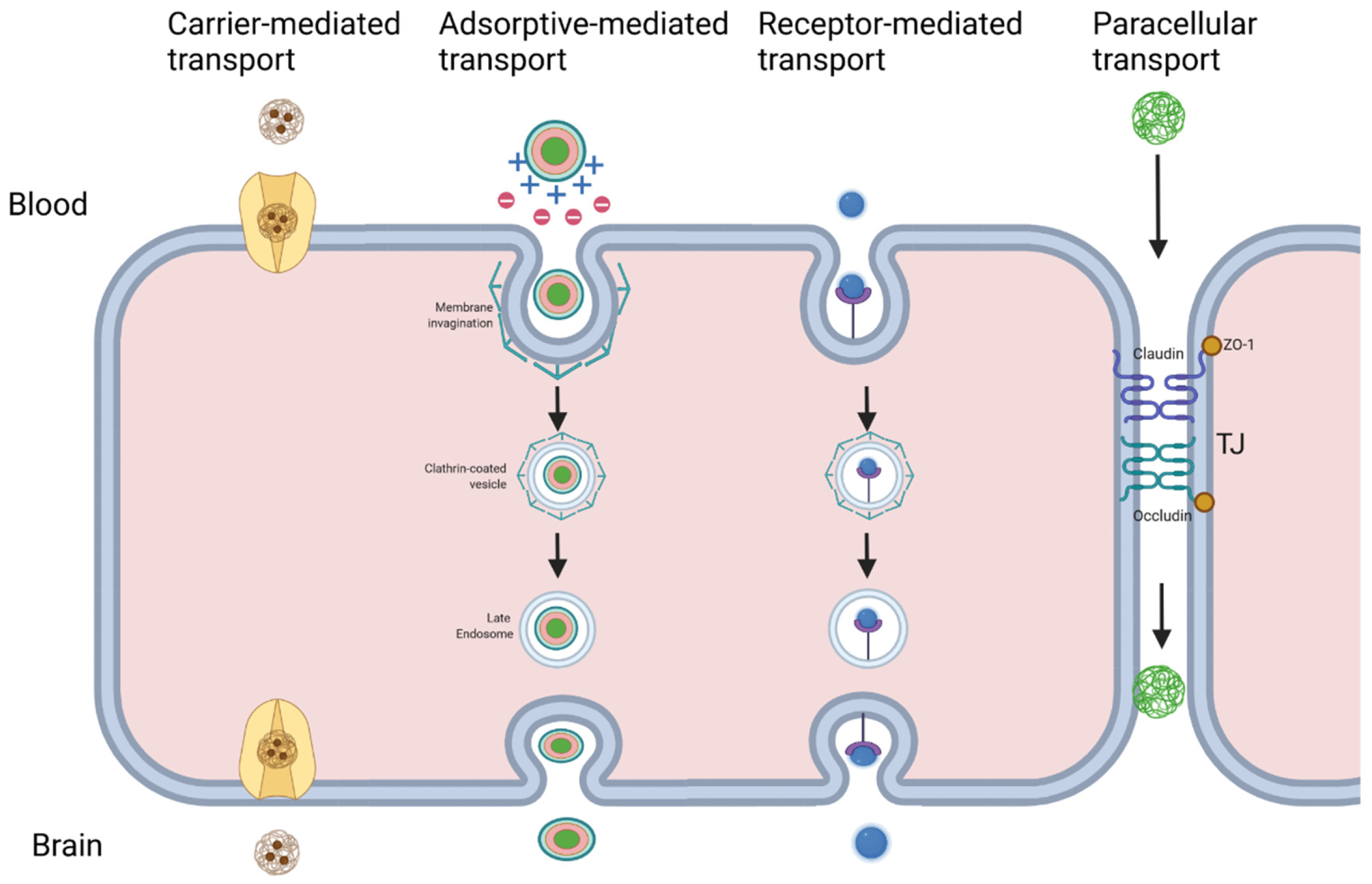
Table 1.
Summary of NPs and select chemotherapeutic ligands investigated for treatment of brain tumors. In addition, NPs are capable of delivering RNA and DNA molecules as gene therapy and can play roles as imaging agents for tumors.
Table 1.
Summary of NPs and select chemotherapeutic ligands investigated for treatment of brain tumors. In addition, NPs are capable of delivering RNA and DNA molecules as gene therapy and can play roles as imaging agents for tumors.
| Nanoparticle Category | Advantages | Chemotherapeutics | References |
|---|---|---|---|
| Polymeric | Stability, biodegradability, biocompatibility, ease of manufacturing, hydrophobic and hydrophilic drug transport, non-immunogenic, low toxicity | 3-bis(2-chloroethyl)-1-nitrosourea | [130,131] |
| Doxorubicin | [132,133] | ||
| Methotrexate | [134] | ||
| Temozolomide | [135] | ||
| Gemcitabine | [136] | ||
| Paclitaxel | [137] | ||
| Liposome | Hydrophobic and hydrophilic drug transport, biocompatible, low toxicity | Doxorubicin | [120] |
| Methotrexate | [138] | ||
| Cisplatin | [139] | ||
| Irinotecan | [140,141] | ||
| Topotecan | [142] | ||
| Paclitaxel | [143] | ||
| Dendrimer | High molecular uniformity, monodispersity, kinetic stability, abundant free functional groups, low toxicity | Methotrexate | [144] |
| Doxorubicin | [145,146] | ||
| Temozolomide | [147] | ||
| Docetaxel | [148] | ||
| Arsenic trioxide | [149] | ||
| Metallic | Contrast imaging agents, surface is readily modifiable, inflammatory cascade increases BBB permeability, hyperthermic effect increases BBB permeability and damage tumors | Doxorubicin | [150,151,152] |
| Cisplatin | [153] | ||
| Paclitaxel | [154] | ||
| Quantum dots | Photoluminescent, photostability, tunable emission/excitation spectra, visualization of individual molecules, readily monitor drug delivery, low toxicity | Topotecan | [155] |
| Doxorubicin | [156] | ||
| Temozolomide | [157] | ||
| Nanogels | Serum stability, uniformity, fluid-like transport properties, bioadehsive, biocompatible, biodegradable, deformable, stimulus-responsive release, low toxicity | Doxorubicin | [158] |
| Cisplatin | [159] | ||
| Methotrexate | [160] | ||
| Paclitaxel | [161] |
Table 2.
Studies on theranostic NPs, investigated for brain cancer, which combine a therapeutic anti-cancer effect with tumor imaging.
Table 2.
Studies on theranostic NPs, investigated for brain cancer, which combine a therapeutic anti-cancer effect with tumor imaging.
| NP Category | Size (nm) | Functional Components | Model | Results | Refs |
|---|---|---|---|---|---|
| Liposome + QD | 182 | Docetaxel (chemo), QD (imaging), transferrin (targeting) | Rats | Sustained drug release >72 h | [270] |
| Carbon Dots | 6–8 | Highly crystalline carbon nanodot (photoacoustic imaging and photothermal therapy) | Mice (U87 glioma cells) | NPs accumulate in tumor cells and image-guided near-infrared-activated photothermal therapy can damage tumor tissue. | [271] |
| Magnetic NP | 12 | Epirubicin (chemo), Fe3O4 core (contrast imaging) | Rat (C6 glioma cells) | FUS can improve uptake across the BBB, magnetic targeting improves tumor targeting, and MRI can monitor magnetic NP distribution. | [272] |
| Silica NP | __ | Doxorubicin (chemo), Cu2 − x Se NP (photoacoustic imaging) | Mice (U87 glioma cells) | FUS can improve uptake across the BBB for tumor-specific targeting and the NPs show contrast enhancement on imaging. | [273] |
| GNP | 56 | Doxorubicin (chemo), Cy5.5 (probe), RRGD peptide (targeting) | Mice (C6 glioma cells) | Effective uptake by glioma cells with co-localization and fluorescent detection of Cy5.5 | [268] |
| Gold + iron oxide-loaded micelle | 100 | Iron oxide (MRI contrast agent), GNP (radiosensitizer) | Mice (U251 GBM cells) | Effective contrast agent for MRI and can show tumor borders of glioblastoma, radiosensitization increases tumor damage from radiation therapy | [274] |
| Iron oxide NP | 43 | Iron oxide (MRI contrast agent), IL1- receptor antagonist (anti-edema agent) | Rats (C6 glioma cells) | IL-1 receptor antagonist reduces peritumoral edema and improves survival, enhanced MRI imaging of tumor | [275] |
| Iron oxide NP | 37 | Iron oxide (MRI contrast agent), Small interfering RNA (gene therapy), temozolomide (chemo) | Mice (T98G GBM cells) | Gene therapy can reduce glioblastoma resistance to temozolomide, therapeutic response can be monitored on MRI | [276] |
| Iron oxide NP | 184 | Iron oxide (MRI contrast agent), doxorubicin (chemo) | C6 glioma cells | MRI showed NP accumulation in tumor cells | [277] |
| Gadolinium-based NP | 120 | Chlorin e6 (photosensitizer), gadolinium (MRI contrast agent) | Mice (C6 glioma cells) | Photodynamic therapy targeted tumor cells, NPs showed contrast enhancement on MRI | [278] |
| Polymeric NP | 40–70 | Doxorubicin (chemo), gadolinium or Hoechst 33342 (imaging agents) | Mice (breast cancer metastasis line) | MRI and fluorescence microscopy confirmed delivery of imaging agents across the BBB, doxorubicin induced apoptosis in metastatic cells | [279] |
| Polymeric NP | 10–200 | Iron oxide (MRI contrast agent), Photofrin (photosensitizer), F3 peptide (targeting) | Rat (9 L glioma cells) | Photodynamic therapy increased survival time, MRI detected NPs in tumor cells | [280] |
Publisher’s Note: MDPI stays neutral with regard to jurisdictional claims in published maps and institutional affiliations. |
© 2022 by the authors. Licensee MDPI, Basel, Switzerland. This article is an open access article distributed under the terms and conditions of the Creative Commons Attribution (CC BY) license (https://creativecommons.org/licenses/by/4.0/).
Share and Cite
MDPI and ACS Style
Hersh, A.M.; Alomari, S.; Tyler, B.M. Crossing the Blood-Brain Barrier: Advances in Nanoparticle Technology for Drug Delivery in Neuro-Oncology. Int. J. Mol. Sci. 2022, 23, 4153. https://doi.org/10.3390/ijms23084153
AMA Style
Hersh AM, Alomari S, Tyler BM. Crossing the Blood-Brain Barrier: Advances in Nanoparticle Technology for Drug Delivery in Neuro-Oncology. International Journal of Molecular Sciences. 2022; 23(8):4153. https://doi.org/10.3390/ijms23084153
Chicago/Turabian StyleHersh, Andrew M., Safwan Alomari, and Betty M. Tyler. 2022. "Crossing the Blood-Brain Barrier: Advances in Nanoparticle Technology for Drug Delivery in Neuro-Oncology" International Journal of Molecular Sciences 23, no. 8: 4153. https://doi.org/10.3390/ijms23084153
Note that from the first issue of 2016, this journal uses article numbers instead of page numbers. See further details here.