Article

Early Inactivation of Membrane Estrogen Receptor Alpha (ERα) Recapitulates the Endothelial Dysfunction of Aged Mouse Resistance Arteries

by
Julie Favre
1,2,3,4,†,
Emilie Vessieres
1,2,3,†,
Anne-Laure Guihot
1,2,3,
Linda Grimaud
1,2,3,
Coralyne Proux
1,2,3,
Laurent Loufrani
1,2,3,
Françoise Lenfant
5,
Coralie Fontaine
5,
Jean-François Arnal
5 and
Daniel Henrion
1,2,3,6,*
1
MitoVasc Department, Team 2 (CarMe) and CARFI Facility, Angers University, F-49045 Angers, France
2
Centre National de la Recherche Scientifique (CNRS), UMR 6015, F-49045 Angers, France
3
Institut National de la Santé et de la Recherche Médicale (INSERM), U1083, F-49045 Angers, France
4
Institut National de la Santé et de la Recherche Médicale (INSERM), UMR S 1121, Centre de Recherche en Biomédecine de Strasbourg (CRBS), F-67084 Strasbourg, France
5
Institut National de la Santé et de la Recherche Médicale (INSERM), U1297, Institut des Maladies Métaboliques et Cardiovasculaires (I2MC), Université Toulouse III Paul Sabatier, CHU Rangueil (University Hospital) de Toulouse, F-31432 Toulouse, France
6
Angers University Hospital (CHU), F-49000 Angers, France
*
Author to whom correspondence should be addressed.
These authors contributed equally to this work.
Int. J. Mol. Sci. 2022, 23(5), 2862; https://doi.org/10.3390/ijms23052862
Submission received: 27 January 2022 / Revised: 28 February 2022 / Accepted: 3 March 2022 / Published: 5 March 2022
(This article belongs to the Special Issue The Role of Estrogen Receptors in Health and Disease)

Abstract

:
Flow-mediated dilation (FMD) of resistance arteries is essential for tissue perfusion but it decreases with ageing. As estrogen receptor alpha (Erα encoded by Esr1), and more precisely membrane ERα, plays an important role in FMD in young mice in a ligand-independent fashion, we evaluated its influence on this arteriolar function in ageing. We first confirmed that in young (6-month-old) mice, FMD of mesenteric resistance arteries was reduced in Esr1−/− (lacking ERα) and C451A-ERα (lacking membrane ERα). In old (24-month-old) mice, FMD was reduced in WT mice compared to young mice, whereas it was not further decreased in Esr1−/− and C451A-ERα mice. Markers of oxidative stress were similarly increased in old WT and C451A-ERα mice. Reduction in oxidative stress with superoxide dismutase plus catalase or Mito-tempo, which reduces mitochondrial superoxide restored FMD to a normal control level in young C451A-ERα mice as well as in old WT mice and old C451A-ERα mice. Estradiol-mediated dilation was absent in old WT mice. We conclude that oxidative stress is a key event in the decline of FMD, and that an early defect in membrane ERα recapitulates phenotypically and functionally ageing of these resistance arteries. The loss of this function could take part in vascular ageing.

1. Introduction

Resistance arteries are the small blood vessels located upstream of capillaries. They control blood delivery to tissues at relevant flow and pressure. Disorders in their structure and function raise capillary pressure, which exacerbates organ damage, favored by age-associated cardio- and cerebrovascular risk factors. The basal tone of these small arteries allows a tight control of local blood flow. It results in part from the interaction between pressure-induced smooth muscle contraction (myogenic tone) and flow-mediated dilation (FMD) due to the activation of endothelial cells by shear stress. A reduced FMD is the hallmark of endothelium dysfunction and FMD is altered very early in cardiovascular and metabolic disorders [1,2,3]. FMD is also progressively reduced in ageing thus amplifying the negative impact of the other risk factors [4,5,6,7].
Both sex and age are independent risk factors for the reduction in endothelial function [8]. Endothelial function decreases with age in healthy men and women with very low cardiovascular risk [4,5]. An age-dependent reduction in FMD is also observed in coronary human resistance arteries with a shift of the mediators involved in FMD from prostacyclin to NO with ageing whereas H2O2 is the mediator of FMD in patients with coronary artery disease [9]. We recently reported that endothelial ERα contributed to optimize flow (shear stress)-mediated dilation in young healthy mouse resistance arteries [10]. Interestingly, the only known mutation in the gene encoding ERα has been described in one man [11] and the main observed vascular defect was a strong reduction in FMD [12].
ERα belongs to the nuclear receptor superfamily and acts classically as a transcription factor, but it can also exert extranuclear, non-genomic actions by activating rapid membrane-initiated steroid signaling (MISS), as demonstrated specifically in the endothelium [13]. The vascular effects of ERα are mediated by both membrane-associated ERα, mainly through the production of NO by endothelial cells [14], and by the nuclear effects of ERα through the activating function AF2 allowing protection against atherosclerosis and hypertension [15]. Although most of the vascular protective effects of ERα are mediated by AF2-dependent nuclear effects, we have previously shown that FMD is facilitated by membrane-associated ERα-signal transduction in young male and female mice in a ligand-independent mode [10].
Beside the decline in estrogens at menopause, abnormalities in the expression and/or function of ERs in tissues, and particularly in arteries, could contribute to the failure of classic estrogens to protect arteries during ageing [16]. Thus, we investigated FMD in resistance arteries in old male mice (24 months old) in comparison to young mice (6 months old) in two mouse models: (i) totally deficient in ERα (Esr1−/− mice) and (ii) lacking the plasma membrane ERα as the codon for palmitoylable cystein (Cys) 451 of ERα was mutated into alanine (C451A-ERα mice) [14].

2. Results

2.1. Mice Age, Heart Weight, Body Weight, Blood Pressure and Heart Rate

The average age of the two groups of mice, all genotypes together, was 5.97 ± 0.12 months (n = 38 young mice) and 23.53 ± 0.38 months (n = 56 old mice). There was no significant difference in age between the four groups of young or old mice (Figure 1A,B). Similarly, body weight was equivalent between the groups independently of age (Figure 1C,D). The ratio of the left ventricle to the tibia length was not significantly affected by the genotypes or by age (Figure 1E,F). Similarly, systolic blood pressure and heart rate measured using plethysmography were not significantly affected by age and by the genotypes (Figure 1G–J).

2.2. FMD in Mouse Mesenteric Arteries

Stepwise increases in intraluminal flow in perfused and cannulated mesenteric resistance arteries induced vasodilation (FMD, Figure 2A–D).
As expected, FMD was significantly reduced in resistance arteries isolated from young mice lacking ERα compared to young littermate Esr1+/+ mice (Figure 2A). FMD was also significantly lower in old Esr1+/+ mice compared to young Esr1+/+ mice (Figure 2B versus Figure 2A), whereas ageing did not further reduce FMD in Esr1−/− mice (Figure 2B versus Figure 2A).
A similar pattern was observed in C451A-ERα mice, which lack membrane-associated ERα compared to their littermate control C451-WT mice (Figure 2C,D).

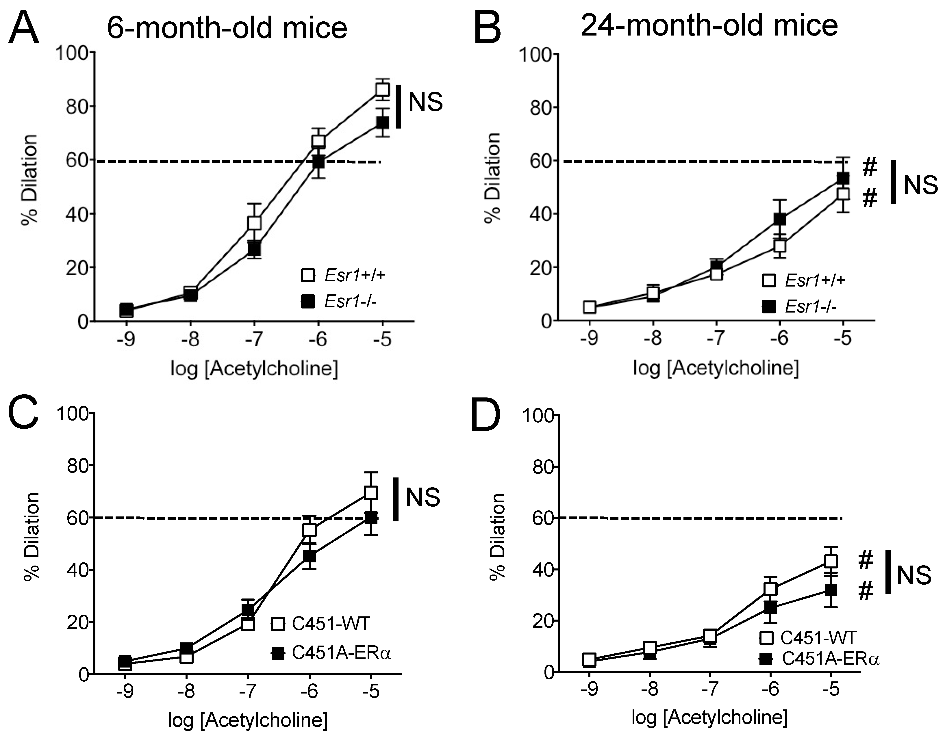
2.3. Agonist-Mediated Endothelium-Dependent Dilation in Mouse Mesenteric Arteries

Acetylcholine-mediated dilation was not significantly affected by the total absence of ERα in Esr1−/− mice (Figure 3A,B) or by its absence at the membrane level in C451A-ERα mice (Figure 3C,D).
Acetylcholine-mediated dilation was significantly reduced in old mice compared to young mice in all the study groups (Figure 3A,C compared to Figure 3B,D, respectively).

2.4. Smooth Muscle-Dependent Contraction in Mouse Mesenteric Arteries

Stepwise increases in intraluminal pressure induced contraction in isolated perfused mesenteric resistance arteries (Figure 4). Pressure-induced contraction or myogenic tone was not significantly affected by the genotype or by ageing in all study groups (Figure 4).

2.5. Wall Structure and Properties of Mouse Mesenteric Arteries

Internal diameter of mesenteric resistance arteries (Figure 5) was not significantly modified by the genotype or by ageing in all study groups. Cross-sectional compliance was attenuated in old mice compared to young mice in all the groups without significant difference between Esr1−/− and C451A-ERα mice and without difference between these mice and their littermate controls (Figure 6).

2.6. Gene Expression Analysis of the Main Pathways Involved in FMD

In order to follow the impact of the absence of membrane ERα on mesenteric arteries in ageing, we measured the expression of genes representative of different main biological pathways which could affect the endothelial response to acute changes in flow or FMD (endothelial function, mechanosensing, oxidative stress, mitochondrial homeostasis, hormone-related genes, purinergic signaling). Considering the impact of ageing, important changes in gene expression were observed both in WT and C451A-ERα mice (Figure 7). They were evidenced by a down-regulation of genes involved in endothelial response and mechanosensing (cluster 1, green). Nevertheless, some genes related to oxidative stress were up-regulated in old mice (p66Shc, Sod1, Sod2, Gpx1). Interestingly, while in young mice the loss of membrane ERα did not significantly affect gene expression levels, different expression profiles were revealed with ageing between WT and C451A-ERα mice. Those included hormone response or metabolism pathways (Esr1, AR, AhR, Cyp1b1, Comt), the renin–angiotensin–aldosterone system (Nr3c2, Ace), mitochondrial homeostasis (Dmnl1, Sirt1) and endothelial response (Sdc4, Kcnma1, Icam1).

2.7. Effect of the Reduction in Oxidative Stress on FMD in Mouse Mesenteric Arteries

We then tested the acute effect of superoxide reduction with SOD plus catalase and of Mito-tempo on FMD. Incubation of mesenteric resistance arteries with SOD and catalase did not affect FMD in young C451-WT mice (Figure 8A), whereas it increased FMD in young C451A-ERα mice (Figure 8B). In 24-month-old mice, SOD and catalase improved FMD in both C451-WT and C451A-ERα mice (Figure 8C,D). A similar pattern was observed with arteries incubated with Mito-tempo which reduces ROS of mitochondrial origin (Figure 8A–D).
These results suggest that the reduced FMD in C451A-ERα as compared to WT mice can be attributed to damages induced by oxidative stress, such as in ageing and or in the absence of membrane ERα mice.

2.8. Effect of Age on Estradiol (E2)-Mediated Dilation in Mouse Mesenteric Arteries

Estradiol induced a concentration-dependent dilation of the resistance mesenteric arteries isolated from young WT mice, whereas no significant dilation was observed in old WT mice (Figure 9).

3. Discussion

The main finding of this study is that the membrane-located ERα has a facilitating role in flow (shear stress)-mediated dilation (FMD) in resistance arteries of male mice, in agreement with our previous work [10], and that this protective effect is lost in ageing. Indeed, FMD was reduced by approximately 30% in mice lacking membrane ERα, whereas in WT mice a similar reduction was observed only at the age of 24 months. However, in mice lacking membrane ERα FMD did not further decrease with age. Thus, the reduction in FMD due to the absence of membrane ERα could be presented as a premature vascular ageing. Furthermore, reducing oxidative stress restored FMD in both young mice lacking membrane ERα and in old WT mice. Thus, it is possible that membrane ERα reduces oxidative stress to facilitate FMD in young mice and that this effect is lost with ageing, due to a reduction in membrane ERα expression or function
We investigated FMD in resistance arteries as they control local blood flow delivery to all tissues and disorders of these small arteries induce organ damage as seen in cardiovascular, metabolic and cerebrovascular disorders [17,18,19]. Resistance artery tone is counteracted by FMD and a reduced FMD is the hallmark of vascular disorders in a large number of diseases [20]. The present study showed a reduction in both FMD and receptor-dependent (acetylcholine) endothelium-mediated dilation in 24-month-old mice. This is in agreement with previous works on humans [8] and on animals [3,21,22] showing reduced FMD and more generally altered endothelium-dependent dilation in ageing. This marked alteration in endothelium-dependent dilation could be due to excessive oxidative stress as shown by an increase in the expression level of genes involved in oxidative stress observed in mesenteric resistance arteries isolated from WT and C451A-ERα mice (Figure 7), in agreement with previous studies [3,23,24]. In addition, myogenic tone was reduced in old mice as previously shown in male and female mice mesenteric arteries [25] and in male rat coronary [26] and skeletal muscle arteries [27]. Ageing was also associated with a reduced arterial compliance suggesting a change in wall structure. Indeed, arterial stiffening has been demonstrated in mesenteric resistance arteries in 2-year-old male mice [28] and it is well demonstrated in human ageing [29,30].
In agreement with our previous study [10], the present work confirmed the importance of the membrane-associated ERα in FMD. This effect also agrees with a single observation in one young man (31 years old). The lack of functional ERα in this man [11] was associated with a selective reduction in acute FMD [12]. Furthermore, estrogens through the nuclear AF2 function of ERα, were shown to play a key role in flow-mediated outward remodeling [31,32,33,34]. This latter is a chronic adaptation of the arterial wall leading to an increase in lumen diameter and wall mass in response to a chronic rise in blood flow in vivo [3,35]. This remodeling is observed in collateral arteries growth in ischemic diseases [35].
The vascular protection provided by membrane-associated ERα involves two arms, with a ligand-dependent pathway leading to acute NO-dependent vasodilation and involved in endothelial repair [14,36] and a ligand-independent pathway potentiating the acute response to flow or FMD (present study and [10]). By contrast, the protective effect of E2 on FMD described in pathological conditions or in menopaused women involves the ligand-dependent pathway and mainly the nuclear functions of ERα. For example, FMD, reduced in post-menopausal women, is improved by a chronic treatment with estradiol (E2) or SERMs [37,38]. Similarly, in old female rats, FMD is reduced and can be improved by estrogen supplementation [39]. On the other hand, in healthy conditions, FMD is not different between men and women and E2 does not influence FMD in healthy conditions [40]. Despite the absence of ERα, we found no significant change in body weight, left ventricle weight, systolic blood pressure and heart rate in mice, in agreement with our previous work [15]. Nevertheless, these mice develop a greater hypertension when perfused with angiotensin II [15]. Similarly, the young man with a disruptive mutation in ERα was normotensive but more susceptible to atherosclerosis [41]
Besides membrane-located ERα, G-protein coupled estrogen receptor (GPER) is also involved in the acute response to estrogens [42,43]. Although, our previous work demonstrating the role of membrane ERα in FMD in young mice has excluded a possible role for GPER [10], its involvement in the reduction in FMD observed in ageing remains to be investigated. Indeed, GPER is associated with ageing of the cardiovascular system [44] with a role in endothelial ageing [45,46]. Thus, further investigation is needed to define the role of GPER in flow-dependent signaling in vascular ageing.
The risk of cardiovascular diseases is higher in men than in women, and the protection due to estrogen in women is progressively lost after menopause. Estrogen substitution therapy has proved to be efficient in reducing this risk although caution should be taken [13,47]. In the present study, in agreement with our previous work [10], we identified a new pathway protecting the vascular tree through the involvement of non-nuclear or membrane-associated ERα in FMD. Thus, estrogen and ERα would have a dual beneficial effect on the endothelium through the activation of eNOS expression level and NO production by E2 [13] and through a reduction in ROS production in a ligand-independent mode (present study and [10]). Noteworthy, both are reduced with ageing. E2 level decreases after menopause and flow-mediated ERα-dependent reduction in ROS production is also lost in ageing (present study). Nevertheless, although we can envision a weak impact of the former effect in our study on males, the precise pathway involved in this latter effect remains to be better defined. Indeed, this question is difficult to address as membrane ERα represents a very small percentage of the total ERα and it is barely observable using immunohistochemistry [13]. Membrane ERα is anchored to the plasma membrane through palmitoylation of its cysteine in position 451 in the mouse [14] and a post-translational dysregulation of cysteine involved in palmitoylation has been reported in ageing [48]. We have previously shown that E2-dependent acute vasodilation is mediated by membrane ERα through activation of NO production [14]. Interestingly, E2-mediated dilation was lost in old WT mice suggesting a loss of functional membrane ERα. This observation agrees with the reduced FMD found in old WT mice, which would thus be equivalent to the C451A-ERα mice which lack membrane ERα. In agreement, endothelial ageing has been shown to be associated with reduced NO production due to decreased eNOS expression and to decreased availability of L-arginine and tetrahydrobiopterin, the cofactor of eNOS [49].
The selective reduction in FMD observed in young C451A-ERα mice could represent a premature ageing of the endothelium. In agreement, FMD is early and selectively reduced in ageing in association with reduced NO bioavailability due to excessive ROS production [22,50]. Interestingly, we found that several genes involved in endothelial response and mechanosensing were down-regulated during ageing, whereas genes mainly related to oxidative stress were up-regulated in the mesenteric arteries of old mice. These observations confirm previous works [51,52]. While no differences in gene expression were found in young mice groups, it is worth noting a limitation of age-related down-regulation of several genes in old C451A-ERα mice (Figure 7, see orange and blue boxes). Thus, we may hypothesize that the functional loss of membrane-located ERα in old WT mice further affected the expression level of genes that could potentially worsen the endothelial response to flow such as genes of the renin–angiotensin–aldosterone system and of mitochondrial homeostasis (Figure 7). In agreement, we observed that Mito-tempo which reduces ROS produced by the mitochondria improved FMD in mature and old C451A-ERα mice as well as in old WT mice. Thus, mitochondrial ROS could be involved in the reduction in FMD, in agreement with previous works showing a role of these organelles in the production of ROS in ageing associated with cardiovascular disorders [53]. The pathway linking membrane-located ERα to a reduction in mitochondria-dependent ROS production remains to be further investigated. Nevertheless, mitochondrial fusion and fission are sensitive to shear stress in cultured human and bovine endothelial cells [54] and shear stress-dependent Ca2+ mobilization in human endothelial cells relies on mitochondria-dependent activation of endoplasmic reticulum channels [55]. In cerebral endothelial cells, activation of ERα induces a decrease in mitochondrial ROS production, possibly through up-regulation of cytochrome C activity [56].
Thus, targeting the pathway activated by membrane ERα in response to flow could be an attractive way to reduce oxidative damages in both healthy and diseased ageing through a reduction in ROS production and restoration of an efficient FMD in resistance arteries. Nevertheless, targeting membrane ERα per se could be useless as our results suggest that it is absent, or at least functionally deficient, in old mice.
In conclusion, our findings confirm that membrane-located ERα signaling takes part in FMD through a reduction in oxidative stress thus facilitating NO-dependent dilation. In addition, the present work suggests that this protective effect of membrane-located ERα could be lost in ageing and further suggests that an early alteration in this pathway may represent premature vascular ageing as observed in young mice lacking membrane-located ERα with a reduction in FMD equivalent to that seen in old mice.

4. Materials and Methods

4.1. Animal Protocol

We used 6-month-old and 24-month-old mice lacking the gene encoding for ERα (Esr1−/− compared to Esr1+/+ mice) [57] and mice in which the codon encoding the palmitoylable cystein (Cys) 451 of ERα was mutated into alanine (C451A-ERα mice compared to C451-WT mice) [14]. Littermate mice were used as control (wild-type, WT or +/+) in each group.
As previously described [15], systolic blood pressure (SBP) was measured on conscious mice using a non-invasive and fully automated and computerized tail-cuff method (photoplethysmograph BP-2000 Blood Pressure Analysis System™, Visitech Systems, Apex, NC, USA). The means of 5-day measurements were computed after one-week adaptation period.
Mice were euthanized using a CO2 chamber and the mesentery was quickly removed and placed in ice-cold physiological salt solution (PSS). Several segments of mesenteric resistance arteries were isolated for the functional and biochemical studies.
The experiment complied with the European Community standards on the care and use of laboratory animals and the Guide for the Care and Use of Laboratory Animals published by the US National Institutes of Health (NIH Publication No. 85–23, revised 1996). The protocol was approved by the regional ethics committee (“Comité d’éthique en Expérimentation Animale des Pays de la Loire”, authorization # CEEA PdL 2012.141).

4.2. Flow-Mediated Dilation in Mesenteric Arteries In Vitro

Arterial segments, approximately 200 µm in internal diameter, were cannulated at both ends on glass micro-cannulae and mounted in a video-monitored perfusion system (Living System, LSI, Burlington, VT, USA) [58]. The arterial segment was bathed in a 5 mL organ bath containing a physiological salt solution (PSS, pH: 7.4, pO2:160 mmHg and pCO2: 37 mmHg) [59]. Perfusion of the artery was obtained with 2 peristaltic pumps, one controlling flow and one under the control of a pressure-servo control system (LSI, Burlington, VT, USA) allowing the control of pressure [59]. Pressure at both ends of the arterial segment was monitored using pressure transducers (MP-4 system, LSI, Burlington, VT, USA). To measure flow-mediated dilation (FMD), pressure was set at 75 mmHg and arterial tone was increased with phenylephrine (1 µmol/L). Flow (3 to 50 µL per min) was then generated through the distal pipette with a peristaltic pump [59].
In separate series of experiments, FMD was measured before and after incubation (20 min) of the arteries with superoxide dismutase (SOD, 120 U/mL) plus catalase (80 U/mL) [60] or Mito-Tempo (1 µmol/L) [61].
Other segments of mesenteric arteries were used for a cumulative concentration–response curve (CRC) to acetylcholine (10−9 to 10−5 mol/L) or to estradiol (E2, 10−9 to 10−7 mol/L) after precontraction of the arterial segment with phenylephrine (1µmol/L) to contract the arteries by approximately 50%.
Myogenic tone (MT) was determined in response to stepwise increases in intra luminal pressure from 10 to 125 mmHg using a video-monitored perfusion system as described above. MT at a given perfusion pressure was defined as the magnitude of the percent myogenic tone (%MT) at that pressure. The %MT was expressed by the active (AD) and passive vessel diameters (PD) such that %MT = [(PD−AD)/PD]·100% [62].
At the end of each experiment, arteries were bathed in a Ca2+-free PSS containing ethylene-bis-(oxyethylenenitrolo) tetra-acetic acid (2 mmol/L) and sodium nitroprusside (10 µmol/L). Pressure was then increased in steps from 10 to 125 mmHg, in the absence of flow, to determine passive arterial diameter and passive mechanical properties of the arterial wall, as previously described [60].

4.3. Quantitative Real-Time PCR

Gene expression was investigated using quantitative polymerase chain reaction after reverse transcription of total RNA (RT-qPCR). Mesenteric arteries were stored at −20 °C in RNAlater Stabilization Reagent (Qiagen, Valencia, CA, USA) until use. RNA was extracted using the RNeasy® Micro Kit (Qiagen, Valencia, CA, USA) following the manufacturer’s instructions. RNA extracted (300 ng) was used to synthesize cDNA using the QuantiTect® Reverse Transcription Kit (Qiagen, Valencia, CA, USA). RT-qPCR was performed with Sybr® Select Master Mix (Applied Biosystems Inc., Lincoln, CA, USA) reagent using a LightCycler 480 Real-Time PCR System (Roche, Branchburg, NJ, USA). Primer sequences are shown in Table 1. Hprt, ActB and Gusb were used as housekeeping genes. Analysis was not performed when Ct values exceeded 35. Results were expressed as: 2(Ct target-Ct housekeeping gene) and heatmaps of expression values were all generated using MeV. Expression values shown within the heatmaps were normalized per mRNA as fold changes of means of young C451-WT mice.

4.4. Statistical Analysis

Results were expressed as means ± SEM. Significance of the differences between groups was determined by analysis of variance (two-way ANOVA for consecutive measurements for pressure–diameter curves, myogenic, FMD and CRC to ACh) followed by a Bonferroni test. The Mann–Whitney test was used for the other comparisons. Probability values less than 0.05 were considered significant.

Author Contributions

Conceptualization, D.H., J.F. and J.-F.A.; methodology, J.F, E.V. and D.H.; software, J.F.; validation, J.F., E.V., L.G., C.P. and A.-L.G.; formal analysis, J.F. and E.V.; investigation, J.F., E.V., L.G., C.P. and A.-L.G.; resources, E.V.; data curation, J.F., E.V. and D.H.; writing—original draft preparation, J.F. and D.H.; writing—review and editing, D.H., J.F., C.F., F.L., L.L. and J.-F.A.; visualization, J.F, E.V. and D.H.; supervision, D.H.; project administration, E.V. and D.H.; funding acquisition, D.H. and J.-F.A. All authors have read and agreed to the published version of the manuscript.

Funding

The work performed in MitoVasc was supported in part by the foundation for medical research (Fondation pour la Recherche Médicale, Mitoshear project, contract FRM-DPC20171138957) and the national agency for research (ANR, Estroshear project, contract # ANR-18-CE14-0016-01). J.F. was supported by a Lefoulon-Delalande fellowship. The work performed in Toulouse, INSERM U1297, was supported in part by INSERM, Université de Toulouse III, the national agency for research (ANR, Estroshear project, contract # ANR-18-CE14-0016-01), Fondation pour la Recherche Médicale (Equipe FRM DEQ20160334924) and Fondation de France (JFA, contract 00086486).

Institutional Review Board Statement

The experiments complied with the European Community standards for the care and use of laboratory animals and the Guide for the Care and Use of Laboratory Animals published by the US National Institutes of Health (NIH Publication No. 85–23, revised in 1996). The protocol was approved by the regional (Pays de la Loire) ethics committee (permits #14335, #16740, and #16108).

Informed Consent Statement

Not applicable.

Data Availability Statement

The data presented in this study are available on request from the corresponding author.

Acknowledgments

Esr1−/− mice and the corresponding littermate WT mice were kindly provided by P. Chambon and A. Krust (Institute of Genetics and Molecular and Cellular Biology, Strasbourg, France).

Conflicts of Interest

The authors declare no conflict of interest.

References

  1. Rizzoni, D.; Rosei, E.A. Small artery remodeling in hypertension and diabetes. Curr. Hypertens. Rep. 2006, 8, 90–95. [Google Scholar] [CrossRef] [PubMed]
  2. Stoner, L.; Sabatier, M.J. Use of ultrasound for non-invasive assessment of flow-mediated dilation. J. Atheroscler. Thromb. 2012, 19, 407–421. [Google Scholar] [CrossRef] [PubMed] [Green Version]
  3. Chehaitly, A.; Vessieres, E.; Guihot, A.-L.; Henrion, D. Flow-mediated outward arterial remodeling in aging. Mech. Ageing Dev. 2020, 194, 111416. [Google Scholar] [CrossRef] [PubMed]
  4. Königstein, K.; Wagner, J.; Frei, M.; Knaier, R.; Klenk, C.; Carrard, J.; Schwarz, A.; Hinrichs, T.; Schmidt-Trucksäss, A. Endothelial function of healthy adults from 20 to 91 years of age: Prediction of cardiovascular risk by vasoactive range. J. Hypertens. 2021, 39, 1361–1369. [Google Scholar] [CrossRef]
  5. Majerczak, J.; Grandys, M.; Frołow, M.; Szkutnik, Z.; Zakrzewska, A.; Niżankowski, R.; Duda, K.; Chlopicki, S.; Zoladz, J.A. Age-Dependent Impairment in Endothelial Function and Arterial Stiffness in Former High Class Male Athletes Is No Different to That in Men With No History of Physical Training. J. Am. Heart Assoc. 2019, 8, e012670. [Google Scholar] [CrossRef] [PubMed]
  6. Lane-Cordova, A.D.; Ranadive, S.M.; Kappus, R.M.; Cook, M.D.; Phillips, S.A.; Woods, J.A.; Wilund, K.R.; Baynard, T.; Fernhall, B. Aging, not age-associated inflammation, determines blood pressure and endothelial responses to acute inflammation. J. Hypertens. 2016, 34, 2402–2409. [Google Scholar] [CrossRef]
  7. Black, M.A.; Cable, N.; Thijssen, D.H.J.; Green, D.J. Impact of age, sex, and exercise on brachial artery flow-mediated dilatation. Am. J. Physiol. Circ. Physiol. 2009, 297, H1109–H1116. [Google Scholar] [CrossRef]
  8. Deanfield, J.E.; Halcox, J.P.; Rabelink, T.J. Endothelial function and dysfunction: Testing and clinical relevance. Circulation 2007, 115, 1285–1295. [Google Scholar] [CrossRef]
  9. Beyer, A.M.; Zinkevich, N.; Miller, B.; Liu, Y.; Wittenburg, A.L.; Mitchell, M.; Galdieri, R.; Sorokin, A.; Gutterman, D.D. Transition in the mechanism of flow-mediated dilation with aging and development of coronary artery disease. Basic Res. Cardiol. 2017, 112, 5. [Google Scholar] [CrossRef]
  10. Favre, J.; Vessieres, E.; Guihot, A.-L.; Proux, C.; Grimaud, L.; Rivron, J.; Garcia, M.C.; Réthoré, L.; Zahreddine, R.; Davezac, M.; et al. Membrane estrogen receptor alpha (ERα) participates in flow-mediated dilation in a ligand-independent manner. eLife 2021, 10, e68695. [Google Scholar] [CrossRef]
  11. Smith, E.P.; Boyd, J.; Frank, G.R.; Takahashi, H.; Cohen, R.M.; Specker, B.; Williams, T.C.; Lubahn, D.B.; Korach, K. Estrogen Resistance Caused by a Mutation in the Estrogen-Receptor Gene in a Man. N. Engl. J. Med. 1994, 331, 1056–1061. [Google Scholar] [CrossRef] [PubMed]
  12. Sudhir, K.; Chou, T.M.; Messina, L.M.; Hutchison, S.J.; Korach, K.; Chatterjee, K.; Rubanyi, G.M. Endothelial dysfunction in a man with disruptive mutation in oestrogen-receptor gene. Lancet 1997, 349, 1146–1147. [Google Scholar] [CrossRef]
  13. Arnal, J.F.; Lenfant, F.; Metivier, R.; Flouriot, G.; Henrion, D.; Adlanmerini, M.; Fontaine, C.; Gourdy, P.; Chambon, P.; Katzenellenbogen, B.; et al. Membrane and Nuclear Estrogen Receptor Alpha Actions: From Tissue Specificity to Medical Implications. Physiol. Rev. 2017, 97, 1045–1087. [Google Scholar] [CrossRef] [PubMed]
  14. Adlanmerini, M.; Solinhac, R.; Abot, A.; Fabre, A.; Letron, I.R.; Guihot, A.-L.; Boudou, F.; Sautier, L.; Vessières, E.; Kim, S.H.; et al. Mutation of the palmitoylation site of estrogen receptor α in vivo reveals tissue-specific roles for membrane versus nuclear actions. Proc. Natl. Acad. Sci. USA 2014, 111, E283–E290. [Google Scholar] [CrossRef] [PubMed] [Green Version]
  15. Guivarc’H, E.; Favre, J.; Guihot, A.; Vessières, E.; Grimaud, L.; Proux, C.; Rivron, J.; Barbelivien, A.; Fassot, C.; Briet, M.; et al. Nuclear Activation Function 2 Estrogen Receptor α Attenuates Arterial and Renal Alterations Due to Aging and Hypertension in Female Mice. J. Am. Heart Assoc. 2020, 9, e013895. [Google Scholar] [CrossRef]
  16. Davezac, M.; Buscato, M.; Zahreddine, R.; Lacolley, P.; Henrion, D.; Lenfant, F.; Arnal, J.-F.; Fontaine, C. Estrogen Receptor and Vascular Aging. Front. Aging 2021, 2, 727380. [Google Scholar] [CrossRef]
  17. Levy, B.I.; Schiffrin, E.; Mourad, J.-J.; Agostini, D.; Vicaut, E.; Safar, M.E.; Struijker-Boudier, H.A. Impaired tissue perfusion: A pathology common to hypertension, obesity, and diabetes mellitus. Circulation 2008, 118, 968–976. [Google Scholar] [CrossRef]
  18. Patel, A.; Honoré, E. Polycystins and renovascular mechanosensory transduction. Nat. Rev. Nephrol. 2010, 6, 530–538. [Google Scholar] [CrossRef]
  19. Dunn, K.M.; Nelson, M.T. Neurovascular signaling in the brain and the pathological consequences of hypertension. Am. J. Physiol. Circ. Physiol. 2014, 306, H1–H14. [Google Scholar] [CrossRef] [Green Version]
  20. Struijker-Boudier, H.A.; Rosei, A.E.; Bruneval, P.; Camici, P.G.; Christ, F.; Henrion, D.; Lévy, B.I.; Pries, A.; Vanoverschelde, J.-L. Evaluation of the microcirculation in hypertension and cardiovascular disease. Eur. Heart J. 2007, 28, 2834–2840. [Google Scholar] [CrossRef]
  21. Celermajer, D.S.; Sorensen, K.E.; Spiegelhalter, D.J.; Georgakopoulos, D.; Robinson, J.; Deanfield, J.E. Aging is associated with endothelial dysfunction in healthy men years before the age-related decline in women. J. Am. Coll. Cardiol. 1994, 24, 471–476. [Google Scholar] [CrossRef] [Green Version]
  22. Csiszar, A.; Ungvari, Z.; Edwards, J.G.; Kaminski, P.; Wolin, M.S.; Koller, A.; Kaley, G. Aging-Induced Phenotypic Changes and Oxidative Stress Impair Coronary Arteriolar Function. Circ. Res. 2002, 90, 1159–1166. [Google Scholar] [CrossRef] [PubMed] [Green Version]
  23. Ungvari, Z.; Tarantini, S.; Kiss, T.; Wren, J.D.; Giles, C.B.; Griffin, C.T.; Murfee, W.L.; Pacher, P.; Csiszar, A. Endothelial dysfunction and angiogenesis impairment in the ageing vasculature. Nat. Rev. Cardiol. 2018, 15, 555–565. [Google Scholar] [CrossRef] [PubMed]
  24. Moreau, K.L.; Hildreth, K.L.; Klawitter, J.; Blatchford, P.; Kohrt, W.M. Decline in endothelial function across the menopause transition in healthy women is related to decreased estradiol and increased oxidative stress. GeroScience 2020, 42, 1699–1714. [Google Scholar] [CrossRef]
  25. Gros, R.; Van Wert, R.; You, X.; Thorin, E.; Husain, M. Effects of age, gender, and blood pressure on myogenic responses of mesenteric arteries from C57BL/6 mice. Am. J. Physiol. Circ. Physiol. 2002, 282, H380–H388. [Google Scholar] [CrossRef]
  26. Shipley, R.D.; Muller-Delp, J.M. Aging decreases vasoconstrictor responses of coronary resistance arterioles through endothelium-dependent mechanisms. Cardiovasc. Res. 2005, 66, 374–383. [Google Scholar] [CrossRef] [Green Version]
  27. Kang, L.S.; Kim, S.; Dominguez, J.M., 2nd; Sindler, A.L.; Dick, G.M.; Muller-Delp, J.M. Aging and muscle fiber type alter K+ channel contributions to the myogenic response in skeletal muscle arterioles. J. Appl. Physiol. 2009, 107, 389–398. [Google Scholar] [CrossRef] [Green Version]
  28. Jelinic, M.; Tare, M.; Conrad, K.P.; Parry, L.J. Differential effects of relaxin deficiency on vascular aging in arteries of male mice. Age 2015, 37, 66. [Google Scholar] [CrossRef] [Green Version]
  29. Lacolley, P.; Regnault, V.; Segers, P.; Laurent, S. Vascular Smooth Muscle Cells and Arterial Stiffening: Relevance in Development, Aging, and Disease. Physiol. Rev. 2017, 97, 1555–1617. [Google Scholar] [CrossRef]
  30. Avolio, A.P.; Kuznetsova, T.; Heyndrickx, G.R.; Kerkhof, P.L.M.; Li, J.K.-J. Arterial Flow, Pulse Pressure and Pulse Wave Velocity in Men and Women at Various Ages. Adv. Exp. Med. Biol. 2018, 1065, 153–168. [Google Scholar] [CrossRef]
  31. Tarhouni, K.; Guihot, A.L.; Freidja, M.L.; Toutain, B.; Henrion, B.; Baufreton, C.; Pinaud, F.; Procaccio, V.; Grimaud, L.; Ayer, A.; et al. Key Role of Estrogens and Endothelial Estrogen Receptor α in Blood Flow–Mediated Remodeling of Resistance Arteries. Arter. Thromb. Vasc. Biol. 2013, 33, 605–611. [Google Scholar] [CrossRef] [PubMed] [Green Version]
  32. Tarhouni, K.; Freidja, M.L.; Guihot, A.L.; Vessieres, E.; Grimaud, L.; Toutain, B.; Lenfant, F.; Arnal, J.F.; Loufrani, L.; Henrion, D. Role of estrogens and age in flow-mediated outward remodeling of rat mesenteric resistance arteries. Am. J. Physiol. Circ. Physiol. 2014, 307, H504–H514. [Google Scholar] [CrossRef] [PubMed] [Green Version]
  33. Tarhouni, K.; Guihot, A.-L.; Vessières, E.; Toutain, B.; Procaccio, V.; Grimaud, L.; Loufrani, L.; Lenfant, F.; Arnal, J.-F.; Henrion, D. Determinants of Flow-Mediated Outward Remodeling in Female Rodents: Respective roles of age, estrogens, and timing. Arter. Thromb. Vasc. Biol. 2014, 34, 1281–1289. [Google Scholar] [CrossRef] [PubMed] [Green Version]
  34. Guivarc’H, E.; Buscato, M.; Guihot, A.; Favre, J.; Vessières, E.; Grimaud, L.; Wakim, J.; Melhem, N.; Zahreddine, R.; Adlanmerini, M.; et al. Predominant Role of Nuclear Versus Membrane Estrogen Receptor α in Arterial Protection: Implications for Estrogen Receptor α Modulation in Cardiovascular Prevention/Safety. J. Am. Heart Assoc. 2018, 7, e008950. [Google Scholar] [CrossRef] [PubMed] [Green Version]
  35. Silvestre, J.-S.; Bergaya, S.; Tamarat, R.; Duriez, M.; Boulanger, C.M.; Levy, B.I. Proangiogenic Effect of Angiotensin-Converting Enzyme Inhibition Is Mediated by the Bradykinin B 2 Receptor Pathway. Circ. Res. 2001, 89, 678–683. [Google Scholar] [CrossRef] [Green Version]
  36. Adlanmerini, M.; Fébrissy, C.; Zahreddine, R.; Vessières, E.; Buscato, M.; Solinhac, R.; Favre, J.; Anquetil, T.; Guihot, A.-L.; Boudou, F.; et al. Mutation of Arginine 264 on ERα (Estrogen Receptor Alpha) Selectively Abrogates the Rapid Signaling of Estradiol in the Endothelium Without Altering Fertility. Arter. Thromb. Vasc. Biol. 2020, 40, 2143–2158. [Google Scholar] [CrossRef]
  37. Kublickiene, K.; Svedas, E.; Landgren, B.-M.; Crisby, M.; Nahar, N.; Nisell, H.; Poston, L. Small Artery Endothelial Dysfunction in Postmenopausal Women:In VitroFunction, Morphology, and Modification by Estrogen and Selective Estrogen Receptor Modulators. J. Clin. Endocrinol. Metab. 2005, 90, 6113–6122. [Google Scholar] [CrossRef] [Green Version]
  38. Kublickiene, K.; Fu, X.-D.; Svedas, E.; Landgren, B.-M.; Genazzani, A.R.; Simoncini, T. Effects in Postmenopausal Women of Estradiol and Medroxyprogesterone Alone and Combined on Resistance Artery Function and Endothelial Morphology and Movement. J. Clin. Endocrinol. Metab. 2008, 93, 1874–1883. [Google Scholar] [CrossRef] [Green Version]
  39. LeBlanc, A.J.; Reyes, R.; Kang, L.S.; Dailey, R.A.; Stallone, J.N.; Moningka, N.C.; Muller-Delp, J.M. Estrogen replacement restores flow-induced vasodilation in coronary arterioles of aged and ovariectomized rats. Am. J. Physiol.-Regul. Integr. Comp. Physiol. 2009, 297, R1713–R1723. [Google Scholar] [CrossRef] [Green Version]
  40. Sullivan, J.C.; Rodriguez-Miguelez, P.; Zimmerman, M.A.; Harris, R.A. Differences in angiotensin (1–7) between men and women. Am. J. Physiol. Circ. Physiol. 2015, 308, H1171–H1176. [Google Scholar] [CrossRef]
  41. Sudhir, K.; Chou, T.M.; Chatterjee, K.; Smith, E.P.; Williams, T.C.; Kane, J.P.; Malloy, M.J.; Korach, K.S.; Rubanyi, G.M. Premature coronary artery disease associated with a disruptive mutation in the estrogen receptor gene in a man. Circulation 1997, 96, 3774–3777. [Google Scholar] [CrossRef] [PubMed]
  42. Barton, M.; Filardo, E.J.; Lolait, S.J.; Thomas, P.; Maggiolini, M.; Prossnitz, E.R. Twenty years of the G protein-coupled estrogen receptor GPER: Historical and personal perspectives. J. Steroid Biochem. Mol. Biol. 2018, 176, 4–15. [Google Scholar] [CrossRef] [PubMed] [Green Version]
  43. Fredette, N.C.; Meyer, M.R.; Prossnitz, E.R. Role of GPER in estrogen-dependent nitric oxide formation and vasodilation. J. Steroid Biochem. Mol. Biol. 2017, 176, 65–72. [Google Scholar] [CrossRef] [PubMed] [Green Version]
  44. Prossnitz, E.R. GPER modulators: Opportunity Nox on the heels of a class Akt. J. Steroid Biochem. Mol. Biol. 2018, 176, 73–81. [Google Scholar] [CrossRef] [PubMed]
  45. Meyer, M.R.; Fredette, N.C.; Daniel, C.; Sharma, G.; Amann, K.; Arterburn, J.B.; Barton, M.; Prossnitz, E.R. Obligatory role for GPER in cardiovascular aging and disease. Sci. Signal. 2016, 9, ra105. [Google Scholar] [CrossRef] [PubMed] [Green Version]
  46. Meyer, M.R.; Rosemann, T.; Barton, M.; Prossnitz, E.R. GPER Mediates Functional Endothelial Aging in Renal Arteries. Pharmacology 2017, 100, 188–193. [Google Scholar] [CrossRef]
  47. Valera, M.-C.; Gourdy, P.; Trémollières, F.; Arnal, J.-F. From the Women’s Health Initiative to the combination of estrogen and selective estrogen receptor modulators to avoid progestin addition. Maturitas 2015, 82, 274–277. [Google Scholar] [CrossRef]
  48. Gu, L.; Robinson, R.A.S. Proteomic approaches to quantify cysteine reversible modifications in aging and neurodegenerative diseases. Proteom.-Clin. Appl. 2016, 10, 1159–1177. [Google Scholar] [CrossRef] [Green Version]
  49. Novella, S.; Dantas, A.P.; Segarra, G.; Medina, P.; Hermenegildo, C. Vascular Aging in Women: Is Estrogen the Fountain of Youth? Front. Physiol. 2012, 3, 165. [Google Scholar] [CrossRef] [Green Version]
  50. Brandes, R.P.; Fleming, I.; Busse, R. Endothelial aging. Cardiovasc. Res. 2005, 66, 286–294. [Google Scholar] [CrossRef] [Green Version]
  51. Zhou, J.; Li, Y.-S.; Chien, S. Shear Stress–Initiated Signaling and Its Regulation of Endothelial Function. Arter. Thromb. Vasc. Biol. 2014, 34, 2191–2198. [Google Scholar] [CrossRef] [PubMed] [Green Version]
  52. Souilhol, C.; Serbanovic-Canic, J.; Fragiadaki, M.; Chico, T.J.; Ridger, V.; Roddie, H.; Evans, P.C. Endothelial responses to shear stress in atherosclerosis: A novel role for developmental genes. Nat. Rev. Cardiol. 2020, 17, 52–63. [Google Scholar] [CrossRef] [PubMed]
  53. López-Lluch, G.; Irusta, P.M.; Navas, P.; de Cabo, R. Mitochondrial biogenesis and healthy aging. Exp. Gerontol. 2008, 43, 813–819. [Google Scholar] [CrossRef] [PubMed] [Green Version]
  54. Bretón-Romero, R.; Acín-Perez, R.; Rodríguez-Pascual, F.; Martínez-Molledo, M.; Brandes, R.P.; Rial, E.; Enríquez, J.A.; Lamas, S. Laminar shear stress regulates mitochondrial dynamics, bioenergetics responses and PRX3 activation in endothelial cells. Biochim. Biophys. Acta 2014, 1843, 2403–2413. [Google Scholar] [CrossRef] [Green Version]
  55. Scheitlin, C.G.; Julian, J.A.; Shanmughapriya, S.; Madesh, M.; Tsoukias, N.M.; Alevriadou, B.R. Endothelial mitochondria regulate the intracellular Ca2+ response to fluid shear stress. Am. J. Physiol.-Cell Physiol. 2016, 310, C479–C490. [Google Scholar] [CrossRef] [Green Version]
  56. Razmara, A.; Sunday, L.; Stirone, C.; Wang, X.B.; Krause, D.N.; Duckles, S.P.; Procaccio, V. Mitochondrial Effects of Estrogen Are Mediated by Estrogen Receptor α in Brain Endothelial Cells. J. Pharmacol. Exp. Ther. 2008, 325, 782–790. [Google Scholar] [CrossRef] [Green Version]
  57. Antal, M.C.; Krust, A.; Chambon, P.; Mark, M. Sterility and absence of histopathological defects in nonreproductive organs of a mouse ERbeta-null mutant. Proc. Natl. Acad. Sci. USA 2008, 105, 2433–2438. [Google Scholar] [CrossRef] [Green Version]
  58. Iglarz, M.; Matrougui, K.; Lévy, B.I.; Henrion, D. Chronic blockade of endothelin ETA receptors improves flow dependent dilation in resistance arteries of hypertensive rats. Cardiovasc. Res. 1998, 39, 657–664. [Google Scholar] [CrossRef] [Green Version]
  59. Henrion, D.; Terzi, F.; Matrougui, K.; Duriez, M.; Boulanger, C.M.; Colucci-Guyon, E.; Babinet, C.; Briand, P.; Friedlander, G.; Poitevin, P.; et al. Impaired flow-induced dilation in mesenteric resistance arteries from mice lacking vimentin. J. Clin. Investig. 1997, 100, 2909–2914. [Google Scholar] [CrossRef]
  60. Bouvet, C.; de Chantemele, E.B.; Guihot, A.-L.; Vessières, E.; Bocquet, A.; Dumont, O.; Jardel, A.; Loufrani, L.; Moreau, P.; Henrion, D. Flow-Induced Remodeling in Resistance Arteries from Obese Zucker Rats Is Associated with Endothelial Dysfunction. Hypertension 2007, 50, 248–254. [Google Scholar] [CrossRef] [Green Version]
  61. Freed, J.K.; Beyer, A.M.; Logiudice, J.A.; Hockenberry, J.C.; Gutterman, D.D. Ceramide Changes the Mediator of Flow-Induced Vasodilation from Nitric Oxide to Hydrogen Peroxide in the Human Microcirculation. Circ. Res. 2014, 115, 525–532. [Google Scholar] [CrossRef] [PubMed] [Green Version]
  62. Kauffenstein, G.; Tamareille, S.; Prunier, F.; Roy, C.; Ayer, A.; Toutain, B.; Billaud, M.; Isakson, B.E.; Grimaud, L.; Loufrani, L.; et al. Central Role of P2Y 6 UDP Receptor in Arteriolar Myogenic Tone. Arter. Thromb. Vasc. Biol. 2016, 36, 1598–1606. [Google Scholar] [CrossRef] [PubMed] [Green Version]
Figure 1. Biometric characteristics of the Esr1−/− and C451A-ERα mice. Age (A,B), body weight (C,D), the ratio left ventricle (LV) weight/tibia length (E,F), systolic blood pressure (SBP, G,H) and heart rate (HR, I,J) were measured in young mice (6 months old, A,C,E,G,I) and old mice (24 months old, B,D,F,H,J) Esr1−/− and C451A-ERα mice and their littermate controls (Esr1+/+ or C451-WT). Mean ± SEM is shown (n = 5 to 18 per group). BPM: beats per minute. NS, Mann–Whitney test.
Ijms 23 02862 g001
Figure 2. Flow-mediated dilation in Esr1−/− and C451A-ERα mice. Flow-mediated dilation was determined in mesenteric resistance arteries isolated from young (A,C 6-month-old) and old (B,D 24-month-old) Esr1−/− (A,B) and C451A-ERα (C,D) male mice and their littermate controls (Esr1+/+ or WT). Mean ± SEM is shown (n = 11 Esr1+/+, 10 Esr1−/−, 7 C451-WT, 8 C451A-ERα mice). * p < 0.05, ** p < 0.01, two-way ANOVA for repeated measurements (flow), Esr1−/− or C451A-ERα versus the corresponding WT. # p < 0.05, two-way ANOVA for repeated measurements, old versus young mice within each group.
Ijms 23 02862 g002
Figure 3. Endothelium-dependent dilation induced by acetylcholine. Acetylcholine-mediated dilation was measured in mesenteric resistance arteries isolated from young (A,C 6-month-old) and old (B,D 24-month-old) Esr1−/− (A,B) and C451A-ERα (C,D) male mice and their littermate controls (Esr1+/+ or WT). Mean ± SEM is shown (n = 11 Esr1+/+, 10 Esr1−/−, 6 C451-WT, 6 C451A-ERα mice). NS (not significant), two-way ANOVA for repeated measurements, Esr1−/− or C451A-ERα versus Esr1+/+ or WT, respectively. # p < 0.05, two-way ANOVA for repeated measurements, old versus young mice within each group.
Ijms 23 02862 g003
Figure 4. Pressure-induced myogenic tone in Esr1−/− (A,B) and C451A-ERα mice (C,D). Myogenic tone was measured in mesenteric resistance arteries isolated from young (A,C 6-month-old) and old (B,D 24-month-old) Esr1−/− (A,B) and C451A-ERα (C,D) male mice and their littermate controls (Esr1+/+ or WT). Mean ± SEM is shown (n = 7 Esr1+/+, 8 Esr1−/−, 6 C451-WT and 6 C451A-ERα mice). NS, two-way ANOVA for repeated measurements, Esr1−/− or C451A-ERα versus the corresponding WT group NS, two-way ANOVA for repeated measurements, old versus young.
Ijms 23 02862 g004
Figure 5. Arterial diameter in Esr1−/− (A,B) and C451A-ERα mice (C,D). Passive arterial diameter was measured in mesenteric resistance arteries isolated from young (A,C 6-month-old) and old (B,D 24-month-old) Esr1−/− (A,B) and C451A-ERα (C,D) male mice and their littermate controls (Esr1+/+ or WT). Mean ± SEM is shown (n = 7 Esr1+/+, 8 Esr1−/−, 6 C451-WT, 6 C451A-ERα mice). NS, two-way ANOVA for repeated measurements, Esr1−/− versus Esr1+/+ and C451A-ERα versus C451-WT NS, two-way ANOVA for repeated measurements, old versus young within each group.
Ijms 23 02862 g005
Figure 6. Arterial compliance in Esr1−/− and C451A-ERα mice. Cross-sectional arterial compliance was measured in mesenteric resistance arteries isolated from young (A,C 6-month-old) and old (B,D 24-month-old) Esr1−/− (A,B) and C451A-ERα (C,D) male mice and their littermate controls (Esr1+/+ or WT). Mean ± SEM is shown (n = 7 Esr1+/+, 8 Esr1−/−, 6 C451-WT, 6 C451A-ERα mice). NS, two-way ANOVA for repeated measurements, Esr1−/− or C451A-ERα versus the corresponding WT groups. # p < 0.05, two-way ANOVA for repeated measurements, old versus young.
Ijms 23 02862 g006
Figure 7. Gene expression profile in mesenteric arteries isolated from mice lacking membrane ERα.RT-qPCR gene expression analysis of mesenteric resistance arteries isolated from young (6-month-old: YNG) and old (24-month-old: OLD) C451A-ERα and C451-WT male mice (n = 6–7 mice per group). Two-way ANOVA analysis shown on the figure. Age: p < 0.05 YNG vs. OLD (green and red labels: down-regulated and up-regulated genes vs. YNG, respectively). C451A: p < 0.05 C451-WT vs. C451A-ERα (blue label). Interaction: p < 0.05 between Age and C451A-ERα genotype (orange label).
Ijms 23 02862 g007
Figure 8. Flow-mediated dilation and oxidative stress in mice lacking membrane ERα. Flow-mediated dilation was determined in mesenteric resistance arteries isolated from male young (A,B 6-month-old) and old (C,D 24-month-old) C451A-ERα and their littermate controls C451-WT. Flow-mediated dilation was measured before and after the addition of SOD plus catalase or Mito-tempo in the physiological salt solution bathing the arterial segments (20 min incubation). Mean ± SEM is shown. Two-way ANOVA for repeated measurements, effect of SOD plus catalase or Mito-tempo. p values shown under each graph.
Ijms 23 02862 g008
Figure 9. Estradiol-mediated dilation in wild-type mice. Estradiol (E2)-mediated dilation was determined in mesenteric resistance arteries isolated from young (6-month-old) and old (24-month-old) wild-type (WT) male mice. Mean ± SEM is shown (n = 8 mice per group). ** p = 0.0074, Two-way ANOVA for repeated measurements.
Ijms 23 02862 g009
Table 1. Primer sequences used for the RT-qPCR.
GeneProteinNCBI Reference SequenceForward Sequence (5’–3’)Reverse Sequence (5’–3’)
AceAceNM_009598.2GGAACAAGTCGATGTTAGAGAAGCACAGAGGTACACTGCTTGATCCT
ActBBeta-actinNM_007393.1GCCGGGACCTGACTGACTACTTCTCCTTAATGTCACGCACGAT
Agtr1bAT1R (b)NM_175086.3GTGACATGATCCCCTGACAGTAGTGAGTGAACTGTCTAGCTAAATGC
AhrTranscription factor cyp1b1NM_001314027.1GATGCCAAAGGGCAGCTTATTCCCACCTCCAGCGACTGTGTTT
ArAndrogen receptorNM_013476.4CCAGTCCCAATTGTGTCAAATCCCTGGTACTGTCCAAACG
Cav1Caveolin1NM_007616.2AACGACGACGTGGTCAAGACACAGTGAAGGTGGTGAAGC
ComtCatechol O-methyltransferaseNM_001111062.1CCGCTACCTTCCAGACACACGTTCCCGGGACAATGACA
Cyp1b1Cytochrome P450 1b1NM_009994.1AGCCAGGACACCCTTTCCCCTGAACATCCGGGTATCTG
Dnm1lDrp1NM_001276340.1AGATCGTCGTAGTGGGAACGCCACTAGGCTTTCCAGCACT
Entpd1NtPDase1NM_009848.3CTCCTGCAAGGCTATAACTTCACGCGTTGCTGTCTTTGATCTTG
Esr1ER alphaMN_007956.4GCTCCTAACTTGCTCCTGGACCAGCAACATGTCAAAGATCTCC
Gja4Cx37NM_008120.3TCCTGGGAAAAAGCACTGATCTGTGTCTGTCCAGGTGACG
Gja5Cx40NM_008121.2CAGTGTGATCCTCCTTTTAGGGTTTCCTGCCTCACACTCCTT
Gpx1gPx-1NM_008160.6TTTCCCGTGCAATCAGTTCTCGGACGTACTTGAGGGAAT
GusbGUSBNM_010368.1CTCTGGTGGCCTTACCTGATCAGTTGTTGTCACCTTCACCTC
Hprt terHPRTNM_013556.2TGATAGATCCATTCCTATGACTGTAGAAAGACATTCTTTCCAGTTAAAGTTGAG
Icam1ICAMNM_010493.2GCTACCATCACCGTGTATTCGAGGTCCTTGCCTACTTGCTG
Kcnma1Bkca alpha1NM_001253358.1GTACCTGTGGACCGTTTGCTCGTCCACTGGCTTGAGAGTA
Klf2KLFNM_008452.2CTAAAGGCGCATCTGCGTATAGTGGCGGGTAAGCTCGT
Nos3eNOSNM_008713.4CCAGTGCCCTGCTTCATCGCAGGGCAAGTTAGGATCAG
Nr2c3Nuclear receptor subfamily 3, group C, member 2NM_001083906.1TTCGGAGAAAGAACTGTCCTGCCCAGCTTCTTTGACTTTCG
Opa1Opa1NM_001199177.1ACCAGGAGAAGTAGACTGTGTCAATCTTCAAATAAACGCAGAGGTG
P2rx4P2X4NM_011026.2CCAACACTTCTCAGCTTGGATTGGTCATGATGAAGAGGGAGT
P2ry6P2Y6NM_183168.1TCTTCCATCTTGCATGAGACAGGATGGTGCCATTGTCCT
Pecam1CD31NM_001032378.1CGGTGTTCAGCGAGATCCCGACAGGATGGAAATCACAA
Piezo1PIEZO1NM_001037298.1ATCAAGTGCAGCCGAGAGACTAATGAGGCCTCCCATACCA
PtgisPgI2 synthase, prostacyclin synthaseNM_008968.3AGGAAAAGCACGGTGACATATTCCCACACCACTGTGTCGTAA
Ptgs1COX1NM_008969.3CCTCTTTCCAGGAGCTCACATCGATGTCACCGTACAGCTC
Ptgs2COX2NM_011198.3GGGAGTCTGGAACATTGTGAAGCACATTGTAAGTAGGTGGACTGT
Ptpn1Protein tyrosine phosphatase, non-receptor type 1NM_011201.3CATCATGGAGAAAGGCTCGTCCTGTGTCATCAAAGACCATCT
S1pr1Sphingosine-1-phosphate receptor 1NM_007901.5CGGTGTAGACCCAGAGTCCTAGCTTTCCTTGGCTGGAG
Sdc4Syndecan4NM_011521.2GACCTCCTGGAAGGCAGATAGCTCCTCCGTGTCATCCA
Shc1ap66shc isoform aNM_001113331GGACCCATTCTGCCTCCTCTGCCAGCTTCAGGTTGCTCAT
Sirt1Sirtuin1NM_019812.2CAGTGAGAAAATGCTGGCCTATTGGTGGTACAAACAGGTATTGA
Sod1SOD1NM_011434.1CAGGACCTCATTTTAATCCTCACTGCCCAGGTCTCCAACAT
Sod2SOD2NM_013671.3GACCCATTGCAAGGAACAAGTAGTAAGCGTGCTCCCACAC
TfamTfamNM_009360.4CAAAGGATGATTCGGCTCAGAAGCTGAATATATGCCTGCTTTTC
Trpv4Trpv4NM_022017.3GGCAAGAGTGAAATCTACCAGTACTATACCGAGGACCAACGATCC
Vcam1VCAM1NM_011693.2TGATTGGGAGAGACAAAGCAAACAACCGAATCCCCAACTT
Publisher’s Note: MDPI stays neutral with regard to jurisdictional claims in published maps and institutional affiliations.

