Review

SH2 Domains: Folding, Binding and Therapeutical Approaches

Istituto Pasteur—Fondazione Cenci Bolognetti, Dipartimento di Scienze Biochimiche “A. Rossi Fanelli” and Istituto di Biologia e Patologia Molecolari del CNR, Sapienza Università di Roma, 00185 Rome, Italy
*
Author to whom correspondence should be addressed.
Int. J. Mol. Sci. 2022, 23(24), 15944; https://doi.org/10.3390/ijms232415944
Submission received: 24 October 2022 / Revised: 6 December 2022 / Accepted: 9 December 2022 / Published: 15 December 2022
(This article belongs to the Special Issue Advances in Protein-Protein Interactions)

Abstract

:
SH2 (Src Homology 2) domains are among the best characterized and most studied protein-protein interaction (PPIs) modules able to bind and recognize sequences presenting a phosphorylated tyrosine. This post-translational modification is a key regulator of a plethora of physiological and molecular pathways in the eukaryotic cell, so SH2 domains possess a fundamental role in cell signaling. Consequently, several pathologies arise from the dysregulation of such SH2-domains mediated PPIs. In this review, we recapitulate the current knowledge about the structural, folding stability, and binding properties of SH2 domains and their roles in molecular pathways and pathogenesis. Moreover, we focus attention on the different strategies employed to modulate/inhibit SH2 domains binding. Altogether, the information gathered points to evidence that pharmacological interest in SH2 domains is highly strategic to developing new therapeutics. Moreover, a deeper understanding of the molecular determinants of the thermodynamic stability as well as of the binding properties of SH2 domains appears to be fundamental in order to improve the possibility of preventing their dysregulated interactions.

1. SH2 Domains Are the Archetypical “Readers” of Phosphorylated Tyrosines

Tyrosine phosphorylation is a key post-translational protein modification, with the main function of transducing signals in response to internal or external stimuli [1,2]. Tyrosine phosphorylation occurs mainly through the activity of specialized enzymes, Protein Tyrosine Kinases (PTKs), and it is at the basis of the regulation of a plethora of molecular and physiological cellular pathways, ranging from cell proliferation to differentiation. While PTKs act as “writer” enzymes and catalyze the transfer of a phosphate group from ATP to the substrate, another class of enzymes, Protein Tyrosine Phosphatases (PTPs), act as “erasers” and catalyzes the removal of the phosphate group from a defined tyrosine residue. The cellular activity of PTKs and PTPs is highly regulated and often operates as on-and-off switches activating and/or repressing specific molecular pathways.
In order to link PTKs to downstream signaling proteins and molecules, the presence of protein modules able to recognize and bind phosphorylated tyrosine residues is necessary. Among them, the SH2 (Src Homology 2) domain is one of the best characterized and studied. Other examples of domains that can interact with phosphotyrosine are the PTB (PhosphoTyrosine Binding) domain [3], the C2 domain [4], and the HYB domain [5]. Given their abundance in the human proteome (111 proteins are known to possess at least one SH2 domain in their sequence, for a total of 121 SH2 domains [6]; a list of SH2-containing proteins is reported in Table 1) and their key role in cell signaling, SH2 domains are involved in several human pathologies, ranging from genetic disorders affecting the normal development of tissue and organs [7] to several kinds of diseases and cancer [8,9,10,11]. Since protein-protein interactions are strictly regulated in space and time in the cellular environment, mutations affecting the ability to bind a specific ligand, affecting the thermodynamic stability of the complex and/or the kinetic properties of the binding event, may lead to dysregulation of key molecular and physiological pathways.
In this review article, we analyze the current knowledge about the structural, folding stability, and binding properties of SH2 domains, their roles in the onset of pathological conditions, and the different strategies designed to modulate/inhibit the function of SH2 domains. Our analysis highlights that SH2 domains represent a key target of pharmacological interest for developing new therapeutic strategies. For this reason, a deeper understanding of the molecular determinants of thermodynamic stability as well as of the binding properties of SH2 domains is needed to increase our ability to prevent their dysregulated interactions.

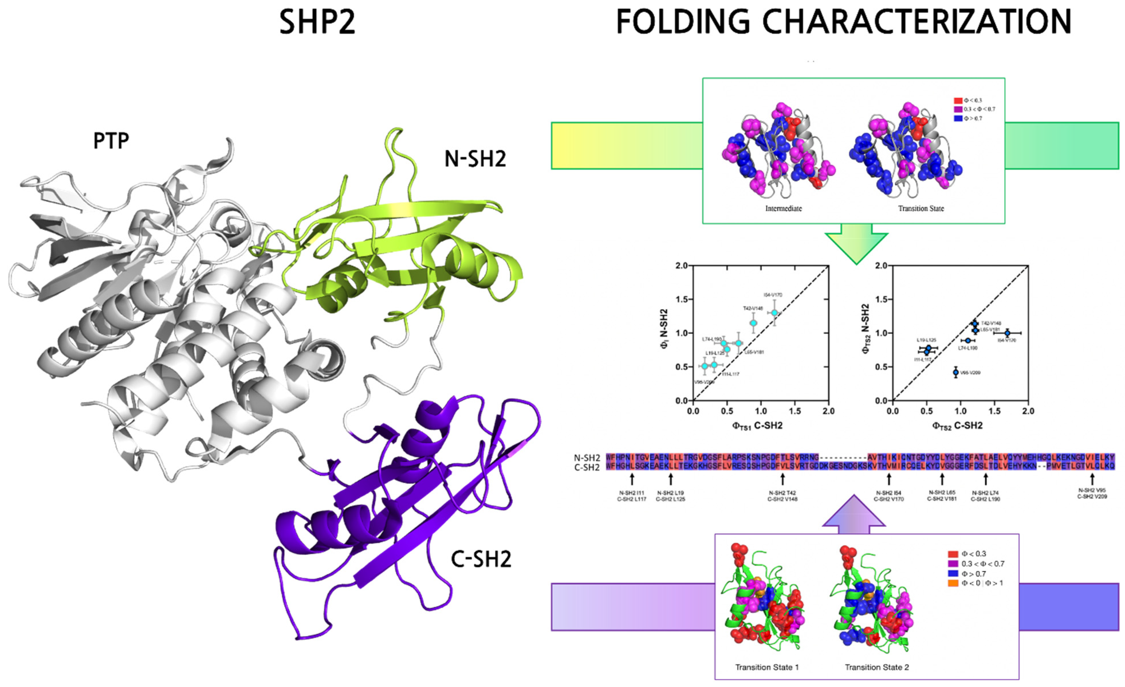
2. Folding Properties of SH2 Domains

SH2 domains are key mediators of the recognition of phosphorylated tyrosines, with the consequent prominent involvement in regulating a plethora of molecular and physiological pathways; however, only a few experimental works characterized the determinants of the thermodynamic stability and the folding of SH2 domains. Comprehending these aspects is fundamental in order to depict the molecular basis of the function of SH2 domains.
The resolution of a high number of SH2 domain three-dimensional structures highlights a highly conserved topology among the domain family [12,13]. They are ~100 residue globular domains, structurally arranged as two α-helices flanking three to five anti-parallel β strands that compose a central β sheet. This highly conserved three-dimensional structure makes SH2 domains perfect model systems for folding studies. In fact, one powerful methodology that allows one to understand the molecular details of the folding mechanism of a given protein is to compare its folding properties with other proteins sharing the same structure but possessing different amino acid sequences.
The folding pathway of the N-SH2 and C-SH2 domains of SHP2 has been characterized in great detail. The analysis of the (un)folding kinetics of N-SH2 revealed the presence of an intermediate along the reaction pathway. In fact, while the typical chevron plots (i.e., a semilogarithmic plot of the observed unfolding and refolding rate constant as a function of the concentration of denaturant) for two-state folding proteins is V-shaped [14], the N-SH2 domain chevron-plot reported a clear curvature in the refolding arm (the so-called roll-over effect) which represents a clear sign of the presence of an intermediate along the reaction [15]. Quantitative analysis of kinetic data allowed us to conclude that the intermediate is a low-energy species transiently populated during the folding reaction [16]. On the other hand, the folding kinetics of the C-SH2 domains obtained at a wide range of experimental conditions were different compared to the N-SH2 domain. In fact, the analysis of the chevron plot revealed a roll-over effect in the unfolding arm, and a rigorous analysis of the kinetic parameters revealed a folding scenario implying the presence of two transition states and a high-energy, never accumulating intermediate.
A useful approach to obtain information about the folding mechanism of a given domain is to gather structural details of the transition state(s) and intermediate(s) that occur along the reaction pathway. However, given their typical elusiveness to an experimental characterization, a structural characterization of transition states and intermediates can be performed only indirectly. A powerful methodology that can be applied to obtain such information is the ϕ-value analysis, an approach that relies on extensively mutating individual residues of the domain and monitoring the effect of mutations on the folding kinetics, inferring the role of each residue in the folding reaction and normalizing it in terms of native-like interactions in the probed state (details about the ϕ-value analysis methodology can be found at [17,18]). Both N-SH2 and C-SH2 were subjected to a ϕ-value analysis [19,20] (Figure 1). The analysis of ϕ-values obtained for the N-SH2 domain revealed that the intermediate state (early event of folding) is highly structured, with native-like interactions further locked in place in the late transition state. Interestingly, a comparison with the ϕ-values obtained for the C-SH2 highlights that the latter experiences a much lower degree of native-like interactions in the early events of folding. This aspect suggests that the two domains that possess an almost perfectly superimposable three-dimensional structure may be characterized by a different degree of residual structure in the denatured state, which influences and dictates the early events of folding. However, quantitative analysis of kinetic data highlights a generally conserved folding mechanism between the C-SH2 and N-SH2 domains.
Other SH2 domains that have been characterized in their folding properties are the SH2 domain of Src [21], the N-terminal and C-terminal SH2 domains of the p85 subunit of PI3K [22,23], and the SH2 domain of Crkl [24]. It is interesting that the SH2 domain from Crkl displays similar folding kinetics compared to the N-SH2 domain of SHP2, with a pronounced roll-over effect in the refolding arm of the chevron plot being compatible with an energetic profile implying the presence of an obligatory intermediate accumulating along the reaction. A peculiar case is represented by the C-SH2 domain of PI3KR (p85 regulatory subunit of PI3K). In this case, the analysis of kinetic data revealed that the domain populates a low-energy intermediate determined by peptidyl-prolyl cis-trans isomerization [23].
It would be tempting to speculate that the presence of intermediate(s) is mandatory for the productive folding of this protein family. However, the folding pathway of the C-SH2 domain of SHP2, which is characterized by a high-energy not accumulating intermediate, and experimental data obtained for the N-SH2 domain of p85 and the SH2 domain of Src that are compatible with a simple two-state folding mechanism [21,22,25] suggest that the presence of multiple energy minima is not obligatory for the folding SH2 domains. Further experimental data based on extensive mutagenesis, as well as on multidomain folding, are crucial for a better understanding of the folding properties of SH2 domains. In fact, since topology and function are tightly correlated in globular domains, deciphering the mechanism by how SH2 domains acquire their three-dimensional structure may unveil new perspectives on the determinants of their function. Moreover, mutations altering the function of the SH2 domain may not occur in the binding pocket (i.e., directly affecting protein-protein interaction) and may result in populating misfolded species that could disrupt SH2 domain function.

3. SH2 Domain Binding Properties

In general, two main events characterize protein-protein interactions, (i) recognition of a specific consensus sequence and (ii) stabilization of the bound complex, whose determinants can vary throughout domain families [26,27,28,29]. For SH2 domains, these aspects are crucial, given their prominent role in mediating the binding of ligands presenting a single specific post-translationally modified residue, binding specificity must occur by the recognition of different consensus sequences flanking the phosphotyrosine [1,30,31]. During the co-evolution with their ligands, members of the SH2 domain family adopted different strategies to specifically recognize the target sequences carrying pTyr residues, even though they preserved the same fold [32]. As observed for mostprotein families, the loops connecting secondary structural elements of SH2 domains are the most flexible and dynamic regions. In addition, recent works have shown that the wide spectrum of specificity observed in SH2 domains can depend on the loop variation [33]. However, the discovery that loops play key roles in the selectivity of SH2 domains not only provides a glimpse of the molecular basis of the SH2 domain/phosphopeptide recognition but also opens new frontiers at the development of specific inhibitors targeting interactions of SH2 domains implicated in pathological conditions [34].

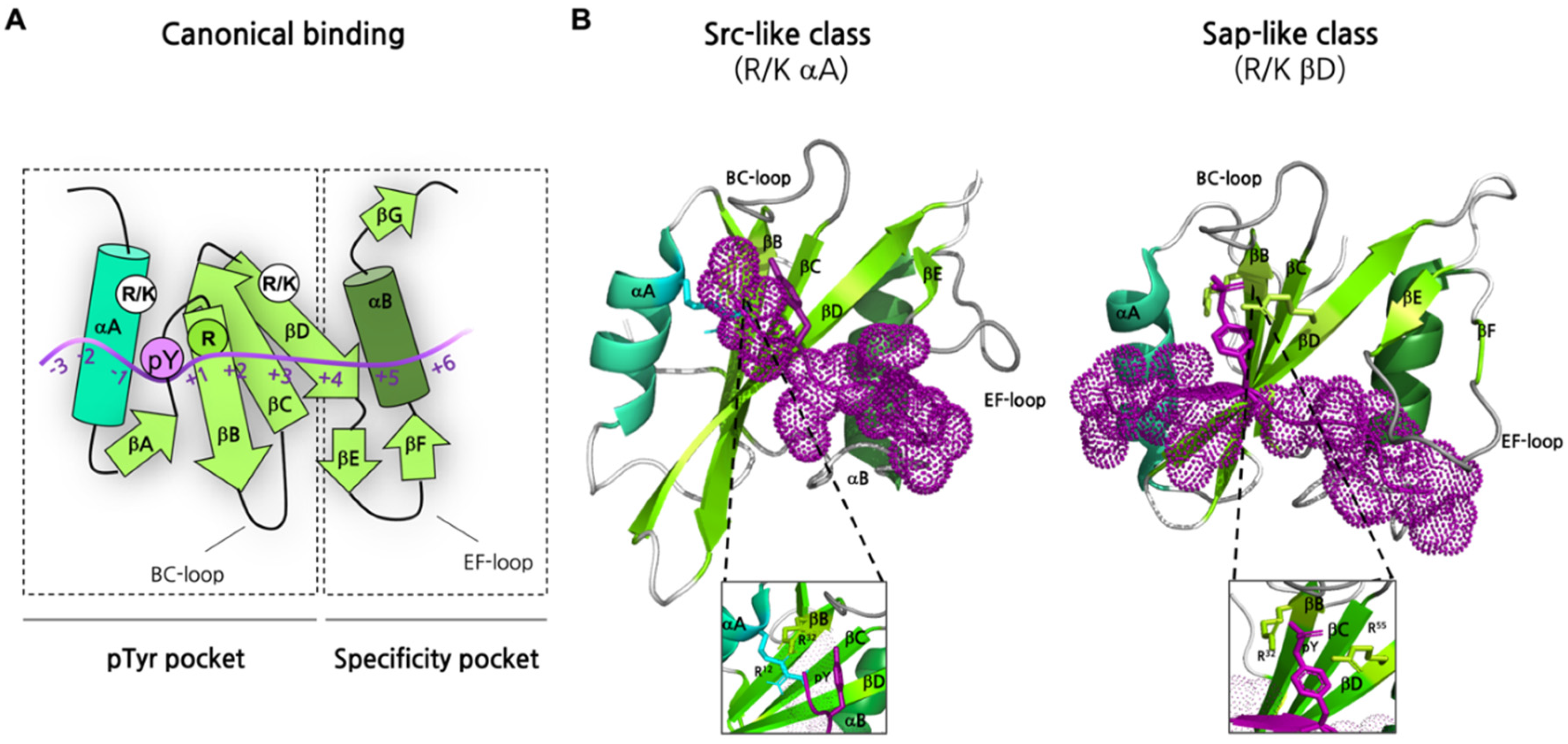
3.1. Defining the Structural Determinants of Recognition and Specificity

SH2 domains are considered key components of the pTyr-dependent signal transduction networks in eukaryotic cells [35], which are triggered by a wide range of external and internal stimuli. In this scenario, SH2 domains mediate the binding events through a fine regulation necessary for proper signaling and rapid cellular response [34,36,37,38].
SH2 domains recognize and bind sequences of ~10 residues, generally referred to as Short Linear Motifs (SLiMs) [39,40]. SLiMs are generally intrinsically disordered, i.e., they do not possess a well-defined three-dimensional structure and are highly dynamic in solution, although they can acquire structure upon binding with their interactor(s) [41,42]. SLiMs recognized by the SH2 domain can be found in structured or disordered protein regions, although disordered regions have been proposed to possess higher biological and physiological significance [43]. In SH2 domains, two structural regions determine the protein-ligand interaction [44]: (i) the pTyr binding site, a groove formed by αA helix, βB/βC/βD strands, and the BC loop, and (ii) the specificity pocket, which is a relatively large hydrophobic pocket mainly delimited by residues of αB helix, βD strand, and BG and EF loops (Figure 2A). The specificity pocket accommodates residues that are C-terminal to the pTyr. It is of interest to notice that the alignment of almost one hundred SH2 domain structures present in the Protein Data Bank (PDB) revealed that the N-terminal region that provides a pTyr-binding pocket is more conserved than the C-terminal half of the SH2 domain, which instead exhibits greater structural variability [35]. In fact, most conserved residues are clustered on the βB strand, where a conserved arginine residue in the FLVR motif (Figure 2B) (Arg βB5 or Arg175 in the v-Src SH2 domain) plays the central role in forming a double hydrogen bond with the phosphate group of pTyr [45,46]. The binding affinities of the SH2 domain for the phosphotyrosine peptide are reported to be in the range of what is typically observed for protein-protein interactions [26], ranging from 10−5 to 10−8 M [16,24,25,47].
Additional residues that are key for phosphopeptide binding are His βD4, Lys βD6, and Arg αA2, which coordinate and anchor the aromatic ring of the phospho-tyrosine [48]. Recent studies have confirmed the importance of the protonation state of His in position βD4 in determining the change in binding affinity over pH. In particular, the His169 of the C-terminal SH2 domain of SHP2 protein and the His60 of CrkL protein is crucial for the binding properties of those SH2 domains. Moreover, thermodynamic studies have established that the ionic interaction between Arg βB5 and pTyr is crucial for the affinity, providing more than half of the total binding energy [49]. Accordingly, our previous kinetic studies have shown a strong dependence on the ionic strength of the aforementioned interaction [16,24,25]. The pioneering work of Cantley and co-workers on the phosphatidylinositol 3-kinase evidenced the key role of residues flanking the phosphotyrosine in determining the specificity of a ligand to a particular SH2 domain [50]. A few years later, another work predicted the binding motifs of twenty-five SH2 domains, first describing the sequence-based and structure-based regulation of the binding [51]. A more recent study defined the determinants of specificity of around two-thirds of the human SH2 domains using the OPAL approach (Oriented Peptide Array Library, a high throughput method to study specificities of protein domains [52]) and identified sequence motifs recognized by different SH2 domains [53]. This work established that most SH2 domains display binding preferences for specific residues at the +2, +3, and +4 positions (relative to pTyr) in the ligand sequence. It is possible to divide SH2 into three different groups based on the consensus sequence recognized, as summarized in Table 2.
Similarly to what is observed for other protein-protein interaction domains, SH2 domains can be isolated or in tandem with other protein binding modules (Figure 3B). Many SH2 domain three-dimensional structures, isolated or in complex with their ligands, have been determined by X-ray and solution NMR (a representative example of the SH2 domain in complex with a peptide mimicking a physiological ligand is reported in Figure 3A), providing a fundamental contribution to the knowledge of the general aspects of the SH2-ligand recognition. The analysis of 63 human SH2 domains highlighted the simultaneous presence of the three binding pockets, which show selectivity for the +2, +3, and +4 residues of the phosphopeptide in all SH2 structures considered [33]. This observation led to hypothesizing a pivotal role of the SH2 domain loops in the control of the accessibility and shape of the binding pockets [33,54]. In particular, the study performed by Huang et al. highlighted that many SH2 domains exhibit specificity for a hydrophobic residue at the +3 position [53]. The nature of residue βD5 is crucial in determining this preference, whereas the +3 binding pocket is shaped by BG and EF loops [12,55]. Differently, 20 SH2 domains, including SH2 Grb2, displayed a preference for an asparagine residue at the +2 site of the ligand [53]. Consistent with the loops hypothesis, the structure of SH2 Grb2 in complex with its target sequence revealed that a tryptophan residue of the EF loop (TrpEF1) occupies the +3 binding pocket, favoring the interaction between Asn+2 of the peptide with βD6 and βE4 residues of the SH2 domain [56,57]. On the other side, SH2 domains listed in the group IIC were reported to be selective for a hydrophobic residue (preferentially Leu or Ile) at +4 position that is accommodated in a pocket formed by five hydrophobic residues (called “pentagon basket”). Interestingly, a Leu or Ile residue of the BG loop forms an intramolecular interaction inside the +4 binding pocket of those SH2 domains, not showing a preference for the +4 residue of the peptide [33,53]. In light of these structural considerations, the hypothesis of EF and BG loops as regulators of SH2 binding pockets has been supported by the engineering of new loops. In fact, by changing the loop sequence and conformation selectivity was altered as expected [33]. It is important to notice that the use of surface loops to determine binding specificity is not a distinct feature of SH2 domains [58,59,60,61,62]. On the other hand, the mechanism by which loops regulate binding pocket accessibility seems to be unique among protein-protein interaction modules [33].

3.2. Biophysical Characterization of SH2 Binding

Although SH2 domains are characterized by highly conserved fold and binding site structure, the molecular basis of selectivity is not completely understood, demanding additional experimental data aimed at characterizing from a thermodynamic and kinetic perspective the interaction occurring between SH2 domains and their ligands. Isothermal Titration Calorimetry (ITC) experiments have been carried out to study the affinity and selectivity of the SH2 domain of Src kinase [49]. A mutational analysis conducted on a peptide binding to the Src SH2 domain revealed that whilst conservative substitutions at +1, +2, and +3 positions resulted in minor destabilization, more drastic mutation to Ala revealed the +3 position as the most contributing to binding free energy. On the other hand, an analysis of the enthalpic change upon mutation showed the positions +1 and +2 to have a prominent role in providing an enthalpic contribution to binding, characterized by the formation of a highly ordered water molecule network [49]. These results provide useful information about the thermodynamics of the reaction, in particular by identifying entropic and enthalpic contribuitions to the change in free energy of the complex. However, equilibrium binding approaches are not feasible for a full understanding of the mechanism underlying the early recognition and the late stabilization events, for which kinetic approaches must be employed.
Stopped-flow fast kinetic binding experiments allowed for measuring the microscopic association and dissociation rate constants (kon and koff, respectively) for the binding reaction of the SH2 domains from SHP2, PI3K, and CrkL proteins with peptides mimicking their physiological ligands [16,24,25,63,64]. Overall, kinetic data confirmed the importance of the electrostatic interactions in the early recognition events, as shown by the decrease of kon values at increasing ionic strength conditions. Moreover, conservative site-directed variants of N-terminal SH2 domains of SHP2 and PI3K were generated and used to determine the energetic contribution of mutated residues to the binding reaction with Gab2608–620 and Gab2448–460, respectively [63,64].
Results from the N-SH2 domain of the SHP2 protein highlighted five mutations (T42S, T52S, I56V, L65A, and L88A) that positively or negatively affected the affinity for Gab2608–620 by influencing the complex stability (as indicated by changes of koff values) rather than the early molecular recognition [63]. The mutation T42S caused a ten-fold destabilization of the complex, with a clear effect on the koff of the reaction, suggesting a prominent role of T42 in the late events of binding. Importantly, a more pronounced effect on the complex stability was also measured for the binding reaction involving the Noonan Syndrome-causing T42A variant of N-SH2 SHP2. In particular, for this reaction, the koff value resulted 100 times lower than what was measured for the interaction of wild-type N-SH2 with Gab2608–620 [65].
A mutational analysis conducted on the N-SH2 of PI3K showed that the affinity of the T48S variant for Gab2448–460 was around 20-fold lower than that of the wild-type form [64]. Interestingly, the T48 residue of N-SH2 PI3K and the T42 residue of N-SH2 SHP2 share the same position in the corresponding 3D structures [PDBs: 4QSY and 2IUH]. However, both kon and koff values of the binding reaction of N-SH2 PI3K with Gab2 were altered upon mutation T48, meaning that the threonine at this position is involved in both formation and stability of the complex. Further mutations of N-SH2 PI3K influencing the kon and/or koff values of the binding reaction with Gab2 peptide were identified. Interestingly, although affecting the kinetics of the reaction, those mutations did not necessarily determine a change of affinity for the ligand. In addition, the energetic coupling between the P74 residue and +3 methionine of the Gab2 peptide was highlighted, suggesting a role for this residue in determining the recognition of non-pTyr residue. Further kinetic experimental data and structural characterization of complexes are demanded to fully understand the determinants of specificity and affinity that allow SH2 domains to discriminate among different pTyr sites in the crowded cellular environment.

3.3. Non-Canonical SH2 Binding

Although it has been well accepted that selectivity relies on the molecular interaction of residues forming the specificity pocket of the SH2 domain with C-terminal residues to the pTyr, different works highlighted unusual pTyr binding modes increasing the spectrum of specificity observed in the SH2 domain family [66] (for which a graphical example is reported in Figure 2B, right panel). For instance, the Arg βB5 residue of the conserved motif FLVR is involved in the formation of a salt bridge with an aspartic acid residue in the C-terminal SH2 domain of p120RasGAP. Consequently, the pTyr recognition is mediated by other residues, such as the basic residues at βD4 and βD6 position, and residues in the BC loop, through a multi-dentate mechanism [67]. On the other hand, in some SH2 domains, Arg βB5 is not present, and the crucial binding of pTyr to this residue cannot occur. In fact, the FLVR arginine is replaced by histidine in the SH2 domain of RIN2 [38] and TYK2 [68] and tryptophan in SH2D5 [38].
TYK2 belongs to the JAK protein family, whose members contain SH2 domains that display a non-canonical binding mode. The JAK proteins are cytoplasmatic multi-domain tyrosine kinases that are associated with the intracellular domains of the cytokine receptors [69]. SH2 domains of JAK proteins specifically bind a glutamate residue instead of the pTyr, supporting the constitutive interaction of the JAK proteins with the cytokine receptors [68,70]. The lack of pTyr recognition caused the Arg residue typically conserved in SH2 domains to be replaced by a His in TYK2 (H474) [68]. Although TYK2 maintains the conserved binding pocket of SH2 domains, the H474 residue does not participate in the interaction with the ligand Ifnar1. The carboxylate group of E497 of Ifnar1 is coordinated by other residues of the binding pocket (S476 and T477). Moreover, the C-terminal hydrophobic residues of the target sequence pack into the hydrophobic groove formed between the EF loop and the βG1/2 hairpin of the SH2 TYK2, resembling the specificity pocket of the typical SH2 domain [68,69]. Another unconventional SH2 domain belongs to the SLAM-associated protein (SAP) (Figure 2B). Its binding pocket is uncommonly elongated and interacts with pY -3 and pY +2 residues of the target motif, forming a “three-pronged plug” interaction [71]. Surprisingly, SH2 SAP also recognizes non-phosphorylated motifs due to the additional binding site, but with a lower affinity compared to the sequences containing pTyr residues [11].
Other factors contributing to binding diversity in SH2 domains are represented by: (i) their ability to bind more than one phosphorylated site from the same ligand, (ii) their incorporation in a multidomain system where each SH2 domain interacts with different phosphorylated sites, (iii) their assembly depending by the oligomeric state of the protein of which they are part of [66]. For example, two phosphorylated sites of Syk are recognized by the SH2 domain of VAV or PLCγ1 [72,73]. Curiously, whilst the first pTyr site is recognized by following the classical “two-pronged plug” mechanism that involves the ArgβB5 residue, the second pTyr motif of Syk interacts with basic residues of the βD strand and BG loop of the SH2 domain. An example of a protein containing tandem SH2 domains is the ZAP-70 tyrosine-kinase. This SH2 tandem recognizes two pTyr sites of the ITAM cytoplasmatic tails. In particular, the C-terminal SH2 binds one pTyr site in a canonical “bidentate” way, whereas the N-terminal SH2 uses the ArgβB5 residue to coordinate the other pTyr and additional residues coming from C-SH2 to form the second dock site of its binding motif [74]. Moreover, both binding affinity and specificity can be influenced by the oligomeric state of the protein incorporating SH2 domains. This has been revealed in the adaptor protein APS, where the SH2 domain forms a dimer capable of binding four pTyr residues of insulin receptor tyrosine kinase. Interestingly, in the dimer, the canonical specificity pocket of one SH2 domain is interrupted by the long αB helix of the other SH2 domain; thus, the interaction with the ligand is mediated only by the pTyr residues recognition [75].

3.4. Biomolecular Condensate Formation Mediated by SH2 Domains

It has been recently discovered that the interactions between proteins do not form only simple protein-ligand complexes but can lead to the formation of highly ordered ensembles characterized by the formation of liquid droplet biomolecular clusters (or condensates) that regulate the physiological activity of the proteins involved [76]. The general properties of these protein condensates and the mechanisms by which they form are beyond the scope of this paper and have been diffusely reviewed elsewhere (see, for example, [77,78,79,80]). What appears clear is that the functions of protein interaction modules are highly dependent on the cellular context [81], so different mechanisms of ligand binding may follow changes in the intracellular microenvironment.
One of the fundamental aspects of the formation of such condensates is their correlation to post-translational modifications [82,83], which determine temporal/spatial regulation of the clustering of specific proteins in response to external stimuli. For example, activation of RTKs signaling pathways determines the phosphorylation of specific tyrosine residues on the receptors and other substrates. It has been shown that SH2 domain-containing proteins, in particular adapter proteins such as Grb2, can drive Liquid-Liquid Phase Separation (LLPS) and protein condensates formation by binding different partners through their PPI domains (two SH3 domains and one SH2 domain in the case of Grb2) [84], thus highlighting a prominent role of protein phosphorylation in such processes. In fact, while Grb2 is recruited through its SH2 domain, additional scaffolding proteins such as Sos1 [85] and Gab2 [86,87] are recognized by Grb2 and provide sites for other specific interactions. Given that tyrosine phosphorylation and protein condensate formation are tightly correlated, and following what has been discussed in previous paragraphs, understanding the molecular basis of the SH2 domain binding mechanism is of primary importance to depict how these domains regulate signal transduction through the productive LLPS, and how their impairment could lead to the disruption of protein condensates.

4. The Pathological Role of SH2-Containing Proteins

As mentioned above, SH2-containing proteins are key components of signal transduction pathways, such as RAS-MAPK, JAK/STAT, and PI3K/AKT pathways. Dysregulation of such pathways due to mutations or altered expression of SH2-containing proteins induces several disorders and diseases. Thus, targeting SH2 domains is an interesting therapeutic strategy for drug design and development. In this section, we focus on diseases related to SH2-containing proteins, such as Noonan Syndrome (NS), cancers, and autoimmune diseases.

4.1. Noonan Syndrome: A Genetic Disorder Due to Mutations Affecting SH2 Domains from SHP2

Noonan syndrome (NS) (OMIM 163950) is an autosomal dominant disorder characterized by unusual facial features, short stature, and congenital cardiopathies. Other symptoms such as mental retardation, Webbed neck, cryptorchidism, chest deformity, and bleeding diathesis are also commonly associated with this disease. The prevalence of the disease is about 1 in 1000–2500 live births [88,89]. This genetic condition is caused by the hyperactivation of the RAS-MAPK molecular pathway. The mutations of a multi-domain phosphatase called SHP2 (PTPN11) protein induce 50% of NS cases.
SHP2 protein is composed of two SH2 domains (N-SH2 and C-SH2) followed by a PTP domain which retains the catalytical activity, and a C-terminal tail [90]. SHP2 possesses a particular mechanism of action, switching between two conformational states. The inactive state (typical of basal conditions) is characterized by a closed autoinhibited conformation in which the N-SH2 DE loop binds to the active site of the PTP domain. Upon interactions with binding partners, the active state adopts an open conformation, with the N-SH2 that moves away from the PTP active site, making it available for substrate recognition and turnover [91,92]. 40 NS-associated SHP2 mutations affecting 30residues close to or part of the N-SH2 domain–PTP domain interface have been described [93]. Mutations of this multi-domain phosphatase protein represent the major cause of Noonan Syndrome (NS) [94]. Currently, there are no effective treatments for such a genetic condition. Thus, understanding the mechanisms underlying SH2-containing-proteins functions on the development of such disease will determine the development of inhibitors of such proteins.
It is of interest to notice that the majority of the mutations related to the onset of NS occur mostly at the interface between N-SH2 and PTP domains, destabilizing the inactive form of SHP2, provoking the disruption of the autoinhibitory effect and leading to a hyperactivated phosphatase with a consequent gain-of-function (GOF) that enhances the RAS/MAPK pathway activation [93,94]. However, one NS-causing mutation, T42A, is located in the binding pocket of the N-SH2 domain, dramatically increasing enzyme activity [8,95]. Recently our group characterized quantitatively the effect of the T42A mutation on the binding of the isolated N-SH2 domain to a peptide mimicking one of its physiological binders, Gab2 [65]. The interaction between N-SH2 of SHP2 and Gab2 causes the release of its auto-inhibited conformation, triggering signal transduction and being crucial for enzyme activation in the cellular milieu [96]. Kinetic data revealed that the mutation impairs the ability of the N-SH2 domain to release the ligand, consequently promoting the hyperactivation of SHP2 phosphatase activity.

4.2. SH2-Containing Proteins and Immunodeficiency Disorders

Severe combined immunodeficiency (SCID) (OMMIM: 600,802) is the name of an ensemble of disorders with several genetic causes. Indeed, SCID is an autosomal recessive disorder caused by the mutations of specific genes. The disease is characterized by severe pulmonary infections, chronic diarrhea, failure to thrive, and persistent candidiasis. The prevalence of such a disorder is about 1 to 58,000 live births [97]. One of the causes is the mutation of the gene encoding ZAP-70. ZAP-70 (Zeta chain-associated protein kinase) is involved in signaling pathways that are important for the development and activation of T cells [74]. ZAP-70 is a 70kDa protein kinase, and it is normally expressed in the proximity of the surface of the membrane of lymphocytes. The protein is constituted of a tandem SH2-domain (N-SH2 and C-SH2), followed by a kinase domain connected to C-SH2 by an SH2-Kinase linker [74]. The pathogenic mutations are located throughout the gene and mostly in the kinase domains. However, as the phosphorylation of Tyr 315 and Tyr319, located in the SH2 kinase linker, is crucial for ZAP-70 activity, their mutations into phenylalanine lead to the inactivation of ZAP-70 downstream signaling events [98]. In addition, the missense mutation P80Q provokes a destabilization of the SH2 domain, leading to a severe SCID [99]. Moreover, mutations of the gene encoding ZAP-70 induce the production of unstable ZAP-70, leading to the abrogation of CD8+ T cell production and inactivation of CD4+ T cells [100]. Consequently, individuals with such mutations are more prone to infections, as their immune system is weakened.
X-linked agammaglobulinemia (XLA) is a primary immunodeficiency genetic disorder affecting B-lymphocyte development, with an estimated prevalence of 1:200,000 to 379,000 live births and ranked 5th in primary immunodeficiency [101]. In XLA patients, a reduced number of mature circulating B cells can be found, as well as lower serum Ig levels and disrupted lymphoid architecture. XLA is caused by mutations in Bruton’s tyrosine kinase (Btk), involved in B-cell receptor signaling. Around 20% of XLA-causing mutations rest on the SH2 domain and decrease protein stability [102]. Indeed, Btk is constituted of five different protein interacting domains, including SH3, SH2, and kinase domains. Even though the phosphotyrosine sites are located on the SH3 and KD domains, the SH2 domain of Btk has been reported to be critical for phospholipase C-gamma phosphorylation, and mutations of this domain cause XLA [103]. In fact, the main mutations on the SH2 domain of Btk observed in XLA patients are R288Q, R288W, L295P, R307G, R307T, Y334S, Y361C, L369F, and 1370M [103]. Tzeng and colleagues highlighted the inability of SH2 Btk mutants to bind phosphopeptide ligands, which could explain XLA development. In addition, Btk and kinase domain (KD) allosteric interaction possesses a crucial role in kinase activation, showing how XLA mutations in the SH2 domain cause the loss of function phenotype [104]. The absence of this functional protein leads to failure of B-cell development that incapacitates antibody production and subsequently leads to recurrent bacterial infections.

4.3. SH2-Containing Proteins and Cancer

Several SH2-containing proteins are involved in the development of certain types of cancers, such as breast cancer (Grb2, Grb7, STAT3, and Src), liver cancer (STAT3), Prostate cancer (STAT3, STAP2), Chronic Myelogenous Leukemia, CML (Bcl/Abl). The aberrant activation of signaling pathways (RAS/MAPK, PI3K/AKT, JAK/STAT), including cytokines and growth factors, has a key role in the onset of those types of cancers, promoting metastasis, angiogenesis, and cancer cell division or proliferation. The protein-protein interactions involved in the activation of these pathways are mainly mediated directly by SH2 domains (Figure 4).
Prostate cancer is one of the most common malignancies diagnosed in men (268,490 new cases) [105], causing approximately 350,000 death per year [106]. Prostate cancer progresses in a multi-step fashion, and androgen deprivation therapy is used for advanced and metastatic phases of the disease. MST2 kinase of the Hippo pathway phosphorylates STAT3 via its tyrosine residue located on its SH2 domain [107]. This hinders the SH2 domain of STAT3 from interacting with another phosphorylated counterpart, affecting STAT3 dimerization and activation by IL-6. Thus, mutations occurring on that tyrosine residue increase STAT3 activity and IL-6 expression. Indeed, by investigating the activity of MST2 on STAT3, Tang and colleagues highlighted the effect of the Hippo pathway on prostate cancer through the monomerization of STAT3 [107]. Previous studies carried out by Antonioli and colleagues reported in 2013 that hyperactivation of STAT3 plays a critical role in malignant initiation, tumor progression, and metastatic dissemination [108]. Another SH2-containing protein involved in prostate cancer is the STAP-2 protein [109]. STAP2 is a signal-transducing adaptor protein 2 that promotes prostate cancer growth by enhancing EGFR stabilization [110].
Liver cancer is one the deadliest cancers, with approximately 906,000 new cases and 830,000 deaths [111]. Chronic infections with Hepatitis B and C Viruses, HBV and HCV, Cirrhosis, excessive alcohol consumption, and obesity are the key risk factors for disease development. Melvin and colleagues reported that STAT3 affects the progression of liver cancer [112]. The Nemo-like kinase involved in the Ras/MAPK pathway is found to inhibit the phosphorylation of STAT3 by competing with its physiological binder GP130 (via YXXQ motif). GP130 activates STAT3 by recruiting and interacting with the SH2 domain of STAT3 [111], blocking JAK1/STAT3 interaction and consequently dysregulating the downstream events of the JAK/STAT pathway.
Chronic Myeloid Leukemia (CML) results from the neoplastic transformation originating from hematopoietic stem cells. The incidence of CML is approximately 1.6 per 100,000 population. The initial chronic phase (CP) shows an expansion of granulocytic cells, while the progression to the blast phase (BP) is characterized by a block of cell differentiation resulting in the presence of 30% or more myeloid or lymphoid blast cells in peripheral blood or bone marrow, or extramedullary infiltrates of blast cells. The hallmark genetic abnormality of CML is a t(9;22)(q34;q11) translocation in a ‘Philadelphia chromosome,’ containing a breakpoint cluster region (BCR) sequence, and chromosome 9, containing Abelson proto-oncogene (ABL) sequence. The N-terminal region of c-Abl is replaced in the Bcr-Abl fusion protein by bcr-encoded sequences inducing constitutive activation of Abl with a dysregulated tyrosine kinase activity and a consequent oncogenic ability [113]. The role of the Src homology 2 (SH2) domain of Bcr/Abl in transformation has been extensively studied. The precise SH2/N-lobe interaction is required for full activation of c-Abl. In CML cells, active Bcr/Abl phosphorylates BCR’s Y177, recruiting Grb2 through the interaction with the SH2-Grb2 domain [114]. This strong interaction leads to the recruitment of SOS at the myeloid cell membrane determining activation of the Ras/MAPK pathway and then a potent oncogenic proliferation (A schematic representation of the pathway is reported in Figure 4).
The disease has been correlated to expression levels of SH2 domain-containing protein tyrosine phosphatase-1 (SHP1). This protein is widely expressed in hematopoietic cells assuming a significant role in the regulation of Bcr-Abl, leading to uncontrolled cell proliferation and, additionally, to a reduced expression of the tumor suppressor SHIP1, a human inositol-5-phosphatase [115]. A marked decrease of this protein in CML cell lines and BP compared with CP provides evidence for a significant biological role in disease progression [116]. STAP-1 is a novel Bcr-Abl binding partner, inhibiting the apoptosis of CML stem cells by upregulating BCL-2 and BCL-xL anti-apoptotic genes [117]. This effect is obtained through the activation of STAT5, mediated by the SH3 and SH2 domains of Bcr-Abl [118].

5. Pharmacological Strategies Aimed to Regulate/Inhibit SH2 Domains

5.1. Peptidomimetics and Small Molecules Inhibitors

Many strategies have been proposed to change the functionality of SH2 domain-containing proteins, such as ABL, BTK, GRB2, SHP2, SRC, and STAT. However, the design of the appropriate interfering molecule must face the concentrated polar charges present in the phosphotyrosine binding pocket, as well as the very well-conserved amino acid sequences in the SH2 domains of different proteins. These aspects required a great effort in designing molecules able to permeate the hydrophobic cell membrane and bind the SH2 domain of a specific protein. Nevertheless, in the last decades, in the search for high-affinity SH2 inhibitors that are specific, stable, and cell-permeable, many different effective compounds have been produced.Historically, the first approaches aimed at blocking the SH2 function were focused on targeting the SH2 phosphotyrosine binding site [119]. Initial compounds tested were the pTyr-mimicking inhibitors (p-Tyr-isosteres), characterized by the presence of a phosphorylated Tyr with the phosphate bridging oxygen replaced by a methylene unit, thus ensuring stability to hydrolysis by phosphatases [120]. Many peptidomimetics-based inhibitors have been developed containing a phosphonomethyl phenylalanine residue (Pmp). For example, a hydroxy benzyl phosphinate has been developed, displaying a better cell permeability than other Pmp peptidomimetics due to its reduced negative charges and binding with relatively high affinity (KD of 0.53 μM) toward the SH2 domain of Grb2 protein [121,122]. More recently, peptidomimetics, with enhanced cell penetration, containing 4-phosphonodifluoromethylcinnamate have been developed by masking the phosphonate hydroxyls with reversible pivaloyloxymethyl protecting groups. A derivative of these compounds has been found to have a very high affinity to STAT3 SH2 (IC50 of 162 nM), and to inhibit the STAT3 phosphorylation, essential for its dimerization-dependent function(s), at 100 nM in MDA-MB-468 breast cancer cell line [123]. Moreover, the compound showed a very high grade of selectivity for STAT3 SH2, not inhibiting PI3K, Src SH2, and poorly binding to other STAT SH2 domains [123,124,125]. This result was mainly achieved thanks to the mimic amino acids sequence present in the compound, in which the auxiliary specificity elements (i.e., glutamine mimics) can be placed in the binding pocket of the STAT3 SH2 domain but not in those of the other SH2-containing proteins [124].
A different strategy based on peptidomimetics was employed for Syk kinase (ZAP-70). In this protein, the simultaneous binding of the two Syk SH2 domains to a dually phosphorylated receptor triggers allosteric rearrangement and activation of the kinase [126]. Thus, peptidomimetic inhibitors were developed to contain two distinct phosphorylated binding regions, separated by a flexible linker which acquired a pivotal role in assuring binding efficiency to the two Syk SH2 domains [127]. With this aim, photo-switchable rigid linkers were explored to assess the effects of cis-trans isomerization on binding affinity. By incorporating a well-studied photo-switchable core, 4-aminomethyl phenylazobenzoic acid (AMPB), as a linker between the two phosphorylated regions, it has been possible to achieve a compound with KD = 65 nM for the trans isomer obtained with visible light irradiation, and KD = 860 nM for cis isomer obtained with irradiation at 366 nm [128]. Even with still some drawbacks that need to be solved [129], photo-switchable systems could represent a novel strategy for developing inducible inhibitors.
Another interesting approach has been focused on the research of constrained peptides (bicyclic) that mimic pTyr using discontinuous epitope-binding surfaces and that aim to overcome the main problem of peptides and peptidomimetics, linked to poor cytosolic absorption by removing the charged phosphate group (non-phosphate peptide) with a carboxylate group [130]. These efforts have led to the development of the BC1 compound, a bicyclic peptide able to inhibit Grb2-SH2 with an IC50 of 350 nM. Unfortunately, even if BC1 demonstrated good cellular uptake in breast cancer cells, no antiproliferative effect was observed. This compound could bind anti-pTyr antibodies, demonstrating that a carboxylate group, instead of phosphate, could successfully mimic pTyr [131,132].
Peptide and peptidomimetics often performed poorly in cell-based assays, mainly due to poor cytosolic penetration or the absence of biological activity. The development of small molecule inhibitors might overcome these issues, possibly acting as pTyr-mimics but with better cytosolic penetration. One of the most promising classes of compounds as small molecule pTyr isostere seems to be the sulfonamide class [133]. A compound found after a screening of 920,000 molecules and then further improved (C188-9) was tested as a small molecule inhibitor of the STAT3 SH2 domain [134]. The C188-9 molecule showed a KD of 4.7 nM, and its treatment (100 mg/kg/day) in mouse xenograft models with UM-SCC-17B head and neck squamous cell carcinoma determines a significant reduction in tumor size [134]. Additionally, under the new name of TTI-101, it entered the phase I clinical trial for Tvardi Therapeutics.

5.2. Allosteric Inhibitors

Unfortunately, so far, very few of the strategies above-described produced compounds that entered clinical trials or have effective results in limiting or influencing the progression of pathologies without excessive toxicity (off-targets). For these reasons, new pharmacological strategies have been developed to avoid all the problems-related pitfalls in targeting the SH2 p-Tyr binding site. One of the most promising is focused on targeting the SH2 domain allosterically.
There are several protein systems in which SH2 domain(s) act as a regulator of the activity of an entire protein, often determining the switching from inactive to active conformation [104,126,135]. An interesting and well-studied example is represented by the SHP2 protein, which plays a central role in mediating signal transduction downstream of receptor tyrosine kinases (RTKs) such as EGFR, ERBB2, c-MET, and FLT3) or FRS2 protein (STAP) [136]. Considering the SHP2 structural and functional properties (described in paragraph 4.1), the possibility of pharmacologically stabilizing the autoinhibited conformation to block the cellular signaling became an attractive approach to fighting several types of cancers [136] or syndromes (e.g., Noonan syndrome) [95]. Indeed, these pathologies are often characterized by the presence of activating mutations of SHP2 (e.g., E76Q, F285S, S502P, D61V, E76K) mainly located at the interface between N-SH2 and PTP domain, and able to abolish the inhibitory interaction between its N-SH2 domain and phosphatase domain. For this reason, there were extensive efforts in searching for compounds able to target this interface to stabilize the autoinhibited conformation.
One of these inhibitors, SHP099, was found after a screening and improvement of a library of 100,000 compounds [137]. SHP099 is a small compound belonging to the class of pyrazinyl molecules. The crystal structure of SHP099 in complex with SHP2, at 1.7Å resolution, revealed that SHP2 was in an auto-inhibited, inactive conformation with the N-terminal SH2 domain blocking the active site, bound to the central tunnel formed at the interface of the N-SH2, C-SH2, and PTP domains. Key interactions included Hydrogen bonds involving residues Arg111 and Phe113 were of key importance. Those residues are physically located on the linker between the N-SH2 and C-SH2 domains, as well as Glu250 from the PTP domain. The dichlorophenyl group of SHP099 explored hydrophobic interactions with Leu254, Gln257, Pro491, and Gln495 side chains of the PTP domain. No activity was reported versus SHP1, the closest homolog of SHP2, sharing 61% amino acid sequence identity, supporting its high degree of target selectivity. The selectivity of SHP099 for SHP2 over SHP1 might be due to the repositioning of the linker between the two SH2 domains in the homolog SHP1. This change would remove key SHP099 interactions (with Arg109 in SHP1 and Arg111 in SHP2) and yield a significantly larger central tunnel, unfavorable for an SHP099 effect. SHP099 was reported to be a highly potent (IC50 = 0.071 μM) SHP2 inhibitor. It suppressed the RAS-ERK signaling with an IC50 of about 25 μM and inhibited the proliferation of receptor-tyrosine-kinase-driven human hematopoietic or colon rectal cancer cells in vitro. It was shown to be efficacious in controlling tumor growth in mouse tumor xenograft models [137]. In addition, evidence has been reported on the efficacy of SHP009 to inhibit the mutant E76A SHP2, the form frequently identified in Noonan syndrome and leukemia, for which an IC50 of 0.12 μM has been measured [138].
In search of optimization of the pyrazine class of allosteric SHP2 inhibitors, TNO155 has been synthesized. This molecule, strictly related to SHP099, is a potent inhibitor of SHP2 (IC50 = 0.011 μM), with high cellular potency (p-ERK IC50 = 0.011 μM; antiproliferation of esophageal cancer cells KYSE-520 IC50 = 0.100 μM) and high solubility (0.736 mM). It can bind to the tunnel site of SHP2, although with interactions different from those reported for SNP99. Antitumor effects could also be recorded in the EGFR-driven esophageal carcinoma xenograft model, KYSE-520, at a dose lower than those of SNP099; as SNP099, it did not cause body weight loss, but differently from SNP099, it proved negative in vitro phototoxicity tests [139]. TNO155 has recently entered phase I clinical trial for advanced solid tumors both as a single agent and in combination therapies with narzatinib (inhibitor of the tyrosine kinase activity of the EGFR bearing activating mutations such as Tyr790Met), ribociclib, (selective inhibitor of CDKs 4and 6) or spartalizumab (humanized monoclonal antibody against PD-1) [140].
Another SHP2 allosteric inhibitor molecule, LY6, was found by exploiting a combination of computational drug design and experimental assay. LY6 was rationale-designed to bind an alternative pocket present between C-SH2 and PTP domain and, similarly to SHP099, to block SHP2 in the inactive state. Even if the co-crystal structure of this compound with SHP2 is still lacking, structure–activity relationship analyses of optimized derivatives suggest that the main binding mode is characterized by strictly connecting the C-SH2 and PTP domain. Additionally, despite LY6 inhibiting wild-type SHP2 and SPH2 E76K with low affinity (IC50 9 μM and 7.7 μM, respectively), it seems to be highly selective for SHP2 rather than SHP1 (about 7-fold). In Ba/F3, an IL-3-dependent murine pro-B lymphoma cell line, LY6 suppressed cell proliferation and inhibited the IL-3-induced phosphorylation of Erk, Akt, Jak2, and Stat5. Also, mouse and patients cells with Juvenile Myelomonocytic Leukemia (JMML) bearing E76K SHP2 are more sensitive to LY6 than wild-type cells, highlighting its possible use for the treatment of PTPN11-associated malignancies [141].
The possibility for dual allosteric inhibition of SHP2 has also been evaluated, considering that additional allosteric binding sites have been predicted on both sides of the N-SH2/PTP domain interface, i.e., the “latch” binding site, located approximately 20 Å from the tunnel, and the “groove” site on the opposite side of the SHP2 protein. The triazolo-quinazolinone compounds SHP244, 844, and 504 have been reported to bind the latch binding site and inhibit the activity of near full-length SHP21-525 protein with IC50 values of 60, 18.9, and 21 µM, respectively. X-ray structure analysis of SHP2 with SHP099, bound at the tunnel site, and SHP244, 844, and 504, at the latch binders, showed that each of the three triazolo-quinazolinone compounds bound to SHP2 simultaneously with SHP099. Although their efficacy was much lower than those of SHP099 (IC50 0.070 μM), co-treatment of KYSE-520 cells with SHP099 (0.2 μM) and SHP504 (30 μM) has been shown to result in a stronger downregulation of DUSP6, a pharmacodynamic marker of the MAPK signal downstream of SHP2, with respect to the effect, compared to each of the single agents, thus paving the way to new possible combined approaches [142].
To summarize, a list of effective molecules on the SH2 domain binding is reported below, in Table 3.

6. Conclusions

SH2 domains are prototypical “readers” of phosphorylated tyrosine residues. Since tyrosine phosphorylation is a post-translational modification that regulates several physiological and molecular pathways in the eukaryotic cell, SH2 domains represent a fundamental piece in the intricate puzzle of cell signaling. As a consequence, dysregulation of SH2-domains mediated PPIs are rigorously involved in the onset of several pathologies. In this review paper, we summarized the current knowledge about the folding and binding properties of SH2 domains, as well as their roles played in pathogenesis. A great effort has been made over the years to understand the determinants of the function of SH2 domains in terms of binding mechanisms with their ligands and regulation of SH2 domain-mediated molecular pathways. However, while through structural and biophysical approaches, we could understand the role of residues located in loops and binding pockets in determining affinity and specificity for their ligands, we still have to investigate the role of single residues in the early events of the association and late events of complex stabilization of the binding event. This could represent a fundamental step toward a better comprehension of the effect of pathological mutations on the binding of SH2 domains and, consequently, to the design of pharmaceutical strategies aimed at regulating their (mis)functions. In particular, a synergistic approach based on site-directed mutagenesis, binding experiments, and molecular dynamic simulations (which could take advantage of a large amount of structural information available about SH2 domains) it would be possible to map, at an almost atomic level, the contribution of single residue side-chains on the binding of the inhibiting molecules, providing useful insights toward the design of inhibitor molecules displaying increased affinity and performance. Our analysis leads to the evidence that the intense pharmacological interest in SH2 domains is of fundamental importance to developing new therapeutics and that further analysis of their binding mechanisms and the determinants of specificity is demanded to improve our ability to inhibit/regulate SH2-mediated protein-protein interactions.

Author Contributions

Conceptualization, S.G. and A.T.; Writing—Original Draft Preparation, A.D., D.S., F.M., C.N., V.P., L.P., L.M., P.P., S.G. and A.T.; Writing—Review & Editing, S.G. and A.T. All authors have read and agreed to the published version of the manuscript.

Funding

Work partly supported by grants from the Italian Ministero dell’Istruzione dell’Università e della Ricerca (Progetto di Interesse “Invecchiamento” to S.G.), Sapienza University of Rome (RP11715C34AEAC9B and RM1181641C2C24B9, RM11916B414C897E, RG12017297FA7223 to S.G, AR22117A3CED340A to C.N.), by an ACIP grant (ACIP 485–21) from Institut Pasteur Paris to S.G., the Associazione Italiana per la Ricerca sul Cancro (Individual Grant—IG 24551 to S.G.), the Regione Lazio (Progetti Gruppi di Ricerca LazioInnova A0375-2020-36559 to S.G.), European Union’s Horizon 2020 research and Innovation programme under the Marie Skłodowska-Curie Grant Agreement UBIMOTIF No 860517 (to S.G.) the Istituto Pasteur Italia, “Teresa Ariaudo Research Project” 2018, and “Research Program 2022–2023 Under 45 Call 2020” (to A.T.).

Institutional Review Board Statement

Not applicable.

Informed Consent Statement

Not applicable.

Data Availability Statement

No new data were created or analyzed in this study. Data sharing is not applicable to this article.

Conflicts of Interest

The authors declare no conflict of interest.

References

  1. Pawson, T.; Nash, P. Assembly of Cell Regulatory Systems through Protein Interaction Domains. Science 2003, 300, 445–452. [Google Scholar] [CrossRef] [PubMed] [Green Version]
  2. Hunter, T. Tyrosine Phosphorylation: Thirty Years and Counting. Curr. Opin. Cell Biol. 2009, 21, 140–146. [Google Scholar] [CrossRef] [Green Version]
  3. Kavanaugh, W.M.; Turck, C.W.; Williams, L.T. PTB Domain Binding to Signaling Proteins through a Sequence Motif Containing Phosphotyrosine. Science 1995, 268, 1177–1179. [Google Scholar] [CrossRef] [PubMed]
  4. Benes, C.H.; Wu, N.; Elia, A.E.H.; Dharia, T.; Cantley, L.C.; Soltoff, S.P. The C2 Domain of PKCdelta Is a Phosphotyrosine Binding Domain. Cell 2005, 121, 271–280. [Google Scholar] [CrossRef] [Green Version]
  5. Mukherjee, M.; Chow, S.Y.; Yusoff, P.; Seetharaman, J.; Ng, C.; Sinniah, S.; Koh, X.W.; Asgar, N.F.M.; Li, D.; Yim, D.; et al. Structure of a Novel Phosphotyrosine-Binding Domain in Hakai That Targets E-Cadherin. EMBO J. 2012, 31, 1308–1319. [Google Scholar] [CrossRef] [PubMed] [Green Version]
  6. Liu, B.A.; Shah, E.; Jablonowski, K.; Stergachis, A.; Engelmann, B.; Nash, P.D. The SH2 Domain–Containing Proteins in 21 Species Establish the Provenance and Scope of Phosphotyrosine Signaling in Eukaryotes. Sci. Signal. 2011, 4, ra83. [Google Scholar] [CrossRef] [Green Version]
  7. Tartaglia, M.; Gelb, B.D. Noonan Syndrome and Related Disorders: Genetics and Pathogenesis. Annu. Rev. Genom. Hum. Genet. 2005, 6, 45–68. [Google Scholar] [CrossRef]
  8. Keilhack, H.; David, F.S.; McGregor, M.; Cantley, L.C.; Neel, B.G. Diverse Biochemical Properties of Shp2 Mutants. Implications for Disease Phenotypes. J. Biol. Chem. 2005, 280, 30984–30993. [Google Scholar] [CrossRef] [Green Version]
  9. Niihori, T.; Aoki, Y.; Ohashi, H.; Kurosawa, K.; Kondoh, T.; Ishikiriyama, S.; Kawame, H.; Kamasaki, H.; Yamanaka, T.; Takada, F.; et al. Functional Analysis of PTPN11/SHP-2 Mutants Identified in Noonan Syndrome and Childhood Leukemia. J. Hum. Genet. 2005, 50, 192–202. [Google Scholar] [CrossRef] [Green Version]
  10. Higashi, H.; Tsutsumi, R.; Muto, S.; Sugiyama, T.; Azuma, T.; Asaka, M.; Hatakeyama, M. SHP-2 Tyrosine Phosphatase as an Intracellular Target of Helicobacter Pylori CagA Protein. Science 2002, 295, 683–686. [Google Scholar] [CrossRef]
  11. Poy, F.; Yaffe, M.B.; Sayos, J.; Saxena, K.; Morra, M.; Sumegi, J.; Cantley, L.C.; Terhorst, C.; Eck, M.J. Crystal Structures of the XLP Protein SAP Reveal a Class of SH2 Domains with Extended, Phosphotyrosine-Independent Sequence Recognition. Mol. Cell 1999, 4, 555–561. [Google Scholar] [CrossRef] [PubMed]
  12. Waksman, G.; Kominos, D.; Robertson, S.C.; Pant, N.; Baltimore, D.; Birge, R.B.; Cowburn, D.; Hanafusa, H.; Mayer, B.J.; Overduin, M.; et al. Crystal Structure of the Phosphotyrosine Recognition Domain SH2 of V-Src Complexed with Tyrosine-Phosphorylated Peptides. Nature 1992, 358, 646–653. [Google Scholar] [CrossRef] [PubMed]
  13. Waksman, G.; Shoelson, S.E.; Pant, N.; Cowburn, D.; Kuriyan, J. Binding of a High Affinity Phosphotyrosyl Peptide to the Src SH2 Domain: Crystal Structures of the Complexed and Peptide-Free Forms. Cell 1993, 72, 779–790. [Google Scholar] [CrossRef] [PubMed]
  14. Parker, M.J.; Spencer, J.; Clarke, A.R. An Integrated Kinetic Analysis of Intermediates and Transition States in Protein Folding Reactions. J. Mol. Biol. 1995, 253, 771–786. [Google Scholar] [CrossRef] [PubMed]
  15. Gianni, S.; Ivarsson, Y.; Jemth, P.; Brunori, M.; Travaglini-Allocatelli, C. Identification and Characterization of Protein Folding Intermediates. Biophys. Chem. 2007, 128, 105–113. [Google Scholar] [CrossRef] [Green Version]
  16. Bonetti, D.; Troilo, F.; Toto, A.; Travaglini-Allocatelli, C.; Brunori, M.; Gianni, S. Mechanism of Folding and Binding of the N-Terminal SH2 Domain from SHP2. J. Phys. Chem. B 2018, 122, 11108–11114. [Google Scholar] [CrossRef]
  17. Fersht, A.R.; Matouschek, A.; Serrano, L. The Folding of an Enzyme. I. Theory of Protein Engineering Analysis of Stability and Pathway of Protein Folding. J. Mol. Biol. 1992, 224, 771–782. [Google Scholar] [CrossRef]
  18. Fersht, A.R.; Sato, S. Phi-Value Analysis and the Nature of Protein-Folding Transition States. Proc. Natl. Acad. Sci. USA 2004, 101, 7976–7981. [Google Scholar] [CrossRef] [Green Version]
  19. Visconti, L.; Malagrinò, F.; Gianni, S.; Toto, A. Structural Characterization of an On-Pathway Intermediate and Transition State in the Folding of the N-Terminal SH2 Domain from SHP2. FEBS J. 2019, 286, 4769–4777. [Google Scholar] [CrossRef]
  20. Toto, A.; Malagrinò, F.; Nardella, C.; Pennacchietti, V.; Pagano, L.; Santorelli, D.; Diop, A.; Gianni, S. Characterization of Early and Late Transition States of the Folding Pathway of a SH2 Domain. Protein Sci. Publ. Protein Soc. 2022, 31, e4332. [Google Scholar] [CrossRef]
  21. Wildes, D.; Anderson, L.M.; Sabogal, A.; Marqusee, S. Native State Energetics of the Src SH2 Domain: Evidence for a Partially Structured State in the Denatured Ensemble. Protein Sci. Publ. Protein Soc. 2006, 15, 1769–1779. [Google Scholar] [CrossRef] [PubMed] [Green Version]
  22. Visconti, L.; Malagrinò, F.; Toto, A.; Gianni, S. The Kinetics of Folding of the NSH2 Domain from P85. Sci. Rep. 2019, 9, 4058. [Google Scholar] [CrossRef] [PubMed] [Green Version]
  23. Troilo, F.; Malagrinò, F.; Visconti, L.; Toto, A.; Gianni, S. The Effect of Proline Cis-Trans Isomerization on the Folding of the C-Terminal SH2 Domain from P85. Int. J. Mol. Sci. 2019, 21, 125. [Google Scholar] [CrossRef] [PubMed] [Green Version]
  24. Nardella, C.; Toto, A.; Santorelli, D.; Pagano, L.; Diop, A.; Pennacchietti, V.; Pietrangeli, P.; Marcocci, L.; Malagrinò, F.; Gianni, S. Folding and Binding Mechanisms of the SH2 Domain from Crkl. Biomolecules 2022, 12, 1014. [Google Scholar] [CrossRef]
  25. Nardella, C.; Malagrinò, F.; Pagano, L.; Rinaldo, S.; Gianni, S.; Toto, A. Determining Folding and Binding Properties of the C-Terminal SH2 Domain of SHP2. Protein Sci. Publ. Protein Soc. 2021, 30, 2385–2395. [Google Scholar] [CrossRef]
  26. Ivarsson, Y.; Jemth, P. Affinity and Specificity of Motif-Based Protein-Protein Interactions. Curr. Opin. Struct. Biol. 2019, 54, 26–33. [Google Scholar] [CrossRef] [Green Version]
  27. Schreiber, G.; Keating, A.E. Protein Binding Specificity versus Promiscuity. Curr. Opin. Struct. Biol. 2011, 21, 50–61. [Google Scholar] [CrossRef] [Green Version]
  28. Schreiber, G. Kinetic Studies of Protein-Protein Interactions. Curr. Opin. Struct. Biol. 2002, 12, 41–47. [Google Scholar] [CrossRef]
  29. Gianni, S.; Jemth, P. How Fast Is Protein-Ligand Association? Trends Biochem. Sci. 2017, 42, 847–849. [Google Scholar] [CrossRef]
  30. Koch, C.A.; Anderson, D.; Moran, M.F.; Ellis, C.; Pawson, T. SH2 and SH3 Domains: Elements That Control Interactions of Cytoplasmic Signaling Proteins. Science 1991, 252, 668–674. [Google Scholar] [CrossRef]
  31. Pawson, T.; Gish, G.D.; Nash, P. SH2 Domains, Interaction Modules and Cellular Wiring. Trends Cell Biol. 2001, 11, 504–511. [Google Scholar] [CrossRef] [PubMed]
  32. Ran, X.; Song, J. Structural Insight into the Binding Diversity between the Tyr-Phosphorylated Human EphrinBs and Nck2 SH2 Domain. J. Biol. Chem. 2005, 280, 19205–19212. [Google Scholar] [CrossRef] [PubMed] [Green Version]
  33. Kaneko, T.; Huang, H.; Zhao, B.; Li, L.; Liu, H.; Voss, C.K.; Wu, C.; Schiller, M.R.; Li, S.S.-C. Loops Govern SH2 Domain Specificity by Controlling Access to Binding Pockets. Sci. Signal. 2010, 3, ra34. [Google Scholar] [CrossRef] [PubMed]
  34. Machida, K.; Mayer, B.J. The SH2 Domain: Versatile Signaling Module and Pharmaceutical Target. Biochim. Biophys. Acta 2005, 1747, 1–25. [Google Scholar] [CrossRef]
  35. Kaneko, T.; Joshi, R.; Feller, S.M.; Li, S.S. Phosphotyrosine Recognition Domains: The Typical, the Atypical and the Versatile. Cell Commun. Signal. CCS 2012, 10, 32. [Google Scholar] [CrossRef] [Green Version]
  36. Mayer, B.J.; Jackson, P.K.; Baltimore, D. The Noncatalytic Src Homology Region 2 Segment of Abl Tyrosine Kinase Binds to Tyrosine-Phosphorylated Cellular Proteins with High Affinity. Proc. Natl. Acad. Sci. USA 1991, 88, 627–631. [Google Scholar] [CrossRef] [Green Version]
  37. Muller, A.J.; Pendergast, A.M.; Havlik, M.H.; Puil, L.; Pawson, T.; Witte, O.N. A Limited Set of SH2 Domains Binds BCR through a High-Affinity Phosphotyrosine-Independent Interaction. Mol. Cell. Biol. 1992, 12, 5087–5093. [Google Scholar] [CrossRef]
  38. Liu, B.A.; Jablonowski, K.; Raina, M.; Arcé, M.; Pawson, T.; Nash, P.D. The Human and Mouse Complement of SH2 Domain Proteins-Establishing the Boundaries of Phosphotyrosine Signaling. Mol. Cell 2006, 22, 851–868. [Google Scholar] [CrossRef]
  39. Diella, F.; Haslam, N.; Chica, C.; Budd, A.; Michael, S.; Brown, N.P.; Trave, G.; Gibson, T.J. Understanding Eukaryotic Linear Motifs and Their Role in Cell Signaling and Regulation. Front. Biosci. J. Virtual Libr. 2008, 13, 6580–6603. [Google Scholar] [CrossRef] [Green Version]
  40. Neduva, V.; Russell, R.B. Peptides Mediating Interaction Networks: New Leads at Last. Curr. Opin. Biotechnol. 2006, 17, 465–471. [Google Scholar] [CrossRef]
  41. Wright, P.E.; Dyson, H.J. Intrinsically Disordered Proteins in Cellular Signalling and Regulation. Nat. Rev. Mol. Cell Biol. 2015, 16, 18–29. [Google Scholar] [CrossRef] [PubMed]
  42. Toto, A.; Malagrinò, F.; Visconti, L.; Troilo, F.; Pagano, L.; Brunori, M.; Jemth, P.; Gianni, S. Templated Folding of Intrinsically Disordered Proteins. J. Biol. Chem. 2020, 295, 6586–6593. [Google Scholar] [CrossRef] [PubMed] [Green Version]
  43. Ren, S.; Uversky, V.N.; Chen, Z.; Dunker, A.K.; Obradovic, Z. Short Linear Motifs Recognized by SH2, SH3 and Ser/Thr Kinase Domains Are Conserved in Disordered Protein Regions. BMC Genom. 2008, 9 (Suppl. S2), S26. [Google Scholar] [CrossRef] [PubMed] [Green Version]
  44. Marasco, M.; Carlomagno, T. Specificity and Regulation of Phosphotyrosine Signaling through SH2 Domains. J. Struct. Biol. X 2020, 4, 100026. [Google Scholar] [CrossRef]
  45. Songyang, Z.; Shoelson, S.E.; McGlade, J.; Olivier, P.; Pawson, T.; Bustelo, X.R.; Barbacid, M.; Sabe, H.; Hanafusa, H.; Yi, T. Specific Motifs Recognized by the SH2 Domains of Csk, 3BP2, Fps/Fes, GRB-2, HCP, SHC, Syk, and Vav. Mol. Cell. Biol. 1994, 14, 2777–2785. [Google Scholar] [CrossRef]
  46. Liu, X.; Brodeur, S.R.; Gish, G.; Songyang, Z.; Cantley, L.C.; Laudano, A.P.; Pawson, T. Regulation of C-Src Tyrosine Kinase Activity by the Src SH2 Domain. Oncogene 1993, 8, 1119–1126. [Google Scholar]
  47. Ladbury, J.E.; Lemmon, M.A.; Zhou, M.; Green, J.; Botfield, M.C.; Schlessinger, J. Measurement of the Binding of Tyrosyl Phosphopeptides to SH2 Domains: A Reappraisal. Proc. Natl. Acad. Sci. USA 1995, 92, 3199–3203. [Google Scholar] [CrossRef] [Green Version]
  48. Liu, B.A.; Engelmann, B.W.; Nash, P.D. The Language of SH2 Domain Interactions Defines Phosphotyrosine-Mediated Signal Transduction. FEBS Lett. 2012, 586, 2597–2605. [Google Scholar] [CrossRef] [Green Version]
  49. Grucza, R.A.; Bradshaw, J.M.; Fütterer, K.; Waksman, G. SH2 Domains: From Structure to Energetics, a Dual Approach to the Study of Structure-Function Relationships. Med. Res. Rev. 1999, 19, 273–293. [Google Scholar] [CrossRef]
  50. Cantley, L.C.; Auger, K.R.; Carpenter, C.; Duckworth, B.; Graziani, A.; Kapeller, R.; Soltoff, S. Oncogenes and Signal Transduction. Cell 1991, 64, 281–302. [Google Scholar] [CrossRef]
  51. Songyang, Z.; Shoelson, S.E.; Chaudhuri, M.; Gish, G.; Pawson, T.; Haser, W.G.; King, F.; Roberts, T.; Ratnofsky, S.; Lechleider, R.J. SH2 Domains Recognize Specific Phosphopeptide Sequences. Cell 1993, 72, 767–778. [Google Scholar] [CrossRef] [PubMed]
  52. Rodriguez, M.; Li, S.S.-C.; Harper, J.W.; Songyang, Z. An Oriented Peptide Array Library (OPAL) Strategy to Study Protein-Protein Interactions. J. Biol. Chem. 2004, 279, 8802–8807. [Google Scholar] [CrossRef] [PubMed] [Green Version]
  53. Huang, H.; Li, L.; Wu, C.; Schibli, D.; Colwill, K.; Ma, S.; Li, C.; Roy, P.; Ho, K.; Songyang, Z.; et al. Defining the Specificity Space of the Human SRC Homology 2 Domain. Mol. Cell. Proteom. MCP 2008, 7, 768–784. [Google Scholar] [CrossRef] [PubMed] [Green Version]
  54. Liu, H.; Huang, H.; Voss, C.; Kaneko, T.; Qin, W.T.; Sidhu, S.; Li, S.S.-C. Surface Loops in a Single SH2 Domain Are Capable of Encoding the Spectrum of Specificity of the SH2 Family. Mol. Cell. Proteom. MCP 2019, 18, 372–382. [Google Scholar] [CrossRef] [Green Version]
  55. Eck, M.J.; Shoelson, S.E.; Harrison, S.C. Recognition of a High-Affinity Phosphotyrosyl Peptide by the Src Homology-2 Domain of P56lck. Nature 1993, 362, 87–91. [Google Scholar] [CrossRef]
  56. Ogura, K.; Tsuchiya, S.; Terasawa, H.; Yuzawa, S.; Hatanaka, H.; Mandiyan, V.; Schlessinger, J.; Inagaki, F. Solution Structure of the SH2 Domain of Grb2 Complexed with the Shc-Derived Phosphotyrosine-Containing Peptide. J. Mol. Biol. 1999, 289, 439–445. [Google Scholar] [CrossRef]
  57. Rahuel, J.; Gay, B.; Erdmann, D.; Strauss, A.; Garcia-Echeverría, C.; Furet, P.; Caravatti, G.; Fretz, H.; Schoepfer, J.; Grütter, M.G. Structural Basis for Specificity of Grb2-SH2 Revealed by a Novel Ligand Binding Mode. Nat. Struct. Biol. 1996, 3, 586–589. [Google Scholar] [CrossRef]
  58. Zhao, H.; Naganathan, S.; Beckett, D. Thermodynamic and Structural Investigation of Bispecificity in Protein-Protein Interactions. J. Mol. Biol. 2009, 389, 336–348. [Google Scholar] [CrossRef] [Green Version]
  59. Akiva, E.; Itzhaki, Z.; Margalit, H. Built-in Loops Allow Versatility in Domain-Domain Interactions: Lessons from Self-Interacting Domains. Proc. Natl. Acad. Sci. USA 2008, 105, 13292–13297. [Google Scholar] [CrossRef] [Green Version]
  60. Sidhu, S.S.; Fellouse, F.A. Synthetic Therapeutic Antibodies. Nat. Chem. Biol. 2006, 2, 682–688. [Google Scholar] [CrossRef]
  61. Hiipakka, M.; Saksela, K. Versatile Retargeting of SH3 Domain Binding by Modification of Non-Conserved Loop Residues. FEBS Lett. 2007, 581, 1735–1741. [Google Scholar] [CrossRef] [PubMed]
  62. Li, S.S.-C. Specificity and Versatility of SH3 and Other Proline-Recognition Domains: Structural Basis and Implications for Cellular Signal Transduction. Biochem. J. 2005, 390, 641–653. [Google Scholar] [CrossRef] [PubMed] [Green Version]
  63. Visconti, L.; Malagrinò, F.; Pagano, L.; Toto, A. Understanding the Mechanism of Recognition of Gab2 by the N-SH2 Domain of SHP2. Life 2020, 10, 85. [Google Scholar] [CrossRef] [PubMed]
  64. Visconti, L.; Toto, A.; Jarvis, J.A.; Troilo, F.; Malagrinò, F.; De Simone, A.; Gianni, S. Demonstration of Binding Induced Structural Plasticity in a SH2 Domain. Front. Mol. Biosci. 2020, 7, 89. [Google Scholar] [CrossRef]
  65. Toto, A.; Malagrinò, F.; Visconti, L.; Troilo, F.; Gianni, S. Unveiling the Molecular Basis of the Noonan Syndrome-Causing Mutation T42A of SHP2. Int. J. Mol. Sci. 2020, 21, 461. [Google Scholar] [CrossRef] [Green Version]
  66. Jaber Chehayeb, R.; Boggon, T.J. SH2 Domain Binding: Diverse FLVRs of Partnership. Front. Endocrinol. 2020, 11, 575220. [Google Scholar] [CrossRef]
  67. Jaber Chehayeb, R.; Wang, J.; Stiegler, A.L.; Boggon, T.J. The GTPase-Activating Protein P120RasGAP Has an Evolutionarily Conserved “FLVR-Unique” SH2 Domain. J. Biol. Chem. 2020, 295, 10511–10521. [Google Scholar] [CrossRef]
  68. Wallweber, H.J.A.; Tam, C.; Franke, Y.; Starovasnik, M.A.; Lupardus, P.J. Structural Basis of Recognition of Interferon-α Receptor by Tyrosine Kinase 2. Nat. Struct. Mol. Biol. 2014, 21, 443–448. [Google Scholar] [CrossRef] [Green Version]
  69. Ferrao, R.; Lupardus, P.J. The Janus Kinase (JAK) FERM and SH2 Domains: Bringing Specificity to JAK-Receptor Interactions. Front. Endocrinol. 2017, 8, 71. [Google Scholar] [CrossRef] [Green Version]
  70. Zhang, D.; Wlodawer, A.; Lubkowski, J. Crystal Structure of a Complex of the Intracellular Domain of Interferon λ Receptor 1 (IFNLR1) and the FERM/SH2 Domains of Human JAK1. J. Mol. Biol. 2016, 428, 4651–4668. [Google Scholar] [CrossRef]
  71. Hwang, P.M.; Li, C.; Morra, M.; Lillywhite, J.; Muhandiram, D.R.; Gertler, F.; Terhorst, C.; Kay, L.E.; Pawson, T.; Forman-Kay, J.D.; et al. A “Three-Pronged” Binding Mechanism for the SAP/SH2D1A SH2 Domain: Structural Basis and Relevance to the XLP Syndrome. EMBO J. 2002, 21, 314–323. [Google Scholar] [CrossRef] [PubMed]
  72. Chen, C.-H.; Piraner, D.; Gorenstein, N.M.; Geahlen, R.L.; Beth Post, C. Differential Recognition of Syk-Binding Sites by Each of the Two Phosphotyrosine-Binding Pockets of the Vav SH2 Domain. Biopolymers 2013, 99, 897–907. [Google Scholar] [CrossRef] [PubMed] [Green Version]
  73. Groesch, T.D.; Zhou, F.; Mattila, S.; Geahlen, R.L.; Post, C.B. Structural Basis for the Requirement of Two Phosphotyrosine Residues in Signaling Mediated by Syk Tyrosine Kinase. J. Mol. Biol. 2006, 356, 1222–1236. [Google Scholar] [CrossRef] [PubMed]
  74. Hatada, M.H.; Lu, X.; Laird, E.R.; Green, J.; Morgenstern, J.P.; Lou, M.; Marr, C.S.; Phillips, T.B.; Ram, M.K.; Theriault, K. Molecular Basis for Interaction of the Protein Tyrosine Kinase ZAP-70 with the T-Cell Receptor. Nature 1995, 377, 32–38. [Google Scholar] [CrossRef]
  75. Hu, J.; Liu, J.; Ghirlando, R.; Saltiel, A.R.; Hubbard, S.R. Structural Basis for Recruitment of the Adaptor Protein APS to the Activated Insulin Receptor. Mol. Cell 2003, 12, 1379–1389. [Google Scholar] [CrossRef]
  76. Banani, S.F.; Lee, H.O.; Hyman, A.A.; Rosen, M.K. Biomolecular Condensates: Organizers of Cellular Biochemistry. Nat. Rev. Mol. Cell Biol. 2017, 18, 285–298. [Google Scholar] [CrossRef]
  77. Boeynaems, S.; Alberti, S.; Fawzi, N.L.; Mittag, T.; Polymenidou, M.; Rousseau, F.; Schymkowitz, J.; Shorter, J.; Wolozin, B.; Van Den Bosch, L.; et al. Protein Phase Separation: A New Phase in Cell Biology. Trends Cell Biol. 2018, 28, 420–435. [Google Scholar] [CrossRef] [Green Version]
  78. Fuxreiter, M. Protein Interactions in Liquid-Liquid Phase Separation. J. Mol. Biol. 2022, 434, 167388. [Google Scholar] [CrossRef]
  79. Alberti, S.; Dormann, D. Liquid-Liquid Phase Separation in Disease. Annu. Rev. Genet. 2019, 53, 171–194. [Google Scholar] [CrossRef] [Green Version]
  80. Shin, Y.; Brangwynne, C.P. Liquid Phase Condensation in Cell Physiology and Disease. Science 2017, 357, eaaf4382. [Google Scholar] [CrossRef] [Green Version]
  81. Horvath, A.; Miskei, M.; Ambrus, V.; Vendruscolo, M.; Fuxreiter, M. Sequence-Based Prediction of Protein Binding Mode Landscapes. PLoS Comput. Biol. 2020, 16, e1007864. [Google Scholar] [CrossRef] [PubMed]
  82. Wippich, F.; Bodenmiller, B.; Trajkovska, M.G.; Wanka, S.; Aebersold, R.; Pelkmans, L. Dual Specificity Kinase DYRK3 Couples Stress Granule Condensation/Dissolution to MTORC1 Signaling. Cell 2013, 152, 791–805. [Google Scholar] [CrossRef] [PubMed] [Green Version]
  83. Berchtold, D.; Battich, N.; Pelkmans, L. A Systems-Level Study Reveals Regulators of Membrane-Less Organelles in Human Cells. Mol. Cell 2018, 72, 1035–1049.e5. [Google Scholar] [CrossRef] [PubMed] [Green Version]
  84. Lin, C.-W.; Nocka, L.M.; Stinger, B.L.; DeGrandchamp, J.B.; Lew, L.J.N.; Alvarez, S.; Phan, H.T.; Kondo, Y.; Kuriyan, J.; Groves, J.T. A Two-Component Protein Condensate of the EGFR Cytoplasmic Tail and Grb2 Regulates Ras Activation by SOS at the Membrane. Proc. Natl. Acad. Sci. USA 2022, 119, e2122531119. [Google Scholar] [CrossRef]
  85. Chardin, P.; Camonis, J.H.; Gale, N.W.; van Aelst, L.; Schlessinger, J.; Wigler, M.H.; Bar-Sagi, D. Human Sos1: A Guanine Nucleotide Exchange Factor for Ras That Binds to GRB2. Science 1993, 260, 1338–1343. [Google Scholar] [CrossRef]
  86. Ding, C.-B.; Yu, W.-N.; Feng, J.-H.; Luo, J.-M. Structure and Function of Gab2 and Its Role in Cancer (Review). Mol. Med. Rep. 2015, 12, 4007–4014. [Google Scholar] [CrossRef] [Green Version]
  87. Zhao, C.; Yu, D.-H.; Shen, R.; Feng, G.-S. Gab2, a New Pleckstrin Homology Domain-Containing Adapter Protein, Acts to Uncouple Signaling from ERK Kinase to Elk-1. J. Biol. Chem. 1999, 274, 19649–19654. [Google Scholar] [CrossRef] [Green Version]
  88. Noonan, J.A. Hypertelorism with Turner Phenotype. A New Syndrome with Associated Congenital Heart Disease. Am. J. Dis. Child. 1960 1968, 116, 373–380. [Google Scholar] [CrossRef]
  89. Roberts, A.E.; Allanson, J.E.; Tartaglia, M.; Gelb, B.D. Noonan Syndrome. Lancet Lond. Engl. 2013, 381, 333–342. [Google Scholar] [CrossRef] [Green Version]
  90. Bobone, S.; Pannone, L.; Biondi, B.; Solman, M.; Flex, E.; Canale, V.C.; Calligari, P.; De Faveri, C.; Gandini, T.; Quercioli, A.; et al. Targeting Oncogenic Src Homology 2 Domain-Containing Phosphatase 2 (SHP2) by Inhibiting Its Protein-Protein Interactions. J. Med. Chem. 2021, 64, 15973–15990. [Google Scholar] [CrossRef]
  91. Calligari, P.; Santucci, V.; Stella, L.; Bocchinfuso, G. Discriminating between Competing Models for the Allosteric Regulation of Oncogenic Phosphatase SHP2 by Characterizing Its Active State. Comput. Struct. Biotechnol. J. 2021, 19, 6125–6139. [Google Scholar] [CrossRef] [PubMed]
  92. LaRochelle, J.R.; Fodor, M.; Vemulapalli, V.; Mohseni, M.; Wang, P.; Stams, T.; LaMarche, M.J.; Chopra, R.; Acker, M.G.; Blacklow, S.C. Structural Reorganization of SHP2 by Oncogenic Mutations and Implications for Oncoprotein Resistance to Allosteric Inhibition. Nat. Commun. 2018, 9, 4508. [Google Scholar] [CrossRef] [PubMed] [Green Version]
  93. Bentires-Alj, M.; Paez, J.G.; David, F.S.; Keilhack, H.; Halmos, B.; Naoki, K.; Maris, J.M.; Richardson, A.; Bardelli, A.; Sugarbaker, D.J.; et al. Activating Mutations of the Noonan Syndrome-Associated SHP2/PTPN11 Gene in Human Solid Tumors and Adult Acute Myelogenous Leukemia. Cancer Res. 2004, 64, 8816–8820. [Google Scholar] [CrossRef] [PubMed] [Green Version]
  94. Chan, G.; Kalaitzidis, D.; Neel, B.G. The Tyrosine Phosphatase Shp2 (PTPN11) in Cancer. Cancer Metastasis Rev. 2008, 27, 179–192. [Google Scholar] [CrossRef]
  95. Martinelli, S.; Torreri, P.; Tinti, M.; Stella, L.; Bocchinfuso, G.; Flex, E.; Grottesi, A.; Ceccarini, M.; Palleschi, A.; Cesareni, G.; et al. Diverse Driving Forces Underlie the Invariant Occurrence of the T42A, E139D, I282V and T468M SHP2 Amino Acid Substitutions Causing Noonan and LEOPARD Syndromes. Hum. Mol. Genet. 2008, 17, 2018–2029. [Google Scholar] [CrossRef] [Green Version]
  96. Cunnick, J.M.; Dorsey, J.F.; Munoz-Antonia, T.; Mei, L.; Wu, J. Requirement of SHP2 Binding to Grb2-Associated Binder-1 for Mitogen-Activated Protein Kinase Activation in Response to Lysophosphatidic Acid and Epidermal Growth Factor. J. Biol. Chem. 2000, 275, 13842–13848. [Google Scholar] [CrossRef] [Green Version]
  97. Kwan, A.; Abraham, R.S.; Currier, R.; Brower, A.; Andruszewski, K.; Abbott, J.K.; Baker, M.; Ballow, M.; Bartoshesky, L.E.; Bonilla, F.A.; et al. Newborn Screening for Severe Combined Immunodeficiency in 11 Screening Programs in the United States. JAMA 2014, 312, 729. [Google Scholar] [CrossRef]
  98. Yan, Q.; Barros, T.; Visperas, P.R.; Deindl, S.; Kadlecek, T.A.; Weiss, A.; Kuriyan, J. Structural Basis for Activation of ZAP-70 by Phosphorylation of the SH2-Kinase Linker. Mol. Cell. Biol. 2013, 33, 2188–2201. [Google Scholar] [CrossRef] [Green Version]
  99. Candotti, F.; Oakes, S.A.; Johnston, J.A.; Giliani, S.; Schumacher, R.F.; Mella, P.; Fiorini, M.; Ugazio, A.G.; Badolato, R.; Notarangelo, L.D.; et al. Structural and Functional Basis for JAK3-Deficient Severe Combined Immunodeficiency. Blood 1997, 90, 3996–4003. [Google Scholar] [CrossRef] [Green Version]
  100. Sharifinejad, N.; Jamee, M.; Zaki-Dizaji, M.; Lo, B.; Shaghaghi, M.; Mohammadi, H.; Jadidi-Niaragh, F.; Shaghaghi, S.; Yazdani, R.; Abolhassani, H.; et al. Clinical, Immunological, and Genetic Features in 49 Patients With ZAP-70 Deficiency: A Systematic Review. Front. Immunol. 2020, 11, 831. [Google Scholar] [CrossRef]
  101. Väliaho, J.; Smith, C.I.E.; Vihinen, M. BTKbase: The Mutation Database for X-Linked Agammaglobulinemia. Hum. Mutat. 2006, 27, 1209–1217. [Google Scholar] [CrossRef] [PubMed]
  102. Zhou, Q.; Teng, Y.; Pan, J.; Shi, Q.; Liu, Y.; Zhang, F.; Liang, D.; Li, Z.; Wu, L. Identification of Four Novel Mutations in BTK from Six Chinese Families with X-Linked Agammaglobulinemia. Clin. Chim. Acta Int. J. Clin. Chem. 2022, 531, 48–55. [Google Scholar] [CrossRef] [PubMed]
  103. Tzeng, S.-R.; Pai, M.-T.; Wu, C.-W.; Cheng, J.-W.; Lung, F.-D.T.; Roller, P.P.; Lei, B.; Wei, C.-J.; Tu, S.-C.; Chen, S.-H.; et al. Stability and Peptide Binding Specificity of Btk SH2 Domain: Molecular Basis for X-Linked Agammaglobulinemia. Protein Sci. 2000, 9, 2377–2385. [Google Scholar] [CrossRef] [PubMed] [Green Version]
  104. Duarte, D.P.; Lamontanara, A.J.; La Sala, G.; Jeong, S.; Sohn, Y.-K.; Panjkovich, A.; Georgeon, S.; Kükenshöner, T.; Marcaida, M.J.; Pojer, F.; et al. Btk SH2-Kinase Interface Is Critical for Allosteric Kinase Activation and Its Targeting Inhibits B-Cell Neoplasms. Nat. Commun. 2020, 11, 2319. [Google Scholar] [CrossRef]
  105. Available online: https://www.cancer.org/cancer/prostate-cancer/about/key-statistics.html (accessed on 15 November 2022).
  106. Claessens, F.; Helsen, C.; Prekovic, S.; Van den Broeck, T.; Spans, L.; Van Poppel, H.; Joniau, S. Emerging Mechanisms of Enzalutamide Resistance in Prostate Cancer. Nat. Rev. Urol. 2014, 11, 712–716. [Google Scholar] [CrossRef]
  107. Tang, Q.; Fang, J.; Lai, W.; Hu, Y.; Liu, C.; Hu, X.; Song, C.; Cheng, T.; Liu, R.; Huang, X. Hippo Pathway Monomerizes STAT3 to Regulate Prostate Cancer Growth. Cancer Sci. 2022, 113, 2753–2762. [Google Scholar] [CrossRef]
  108. Antonioli, L.; Blandizzi, C.; Pacher, P.; Haskó, G. Immunity, Inflammation and Cancer: A Leading Role for Adenosine. Nat. Rev. Cancer 2013, 13, 842–857. [Google Scholar] [CrossRef]
  109. Matsuda, T.; Oritani, K. Possible Therapeutic Applications of Targeting STAP Proteins in Cancer. Biol. Pharm. Bull. 2021, 44, 1810–1818. [Google Scholar] [CrossRef]
  110. Kitai, Y.; Iwakami, M.; Saitoh, K.; Togi, S.; Isayama, S.; Sekine, Y.; Muromoto, R.; Kashiwakura, J.-I.; Yoshimura, A.; Oritani, K.; et al. STAP-2 Protein Promotes Prostate Cancer Growth by Enhancing Epidermal Growth Factor Receptor Stabilization. J. Biol. Chem. 2017, 292, 19392–19399. [Google Scholar] [CrossRef] [Green Version]
  111. Zhu, X.; Song, G.; Zhang, S.; Chen, J.; Hu, X.; Zhu, H.; Jia, X.; Li, Z.; Song, W.; Chen, J.; et al. Asialoglycoprotein Receptor 1 Functions as a Tumor Suppressor in Liver Cancer via Inhibition of STAT3. Cancer Res. 2022, 82, 3987–4000. [Google Scholar] [CrossRef]
  112. Melvin, W.J.; Audu, C.O.; Davis, F.M.; Sharma, S.B.; Joshi, A.; DenDekker, A.; Wolf, S.; Barrett, E.; Mangum, K.; Zhou, X.; et al. Coronavirus Induces Diabetic Macrophage-Mediated Inflammation via SETDB2. Proc. Natl. Acad. Sci. USA 2021, 118, e2101071118. [Google Scholar] [CrossRef]
  113. Ren, R. Mechanisms of BCR–ABL in the Pathogenesis of Chronic Myelogenous Leukaemia. Nat. Rev. Cancer 2005, 5, 172–183. [Google Scholar] [CrossRef] [PubMed]
  114. Liu, Y.; Jang, H.; Zhang, M.; Tsai, C.-J.; Maloney, R.; Nussinov, R. The Structural Basis of BCR-ABL Recruitment of GRB2 in Chronic Myelogenous Leukemia. Biophys. J. 2022, 121, 2251–2265. [Google Scholar] [CrossRef] [PubMed]
  115. Ruschmann, J.; Ho, V.; Antignano, F.; Kuroda, E.; Lam, V.; Ibaraki, M.; Snyder, K.; Kim, C.; Flavell, R.A.; Kawakami, T.; et al. Tyrosine Phosphorylation of SHIP Promotes Its Proteasomal Degradation. Exp. Hematol. 2010, 38, 392–402.e1. [Google Scholar] [CrossRef]
  116. Amin, H.; Hoshino, K.; Yang, H.; Lin, Q.; Lai, R.; Garcia-Manero, G. Decreased Expression Level of SH2 Domain-Containing Protein Tyrosine Phosphatase-1 (Shp1) Is Associated with Progression of Chronic Myeloid Leukaemia. J. Pathol. 2007, 212, 402–410. [Google Scholar] [CrossRef]
  117. Toda, J.; Ichii, M.; Oritani, K.; Shibayama, H.; Tanimura, A.; Saito, H.; Yokota, T.; Motooka, D.; Okuzaki, D.; Kitai, Y.; et al. Signal-Transducing Adapter Protein-1 Is Required for Maintenance of Leukemic Stem Cells in CML. Oncogene 2020, 39, 5601–5615. [Google Scholar] [CrossRef] [PubMed]
  118. Nieborowska-Skorska, M.; Wasik, M.A.; Slupianek, A.; Salomoni, P.; Kitamura, T.; Calabretta, B.; Skorski, T. Signal Transducer and Activator of Transcription (STAT)5 Activation by BCR/ABL Is Dependent on Intact Src Homology (SH)3 and SH2 Domains of BCR/ABL and Is Required for Leukemogenesis. J. Exp. Med. 1999, 189, 1229–1242. [Google Scholar] [CrossRef] [PubMed]
  119. Elliott, T.S.; Slowey, A.; Ye, Y.; Conway, S.J. The Use of Phosphate Bioisosteres in Medicinal Chemistry and Chemical Biology. MedChemComm 2012, 3, 735. [Google Scholar] [CrossRef]
  120. Domchek, S.M.; Auger, K.R.; Chatterjee, S.; Burke, T.R.; Shoelson, S.E. Inhibition of SH2 Domain/Phosphoprotein Association by a Nonhydrolyzable Phosphonopeptide. Biochemistry 1992, 31, 9865–9870. [Google Scholar] [CrossRef]
  121. Furet, P.; Caravatti, G.; Denholm, A.A.; Faessler, A.; Fretz, H.; García-Echeverría, C.; Gay, B.; Irving, E.; Press, N.J.; Rahuel, J.; et al. Structure-Based Design and Synthesis of Phosphinate Isosteres of Phosphotyrosine for Incorporation in Grb2-SH2 Domain Inhibitors. Part 1. Bioorg. Med. Chem. Lett. 2000, 10, 2337–2341. [Google Scholar] [CrossRef]
  122. Walker, C.V.; Caravatti, G.; Denholm, A.A.; Egerton, J.; Faessler, A.; Furet, P.; García-Echeverría, C.; Gay, B.; Irving, E.; Jones, K.; et al. Structure-Based Design and Synthesis of Phosphinate Isosteres of Phosphotyrosine for Incorporation in Grb2-SH2 Domain Inhibitors. Part 2. Bioorg. Med. Chem. Lett. 2000, 10, 2343–2346. [Google Scholar] [CrossRef] [PubMed]
  123. Mandal, P.K.; Liao, W.S.-L.; McMurray, J.S. Synthesis of Phosphatase-Stable, Cell-Permeable Peptidomimetic Prodrugs That Target the SH2 Domain of Stat3. Org. Lett. 2009, 11, 3394–3397. [Google Scholar] [CrossRef] [PubMed] [Green Version]
  124. Mandal, P.K.; Gao, F.; Lu, Z.; Ren, Z.; Ramesh, R.; Birtwistle, J.S.; Kaluarachchi, K.K.; Chen, X.; Bast, R.C.; Liao, W.S.; et al. Potent and Selective Phosphopeptide Mimetic Prodrugs Targeted to the Src Homology 2 (SH2) Domain of Signal Transducer and Activator of Transcription 3. J. Med. Chem. 2011, 54, 3549–3563. [Google Scholar] [CrossRef] [PubMed] [Green Version]
  125. McMurray, J.S.; Mandal, P.K.; Liao, W.S.; Klostergaard, J.; Robertson, F.M. The Consequences of Selective Inhibition of Signal Transducer and Activator of Transcription 3 (STAT3) Tyrosine705 Phosphorylation by Phosphopeptide Mimetic Prodrugs Targeting the Src Homology 2 (SH2) Domain. JAK-STAT 2012, 1, 263–347. [Google Scholar] [CrossRef] [Green Version]
  126. Kulathu, Y.; Grothe, G.; Reth, M. Autoinhibition and Adapter Function of Syk. Immunol. Rev. 2009, 232, 286–299. [Google Scholar] [CrossRef]
  127. Dekker, F.J.; de Mol, N.J.; Fischer, M.J.E.; Liskamp, R.M.J. Amino Propynyl Benzoic Acid Building Block in Rigid Spacers of Divalent Ligands Binding to the Syk SH2 Domains with Equally High Affinity as the Natural Ligand. Bioorg. Med. Chem. Lett. 2003, 13, 1241–1244. [Google Scholar] [CrossRef]
  128. Kuil, J.; van Wandelen, L.T.M.; de Mol, N.J.; Liskamp, R.M.J. A Photoswitchable ITAM Peptidomimetic: Synthesis and Real Time Surface Plasmon Resonance (SPR) Analysis of the Effects of Cis-Trans Isomerization on Binding. Bioorg. Med. Chem. 2008, 16, 1393–1399. [Google Scholar] [CrossRef]
  129. Kuil, J.; van Wandelen, L.T.M.; de Mol, N.J.; Liskamp, R.M.J. Switching between Low and High Affinity for the Syk Tandem SH2 Domain by Irradiation of Azobenzene Containing ITAM Peptidomimetics. J. Pept. Sci. Off. Publ. Eur. Pept. Soc. 2009, 15, 685–691. [Google Scholar] [CrossRef]
  130. Oligino, L.; Lung, F.D.; Sastry, L.; Bigelow, J.; Cao, T.; Curran, M.; Burke, T.R.; Wang, S.; Krag, D.; Roller, P.P.; et al. Nonphosphorylated Peptide Ligands for the Grb2 Src Homology 2 Domain. J. Biol. Chem. 1997, 272, 29046–29052. [Google Scholar] [CrossRef] [Green Version]
  131. Quartararo, J.S.; Eshelman, M.R.; Peraro, L.; Yu, H.; Baleja, J.D.; Lin, Y.-S.; Kritzer, J.A. A Bicyclic Peptide Scaffold Promotes Phosphotyrosine Mimicry and Cellular Uptake. Bioorg. Med. Chem. 2014, 22, 6387–6391. [Google Scholar] [CrossRef] [Green Version]
  132. Quartararo, J.S.; Wu, P.; Kritzer, J.A. Peptide Bicycles That Inhibit the Grb2 SH2 Domain. Chembiochem Eur. J. Chem. Biol. 2012, 13, 1490–1496. [Google Scholar] [CrossRef] [PubMed]
  133. Cerulli, R.A.; Kritzer, J.A. Phosphotyrosine Isosteres: Past, Present and Future. Org. Biomol. Chem. 2020, 18, 583–605. [Google Scholar] [CrossRef] [PubMed]
  134. Bharadwaj, U.; Eckols, T.K.; Xu, X.; Kasembeli, M.M.; Chen, Y.; Adachi, M.; Song, Y.; Mo, Q.; Lai, S.Y.; Tweardy, D.J. Small-Molecule Inhibition of STAT3 in Radioresistant Head and Neck Squamous Cell Carcinoma. Oncotarget 2016, 7, 26307–26330. [Google Scholar] [CrossRef] [PubMed] [Green Version]
  135. Corbi-Verge, C.; Marinelli, F.; Zafra-Ruano, A.; Ruiz-Sanz, J.; Luque, I.; Faraldo-Gómez, J.D. Two-State Dynamics of the SH3-SH2 Tandem of Abl Kinase and the Allosteric Role of the N-Cap. Proc. Natl. Acad. Sci. USA 2013, 110, E3372–E3380. [Google Scholar] [CrossRef] [Green Version]
  136. Asmamaw, M.D.; Shi, X.-J.; Zhang, L.-R.; Liu, H.-M. A Comprehensive Review of SHP2 and Its Role in Cancer. Cell. Oncol. Dordr. 2022, 45, 729–753. [Google Scholar] [CrossRef]
  137. Chen, Y.-N.P.; LaMarche, M.J.; Chan, H.M.; Fekkes, P.; Garcia-Fortanet, J.; Acker, M.G.; Antonakos, B.; Chen, C.H.-T.; Chen, Z.; Cooke, V.G.; et al. Allosteric Inhibition of SHP2 Phosphatase Inhibits Cancers Driven by Receptor Tyrosine Kinases. Nature 2016, 535, 148–152. [Google Scholar] [CrossRef]
  138. Xie, J.; Si, X.; Gu, S.; Wang, M.; Shen, J.; Li, H.; Shen, J.; Li, D.; Fang, Y.; Liu, C.; et al. Allosteric Inhibitors of SHP2 with Therapeutic Potential for Cancer Treatment. J. Med. Chem. 2017, 60, 10205–10219. [Google Scholar] [CrossRef]
  139. LaMarche, M.J.; Acker, M.; Argintaru, A.; Bauer, D.; Boisclair, J.; Chan, H.; Chen, C.H.-T.; Chen, Y.-N.; Chen, Z.; Deng, Z.; et al. Identification of TNO155, an Allosteric SHP2 Inhibitor for the Treatment of Cancer. J. Med. Chem. 2020, 63, 13578–13594. [Google Scholar] [CrossRef]
  140. Vainonen, J.P.; Momeny, M.; Westermarck, J. Druggable Cancer Phosphatases. Sci. Transl. Med. 2021, 13, eabe2967. [Google Scholar] [CrossRef]
  141. Wu, X.; Xu, G.; Li, X.; Xu, W.; Li, Q.; Liu, W.; Kirby, K.A.; Loh, M.L.; Li, J.; Sarafianos, S.G.; et al. Small Molecule Inhibitor That Stabilizes the Autoinhibited Conformation of the Oncogenic Tyrosine Phosphatase SHP2. J. Med. Chem. 2019, 62, 1125–1137. [Google Scholar] [CrossRef]
  142. Fodor, M.; Price, E.; Wang, P.; Lu, H.; Argintaru, A.; Chen, Z.; Glick, M.; Hao, H.-X.; Kato, M.; Koenig, R.; et al. Dual Allosteric Inhibition of SHP2 Phosphatase. ACS Chem. Biol. 2018, 13, 647–656. [Google Scholar] [CrossRef] [PubMed]
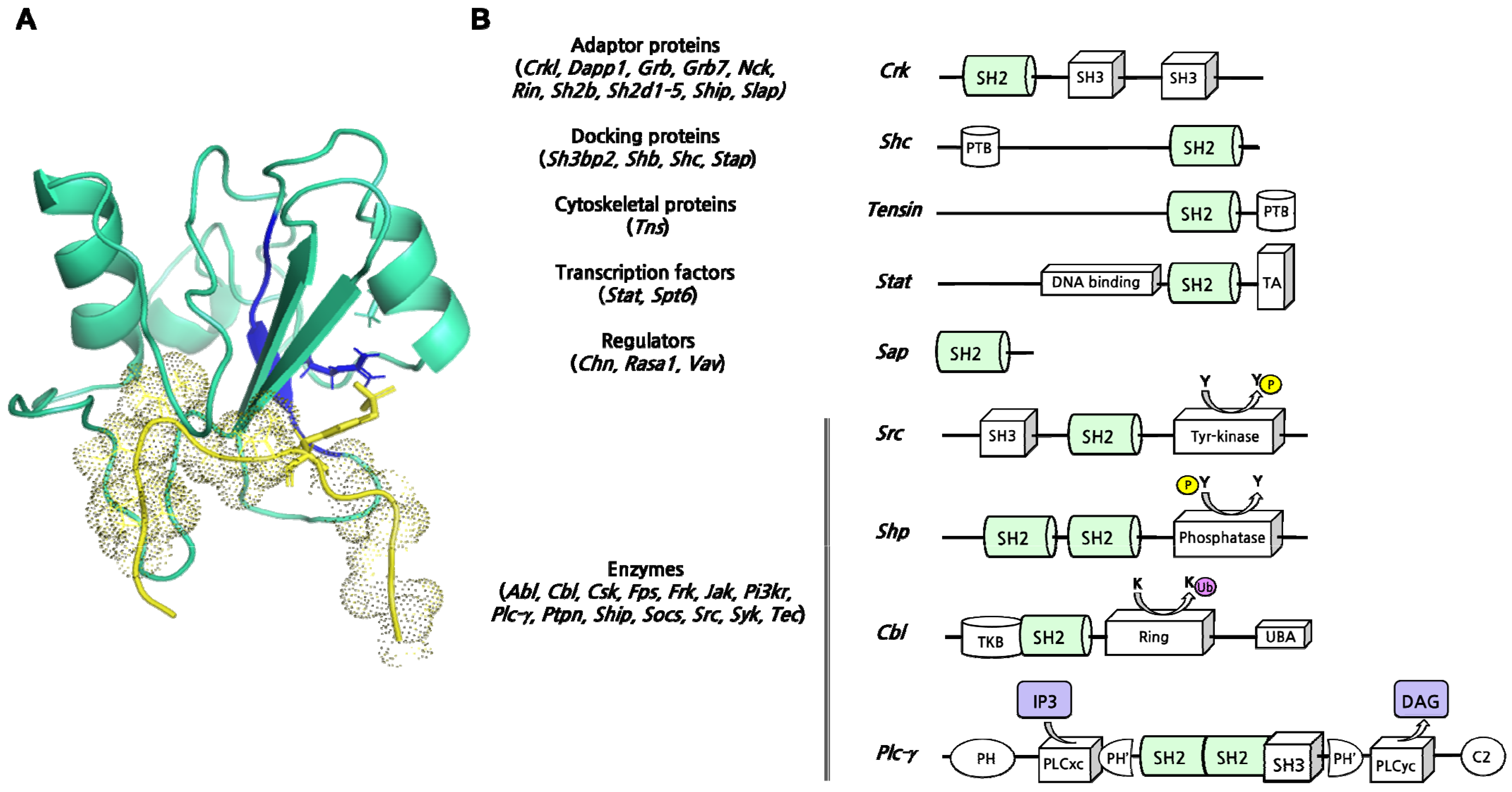
Figure 1. (Left) structure of Shp2 protein composed of two contiguous SH2 domains (N- and C-SH2, in green and purple, respectively) and a PTP catalytic domain. PDBcode: 2shp. (Right) Folding characterization of N-SH2 (reproduced with permission from [19]) and C-SH2 (reproduced under the terms of the Creative Commons Attribution License from [20]) domains of Shp2. The ϕ value analysis carried out using the isolated domains allows for defining the native-like interactions occurring in the transition state(s) of the folding reaction. Data obtained were then used to obtain a Φ vs. Φ plots (in the center) of early (top left panel) and late (top right panel) events of the folding reaction of N-SH2 versus C-SH2 domain (see references and text for details).
Ijms 23 15944 g001
Figure 2. (A) Scheme of a canonical binding reaction between an SH2 domain (in green) and a Tyrosine-phosphorylated peptide (in violet). The peptide is numbered by counting pTyr as 0. In detail, we can distinguish two structural binding elements in the SH2 domain; pTyr pocket—composed by helix αA (in cyano), beta-strands βB, βC, and βD (in light green), and the BC-loop—and a specificity pocket—composed by helix αB (dark green), beta-strands βD, βE, βF and βG (light green) BG loop and the EF loop. Arg32 in the pTyr pocket (green circle) is a highly conserved residue driving the interaction in all SH2 domains. Arg/Lys on αA or βD (white circles) can assist the reaction. (B) Examples of interactions between SH2 domains and pTyr-peptides mediated by Arg32 and an Arg residue on αA in case of Src-like class of binding (left, PDBcode:1sps), and an Arg residue on βD in case of Sap-like class of binding (right, PDBcode:1d4w). Structural details in the squares. The conserved binding motif is highlighted in blue. The motif corresponds to FLVR and YLVR in reported examples of Src-like (canonical) and Sap-like (non-canonical) classes of binding, respectively.
Ijms 23 15944 g002
Figure 3. (A) A representative example of an SH2 domain in complex with a peptide mimicking a physiological ligand (PDBcode 1TCE). In detail, the SH2 domain of Shc (in light cyan) can interact with a tyrosine-phosphorylated peptide from the T cell receptor (in yellow) through a highly conserved sequence FLVRES (highlighted in blue). Arg32 (blue stick) is crucial for the interaction with the pY (yellow stick) harbored by the ligand. (B) Schematic representation of different SH2-domain-containing proteins exerting different molecular functions. In brackets, all protein families split according to the function (see Table 1 for references).
Ijms 23 15944 g003
Figure 4. Oncogenic signaling pathways activated by interaction between the SH2 domain of different proteins (Grb2, p85PI3K, Stat). Phosphorylated sites are highlighted.
Ijms 23 15944 g004
Table 1. A comprehensive list of human SH2-domain-containing proteins. For each protein listed, the number of SH2 domains, the function, and the Uniprot Entry number is reported.
FamilyHuman GenesProtein Short NamesSH2 DomainsMolecular FunctionsUniprot Entry
ABLABL1, ABL2Abl-1, Abl-21Enzyme (Tyrosine-kinase)P00519, P42684
CBLCBL, CBLB, CBLCCbl, Cbl-b, Cbl-c1Enzyme (E3 ubiquitin-protein ligase)P22681, Q13191, Q9ULV8
CHNCHN1, CHN2Chin-1, Chin-21Regulator (GTPase activity activator)P15882, P52757
CRKCRK, CRKLCrk, Crkl1Adaptor proteinP46108, P46109
CSKCSK, MATKCsk; Ctk1Enzyme (Tyrosine-kinase)P41240, P42679
DAPP1DAPP1Dapp-11Adaptor proteinQ9UN19
FPSFPS (FES), FERFps (Fes), Fer1Enzyme (Tyrosine-kinase)P07332, P16591
FRKFRK, BRK, SRMSFrk, Brk-1, Srms1Enzyme (Tyrosine-kinase)P42685, Q8WUW1, Q9H3Y6
GRBGRB2, GADS, GRAPGrb2, Gads, Grap1Adaptor proteinP62993, O75791, Q13588
GRB7GRB7, GRB10, GRB14Grb7, Grb10, Grb141Adaptor proteinQ14451, P0CE43, Q14449
JAKTYK2, JAK1, JAK2, JAK3Tyk2, Jak1, Jak2, Jak31, atypicalEnzyme (Tyrosine-kinase)P29597, P23458, O60674, P52333
NCKNCK1, NCK2Nck1, Nck21Adaptor proteinP16333, O43639
PI3KRPIK3R1, PIK3R2, PIK3R3p85A, p85B, p55G2Enzyme (1-phosphatidylinositol-3-kinase)P27986, O00459, Q92569
PLCgPLCG1, PLCG2Plcg1, Plcg22Enzyme (phosphatidylinositol phospholipase)P19174, P16885,
PTPNPTPN6, PTPN11Shp2 (Ptp)2Enzyme (Tyrosine phosphatase)Q06124
RASA1RASA1Gap (RasGap, p120RasGap)2Regulator (GTPase activity inhibitor)P20936
RINRIN1, RIN2, RIN3Rin1, Rin2, Rin31Regulator (Ras effector)Q13671, Q8WYP3, Q8TB24
SH2BAPS, LNK, SH2BAps, Lnk, Sh2b1Adaptor proteinO14492, Q9UQQ2, Q9NRF2
SH2D1SH2D1A, SH2D1BSh2d1A (Sap), Sh2d1B 1Adaptor proteinO60880, O14796
SH2D2SH2D2A, HSH2D, SH2D7Sh2d2a, Hsh2d, Sh2d71Adaptor proteinQ9NP31, Q96JZ2, A6NKC9
SH2D3SH2D3A, SH2D3C, BCAR3Sh2d3a, Sh2d3c, Bcar31Adaptor proteinQ9BRG2, Q9QZS8, Q9QZK2
SH2D4SH2D4A, SH2D4BSh2d4a, Sh2d4b1Adaptor proteinQ9H788, Q5SQS7
SH2D5SH2D5Sh2d51Adaptor proteinQ6ZV89
SH3BP2SH3BP2Sh3bp2 (3bp-2)1Docking proteinP78314
SHBSHB, SHD, SHE, SHFShb, Shd, She, Shf1Docking proteinQ15464, Q96IW2, Q5VZ18, Q7M4L6
SHCSHC1, SHC2, SHC3, SHC4Shc1, Shc2, Shc3, Shc41Docking proteinP29353, P98077, Q92529, Q6S5L8
SHIPSHIP1, SHIP2Ship-1, Ship-21Enzyme (Phosphatidylinositol (PtdIns) phosphatase)P97573, O15357
SLAPSLAP, SLAP2Slap-1, Slap-21Adaptor proteinQ13239, Q9H6Q3
SLP76BLNK, LCP2, CLNK, SCIMPSlnk, Blnk, Slp76, Mist, Slp65/Slp761Adaptor proteinQ8WV28, Q13094, Q7Z7G1, Q6UWF3
SOCSSOCS1, SOCS2, SOCS3, SOCS4, SOCS5, SOCS6, SOCS7, CISHSocs1, Socs2, Socs3, Socs4, Socs5, Socs6, Socs7, Cish1Enzyme (protein ubiquitination)O15524, O35717, O14543, Q8WXH5, O75159, O14544, O14512, Q9NSE2
SRCSRC, FYN, LCK, FGR, YES, LYN, HCK, BLKSrc, Fyn, Lck, Fgr, Yes, Lyn, Hck, Blk1Enzyme (Tyrosine-kinase)P12931, P06241, P06239, P09769, P07947, P07948, P08631, P51451
STAPBRDG1, BKSBrdg1 (Stap-1), Bks (Stap-2) 1Docking proteinQ9ULZ2, Q9UGK3
STATSTAT1, STAT2, STAT3, STAT4, STAT5, STAT5B, STAT6Stat1, Stat2, Stat3, Stat4, Stat5, Stat5b, Stat61Transcription factorP42224, P52630, P40763, Q14765, P42229, P51692, P42226
SPT6SUPT6HSupt6h (Spt6)1Transcription factorP42226
SYKZAP70, SYKZap-70, p72-syk2Enzyme (Tyrosine-kinase)P43403, P43405
TECBMX, TEC, BTK, ITK, TXKBmx, Tec, Btk, Itk, Txk1Enzyme (Tyrosine-kinase)P51813, P42680, Q06187, Q08881, P42681
TNSTNS1, TENS2, TNS3, TNS4Tensin-1, Tensin-2, Tensin-3, Tensin-41Cytoskeletal proteinQ63HR2, Q63HR2, Q68CZ2, Q8IZW8
VAVVAV1, VAV2, VAV3Vav1, Vav2, Vav31Regulator (Guanine-nucleotide releasing factor)P15498, P52735, Q9UKW4
Table 2. List of SH2 domains whose structures are currently available (adapted and updated from [53]). The abbreviation pY indicates the phosphotyrosine, σ an acidic residue, x an undefined residue, Ψ a hydrophobic residue, and n/a not available information.
GroupSH2 DomainβD5MotifPeptide Specificity Residues
IIASRC, FYN, FRK, LCK, HCK, BLK, ABL1,
ABL2, ITK, BTK, TXK, ZAP70_N & C,
SYK_N &C, NCK1, NCK2, BRK, YES, LYN
Y/FpY-σ-σ-ψ+3
IBSAP, EAT2, SHIP1, SHIP2, CRK, CRKL,
RasGAP_C, CHK, BRK
Y/FpY-x-x-ψ+3
ICGRB2, GADS, GRB7, GRB10, GRB14, ALX,
FES, BMX, CSK, SH3BP2, HSH2D, TENC1,
FER, SRM
Y/FpY-x-N+2
IEα-Chimaerin, β-ChimaerinY/Fn/a?
IIIIAVAV, VAV2, PI3K-p85α_N & C, PLC-γ1_C,
PLC-γ2_N & C, SHP-1_N & C, SHP2_N & C,
SOCS2 & 3 & 4 & 6
I/C/L/V/A/TpY-ψ-x-ψ+3
IIBAPS, SHB2, SHC1, BLNKL/IpY-[E/D/x]-x-ψ+3
IICBRDG1, BKS, CBLY/TpY-x-x-x-ψ+4
IIDSPT6VpY-[P/I]-P-[K/R]-M?
IIIIIISTAT1 & 3 & 5AV/LpY-x-x-Q+3
Table 3. List of effective SH2-domain inhibitors (* Interactions with different amino acid residues).
Inhibitors ClassesActive Moiety or Binding SiteInhibitor NameTargeted ProteinKD° or IC50#References
Peptidomimetis
pTyr-isosteresPhosphonomethyl-PhePmpPI 3-kinase p850.01–0.02 μM°[120]
Hydroxy benzyl phosphinate Grb20.53 μM °[121,122]
4-phosphonodifluoromethylcinnamate Stat30.16 μM #[124,125]
SulfonamidesC188-9Stat3 [133,134]
(TTI-101)
Photo-switchable linkers4-aminomethyl phenylazobenzoic acid Zap-700.065–0.8 μM °[126,127,128,129]
Bicyclic peptides Carboxylate groupBC1Grb20.35 μM #[130,131,132]
Allosteric inhibitors
PyrazinesN-SH2/C-SH2/PTP interfaceSHP099 *Shp20.071 μM #[137,138]
N-SH2/C-SH2/PTP interfaceTNO155 *Shp2 0.011 μM #[139,140]
C-SH2/PTP interfaceLY6Shp2 9 μM #[141]
Triazolo-quinazolinones“latch” N-SH2/PTP binding siteSHP244Shp2 60 μM #[142]
“latch” N-SH2/PTP binding siteSHP504Shp2 21 μM #[142]
“latch” N-SH2/PTP binding siteSHP844Shp2 18.9 μM #[142]
Publisher’s Note: MDPI stays neutral with regard to jurisdictional claims in published maps and institutional affiliations.

