
Magnetic Micellar Nanovehicles: Prospects of Multifunctional Hybrid Systems for Precision Theranostics
 , and
, and
Abstract
:1. Introduction
2. Fabrication of Magnetic Polymeric Micelles
2.1. Types of Polymers Used in the Shell of Magnetic Polymeric Micelles
2.1.1. Synthetic Polymers
2.1.2. Natural-Origin Polymers
2.2. Types of Copolymers Used in the Core of Magnetic Polymeric Micelles
2.2.1. Synthetic Polymers
2.2.2. Small Molecules
2.2.3. Proteins
2.3. Intrinsically Amphiphilic Natural Polysaccharides
2.4. Magnetic Nanoparticles
2.4.1. The Impact of Magnetic Compliance
2.4.2. Magnetic Nanoparticles Synthesis
2.4.3. Shape and the Core Composition of MNPs
2.4.4. Biocompatibility of MNPs
2.5. Self-Assembly Architectures of Amphiphilic Copolymers
2.6. Techniques for the Preparation of Magnetic Polymeric Micelles
3. Magnetic Micelles in Cell and Tissue Targeting

| Amphiphilic Co-Polymer | MNPs | Therapeutic Agent | Target Ligand | Approach | Outcomes | Ref. |
|---|---|---|---|---|---|---|
| Imaging | ||||||
| GCPQ | Fe3O4 | - | - | MRI (liver vasculature) |
| [21] |
| DSPE-PEG | Fe3O4 | - | - | MRI (liver) |
| [87] |
| PDLA-b-PEG/PLLA-b-PEG | MnFe2O4 (s,c) MnFe2O4@Fe3O4 | - | - | MRI |
| [49] |
| P4VP-b-PEG | Fe3O4 | - | - | MRI |
| [90] |
| (CA)4-Lys3-PEG | Fe3O4 | - | - | MRI |
| [79] |
| WPU-BPLP-WPU | Fe3O4 | DOX | Y1R | MRI |
| [91] |
| (CA)2-Lys-(PAsp(DMA)) | Fe3O4 | - | - | stem cell labeling MRI of transplanted NSCs |
| [75] |
| Levan | Fe3O4 | - | - | MRI cell tracking intracellular magnetic actuators |
| [38] |
| Theranostic potential | ||||||
| Therapeutics delivery and responsive release | ||||||
| PLGA-PEG | Fe3O4 | QCT | - | drug delivery |
| [77] |
| PCL-b-PEG | Fe3O4 | NPX | - | magnetic drug delivery |
| [76] |
| Zein-LF | Fe3O4 | DAS | - | magnetic drug delivery pH-release |
| [24] |
| PCCL-b-PEG | Fe3O4 | PTX | - | magnetic drug delivery magnetic and pH-release |
| [92,93,94] |
| PCL-b-PEG | Fe3O4 | QCT | - | pH-release |
| [95] |
| Octyl-g-HTCC/Octyl-g-PEG-HTCC | Fe3O4 | PTX | - | pH-release |
| [22] |
| P(NIPAAm-co-DMAAm-co-UA) | Fe3O4 | Hesp | - | pH-release |
| [96] |
| PSar-b-PCys(SO2Et) | Fe2O3 | Iron (Fe2O3) | - | redox-release |
| [82] |
| PCL-b-PEG | Fe3O4 | DOX | - | redox-release |
| [97] |
| PLA-PEG/PLA-CHI-Spm | Fe3O4 | siRNA PTX | FA T7 peptide | dual therapeutics delivery pH-release |
| [98] |
| Imaging/Therapeutics delivery/Combined therapies | ||||||
| PCL-b-PEG | Fe3O4 | TAM | - | MRI drug delivery |
| [99] |
| PS-b-PAA-b-PEG | Fe3O4 | DC | - | MRI/optical imaging drug delivery |
| [2] |
| DSPE-PEG | Fe3O4 | PTX | - | MRI drug delivery |
| [88] |
| OCL-Bz-b-PEG | Fe3O4 | QCT | - | MRI magnetic drug delivery |
| [84] |
| PLA-PEG | Fe3O4 | DOX | MRI pH-release |
| [86] | |
| PCL-b-PEG | Fe3O4 | DOX | PBA | optical imaging magnetic drug delivery pH-release |
| [85] |
| PLGA-b-PEI-b-PEG | Fe3O4 | DOX | cRGD | MRI-guided therapy pH-release |
| [100] |
| PAsp(DBA-co-DIP)-b-PEG | Fe3O4 | DOX | - | MRI/optical imaging pH release |
| [1] |
| PCL-b-PAELG | Fe3O4 | DOX | Gal/Lac | MRI redox release |
| [83] |
| PCL-b-HA | Fe3O4 | DOX | - | MRI redox release |
| [18] |
| PZLL-g-HA | Fe3O4 | DOX | - | MRI redox release |
| [19] |
| PEO-b-PPO-b-PEO (Pluronic F127) | Zn1.15Fe1.85O4 | OA-R837 | OVA257-264 | MRI/optical imaging magnetic delivery redox release |
| [55] |
| PAsp(MEA-co-DIP)-b-PEG | Fe3O4 | SF | AbGPC3 | MRI pH and redox release |
| [101] |
| PCL-b-PGA | Fe3O4 | DOX | - | MRI pH and redox release |
| [81] |
| PEG-PU-PCL-PU-PEG | Fe3O4 | DOX | FA | MRI pH- and redox-release |
| [89] |
| (CA)2-Lys-(PAsp(DMA)) | Fe3O4 | siRNA/ASO | - | MRI tracking of NSCs neuronal differentiation |
| [12] |
| CAM-HA (PLL coating) | Fe3O4 | plasmid (pLuc) | - | MRI-guided gene delivery |
| [33] |
| OAMAM-b-DEX | Fe3O4 | BPD | - | MRI photo release PDT |
| [23] |
| PCL-b-PEG | Mn0.6Zn0.4Fe2O4 | - | HA | MRI radiotherapy MHT |
| [54] |
| C16-g-HA | Fe3O4 | docetaxel | - | MRI photo-thermal therapy thermo release |
| [20] |
| PPI-b-TEGME | Fe3O4 | DOX | - | MHT thermo release |
| [8] |
| PHEP-b-PEG | Fe3O4 (c) | emodin | - | MRI magnetic targeting MHT thermo release |
| [31] |
| P(AAm-co-AN)-g-PEG | Fe3O4 | DOX | A54 | hyperthermia (microwave) thermo- release |
| [102] |
| P(AAm-co-AN)-g-PEG | Fe3O4 | DOX | - | NIR imaging photo-thermal therapy thermo release pH release |
| [32] |
| PHEMA-b-PEG | Mn0.6Zn0.4Fe2O4 | Pt(IV) | - | MRI pH and redox release magnetic targeting MHT |
| [16] |
| PAE-b-PEG/DPPC | Fe3O4 | DOX | - | MRI/optical/photoacoustic imaging MHT photo-thermal therapy pH- and thermo- release chemodynamic therapy |
| [103] |
4. Magnetic Micelle Applications
4.1. Imaging
4.2. Delivery and Release of Therapeutics
4.2.1. Magnetic Targeting
4.2.2. Thermal Stimulation
4.2.3. MPM Responsiveness to Extracellular and Intracellular Stimuli
4.3. Oligonucleotides Delivery
4.4. Theranostics
5. Conclusions and Future Perspectives
Author Contributions
Funding
Conflicts of Interest
Abbreviations
References
- Li, B.; Cai, M.; Lin, L.; Sun, W.; Zhou, Z.; Wang, S.; Wang, Y.; Zhu, K.; Shuai, X. MRI-visible and pH-sensitive micelles loaded with doxorubicin for hepatoma treatment. Biomater. Sci. 2019, 7, 1529–1542. [Google Scholar] [CrossRef] [PubMed]
- Bastakoti, B.P.; Bentley, J.; McLaurin, D.; Yusa, S.-I.; Shaji, S.; Mucha, N.R.; Kumar, D.; Ahamad, T. Synthesis of magnetite loaded fluorescence micelles of triblock copolymer. J. Mol. Liq. 2020, 305, 1841–1863. [Google Scholar] [CrossRef]
- Terzopoulou, Z.; Zamboulis, A.; Koumentakou, I.; Michailidou, G.; Noordam, M.J.; Bikiaris, D.N. Biocompatible Synthetic Polymers for Tissue Engineering Purposes. Biomacromolecules 2022, 23, 1841–1863. [Google Scholar] [CrossRef] [PubMed]
- Bolívar-Monsalve, E.J.; Alvarez, M.M.; Hosseini, S.; Espinosa-Hernandez, M.A.; Ceballos-González, C.F.; Sanchez-Dominguez, M.; Shin, S.R.; Cecen, B.; Hassan, S.; Di Maio, E.; et al. Engineering bioactive synthetic polymers for biomedical applications: A review with emphasis on tissue engineering and controlled release. Mater. Adv. 2021, 2, 4447–4478. [Google Scholar] [CrossRef]
- Thi, T.T.H.; Pilkington, E.H.; Nguyen, D.H.; Lee, J.S.; Park, K.D.; Truong, N.P. The Importance of Poly(ethylene glycol) Alternatives for Overcoming PEG Immunogenicity in Drug Delivery and Bioconjugation. Polymers 2020, 12, 298. [Google Scholar] [CrossRef] [Green Version]
- Suk, J.S.; Xu, Q.; Kim, N.; Hanes, J.; Ensign, L.M. PEGylation as a strategy for improving nanoparticle-based drug and gene delivery. Adv. Drug Deliv. Rev. 2016, 99 Pt A, 28–51. [Google Scholar] [CrossRef] [Green Version]
- Birke, A.; Ling, J.; Barz, M. Polysarcosine-containing copolymers: Synthesis, characterization, self-assembly, and applications. Prog. Polym. Sci. 2018, 81, 163–208. [Google Scholar] [CrossRef]
- Wang, Q.; Xiao, J.; Su, Y.; Huang, J.; Li, J.; Qiu, L.; Zhan, M.; He, X.; Yuan, W.; Li, Y. Fabrication of thermoresponsive magnetic micelles from amphiphilic poly(phenyl isocyanide) and Fe3O4 nanoparticles for controlled drug release and synergistic thermochemotherapy. Polym. Chem. 2021, 12, 2132–2140. [Google Scholar] [CrossRef]
- Zhao, J.; Lee, V.E.; Liu, R.; Priestley, R.D. Responsive Polymers as Smart Nanomaterials Enable Diverse Applications. Annu. Rev. Chem. Biomol. Eng. 2019, 10, 361–382. [Google Scholar] [CrossRef]
- Zakeri, A.; Kouhbanani, M.A.J.; Beheshtkhoo, N.; Beigi, V.; Mousavi, S.M.; Hashemi, S.A.R.; Zade, A.K.; Amani, A.M.; Savardashtaki, A.; Mirzaei, E.; et al. Polyethylenimine-based nanocarriers in co-delivery of drug and gene: A developing horizon. Nano Rev. Exp. 2017, 9, 1488497. [Google Scholar] [CrossRef]
- Lungwitz, U.; Breunig, M.; Blunk, T.; Göpferich, A. Polyethylenimine-based non-viral gene delivery systems. Eur. J. Pharm. Biopharm. 2005, 60, 247–266. [Google Scholar] [CrossRef]
- Lin, B.; Lu, L.; Wang, Y.; Zhang, Q.; Wang, Z.; Cheng, G.; Duan, X.; Zhang, F.; Xie, M.; Le, H.; et al. Nanomedicine Directs Neuronal Differentiation of Neural Stem Cells via Silencing Long Noncoding RNA for Stroke Therapy. Nano Lett. 2021, 21, 806–815. [Google Scholar] [CrossRef] [PubMed]
- Pegg, A.E. The function of spermine. IUBMB Life 2014, 66, 8–18. [Google Scholar] [CrossRef]
- Tang, L.; Wang, L.; Yang, X.; Feng, Y.; Li, Y.; Feng, W. Poly(N-isopropylacrylamide)-based smart hydrogels: Design, properties and applications. Prog. Mater. Sci. 2020, 115, 100702. [Google Scholar] [CrossRef]
- Wei, H.; Cheng, S.-X.; Zhang, X.-Z.; Zhuo, R.-X. Thermo-sensitive polymeric micelles based on poly(N-isopropylacrylamide) as drug carriers. Prog. Polym. Sci. 2009, 34, 893–910. [Google Scholar] [CrossRef]
- Qu, Y.; Wang, Z.; Sun, M.; Zhao, T.; Zhu, X.; Deng, X.; Zhang, M.; Xu, Y.; Liu, H. A Theranostic Nanocomplex Combining with Magnetic Hyperthermia for Enhanced Accumulation and Efficacy of pH-Triggering Polymeric Cisplatin(IV) Prodrugs. Pharmaceuticals 2022, 15, 480. [Google Scholar] [CrossRef]
- Atanase, L.I. Micellar Drug Delivery Systems Based on Natural Biopolymers. Polymers 2021, 13, 477. [Google Scholar] [CrossRef]
- Yang, H.; Wang, N.; Mo, L.; Wu, M.; Yang, R.; Xu, X.; Huang, Y.; Lin, J.; Zhang, L.-M.; Jiang, X. Reduction sensitive hyaluronan-SS-poly(ε-caprolactone) block copolymers as theranostic nanocarriers for tumor diagnosis and treatment. Mater. Sci. Eng. C 2018, 98, 9–18. [Google Scholar] [CrossRef] [PubMed]
- Yang, H.; Miao, Y.; Chen, L.; Li, Z.; Yang, R.; Xu, X.; Liu, Z.; Zhang, L.-M.; Jiang, X. Redox-responsive nanoparticles from disulfide bond-linked poly-(N-ε-carbobenzyloxy-l-lysine)-grafted hyaluronan copolymers as theranostic nanoparticles for tumor-targeted MRI and chemotherapy. Int. J. Biol. Macromol. 2020, 148, 483–492. [Google Scholar] [CrossRef]
- Zheng, S.; Han, J.; Jin, Z.; Kim, C.-S.; Park, S.; Kim, K.-P.; Park, J.-O.; Choi, E. Dual tumor-targeted multifunctional magnetic hyaluronic acid micelles for enhanced MR imaging and combined photothermal-chemotherapy. Colloids Surf. B Biointerfaces 2018, 164, 424–435. [Google Scholar] [CrossRef]
- Hobson, N.J.; Weng, X.; Siow, B.; Veiga, C.; Ashford, M.; Thanh, N.T.; Schätzlein, A.G.; Uchegbu, I.F. Clustering superparamagnetic iron oxide nanoparticles produces organ-targeted high-contrast magnetic resonance images. Nanomedicine 2019, 14, 1135–1152. [Google Scholar] [CrossRef] [PubMed]
- Chu, L.; Zhang, Y.; Feng, Z.; Yang, J.; Tian, Q.; Yao, X.; Zhao, X.; Tan, H.; Chen, Y. Synthesis and application of a series of amphipathic chitosan derivatives and the corresponding magnetic nanoparticle-embedded polymeric micelles. Carbohydr. Polym. 2019, 223, 114966. [Google Scholar] [CrossRef] [PubMed]
- Yan, L.; Luo, L.; Amirshaghaghi, A.; Miller, J.; Meng, C.; You, T.; Busch, T.M.; Tsourkas, A.; Cheng, Z. Dextran-Benzoporphyrin Derivative (BPD) Coated Superparamagnetic Iron Oxide Nanoparticle (SPION) Micelles for T2-Weighted Magnetic Resonance Imaging and Photodynamic Therapy. Bioconjug. Chem. 2019, 30, 2974–2981. [Google Scholar] [CrossRef] [PubMed]
- Sabra, S.A.; Sheweita, S.A.; Haroun, M.; Ragab, D.; Eldemellawy, M.A.; Xia, Y.; Goodale, D.; Allan, A.L.; Elzoghby, A.O.; Rohani, S. Magnetically Guided Self-Assembled Protein Micelles for Enhanced Delivery of Dasatinib to Human Triple-Negative Breast Cancer Cells. J. Pharm. Sci. 2018, 108, 1713–1725. [Google Scholar] [CrossRef] [PubMed]
- Mano, J.F.; Silva, G.A.; Azevedo, H.S.; Malafaya, P.B.; Sousa, R.A.; Silva, S.S.; Boesel, L.F.; Oliveira, J.M.; Santos, T.C.; Marques, A.P.; et al. Natural origin biodegradable systems in tissue engineering and regenerative medicine: Present status and some moving trends. J. R. Soc. Interface 2007, 4, 999–1030. [Google Scholar] [CrossRef] [PubMed] [Green Version]
- Kogan, G.; Šoltés, L.; Stern, R.; Gemeiner, P. Hyaluronic acid: A natural biopolymer with a broad range of biomedical and industrial applications. Biotechnol. Lett. 2006, 29, 17–25. [Google Scholar] [CrossRef]
- Fakhari, A.; Berkland, C. Applications and emerging trends of hyaluronic acid in tissue engineering, as a dermal filler and in osteoarthritis treatment. Acta Biomater. 2013, 9, 7081–7092. [Google Scholar] [CrossRef] [PubMed] [Green Version]
- Hinchliffe, J.; Madappura, A.P.; Mohamed, S.S.; Roy, I. Biomedical Applications of Bacteria-Derived Polymers. Polymers 2021, 13, 1081. [Google Scholar] [CrossRef] [PubMed]
- Monchaux, E.; Vermette, P. Development of Dextran-Derivative Arrays To Identify Physicochemical Properties Involved in Biofouling from Serum. Langmuir 2007, 23, 3290–3297. [Google Scholar] [CrossRef] [PubMed]
- Drago-Serrano, M.E.; Campos-Rodríguez, R.; Carrero, J.C.; De La Garza, M. Lactoferrin: Balancing Ups and Downs of Inflammation Due to Microbial Infections. Int. J. Mol. Sci. 2017, 18, 501. [Google Scholar] [CrossRef] [PubMed]
- Song, Y.; Li, D.; Lu, Y.; Jiang, K.; Yang, Y.; Xu, Y.; Dong, L.; Yan, X.; Ling, D.; Yang, X.; et al. Ferrimagnetic mPEG-b-PHEP copolymer micelles loaded with iron oxide nanocubes and emodin for enhanced magnetic hyperthermia–chemotherapy. Natl. Sci. Rev. 2020, 7, 723–736. [Google Scholar] [CrossRef] [PubMed] [Green Version]
- Wu, L.; Zong, L.; Ni, H.; Liu, X.; Wen, W.; Feng, L.; Cao, J.; Qi, X.; Ge, Y.; Shen, S. Magnetic thermosensitive micelles with upper critical solution temperature for NIR triggered drug release. Biomater. Sci. 2019, 7, 2134–2143. [Google Scholar] [CrossRef] [PubMed]
- Thomas, R.G.; Muthiah, M.; Moon, M.; Park, I.-K.; Jeong, Y.Y. SPION loaded poly(l-lysine)/hyaluronic acid micelles as MR contrast agent and gene delivery vehicle for cancer theranostics. Macromol. Res. 2017, 25, 446–451. [Google Scholar] [CrossRef]
- Shukla, R.; Cheryan, M. Zein: The industrial protein from corn. Ind. Crop. Prod. 2001, 13, 171–192. [Google Scholar] [CrossRef]
- Paliwal, R.; Palakurthi, S. Zein in controlled drug delivery and tissue engineering. J. Control. Release 2014, 189, 108–122. [Google Scholar] [CrossRef] [PubMed]
- Labib, G. Overview on zein protein: A promising pharmaceutical excipient in drug delivery systems and tissue engineering. Expert Opin. Drug Deliv. 2017, 15, 65–75. [Google Scholar] [CrossRef]
- Raza, A.; Hayat, U.; Bilal, M.; Iqbal, H.M.; Wang, J.-Y. Zein-based micro- and nano-constructs and biologically therapeutic cues with multi-functionalities for oral drug delivery systems. J. Drug Deliv. Sci. Technol. 2020, 58, 101818. [Google Scholar] [CrossRef]
- Park, J.C.; Park, T.Y.; Kim, D.H.; Cha, H.J.; Seo, J.H. Multifunctional nanocomposite clusters enabled by amphiphilic/bioactive natural polysaccharides. Chem. Eng. J. 2019, 379, 122406. [Google Scholar] [CrossRef]
- Srikanth, R.; Reddy, C.H.S.S.S.; Siddartha, G.; Ramaiah, M.J.; Uppuluri, K.B. Review on production, characterization and applications of microbial levan. Carbohydr. Polym. 2015, 120, 102–114. [Google Scholar] [CrossRef]
- Sezer, A.D.; Kazak, H.; Öner, E.T.; Akbuğa, J. Levan-based nanocarrier system for peptide and protein drug delivery: Optimization and influence of experimental parameters on the nanoparticle characteristics. Carbohydr. Polym. 2011, 84, 358–363. [Google Scholar] [CrossRef]
- Yaman, S.; Anil-Inevi, M.; Ozcivici, E.; Tekin, H.C. Magnetic Force-Based Microfluidic Techniques for Cellular and Tissue Bioengineering. Front. Bioeng. Biotechnol. 2018, 6, 192. [Google Scholar] [CrossRef] [PubMed] [Green Version]
- Mahmoudi, M.; Sant, S.; Wang, B.; Laurent, S.; Sen, T. Superparamagnetic iron oxide nanoparticles (SPIONs): Development, surface modification and applications in chemotherapy. Adv. Drug Deliv. Rev. 2011, 63, 24–46. [Google Scholar] [CrossRef] [PubMed] [Green Version]
- Mosayebi, J.; Kiyasatfar, M.; Laurent, S. Synthesis, Functionalization, and Design of Magnetic Nanoparticles for Theranostic Applications. Adv. Health Mater. 2017, 6, 1700306. [Google Scholar] [CrossRef] [PubMed]
- Nguyen, V.T.A.; Gauthier, M.; Sandre, O. Templated Synthesis of Magnetic Nanoparticles through the Self-Assembly of Polymers and Surfactants. Nanomaterials 2014, 4, 628–685. [Google Scholar] [CrossRef] [Green Version]
- Park, J.; An, K.; Hwang, Y.; Park, J.-G.; Noh, H.-J.; Kim, J.-Y.; Park, J.-H.; Hwang, N.-M.; Hyeon, T. Ultra-large-scale syntheses of monodisperse nanocrystals. Nat. Mater. 2004, 3, 891–895. [Google Scholar] [CrossRef] [PubMed]
- Wu, L.; Mendoza-Garcia, A.; Li, Q.; Sun, S. Organic Phase Syntheses of Magnetic Nanoparticles and Their Applications. Chem. Rev. 2016, 116, 10473–10512. [Google Scholar] [CrossRef] [PubMed]
- Li, Y.; Wang, N.; Huang, X.; Li, F.; Davis, T.P.; Qiao, R.; Ling, D. Polymer-Assisted Magnetic Nanoparticle Assemblies for Biomedical Applications. ACS Appl. Bio Mater. 2019, 3, 121–142. [Google Scholar] [CrossRef] [PubMed] [Green Version]
- Xiao, Y.; Du, J. Superparamagnetic nanoparticles for biomedical applications. J. Mater. Chem. B 2019, 8, 354–367. [Google Scholar] [CrossRef]
- Niu, H.; Li, J.; Cai, Q.; Wang, X.; Luo, F.; Gong, J.; Qiang, Z.; Ren, J. Molecular Stereocomplexation for Enhancing the Stability of Nanoparticles Encapsulated in Polymeric Micelles for Magnetic Resonance Imaging. Langmuir 2020, 36, 13881–13889. [Google Scholar] [CrossRef]
- Pardo, A.; Pujales, R.; Blanco, M.; Villar-Alvarez, E.M.; Barbosa, S.; Taboada, P.; Mosquera, V. Analysis of the influence of synthetic paramaters on the structure and physico-chemical properties of non-spherical iron oxide nanocrystals and their biological stability and compatibility. Dalton Trans. 2015, 45, 797–810. [Google Scholar] [CrossRef] [PubMed]
- Nemati, Z.; Alonso, J.; Rodrigo, I.; Das, R.; Garaio, E.; García, J.A.; Orue, I.; Phan, M.-H.; Srikanth, H. Improving the Heating Efficiency of Iron Oxide Nanoparticles by Tuning Their Shape and Size. J. Phys. Chem. C 2018, 122, 2367–2381. [Google Scholar] [CrossRef]
- Das, R.; Alonso, J.; Nemati Porshokouh, Z.; Kalappattil, V.; Torres, D.; Phan, M.-H.; Garaio, E.; García, J.Á.; Sanchez Llamazares, J.L.; Srikanth, H. Tunable High Aspect Ratio Iron Oxide Nanorods for Enhanced Hyperthermia. J. Phys. Chem. C 2016, 120, 10086–10093. [Google Scholar] [CrossRef]
- Cho, M.; Cervadoro, A.; Ramirez, M.R.; Stigliano, C.; Brazdeikis, A.; Colvin, V.L.; Civera, P.; Key, J.; Decuzzi, P. Assembly of Iron Oxide Nanocubes for Enhanced Cancer Hyperthermia and Magnetic Resonance Imaging. Nanomaterials 2017, 7, 72. [Google Scholar] [CrossRef] [PubMed] [Green Version]
- Wang, Y.; Zou, L.; Qiang, Z.; Jiang, J.; Zhu, Z.; Ren, J. Enhancing Targeted Cancer Treatment by Combining Hyperthermia and Radiotherapy Using Mn–Zn Ferrite Magnetic Nanoparticles. ACS Biomater. Sci. Eng. 2020, 6, 3550–3562. [Google Scholar] [CrossRef] [PubMed]
- Li, J.; Ren, H.; Sun, Y.; Liu, G.; Yang, X.; Qiu, Q.; Ding, Y.; Lovell, J.F.; Zhang, Y. Magnetic Metal Micelles for Enhanced Delivery of Self-Immolating CD8+ T-Cell Epitopes for Cancer Immunotherapy. Chem. Mater. 2021, 33, 9780–9794. [Google Scholar] [CrossRef]
- Pardo, A.; Pelaz, B.; Gallo, J.; Bañobre-López, M.; Parak, W.J.; Barbosa, S.; del Pino, P.; Taboada, P. Synthesis, Characterization, and Evaluation of Superparamagnetic Doped Ferrites as Potential Therapeutic Nanotools. Chem. Mater. 2020, 32, 2220–2231. [Google Scholar] [CrossRef]
- Kandasamy, G.; Maity, D. Recent advances in superparamagnetic iron oxide nanoparticles (SPIONs) for in vitro and in vivo cancer nanotheranostics. Int. J. Pharm. 2015, 496, 191–218. [Google Scholar] [CrossRef] [PubMed]
- Singh, N.; Jenkins, G.J.; Asadi, R.; Doak, S.H. Potential toxicity of superparamagnetic iron oxide nanoparticles (SPION). Nano Rev. 2010, 1, 5358. [Google Scholar] [CrossRef] [Green Version]
- Ankamwar, B.; Lai, T.C.; Huang, J.H.; Liu, R.S.; Hsiao, M.; Chen, C.H.; Hwu, Y.K. Biocompatibility of Fe3O4 nanoparticles evaluated by in vitro cytotoxicity assays using normal, glia and breast cancer cells. Nanotechnology 2010, 21, 075102. [Google Scholar] [CrossRef] [PubMed]
- Miri, A.; Najafzadeh, H.; Darroudi, M.; Miri, M.J.; Kouhbanani, M.A.J.; Sarani, M. Iron Oxide Nanoparticles: Biosynthesis, Magnetic Behavior, Cytotoxic Effect. ChemistryOpen 2021, 10, 327–333. [Google Scholar] [CrossRef] [PubMed]
- Alromi, D.A.; Madani, S.Y.; Seifalian, A. Emerging Application of Magnetic Nanoparticles for Diagnosis and Treatment of Cancer. Polymers 2021, 13, 4146. [Google Scholar] [CrossRef] [PubMed]
- Frtús, A.; Smolková, B.; Uzhytchak, M.; Lunova, M.; Jirsa, M.; Kubinová, Š.; Dejneka, A.; Lunov, O. Analyzing the mechanisms of iron oxide nanoparticles interactions with cells: A road from failure to success in clinical applications. J. Control. Release 2020, 328, 59–77. [Google Scholar] [CrossRef] [PubMed]
- Reinert, M.; Schlachter, E.K.; Bregy, A.; Lönnfors-Weitzel, T.; Vajtai, I.; Bernau, V.J.; Weitzel, T.; Mordasini, P.; Slotboom, J.; Herrmann, G.; et al. Metabolic pathway and distribution of superparamagnetic iron oxide nanoparticles: In vivo study. Int. J. Nanomed. 2011, 6, 1793–1800. [Google Scholar] [CrossRef] [PubMed] [Green Version]
- Patsula, V.; Tulinska, J.; Trachtová, Š.; Kuricova, M.; Liskova, A.; Španová, A.; Ciampor, F.; Vavra, I.; Rittich, B.; Ursinyova, M.; et al. Toxicity evaluation of monodisperse PEGylated magnetic nanoparticles for nanomedicine. Nanotoxicology 2019, 13, 510–526. [Google Scholar] [CrossRef] [PubMed]
- Lewis, C.S.; Torres, L.; Miyauchi, J.T.; Rastegar, C.; Patete, J.M.; Smith, J.M.; Wong, S.S.; Tsirka, S.E. Absence of cytotoxicity towards microglia of iron oxide (α-Fe2O3) nanorhombohedra. Toxicol. Res. 2016, 5, 836–847. [Google Scholar] [CrossRef] [PubMed] [Green Version]
- Van de Walle, A.; Fromain, A.; Sangnier, A.P.; Curcio, A.; Lenglet, L.; Motte, L.; Lalatonne, Y.; Wilhelm, C. Real-time in situ magnetic measurement of the intracellular biodegradation of iron oxide nanoparticles in a stem cell-spheroid tissue model. Nano Res. 2020, 13, 467–476. [Google Scholar] [CrossRef]
- Valdiglesias, V.; Bertólez, N.F.; Kiliç, G.; Costa, C.; Costa, S.; Fraga, S.; Bessa, M.J.; Pasaro, E.; Teixeira, J.P.; Laffon, B. Are iron oxide nanoparticles safe? Current knowledge and future perspectives. J. Trace Elements Med. Biol. 2016, 38, 53–63. [Google Scholar] [CrossRef] [Green Version]
- Wu, Q.; Jin, R.; Feng, T.; Liu, L.; Yang, L.; Tao, Y.; Anderson, J.M.; Ai, H.; Li, H. Iron oxide nanoparticles and induced autophagy in human monocytes. Int. J. Nanomed. 2017, 12, 3993–4005. [Google Scholar] [CrossRef] [Green Version]
- Jin, R.; Liu, L.; Zhu, W.; Li, D.; Yang, L.; Duan, J.; Cai, Z.; Nie, Y.; Zhang, Y.; Gong, Q.; et al. Iron oxide nanoparticles promote macrophage autophagy and inflammatory response through activation of toll-like Receptor-4 signaling. Biomaterials 2019, 203, 23–30. [Google Scholar] [CrossRef]
- Zanganeh, S.; Hutter, G.; Spitler, R.; Lenkov, O.; Mahmoudi, M.; Shaw, A.; Pajarinen, J.S.; Nejadnik, H.; Goodman, S.; Moseley, M.; et al. Iron oxide nanoparticles inhibit tumour growth by inducing pro-inflammatory macrophage polarization in tumour tissues. Nat. Nanotechnol. 2016, 11, 986–994. [Google Scholar] [CrossRef]
- Chin, Y.-C.; Yang, L.-X.; Hsu, F.-T.; Hsu, C.-W.; Chang, T.-W.; Chen, H.-Y.; Chen, L.Y.-C.; Chia, Z.C.; Hung, C.-H.; Su, W.-C.; et al. Iron oxide@chlorophyll clustered nanoparticles eliminate bladder cancer by photodynamic immunotherapy-initiated ferroptosis and immunostimulation. J. Nanobiotechnol. 2022, 20, 373. [Google Scholar] [CrossRef] [PubMed]
- Vogt, C.; Pernemalm, M.; Kohonen, P.; Laurent, S.; Hultenby, K.; Vahter, M.; Lehtiö, J.; Toprak, M.S.; Fadeel, B. Proteomics Analysis Reveals Distinct Corona Composition on Magnetic Nanoparticles with Different Surface Coatings: Implications for Interactions with Primary Human Macrophages. PLoS ONE 2015, 10, e0129008. [Google Scholar] [CrossRef] [PubMed] [Green Version]
- Müller, K.; Skepper, J.N.; Posfai, M.; Trivedi, R.; Howarth, S.; Corot, C.; Lancelot, E.; Thompson, P.W.; Brown, A.P.; Gillard, J.H. Effect of ultrasmall superparamagnetic iron oxide nanoparticles (Ferumoxtran-10) on human monocyte-macrophages in vitro. Biomaterials 2007, 28, 1629–1642. [Google Scholar] [CrossRef] [PubMed]
- Wu, J.; Zhu, J.; Wu, Q.; An, Y.; Wang, K.; Xuan, T.; Zhang, J.; Song, W.; He, H.; Song, L.; et al. Mussel-Inspired Surface Immobilization of Heparin on Magnetic Nanoparticles for Enhanced Wound Repair via Sustained Release of a Growth Factor and M2 Macrophage Polarization. ACS Appl. Mater. Interfaces 2021, 13, 2230–2244. [Google Scholar] [CrossRef]
- Lu, L.; Wang, Y.; Cao, M.; Chen, M.; Lin, B.; Duan, X.; Zhang, F.; Mao, J.; Shuai, X.; Shen, J. A novel polymeric micelle used for in vivo MR imaging tracking of neural stem cells in acute ischemic stroke. RSC Adv. 2017, 7, 15041–15052. [Google Scholar] [CrossRef] [Green Version]
- Karami, Z.; Sadighian, S.; Rostamizadeh, K.; Hosseini, S.H.; Rezaee, S.; Hamidi, M. Magnetic brain targeting of naproxen-loaded polymeric micelles: Pharmacokinetics and biodistribution study. Mater. Sci. Eng. C 2019, 100, 771–780. [Google Scholar] [CrossRef] [PubMed]
- Ashjari, M.; Panahandeh, F.; Niazi, Z.; Abolhasani, M.M. Synthesis of PLGA–mPEG star-like block copolymer to form micelle loaded magnetite as a nanocarrier for hydrophobic anticancer drug. J. Drug Deliv. Sci. Technol. 2020, 56, 101563. [Google Scholar] [CrossRef]
- Kubisa, P.; Lapienis, G.; Biela, T. Star-shaped copolymers with PLA – PEG arms and their potential applications as biomedical materials. Polym. Adv. Technol. 2021, 32, 3857–3866. [Google Scholar] [CrossRef]
- Li, W.-J.; Wang, Y.; Liu, Y.; Wu, T.; Cai, W.-L.; Shuai, X.-T.; Hong, G.-B. Preliminary Study of MR and Fluorescence Dual-mode Imaging: Combined Macrophage-Targeted and Superparamagnetic Polymeric Micelles. Int. J. Med Sci. 2018, 15, 129–141. [Google Scholar] [CrossRef] [Green Version]
- Hickey, R.J.; Haynes, A.S.; Kikkawa, J.M.; Park, S.-J. Controlling the Self-Assembly Structure of Magnetic Nanoparticles and Amphiphilic Block-Copolymers: From Micelles to Vesicles. J. Am. Chem. Soc. 2011, 133, 1517–1525. [Google Scholar] [CrossRef]
- Yang, T.; Niu, D.; Chen, J.; He, J.; Yang, S.; Jia, X.; Hao, J.; Zhao, W.; Li, Y. Biodegradable organosilica magnetic micelles for magnetically targeted MRI and GSH-triggered tumor chemotherapy. Biomater. Sci. 2019, 7, 2951–2960. [Google Scholar] [CrossRef] [PubMed]
- Bauer, T.A.; Horvat, N.K.; Marques, O.; Chocarro, S.; Mertens, C.; Colucci, S.; Schmitt, S.; Carrella, L.M.; Morsbach, S.; Koynov, K.; et al. Core Cross-Linked Polymeric Micelles for Specific Iron Delivery: Inducing Sterile Inflammation in Macrophages. Adv. Healthc. Mater. 2021, 10, 2100385. [Google Scholar] [CrossRef] [PubMed]
- Yang, H.-K.; Bao, J.-F.; Mo, L.; Yang, R.-M.; Xu, X.-D.; Tang, W.-J.; Lin, J.-T.; Wang, G.-H.; Zhang, L.-M.; Jiang, X.-Q. Bioreducible amphiphilic block copolymers based on PCL and glycopolypeptide as multifunctional theranostic nanocarriers for drug delivery and MR imaging. RSC Adv. 2017, 7, 21093–21106. [Google Scholar] [CrossRef] [Green Version]
- Srisa-Nga, K.; Mankhetkorn, S.; Okonogi, S.; Khonkarn, R. Delivery of Superparamagnetic Polymeric Micelles Loaded With Quercetin to Hepatocellular Carcinoma Cells. J. Pharm. Sci. 2019, 108, 996–1006. [Google Scholar] [CrossRef]
- Gao, Y.; Zhao, J.; Zhang, X.; Wei, X.; Xiong, X.; Guo, X.; Zhou, S. A rod bacterium-like magnetic polymer micelle for strongly enhancing selective accumulation and internalization of nanocarriers. J. Mater. Chem. B 2017, 5, 4943–4954. [Google Scholar] [CrossRef]
- Jiang, Y.; Lee, J.; Seo, J.-M.; Davaa, E.; Shin, K.-J.; Yang, S.-G. Enhanced thermodynamic, pharmacokinetic and theranostic properties of polymeric micelles via hydrophobic core-clustering of superparamagnetic iron oxide nanoparticles. Biomater. Res. 2022, 26, 8. [Google Scholar] [CrossRef]
- Din, I.M.P.; Balas, M.; Hermenean, A.; Elst, L.V.V.; Laurent, S.; Burtea, C.; Cinteza, L.O.; Dinischiotu, A. Novel Polymeric Micelles-Coated Magnetic Nanoparticles for In Vivo Bioimaging of Liver: Toxicological Profile and Contrast Enhancement. Materials 2020, 13, 2722. [Google Scholar] [CrossRef]
- Upponi, J.R.; Jerajani, K.; Nagesha, D.K.; Kulkarni, P.; Sridhar, S.; Ferris, C.; Torchilin, V.P. Polymeric micelles: Theranostic co-delivery system for poorly water-soluble drugs and contrast agents. Biomaterials 2018, 170, 26–36. [Google Scholar] [CrossRef]
- Wei, J.; Shuai, X.; Wang, R.; He, X.; Li, Y.; Ding, M.; Li, J.; Tan, H.; Fu, Q. Clickable and imageable multiblock polymer micelles with magnetically guided and PEG-switched targeting and release property for precise tumor theranosis. Biomaterials 2017, 145, 138–153. [Google Scholar] [CrossRef]
- Zou, Y.; Li, Y.; Xu, J.; Huang, X.; Chen, D. Heavily superparamagnetic magnetite-loaded polymeric worm-like micelles that have an ultrahigh T2 relaxivity. Polym. Chem. 2020, 11, 6134–6138. [Google Scholar] [CrossRef]
- Jiang, Z.; Tian, Y.; Shan, D.; Wang, Y.; Gerhard, E.; Xia, J.; Huang, R.; He, Y.; Li, A.; Tang, J.; et al. pH protective Y1 receptor ligand functionalized antiphagocytosis BPLP-WPU micelles for enhanced tumor imaging and therapy with prolonged survival time. Biomaterials 2018, 170, 70–81. [Google Scholar] [CrossRef] [PubMed]
- Liu, Y.; Li, Y.; Rao, Z.; Xu, J.; Zhao, S.; Zhao, C.; Zhu, H.; Hao, J.; Yang, T.; Yang, Y. Physiochemical properties and paclitaxel release behaviors of dual-stimuli responsive copolymer-magnetite superparamagnetic nanocomposites. Nanotechnology 2018, 30, 105602. [Google Scholar] [CrossRef] [PubMed]
- Li, X.; Yang, Y.; Jia, Y.; Pu, X.; Yang, T.; Wang, Y.; Ma, X.; Chen, Q.; Sun, M.; Wei, D.; et al. Enhanced tumor targeting effects of a novel paclitaxel-loaded polymer: PEG–PCCL-modified magnetic iron oxide nanoparticles. Drug Deliv. 2017, 24, 1284–1294. [Google Scholar] [CrossRef] [PubMed] [Green Version]
- Li, Y.; Liu, Y.; Zhu, H.; Shuai, S.; Zhao, C.; Zhou, K.; Ge, W.; Hao, J. Degradation of methoxy-poly(ethylene glycol)-block-poly(α-carboxyl-ε-caprolactone)/magnetite nanocomposites in vitro polymer degradation and stability. Polym. Degrad. Stab. 2020, 177, 109191. [Google Scholar] [CrossRef]
- Rahmati, M.-A.; Rashidzadeh, H.; Hosseini, M.-J.; Sadighian, S.; Kermanian, M. Self-assembled magnetic polymeric micelles for delivery of quercetin: Toxicity evaluation on isolated rat liver mitochondria. J. Biomater. Sci. Polym. Ed. 2021, 33, 279–298. [Google Scholar] [CrossRef]
- Wang, W.-J.; Huang, Y.-C.; Su, C.-M.; Ger, T.-R. Multi-Functional Drug Carrier Micelles With Anti-inflammatory Drug. Front. Chem. 2019, 7, 93. [Google Scholar] [CrossRef]
- Mousavi, S.-D.; Maghsoodi, F.; Panahandeh, F.; Yazdian-Robati, R.; Reisi-Vanani, A.; Tafaghodi, M. Doxorubicin delivery via magnetic nanomicelles comprising from reduction-responsive poly(ethylene glycol)-b-poly(ε-caprolactone) (PEG-SS-PCL) and loaded with superparamagnetic iron oxide (SPIO) nanoparticles: Preparation, characterization and simulation. Mater. Sci. Eng. C 2018, 92, 631–643. [Google Scholar] [CrossRef]
- Amani, A.; Dustparast, M.; Noruzpour, M.; Zakaria, R.A.; Ebrahimi, H.A. Design and Invitro Characterization of Green Synthesized Magnetic Nanoparticles Conjugated with Multitargeted Poly Lactic Acid Copolymers for Co-Delivery of siRNA and Paclitaxel. Eur. J. Pharm. Sci. 2021, 167, 106007. [Google Scholar] [CrossRef]
- Zarei, S.; Sadighian, S.; Rostamizadeh, K.; Khalkhali, M. Theragnostic Magnetic Core-Shell Nanoparticle as Versatile Nanoplatform for Magnetic Resonance Imaging and Drug Delivery. Biointerface Res. Appl. Chem. 2021, 11, 13276–13289. [Google Scholar]
- Xiao, Z.; Chan, L.; Zhang, D.; Huang, C.; Mei, C.; Gao, P.; Huang, Y.; Liang, J.; He, L.; Shi, C.; et al. Precise delivery of a multifunctional nanosystem for MRI-guided cancer therapy and monitoring of tumor response by functional diffusion-weighted MRI. J. Mater. Chem. B 2019, 7, 2926–2937. [Google Scholar] [CrossRef]
- Cai, M.; Li, B.; Lin, L.; Huang, J.; An, Y.; Huang, W.; Zhou, Z.; Wang, Y.; Shuai, X.; Zhu, K. A reduction and pH dual-sensitive nanodrug for targeted theranostics in hepatocellular carcinoma. Biomater. Sci. 2020, 8, 3485–3499. [Google Scholar] [CrossRef] [PubMed]
- Li, W.-S.; Wang, X.-J.; Zhang, S.; Hu, J.-B.; Du, Y.-L.; Kang, X.-Q.; Xu, X.-L.; Ying, X.-Y.; You, J.; Du, Y.-Z. Mild microwave activated, chemo-thermal combinational tumor therapy based on a targeted, thermal-sensitive and magnetic micelle. Biomaterials 2017, 131, 36–46. [Google Scholar] [CrossRef] [PubMed]
- Liu, N.; Wu, L.; Zuo, W.; Lin, Q.; Liu, J.; Jin, Q.; Xiao, Z.; Chen, L.; Zhao, Y.; Zhou, J.; et al. pH/Thermal-Sensitive Nanoplatform Capable of On-Demand Specific Release to Potentiate Drug Delivery and Combinational Hyperthermia/Chemo/Chemodynamic Therapy. ACS Appl. Mater. Interfaces 2022, 14, 29668–29678. [Google Scholar] [CrossRef] [PubMed]
- Misra, S.; Hascall, V.C.; Markwald, R.R.; Ghatak, S. Interactions between Hyaluronan and Its Receptors (CD44, RHAMM) Regulate the Activities of Inflammation and Cancer. Front. Immunol. 2015, 6, 201. [Google Scholar] [CrossRef] [Green Version]
- Nguyen, L.H.; Gao, M.; Lin, J.; Wu, W.; Wang, J.; Chew, S.Y. Three-dimensional aligned nanofibers-hydrogel scaffold for controlled non-viral drug/gene delivery to direct axon regeneration in spinal cord injury treatment. Sci. Rep. 2017, 7, srep42212. [Google Scholar] [CrossRef] [Green Version]
- Kim, C.-J.; Si, Z.; Reghu, S.; Guo, Z.; Zhang, K.; Li, J.; Chan-Park, M.B. DNA-derived nanostructures selectively capture gram-positive bacteria. Drug Deliv. Transl. Res. 2021, 11, 1438–1450. [Google Scholar] [CrossRef]
- Schmidtke, C.; Eggers, R.; Zierold, R.; Feld, A.; Kloust, H.; Wolter, C.; Ostermann, J.; Merkl, J.-P.; Schotten, T.; Nielsch, K.; et al. Polymer-Assisted Self-Assembly of Superparamagnetic Iron Oxide Nanoparticles into Well-Defined Clusters: Controlling the Collective Magnetic Properties. Langmuir 2014, 30, 11190–11196. [Google Scholar] [CrossRef]
- Gadekar, V.; Borade, Y.; Kannaujia, S.; Rajpoot, K.; Anup, N.; Tambe, V.; Kalia, K.; Tekade, R.K. Nanomedicines accessible in the market for clinical interventions. J. Control. Release 2020, 330, 372–397. [Google Scholar] [CrossRef]
- El-Boubbou, K. Magnetic iron oxide nanoparticles as drug carriers: Preparation, conjugation and delivery. Nanomedicine 2018, 13, 929–952. [Google Scholar] [CrossRef]
- Sun, C.; Lee, J.S.H.; Zhang, M. Magnetic nanoparticles in MR imaging and drug delivery. Adv. Drug Deliv. Rev. 2008, 60, 1252–1265. [Google Scholar] [CrossRef] [Green Version]
- Bordat, A.; Boissenot, T.; Nicolas, J.; Tsapis, N. Thermoresponsive polymer nanocarriers for biomedical applications. Adv. Drug Deliv. Rev. 2018, 138, 167–192. [Google Scholar] [CrossRef]
- Wang, H.; Ding, S.; Zhang, Z.; Wang, L.; You, Y. Cationic micelle: A promising nanocarrier for gene delivery with high transfection efficiency. J. Gene Med. 2019, 21, e3101. [Google Scholar] [CrossRef] [PubMed] [Green Version]
- Navarro, G.; Pan, J.; Torchilin, V.P. Micelle-like Nanoparticles as Carriers for DNA and siRNA. Mol. Pharm. 2015, 12, 301–313. [Google Scholar] [CrossRef] [PubMed] [Green Version]
- Lao, Y.-H.; Li, M.; Gao, M.A.; Shao, D.; Chi, C.-W.; Huang, D.; Chakraborty, S.; Ho, T.-C.; Jiang, W.; Wang, H.-X.; et al. HPV Oncogene Manipulation Using Nonvirally Delivered CRISPR/Cas9 or Natronobacterium gregoryi Argonaute. Adv. Sci. 2018, 5, 1700540. [Google Scholar] [CrossRef] [PubMed]


| Hydrophilic Segment (Shell) | Hydrophobic Segment (Core) | |
|---|---|---|
| Synthetic polymers | Synthetic polymers | |
|
|
|
| Natural polymers | Natural polymers/molecules | |
|
| |
| Co-Polymer Type | Examples of Co-Polymer | Micelle | |
|---|---|---|---|
| Di-block | |||
A-B | PCL-b-PEG PCL-b-PGA PCL-b-HA PCL-b-PAELG PLA-b-PEG P4VP-b-PEG DSPE-b-PEG | PHEP-b-PEG PAsp(DBA-co-DIP)-b-PEG PAsp(MEA-co-DIP)-b-PEG PAE-b-PEG PCys(SO2Et)-b-PSar PHEMA-b-PEG PS-b-DNA | 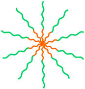 |
| Tri-block | |||
A-B-A | PEO-b-PPO-b-PEO (Pluronic F127) PNIPAM-b-PCL-b-PNIPAM | 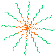 | |
A-B-C | PS-b-PAA-b-PEG PLGA-b-PEI-b-PEG | 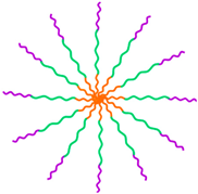 | |
| Star-like | |||
A2B3 | PLGA-PEG |  | |
| Graft | |||
 | C16-g-HA P(AAm-co-AN)-g-PEG PZLL-g-HA Octyl-g-HTCC Octyl-g-PEG-HTCC GCPQ CAM-g-HA PLA-g-CHI-g-Spm | 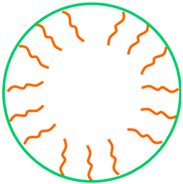 | |
| Telodendrimer | |||
 | (CA)4-Lys3-PEG (CA)2-Lys-(PAsp(DMA)) OAMAM-b-DEX |  | |
Publisher’s Note: MDPI stays neutral with regard to jurisdictional claims in published maps and institutional affiliations. |
© 2022 by the authors. Licensee MDPI, Basel, Switzerland. This article is an open access article distributed under the terms and conditions of the Creative Commons Attribution (CC BY) license (https://creativecommons.org/licenses/by/4.0/).
Share and Cite
Miranda, M.S.; Almeida, A.F.; Gomes, M.E.; Rodrigues, M.T. Magnetic Micellar Nanovehicles: Prospects of Multifunctional Hybrid Systems for Precision Theranostics. Int. J. Mol. Sci. 2022, 23, 11793. https://doi.org/10.3390/ijms231911793
Miranda MS, Almeida AF, Gomes ME, Rodrigues MT. Magnetic Micellar Nanovehicles: Prospects of Multifunctional Hybrid Systems for Precision Theranostics. International Journal of Molecular Sciences. 2022; 23(19):11793. https://doi.org/10.3390/ijms231911793
Chicago/Turabian StyleMiranda, Margarida S., Ana F. Almeida, Manuela E. Gomes, and Márcia T. Rodrigues. 2022. "Magnetic Micellar Nanovehicles: Prospects of Multifunctional Hybrid Systems for Precision Theranostics" International Journal of Molecular Sciences 23, no. 19: 11793. https://doi.org/10.3390/ijms231911793