Article

Glucocorticoid Receptor Regulates TNFSF11 Transcription by Binding to Glucocorticoid Responsive Element in TNFSF11 Proximal Promoter Region

The Chair of Clinical Biochemistry, Faculty of Pharmacy, University of Ljubljana, Aškerčeva cesta 7, 1000 Ljubljana, Slovenia
*
Author to whom correspondence should be addressed.
Int. J. Mol. Sci. 2021, 22(3), 1054; https://doi.org/10.3390/ijms22031054
Submission received: 5 December 2020 / Revised: 13 January 2021 / Accepted: 18 January 2021 / Published: 21 January 2021
(This article belongs to the Special Issue Regulation of Bone Mineral Homeostasis)

Abstract

:
Glucocorticoid osteoporosis is a serious side effect of long term glucocorticoid uptake and it is caused by osteoblast apoptosis and imbalance in the major bone remodeling pathway RANK/RANKL/OPG. The impact of glucocorticoid on the maintenance of RANK/RANKL/OPG is well explored; dexamethasone was shown to disturb the ratio between OPG and RANKL level by decreasing the expression level of OPG and increasing level of RANKL. Here, were aimed to decipher whether glucocorticoid receptor directly influences RANKL promoter activity and its transcriptional regulation. We demonstrate that overexpression of glucocorticoid receptor (GR) NR3C1 increased RANKL promoter activity in human osteosarcoma, cervical cancer (2-fold) and adenocarcinoma cells (4.5-fold). Mutational analysis revealed that +352 site in the RANKL promoter is functional glucocorticoid responsive element (GRE) since the effect of GR on RANKL promoter activity was diminished by mutation at this site. Overexpression of NR3C1 upregulated RANKL mRNA expression 1.5-fold in human A549 and HOS cells. On the other hand silencing of NR3C1 caused slight decrease in RANKL mRNA level, suggesting that NR3C1 directly accounts for RANKL transcriptional regulation. Using electrophoretic mobility shift assay we demonstrate that NR3C1 binds to the proximal RANKL promoter region. Our study provides evidences that NR3C1 directly upregulates RANKL transcription in human cell lines and connects the missing link in the mechanism of RANK/RANKL/OPG imbalance of glucocorticoid induced osteoporosis.

1. Introduction

The Glucocorticoid induced osteoporosis (GIO) is the most frequent cause of secondary osteoporosis [1,2,3]. Patients treated with glucocorticoids have increased risk for bone fracture and increased bone resorption [4,5,6]. Glucocorticoid-induced bone loss is primarily caused by apoptosis of osteoblast and secondarily by increase in bone resorption. Increased bone resorption at the early phase of GIO is attributed with increased osteoclast formation and activity of mature osteoclasts [7,8]. At the later stage of GIO, long term bone loss is associated with decrease in bone formation [9,10]. Osteoclasts are monocyte derived multinuclear cells responsible for bone absorption. Osteoclast are formed through osteoclastogenesis induced by osteoblast secreted receptor activator of nuclear factor κ B ligand (RANKL), which is a key signaling molecule that induces differentiation of bone macrophages into mature osteoclasts, and their activation. RANKL induces osteoclastogenesis by binding to RANK receptor on osteoclast precursor cells. Tight balance between osteoclast and osteoblast formation and degradation prerequisites proper bone formation and resorption processes. The key system regulating those bone remodelling processes is a RANK/RANKL/OPG pathway. Upregulation of RANKL in osteoblasts, increases RANKL secretion that activates osteoclasts formation and an increase in bone resorption [11]. Osteoprotegerin (OPG) is a soluble decoy receptor for RANKL and prevents its interactions with RANK, thus inhibits osteoclastogenesis and bone resorption. In osteoporosis, RANKL expression in bone tissues is upregulated, which leads to stronger bone resorption and decrease in bone mineral density [12].
RANKL belongs to the tumour necrosis factor (TNF) family of cytokines, and is encoded by TNFSF11 gene. It is found as membrane-bound and soluble forms, the latter of which is produced by alternative splicing or by proteolytic cleavage of membrane-bound RANKL [13,14,15]. RANKL is involved in numerous physiological processes as well as bone metabolism, such as in T-cell functions, and lymph node and mammary gland development [16,17,18,19]. RANKL is expressed in cells involved in bone formation (e.g., osteoblasts, their precursors, and osteocytes), immune cells, malignant cells, and other cell types [20]. RANKL is also involved in pathological processes, such as migration of tumor cells and development of bone metastases in diseases like breast, lung and prostate cancers [21,22,23,24,25]. RANKL expression is regulated by parathyroid hormone (PTH), vitamin 1,25(OH)2D3, Runt-related transcription factor 2 (Runx2), interleukin (IL)-6-type cytokines [26,27], molecules of the Wnt/β-catenin signaling pathway [28,29,30,31,32,33,34,35], and c-Fos in activated T cells that act through proximal or/and distal promoter regions [20]. We demonstrated that RANKL gene expression is also mediated by gender specific transcription factor SRY [12].
Glucocorticoid receptor (GR) has been found in most of the human cells and can regulate transcription of hundreds of genes depending on the pathophysiological state of cell [36,37]. GR is encoded by NR3C1 gene, which is transcribed into two isoforms, GR-α and GR-β that modulate transcriptional regulation either directly or through transactivation with other transcription factors [1,38]. When glucocorticoid (GC) (e.g., dexamethasone) binds GR, causes its activation, and translocation into the nucleus. In the nucleus, GR transactivates or represses promoters with glucocorticoid response elements (GRE) [39]. Besides direct binding to GREs, GR can modulate gene expression by binding to other transcription factors in the process named tethering [38,40].
Glucocorticoid dexamethasone was shown to cause imbalance in RANK/RANKL/OPG pathway and bone resorption by reduction of osteoprotegerin (OPG) level and also by modifying RANKL level [39,41,42]. The impact of dexamethasone on RANKL expression varies between the cell types depending on their nature of origin [39]. In calvaria, dexamethasone upregulates OPG [7]. In human osteoblastic cell lines, dexamethasone downregulated OPG and upregulated RANKL [8,43]. The level of OPG and RANKL expression is influenced by time and concentration of dexamethasone [8]. In human primary osteoblasts, dexamethasone was shown to decrease OPG and increase RANKL expression level [44,45]. Dexamethasone itself also caused slight increase in RANKL gene expression in bone-marrow-derived osteoblastic/stromal cell line ST2, whereas OPG was strongly downregulated in this cell line [41]. In mice treated with GC, bone loss and osteoclast formation was prevented by treatment with denosumab, anti-RANKL antibodies, suggesting direct impact of GC on RANKL induced osteoclastogenesis [46]. Kitazawa et al. has identified dexamethasone and vitamin D3 responsive elements in the mouse proximal RANKL promoter region suggesting that GC can directly impact RANKL transcriptional regulation [41,47]. The authors suggested that −642/−628 putative GRE half sites (TTTCCTGACTGTTCC) or AP-1 binding site are dexamethasone responsive sites [41]. A strong synergistic effect of vitD3 and dexamethasone suggested that VDR-RXRb heterodimer is capable of forming more durable/effective complexes with cofactors in the presence of GR [47]. Moreover, a recent mouse model study revealed that monomeric GR suffice for upregulation of RANKL expression in osteoblasts implying that GC upregulate RANKL expression through monomeric GR [48]. All these data demonstrate that RANKL expression is influenced by GCs that act through GR pathway. Whether GR activates RANKL promoter directly by DNA binding or by tethering with other transcription factors remains unknown.
Here, we explored whether GR directly upregulates human RANKL expression. We demonstrate that GR increases human RANKL promoter activity in human A549, HeLa and HOS cells via at least two GRE sites in the human RANKL promotor region; −642, and +352 relative to transcription start site (TSS). We provide an evidence that GR activates RANKL promoter through GRE site at +352, since the mutated promoter was not affected by overexpression of GR. This is the first study demonstrating that GR can directly regulate human RANKL gene by binding to its proximal promoter region. We show that GR caused upregulation of RANKL in human adenocarcinoma A549 and human osteosarcoma HOS cells. Altogether, our results indicate that GR directly activates human RANKL promoter and regulates RANKL expression.

2. Results

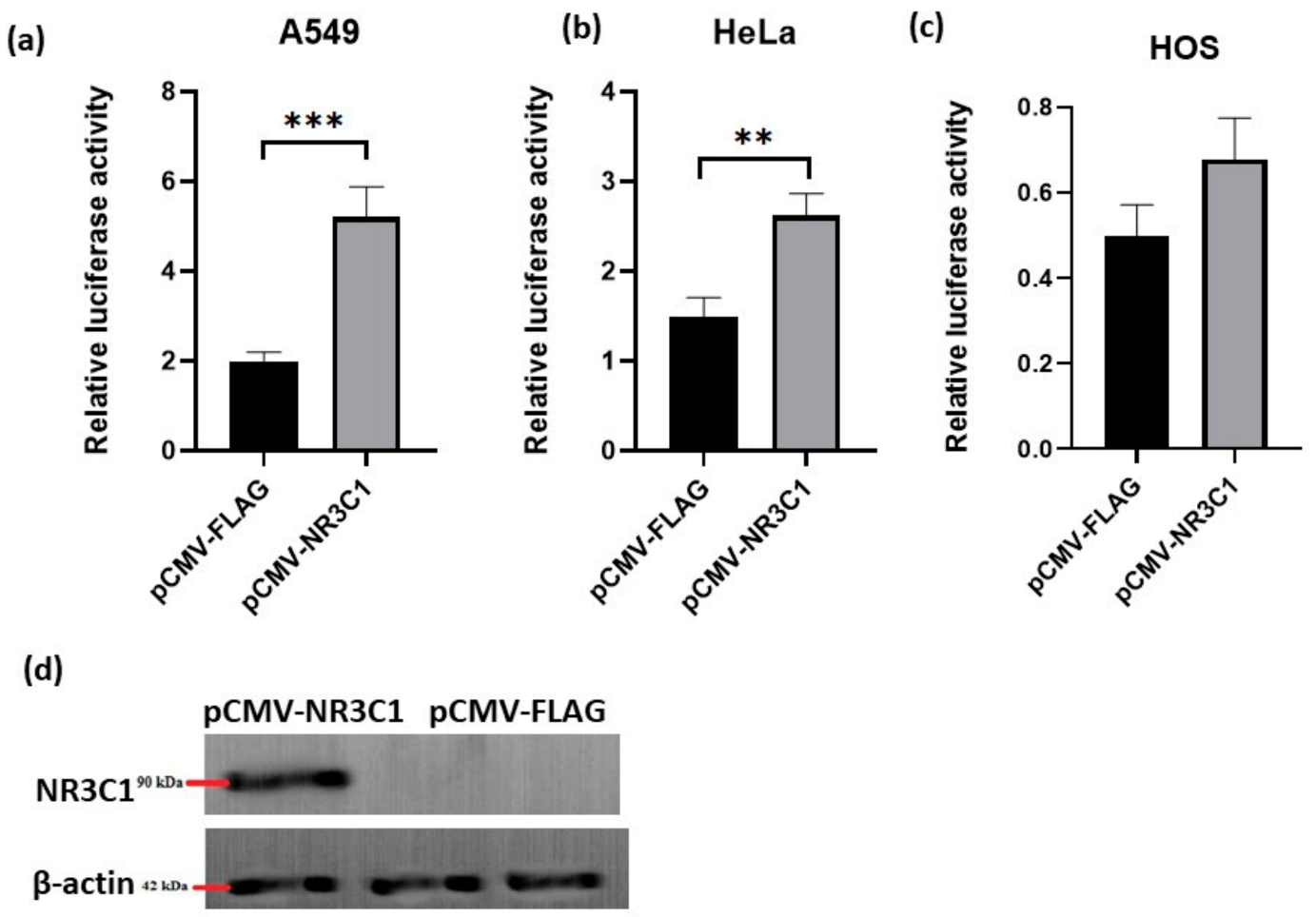
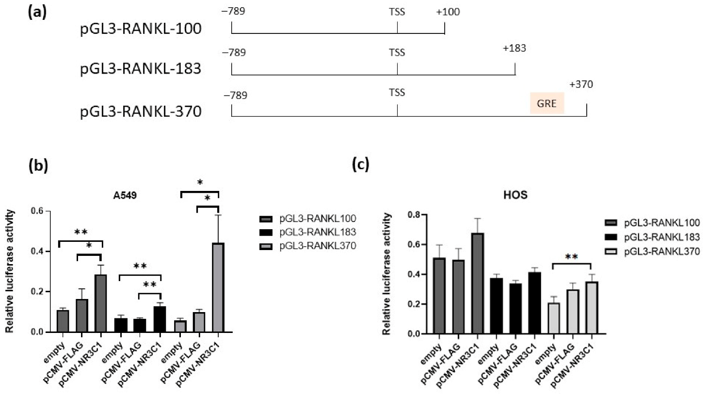
2.1. Overexpressed Glucocorticoid Receptor NR3C1 Increases RANKL Promoter Activity in A549, HOS and HeLa Cells

Here, were aimed to examine whether glucocorticoid receptor directly influences RANKL promoter activity and its transcriptional regulation. Since the expression of RANKL gene plays a crucial role in bone remodelling processes and it is modulated in various cancers [49], three different human cell lines were included in the experiments, human lung cancer cells (A549), human osteosarcoma cells (HOS) and human cervical cancer cells (HeLa). To examine the impact of glucocorticoid receptor NR3C1 on the activity of RANKL promoter, cells were co-transfected with plasmids encoding glucocorticoid receptor NR3C1 (pCMV-NR3C1) and pGL3 plasmid with 898 bp RANKL proximal promoter region (plasmid pGL3-RANKL-100 (region −789 to +100 relative to transcription start site [12]). Promoter activity was measured by luciferase assays 24 h after transfection. In A549 cells, in the presence of overexpressed NR3C1 promoter activity of pGL3-RANKL-100 increased 2.6 fold (p = 0.0008) in comparison with an empty vector (Figure 1a). In HeLa cells, overexpression of NR3C1 caused 1.7 fold (p = 0.0063) increase in promoter activity in comparison with an empty vector (Figure 1b). In HOS cells, overexpression of NR3C1 increased the promoter activity when comparing to an empty vector, yet the change was not significant (Figure 1c). Overexpression of glucocorticoid receptor NR3C1 protein was verified by immunoblot (Figure 1d). Increase in RANKL promoter activity in the presence of NR3C1 suggests that RANKL promoter is directly inducible by glucocorticoid receptor.
In bioinformatics analysis, we identified GRE site in the RANKL exon 1 region +342\+352 relative to transcription start site (Figure 2a). Next, we prepared plasmids harbouring different lengths of RANKL promoter region, also plasmid including newly identified GRE site (plasmids pGL3-RANKL-183, pGL3-RANKL-370) (Figure 2a). A549 and HOS cells were transfected with promoter length mutations (plasmids pGL3-RANKL-100 or pGL3-RANKL-183 or pGL3-RANKL-370) and pCMV-NR3C1 or an empty plasmid (pCMV-FLAG) and the luciferase activity was measured 24 h after transfection. Again, in A549 cells, overexpression of NR3C1 caused significant increase in promoter activity for all three lengths of the RANKL promoter. Promoter activity of plasmid pGL3-RANKL-100 (region −789 to +100 relative to TSS) increased 1.7 fold in comparison with an empty vector (p = 0.0376), pGL3-RANKL-183 (region −789 to +183 relative to TSS) 1.9 fold (p = 0.0024) and plasmid pGL3-RANKL-370 (region −789 to +370 relative to TSS) which includes novel GRE site 4.5 fold (p = 0.0224) in comparison with an empty vector (Figure 2b). Promoter activity of the RANKL promoter region that includes putative GRE site at +352 (pGL3-RANKL370) was 2.3-fold higher than that of a shorter promoter region (−789 to +183) (pGL3RANKL-183), suggesting that putative GRE site at +352 is a functional GRE site and that overexpression of GR increases RANKL promoter activity by binding to this site. Promoter activity of RANKL promoter length mutations also increased in the presence of GR in HOS cells, yet changes were not significant (Figure 2c).

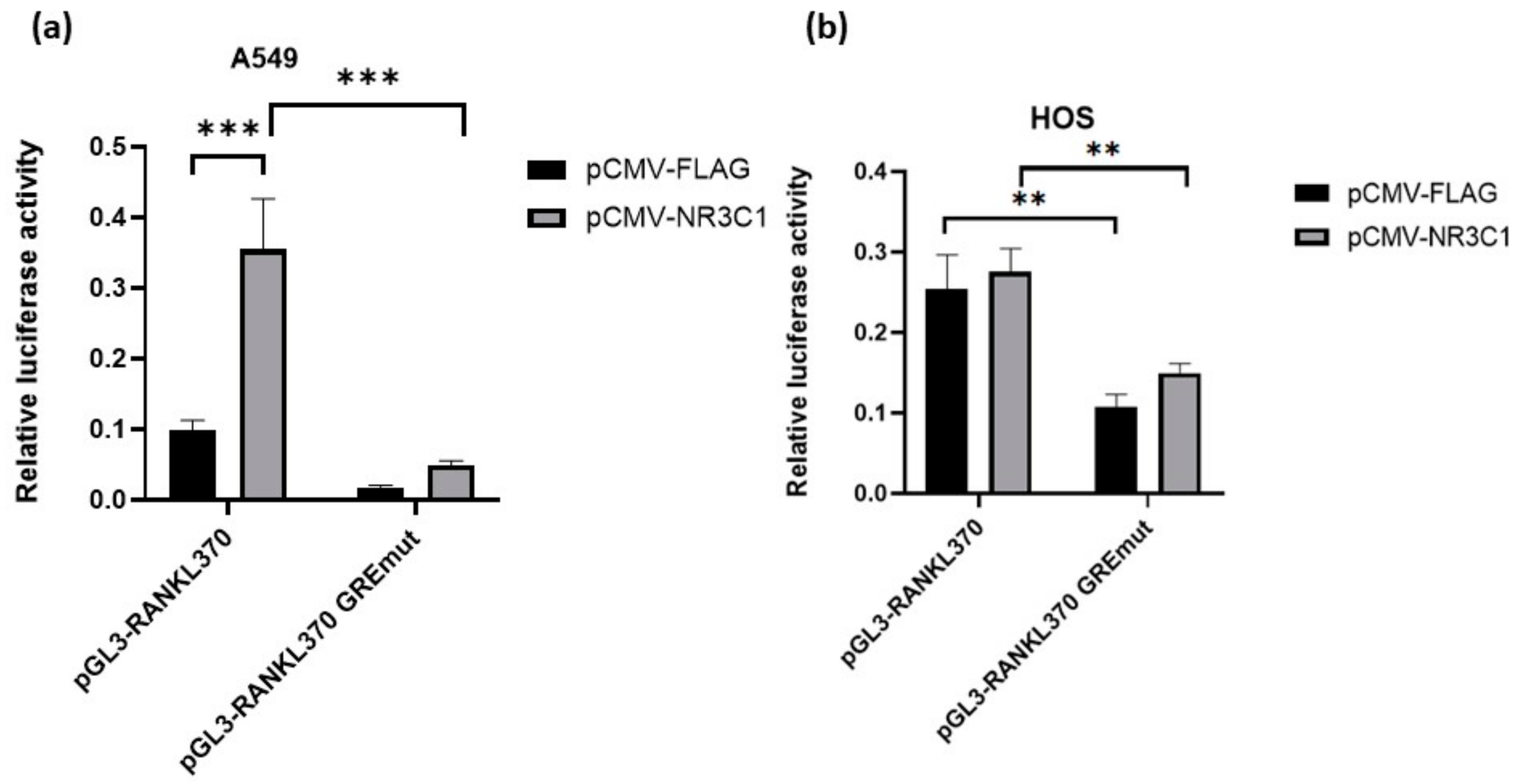
2.2. Mutation in Glucocorticoid Responsive Element Alleviates Effect of Glucocorticoid Receptor on RANKL Promoter Activity

Next, to examine whether GRE site directly influences the RANKL promoter activity, we introduced mutation in the putative GRE site (+352 relative to TSS) (pGL3-RANKL370 GREmut plasmid) and performed luciferase assays in A549 and HOS cells. Overexpression of NR3C1 did not cause significant change in promoter activity of pGL3-RANKL370-GREmut in comparison with an empty vector in any of the tested cells confirming that GRE at +352 is important for induction of RANKL promoter activity by NR3C1. In fact, introduction of mutation at GRE site caused 7.9-fold (p < 0.0001) and 1.8-fold (p = 0.009) lower promoter activity in the presence of GR in A549 and HOS cells, respectively (Figure 3). Interestingly, even in the absence of overexpressed NR3C1, wild type promoter activity was 5-fold and 2.3-fold higher than in the GRE mutated promoter in both cell types in A549 and HOS cells, respectively. Higher promoter activity of the wild type promoter in comparison with the GREmut sequence in the absence of overexpressed NR3C1 implies that GRE, at +352 site, is important for regulation of RANKL promoter activity and that endogenous transcription factors contribute to activation of RANKL promoter in this region. To summarize, our results indicate that GRE site (+352 relative to TSS) is important for GR activation of RANKL transcription in HOS and A549 cells. Moreover, endogenous transcription factors are responsible for activation of RANKL promoter (in the region +183 to +370), and this effect is potentiated by overexpressed GR.

2.3. Glucocorticoid Receptor Binds to the RANKL Proximal Promoter Region

To examine whether GR directly binds to the putative GRE binding site at +352 relative to TSS of the human RANKL proximal promoter region, we performed electrophoretic mobility shift assay. For that purpose, HEK293T cells were transfected with pCMV-NR3C1 plasmid and nuclear extracts were isolated. Nuclear extracts with overexpressed GR or prepared from untransfected cells were incubated with biotinylated probe with GRE sequence at position +352. In parallel, incubations with unlabelled competitive probe (the same sequence as biotinylated probe) and with mutated probe (with mutations at the GRE +352 site) were performed. In case a protein binds to the biotinylated probe, a shift on the gel appears. In the presence of nuclear extract from cells with overexpressed GR, a shift of biotinylated oligonucleotides appeared indicating direct binding of a protein from the nuclear extract to the biotinylated probe (Figure 4a, lane 2). The specificity of the binding was confirmed using unlabelled competitive oligonucleotides in 200-fold molar excess, where a shift disappears (Figure 4a, lane 4). When mutated unlabelled competitive oligonucleotide was added in the reaction, a shift reappeared indicating that unlabelled mutated competitive oligonucleotides did not bind to a protein from the nuclear extract (Figure 4a, lane 5). The presence of overexpressed GR protein in the nuclear extract was confirmed by Western blot (Figure 4b). A shift of labelled probe appeared also in untransfected cells, suggesting that endogenous GR binds to the labelled probe. Indeed, in both cell lysates (with overexpressed and empty cells), a bend resembling to endogenous GR appeared (Figure 4b), overexpressed protein with a higher intensity. This suggests that cells contain sufficient amount of endogenous GR to cause supershift in the EMSA assay. Results of the EMSA experiment indicate that GR directly binds to the RANKL proximal promoter region at site +352 (Figure 4c).

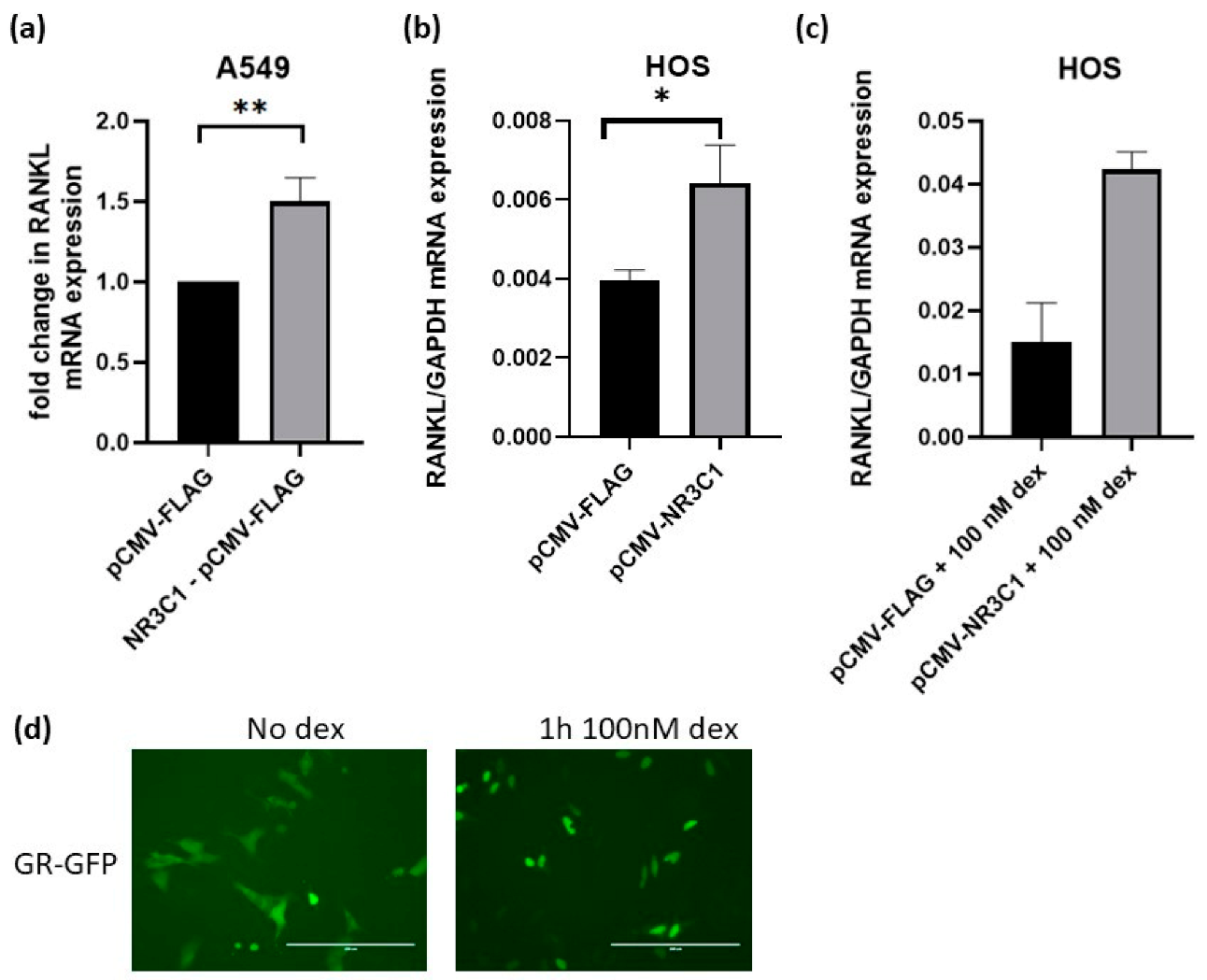
2.4. Overexpression of Glucocorticoid Receptor Upregulates RANKL Expression in A549 and HOS Cells

Since, in our RANKL promoter activity assays glucocorticoid receptor activated RANKL promoter, we next addressed the question whether NR3C1 also increases the mRNA level of RANKL in A549 and HOS cells. For that purpose, A549 and HOS cells were overexpressed with pCMV-NR3C1 plasmid and an empty control plasmid and the level of RANKL mRNA was measured by qPCR 72 h after transfection. The overexpression of NR3C1 caused 1.5-fold (p = 0.0059) and 1.6-fold (p = 0.0163) upregulation of RANKL in A549 and HOS cells, respectively (Figure 5a,b). We hypothesized that addition of dexamethasone will cause stronger effect of GR overexpression on the level of RANKL mRNA. To test this hypothesis, we performed experiment in the presence of dexamethasone. The expression of RANKL was measured after treatment of cells with 100 nM glucocorticoid dexamethasone, and the expression of RANKL was 2.8 fold higher (p = 0.055) in the presence of NR3C1 in comparison with an empty plasmid (Figure 5c). The impact of 100 nM dexamethasone on translocation of GR to the nucleus in shown in Figure 5d. We next hypothesized that silencing of endogenous GR would cause a decrease in RANKL expression in HOS cells. To examine whether silencing of endogenous NR3C1 causes any changes in RANKL expression, NR3C1 was silenced in HOS with siRNA and expression of RANKL was measured 72 h after transfection. In HOS cells, 0.49 fold (p = 0.0004) change in NR3C1 level was observed, which caused 0.38 fold change in RANKL expression (Figure 6a,b) suggesting that silencing of endogenous GR affects the expression of RANKL.

3. Discussion

Glucocorticoid osteoporosis is a serious side effect of long term glucocorticoid uptake and it is caused by imbalance in the major bone remodeling pathway RANK/RANL/OPG. The impact of glucocorticoid on the maintenance of RANK/RANL/OPG is well documented; dexamethasone was shown to disturb the ratio between OPG and RANKL level by downregulation OPG and in some case upregulation of RANKL. In this study, we examined how GR affects transcriptional regulation of human RANKL gene. We show that GR causes an increase in RANKL promoter activity and an increase in RANKL mRNA expression level. We demonstrate that GR acts through direct binding on GRE site at +352 in the RANKL promoter region.
Given that GR affects RANKL expression and plays an important role in GIO, we examined a direct effect of GR on the activity of the human RANKL promoter. Three regions of the human RANKL proximal promoter were analyzed harboring previously reported putative GRE half site (−679; corresponding to mouse −642/−628 site) and putative GRE site at +352 relative to transcription start site. Addition of GR caused higher increase in promoter activity of the region overlapping putative GRE site at +352 in comparison with the region overlapping only putative GRE site at −679. Therefore, our results confirm the results obtained with a mouse RANKL promoter [47] and identify a novel putative GRE site at +352 that is involved in the activation of RANKL promoter. Moreover, our results demonstrate that GRE residing in the untranslated region of RANKL promoter is more important for activation of RANKL promotor activity with GR than GRE at −679 site. Namely, +352 region was more inducible by GR than previously reported putative GRE site at −679. Indeed, promoter activity of the region from −789 to +370 in the presence of NR3C1 was higher in comparison with a shorter region from −789 to +100 suggesting that GRE in the untranslated region plays an important role in transcriptional regulation of RANKL. Using EMSA, we demonstrate that GR from the nuclear extract directly binds to this +352 region of the RANKL promoter confirming the importance of the newly identified GRE in the regulation of RANKL gene. In one fifth of the genes that bear GREs, GR binding sites have been reported to reside upstream from transcription start site, yet small portion of GREs have been also identified in the first intron and exon [50], which we also show here for RANKL gene. Moreover, several GREs in a single gene have also been identified in other genes, which probably enhances the responsiveness to GC [50]. Our results also corroborated with the results of the ENCODE project, where chromatin immunoprecipitation-sequencing revealed NR3C1 binding to RANKL gene in A549 cells treated with 100 nM dexamethasone [51]. Our results indicate that the impact of GR on the RANKL expression is very complex. There are at least two regions in the proximal RANKL promoter region that can be directly affected by GR. This provides important knowledge in the understanding the complexity of glucocorticoid induced osteoporosis.
In our experimental setup, in human osteosarcoma and adenocarcinoma cells, addition of GR caused an increase in RANKL mRNA level which is similar to previously reported results obtained on dexamethasone treated human primary osteoblast [44] and mouse bone marrow stromal cells treated with dexamethasone and vitamin D3 [41,47]. It was demonstrated that vitamin D3 and dexamethasone synergistically increase expression of RANKL in stromal ST2 cells. Dexamethasone was also shown to act synergistically with vitamin D3 to enhance osteoclast formation by increasing RANKL mRNA expression in bone marrow stromal primary and transformed cells [48]. Reports on how glucocorticoids affect RANKL expression differ depending on the investigated cell types and experimental setups. Comparison between activation of RANKL promoter in three different cell types revealed that there is significant difference in transactivation of RANKL promoter in the presence of GR between cell systems and that fold of activation differs between cells. Our results indicate that transcription machinery of particular cell system affects RANKL promoter activity and corroborates with previous results that GC differently affect RANKL expression in different cell types. Besides, different transcriptional machinery, the existence of various mechanism by which GR can transactivate promoters, contribute to those differences.
In vivo relevance of our results was provided by mouse model study, where mice treated with vitamin D3 and dexamethasone showed potentiated osteoclast formation [48]. Treatment of mouse bone marrow stromal cells caused 24-fold increase in RANKL expression 6 days after treatment, and decrease in OPG mRNA level and was also confirmed at the protein level [48]. The level of endogenous GR was unaffected by dexamethasone and vitamin D3 treatment. However, in mice without GR, combination of dexamethasone and vitamin D3, had no impact on the expression of RANKL indicating that dexamethasone and vitamin D3 upregulate RANKL transcription via GR transcriptional activation. Interestingly, in mice where dimerization of GR was hindered, dexamethasone and vitamin D3 still caused upregulation of RANKL implying that monomeric GR suffice for RANKL upregulation and enhanced osteoclastogenesis [48]. Here, we identified GRE site in RANKL promoter with one conserved GRE site (CAGCGTCGCCCTGTTCTTC), which mutation alleviates promoter activation, probably due to hindered binding of GR to DNA. Therefore, we propose that one of the mechanisms by which GR upregulates RANKL expression is via direct binding of monomeric GR unit to the +352 GRE site of the RANKL promoter. Further studies to elucidate whether other transcription factors are involved in this transactivation are required.
In glucocorticoid induced osteoporosis, GC cause bone loss through decrease in osteoblast formation and stimulated osteoclastogenesis. For both processes, balanced ratio in RANK/RANKL/OPG is essential. Here, we focused on the deciphering mechanism behind increase in RANKL level. We demonstrate that overexpression of GR caused activation of RANKL promoter and an increase in RANKL expression in human lung cancer and osteosarcoma cells; probably through direct binding to RANKL promoter at the region +352. We provide a link between upregulation of RANKL in patients treated with GC and activation of RANKL promoter. Our results imply that GC directly influences RANKL expression via GR induced activation of RANKL transcription.

4. Materials and Methods

4.1. Plasmid Constructs

Human proximal RANKL promoter region (from −798 bp to +370 bp relatively to the transcription start site) was PCR amplified from the human genomic DNA with Q5® High-Fidelity DNA Polymerase (New England Biolabs, Ipswich, MA, USA) using RANKL370 f and RANKL370 rev primers (Supplementary Table S1) and cloned through BglII and HindIII sites into the pGL3 basic vector (Promega, Madison, WI, USA). Cloning resulted in the pGL3-RANKL370 plasmid vector that was sequence verified. Mutations in the GRE region of RANKL promoter were introduced with the Quick-Change II Site-Directed Mutagenesis kits (Stratagene, San Diego, CA, USA) using primers listed in the Supplementary Table S1. All the plasmid vectors were sequence verified by Sanger sequencing (GATC, Konstanz, Germany).

4.2. Cell Culturing and Transfections

Human bone osteosarcoma HOS cells (ATCC® CRL-1543™), human cervical cancer HeLa cells (ATCC® CCL-2) and human lung cancer A549 cells (ATCC®CCL-185™) were cultured in Dulbecco’s Modified Eagle Medium (DMEM) supplemented with 10% FBS, 1% glutamine, 1% antimycotic/antibiotic (Gibco, Thermo Fisher Scientific, MA, USA) at 37 °C, 5% CO2 and subcultured according to manufacturer’s procedure.

4.3. Luciferase Reporter Assay

Luciferase activity was measured with the Dual-Luciferase® Reporter (DLR™) Assay System (Promega, Madison, WI, USA) according to the manufacturer’s procedure. Cells were seeded at the density 5 × 10⁴ cells/well in 24 well plates in antibiotic free growth media 24 h prior transfection. Next day, 500 ng of respective pGL3 reporter plasmid harbouring RANKL promoter (pGL3-RANKL370, pGL3-RANKL100, pGL3-RANKL183), its mutant (pGL3-RANKL370 GREMut), positive control (pGL3-SV40) and negative control (pGL3-Basic, empty vector) and together with 15 ng of pRL Renilla plasmid were transfected using 1.5 µL PolyJet™ In Vitro DNA Transfection Reagent (SignaGen Laboratories, Rockville, MD, USA) according to manufacturer’s instructions. Luciferase assays were performed 24 h after transfection. The Firefly (Firefly luciferase is encoded in pGL3 vectors) and Renilla luciferase (Renilla is encoded in pRL vector) activities were measured for each sample. Relative luciferase activity was calculated by normalizing Firefly luciferase activity with the Renilla luciferase. All the experiments were performed at least three times independently.

4.4. Quantitative PCR

Expression of RANKL was analyzed using qPCR with RNA samples obtained in gain-of-function and loss-of-function experiments. The RNA was extracted from cells and the complementary DNAs (cDNAs) were synthesized using High-Capacity cDNA Reverse Transcription kits (Thermo Fisher Scientific, Waltham, MA, USA), with gene expression analyses performed as described below. Expressions of RANKL, NR3C1 were also analyzed using qPCR assays [12]. For qPCR, 5× Hot FirePol EvaGreen qPCR Mix Plus (Solis, BioDyne, Tartu, Estonia) was used, following the manufacturer recommendations, on a LightCycler 480 (Roche Diagnostics, Mannheim, Germany). Concentration of each primer in the qPCR reaction was 150 nM. Nucleotide sequences of primers are listed in Supplementary Table S1. All of the cDNA samples were diluted to the final concentration of 2.5 ng/µL. All of the samples were quantified in triplicate. Dilution series of cDNAs were prepared to create a relative standard curve, and absolute quantification of the data was performed using the second derivative maximum method (LightCycler 480, Software version 1.5; Roche Diagnostics, Mannheim, Germany). All of the data were normalized to the internal housekeeping genes of ribosomal protein, large, P0 (RPLP0) or glyceraldehyde 3-phosphate dehydrogenase (GAPDH).

4.5. Western Blotting

Human bone osteosarcoma cells were seeded in 10-cm2 dishes and transfected with empty pCMV-FLAG expression vector or pCMV-NR3C1 expression vector. The cells were lysed after 24 h and resolved on SDS-PAGE. The proteins were transferred to nitrocellulose membranes, which were later blocked in 5% milk/TTBS, and incubated overnight with the primary antibodies against FLAG (clone M2; Sigma-Aldrich, St. Louis, MO, USA). After the incubation in the primary antibodies, the membranes were washed in TTBS and incubated in the secondary antibodies bound to HRP, for 1 h. Substrate was then added and the proteins were visualized using imaging platform (Uvitec Alliance, Cambridge, UK). Antibodies against beta-actin (A2228, Sigma-Aldrich) were used for loading controls.

4.6. RNA Interference

siRNAs for silencing of NR3C1 (sc-35505) and a negative control (sc-37007) were purchased from Santa Cruz Biotechnology, Dallas, TX, USA. Here, 50 pmol was transfected using Oligofectamine reagent (Invitrogen Life Technologies, Carlsbad, CA, USA). Then, 72 h after transfection, the RNA was isolated using PeqGold RNA extraction kits (VWR, Radnor, PA, USA), reverse transcribed, and subjected to q-PCR for measurements of expressions of the genes of interest. Three biological replicates were analyzed.

4.7. Electrophoretic Mobility Shift Assays

Electrophoretic mobility shift assays were performed using LightShift Chemiluminescent EMSA kits (Thermo Fisher Scientific, Waltham, MA, USA), according to the manufacturer instructions and as previously described [12]. 293T nuclear extracts were prepared using NE-PER Nuclear and Cytoplasmic Extraction Reagents (Thermo Fisher Scientific, Waltham, MA, USA), according to manufacturer instructions. Double-stranded biotinylated oligonucleotides (probe sequence 5′CAGCGTCGCCCTGTTCTTCTATTTCAGAG′3) were prepared by hybridization in a C1000 thermal cycler by stepwise 0.5 °C decreases in the temperature, ranging from 95 °C to 35 °C, over 120 cycles, each 30 s long. Control reaction was performed without nuclear extract. The specificity of the DNA-protein complexes was verified by binding with competitive non-biotinylated DNA or mutated non-biotinylated DNA. The biotinylated oligonucleotides listed in Supplementary Table S1 were used in the EMSA experiments. After the binding reactions, the samples were loaded onto electrophoretic gels and run for 1 h (100 V), then transferred to nylon membranes (30 min, 100 V), and cross-linked on a transilluminator. Biotin-labeled nuclear probes were detected with Chemiluminescent Nucleic Acid Detection Module kits (Pierce, Thermo Scientific, Wilmington, DE, USA), according to the manufacturer instructions.

4.8. Statistical Analysis

All experiments were performed in triplicates. Data were analysed using the GraphPad Prism 8 software (Graph Pad Software Inc., La Jolla, CA, USA). All of the data are presented as mean relative target gene expression after normalization with GAPDH expression. The results in the graphs are depicted as the means and standard error of the means of all three biological repeats. Statistical analyses were carried out by Student’s t-test. The level of significance was taken to be p < 0.05. In case of the luciferase assays, the results of firefly luminescence were normalized with Renilla luminescence values. The significance of the data was analysed by student t-test (two tailed) or ANOVA as implemented in Graphpad Prism 8.0. Data are presented as means with standard errors of the means.

5. Conclusions

In this study we demonstrate for the first time that glucocorticoid receptor can directly regulate human TNFSF11 expression by binding to it proximal promoter region. We identified a novel glucocorticoid response element at the position +352 of the human TNFSF11 gene and show that this site is responsible for GR dependent activation of the human TNFSF11 promoter. Our study provides a novel link between glucocorticoid administration and onset of osteoporosis.

Supplementary Materials

Supplementary Materials can be found at https://www.mdpi.com/1422-0067/22/3/1054/s1. Table S1: List of primers used for plasmid construction, electrophoretic mobility shift assays and mutagenesis.

Author Contributions

Cloning site, specific mutagenesis, plasmid preparation, sequencing, luciferase assays, statistical analysis, RNA isolation: N.L.; study design and conceiving the research: N.L., J.M.; manuscript preparation: N.L., J.M. All authors have read and agreed to the published version of the manuscript.

Funding

This research was funded by Slovenian Research Agency (ARRS) grants: J3-7245, J3-1759, P3-0298.

Data Availability Statement

No applicable.

Acknowledgments

We thank Maja Zupančič, Manca Zupan, Klemen Kodrič and Tina Goršek for their assistance in the laboratory.

Conflicts of Interest

The authors declare no conflict of interest.

Abbreviations

RANKLreceptor activator of nuclear factor κ B ligand
GIOGlucocorticoid induced osteoporosis
GRGlucocorticoid receptor
GREglucocorticoid responsive element
GCglucocorticoid
NR3C1Nuclear Receptor Subfamily 3 Group C Member
OPGosteoprotegerin
EMSAelectrophoretic mobility shift assay

References

  1. Moutsatsou, P.; Kassi, E.; Papavassiliou, A.G. Glucocorticoid receptor signaling in bone cells. Trends Mol. Med. 2012, 18, 348–359. [Google Scholar] [CrossRef]
  2. Chotiyarnwong, P.; McCloskey, E.V. Pathogenesis of glucocorticoid-induced osteoporosis and options for treatment. Nat. Rev. Endocrinol. 2020, 16, 437–447. [Google Scholar] [CrossRef]
  3. Compston, J. Glucocorticoid-induced osteoporosis: An update. Endocrine 2018, 61, 7–16. [Google Scholar] [CrossRef] [Green Version]
  4. Canalis, E.; Mazziotti, G.; Giustina, A.; Bilezikian, J.P. Glucocorticoid-induced osteoporosis: Pathophysiology and therapy. Osteoporos. Int. 2007, 18, 1319–1328. [Google Scholar] [CrossRef]
  5. Ahmad, M.; Hachemi, Y.; Paxian, K.; Mengele, F.; Koenen, M.; Tuckermann, J. A Jack of All Trades: Impact of Glucocorticoids on Cellular Cross-Talk in Osteoimmunology. Front. Immunol. 2019, 10, 2460. [Google Scholar] [CrossRef] [Green Version]
  6. Adami, G.; Saag, K.G. Glucocorticoid-induced osteoporosis update. Curr. Opin. Rheumatol. 2019, 31, 388–393. [Google Scholar] [CrossRef]
  7. Swanson, C.; Lorentzon, M.; Conaway, H.H.; Lerner, U.H. Glucocorticoid regulation of osteoclast differentiation and expression of receptor activator of nuclear factor-kappaB (NF-kappaB) ligand, osteoprotegerin, and receptor activator of NF-kappaB in mouse calvarial bones. Endocrinology 2006, 147, 3613–3622. [Google Scholar] [CrossRef] [PubMed] [Green Version]
  8. Hofbauer, L.C.; Gori, F.; Riggs, B.L.; Lacey, D.L.; Dunstan, C.R.; Spelsberg, T.C.; Khosla, S. Stimulation of osteoprotegerin ligand and inhibition of osteoprotegerin production by glucocorticoids in human osteoblastic lineage cells: Potential paracrine mechanisms of glucocorticoid-induced osteoporosis. Endocrinology 1999, 140, 4382–4389. [Google Scholar] [CrossRef] [PubMed]
  9. Sato, A.Y.; Peacock, M.; Bellido, T. Glucocorticoid Excess in Bone and Muscle. Clin. Rev. Bone Miner. Metab. 2018, 16, 33–47. [Google Scholar] [CrossRef] [PubMed]
  10. Chiodini, I.; Merlotti, D.; Falchetti, A.; Gennari, L. Treatment options for glucocorticoid-induced osteoporosis. Expert Opin. Pharmacother. 2020, 21, 721–732. [Google Scholar] [CrossRef]
  11. Lovsin, N.; Zupan, J.; Marc, J. Genetic effects on bone health. Curr. Opin. Clin. Nutr. Metab. Care 2018, 21, 233–239. [Google Scholar] [CrossRef] [PubMed]
  12. Kodric, K.; Zupan, J.; Kranjc, T.; Komadina, R.; Mlakar, V.; Marc, J.; Lovsin, N. Sex-determining region Y (SRY) attributes to gender differences in RANKL expression and incidence of osteoporosis. Exp. Mol. Med. 2019, 51, 97. [Google Scholar] [CrossRef] [PubMed] [Green Version]
  13. Hikita, A.; Yana, I.; Wakeyama, H.; Nakamura, M.; Kadono, Y.; Oshima, Y.; Nakamura, K.; Seiki, M.; Tanaka, S. Negative regulation of osteoclastogenesis by ectodomain shedding of receptor activator of NF-kappaB ligand. J. Biol. Chem. 2006, 281, 36846–36855. [Google Scholar] [CrossRef] [PubMed] [Green Version]
  14. Ikeda, T.; Kasai, M.; Utsuyama, M.; Hirokawa, K. Determination of three isoforms of the receptor activator of nuclear factor-kappaB ligand and their differential expression in bone and thymus. Endocrinology 2001, 142, 1419–1426. [Google Scholar] [CrossRef]
  15. Nagai, M.; Kyakumoto, S.; Sato, N. Cancer cells responsible for humoral hypercalcemia express mRNA encoding a secreted form of ODF/TRANCE that induces osteoclast formation. Biochem. Biophys. Res. Commun. 2000, 269, 532–536. [Google Scholar] [CrossRef]
  16. Wong, B.R.; Rho, J.; Arron, J.; Robinson, E.; Orlinick, J.; Chao, M.; Kalachikov, S.; Cayani, E.; Bartlett, F.S., 3rd; Frankel, W.N.; et al. TRANCE is a novel ligand of the tumor necrosis factor receptor family that activates c-Jun N-terminal kinase in T cells. J. Biol. Chem. 1997, 272, 25190–25194. [Google Scholar] [CrossRef] [Green Version]
  17. Dougall, W.C.; Glaccum, M.; Charrier, K.; Rohrbach, K.; Brasel, K.; De Smedt, T.; Daro, E.; Smith, J.; Tometsko, M.E.; Maliszewski, C.R.; et al. RANK is essential for osteoclast and lymph node development. Genes Dev. 1999, 13, 2412–2424. [Google Scholar] [CrossRef]
  18. Fata, J.E.; Kong, Y.Y.; Li, J.; Sasaki, T.; Irie-Sasaki, J.; Moorehead, R.A.; Elliott, R.; Scully, S.; Voura, E.B.; Lacey, D.L.; et al. The osteoclast differentiation factor osteoprotegerin-ligand is essential for mammary gland development. Cell 2000, 103, 41–50. [Google Scholar] [CrossRef] [Green Version]
  19. Kong, Y.-Y.; Yoshida, H.; Sarosi, I.; Tan, H.-L.; Timms, E.; Capparelli, C.; Morony, S.; Oliveira-dos-Santos, A.J.; Van, G.; Itie, A.; et al. OPGL is a key regulator of osteoclastogenesis, lymphocyte development and lymph-node organogenesis. Nature 1999, 397, 315–323. [Google Scholar] [CrossRef]
  20. O’Brien, C.A. Control of RANKL Gene Expression. Bone 2010, 46, 911–919. [Google Scholar] [CrossRef] [Green Version]
  21. Zhang, J.; Dai, J.; Qi, Y.; Lin, D.L.; Smith, P.; Strayhorn, C.; Mizokami, A.; Fu, Z.; Westman, J.; Keller, E.T. Osteoprotegerin inhibits prostate cancer–induced osteoclastogenesis and prevents prostate tumor growth in the bone. J. Clin. Investig. 2001, 107, 1235–1244. [Google Scholar] [CrossRef] [PubMed]
  22. Brown, J.M.; Corey, E.; Lee, Z.D.; True, L.D.; Yun, T.J.; Tondravi, M.; Vessella, R.L. Osteoprotegerin and rank ligand expression in prostate cancer. Urology 2001, 57, 611–616. [Google Scholar] [CrossRef]
  23. Huang, L.; Cheng, Y.Y.; Chow, L.T.; Zheng, M.H.; Kumta, S.M. Tumour cells produce receptor activator of NF-kappaB ligand (RANKL) in skeletal metastases. J. Clin. Pathol. 2002, 55, 877–878. [Google Scholar] [CrossRef] [PubMed] [Green Version]
  24. Farrugia, A.N.; Atkins, G.J.; To, L.B.; Pan, B.; Horvath, N.; Kostakis, P.; Findlay, D.M.; Bardy, P.; Zannettino, A.C. Receptor activator of nuclear factor-kappaB ligand expression by human myeloma cells mediates osteoclast formation in vitro and correlates with bone destruction in vivo. Cancer Res. 2003, 63, 5438–5445. [Google Scholar] [PubMed]
  25. Jones, D.H.; Nakashima, T.; Sanchez, O.H.; Kozieradzki, I.; Komarova, S.V.; Sarosi, I.; Morony, S.; Rubin, E.; Sarao, R.; Hojilla, C.V.; et al. Regulation of cancer cell migration and bone metastasis by RANKL. Nature 2006, 440, 692–696. [Google Scholar] [CrossRef] [PubMed] [Green Version]
  26. O’Brien, C.A.; Gubrij, I.; Lin, S.C.; Saylors, R.L.; Manolagas, S.C. STAT3 activation in stromal/osteoblastic cells is required for induction of the receptor activator of NF-kappaB ligand and stimulation of osteoclastogenesis by gp130-utilizing cytokines or interleukin-1 but not 1,25-dihydroxyvitamin D3 or parathyroid hormone. J. Biol. Chem. 1999, 274, 19301–19308. [Google Scholar]
  27. Jilka, R.L.; Hangoc, G.; Girasole, G.; Passeri, G.; Williams, D.C.; Abrams, J.S.; Boyce, B.; Broxmeyer, H.; Manolagas, S.C. Increased osteoclast development after estrogen loss: Mediation by interleukin-6. Science 1992, 257, 88–91. [Google Scholar] [CrossRef]
  28. Holmen, S.L.; Zylstra, C.R.; Mukherjee, A.; Sigler, R.E.; Faugere, M.C.; Bouxsein, M.L.; Deng, L.; Clemens, T.L.; Williams, B.O. Essential role of beta-catenin in postnatal bone acquisition. J. Biol. Chem. 2005, 280, 21162–21168. [Google Scholar] [CrossRef] [Green Version]
  29. Spencer, G.J.; Utting, J.C.; Etheridge, S.L.; Arnett, T.R.; Genever, P.G. Wnt signalling in osteoblasts regulates expression of the receptor activator of NFkappaB ligand and inhibits osteoclastogenesis in vitro. J. Cell Sci. 2006, 119, 1283–1296. [Google Scholar] [CrossRef] [Green Version]
  30. Fujita, K.; Janz, S. Attenuation of WNT signaling by DKK-1 and -2 regulates BMP2-induced osteoblast differentiation and expression of OPG, RANKL and M-CSF. Mol. Cancer 2007, 6, 71. [Google Scholar] [CrossRef] [Green Version]
  31. Wang, F.S.; Ko, J.Y.; Lin, C.L.; Wu, H.L.; Ke, H.J.; Tai, P.J. Knocking down dickkopf-1 alleviates estrogen deficiency induction of bone loss. A histomorphological study in ovariectomized rats. Bone 2007, 40, 485–492. [Google Scholar] [CrossRef] [PubMed]
  32. Aicher, A.; Kollet, O.; Heeschen, C.; Liebner, S.; Urbich, C.; Ihling, C.; Orlandi, A.; Lapidot, T.; Zeiher, A.M.; Dimmeler, S. The Wnt antagonist Dickkopf-1 mobilizes vasculogenic progenitor cells via activation of the bone marrow endosteal stem cell niche. Circ. Res. 2008, 103, 796–803. [Google Scholar] [CrossRef] [PubMed] [Green Version]
  33. Kubota, T.; Michigami, T.; Sakaguchi, N.; Kokubu, C.; Suzuki, A.; Namba, N.; Sakai, N.; Nakajima, S.; Imai, K.; Ozono, K. Lrp6 hypomorphic mutation affects bone mass through bone resorption in mice and impairs interaction with Mesd. J. Bone Miner. Res. 2008, 23, 1661–1671. [Google Scholar] [CrossRef] [PubMed]
  34. Qiang, Y.W.; Chen, Y.; Stephens, O.; Brown, N.; Chen, B.; Epstein, J.; Barlogie, B.; Shaughnessy, J.D., Jr. Myeloma-derived Dickkopf-1 disrupts Wnt-regulated osteoprotegerin and RANKL production by osteoblasts: A potential mechanism underlying osteolytic bone lesions in multiple myeloma. Blood 2008, 112, 196–207. [Google Scholar] [CrossRef] [PubMed]
  35. Suzuki, A.; Ozono, K.; Kubota, T.; Kondou, H.; Tachikawa, K.; Michigami, T. PTH/cAMP/PKA signaling facilitates canonical Wnt signaling via inactivation of glycogen synthase kinase-3beta in osteoblastic Saos-2 cells. J. Cell. Biochem. 2008, 104, 304–317. [Google Scholar] [CrossRef]
  36. Weikum, E.R.; Knuesel, M.T.; Ortlund, E.A.; Yamamoto, K.R. Glucocorticoid receptor control of transcription: Precision and plasticity via allostery. Nat. Rev. Mol. Cell Biol. 2017, 18, 159–174. [Google Scholar] [CrossRef]
  37. Kassel, O.; Herrlich, P. Crosstalk between the glucocorticoid receptor and other transcription factors: Molecular aspects. Mol. Cell. Endocrinol. 2007, 275, 13–29. [Google Scholar] [CrossRef] [Green Version]
  38. Ratman, D.; Vanden Berghe, W.; Dejager, L.; Libert, C.; Tavernier, J.; Beck, I.M.; De Bosscher, K. How glucocorticoid receptors modulate the activity of other transcription factors: A scope beyond tethering. Mol. Cell. Endocrinol. 2013, 380, 41–54. [Google Scholar] [CrossRef]
  39. Hachemi, Y.; Rapp, A.E.; Picke, A.K.; Weidinger, G.; Ignatius, A.; Tuckermann, J. Molecular mechanisms of glucocorticoids on skeleton and bone regeneration after fracture. J. Mol. Endocrinol. 2018, 61, R75–R90. [Google Scholar] [CrossRef]
  40. Scheschowitsch, K.; Leite, J.A.; Assreuy, J. New Insights in Glucocorticoid Receptor Signaling-More Than Just a Ligand-Binding Receptor. Front. Endocrinol. (Lausanne) 2017, 8, 16. [Google Scholar] [CrossRef]
  41. Kondo, T.; Kitazawa, R.; Yamaguchi, A.; Kitazawa, S. Dexamethasone promotes osteoclastogenesis by inhibiting osteoprotegerin through multiple levels. J. Cell. Biochem. 2008, 103, 335–345. [Google Scholar] [CrossRef] [PubMed]
  42. Hartmann, K.; Koenen, M.; Schauer, S.; Wittig-Blaich, S.; Ahmad, M.; Baschant, U.; Tuckermann, J.P. Molecular Actions of Glucocorticoids in Cartilage and Bone During Health, Disease, and Steroid Therapy. Physiol. Rev. 2016, 96, 409–447. [Google Scholar] [CrossRef] [Green Version]
  43. Harding, G.; Mak, Y.T.; Evans, B.; Cheung, J.; MacDonald, D.; Hampson, G. The effects of dexamethasone and dehydroepiandrosterone (DHEA) on cytokines and receptor expression in a human osteoblastic cell line: Potential steroid-sparing role for DHEA. Cytokine 2006, 36, 57–68. [Google Scholar] [CrossRef]
  44. Sivagurunathan, S.; Muir, M.M.; Brennan, T.C.; Seale, J.P.; Mason, R.S. Influence of glucocorticoids on human osteoclast generation and activity. J. Bone Miner. Res. 2005, 20, 390–398. [Google Scholar] [CrossRef]
  45. Rauner, M.; Goettsch, C.; Stein, N.; Thiele, S.; Bornhaeuser, M.; De Bosscher, K.; Haegeman, G.; Tuckermann, J.; Hofbauer, L.C. Dissociation of osteogenic and immunological effects by the selective glucocorticoid receptor agonist, compound A, in human bone marrow stromal cells. Endocrinology 2011, 152, 103–112. [Google Scholar] [CrossRef] [PubMed]
  46. Hofbauer, L.C.; Zeitz, U.; Schoppet, M.; Skalicky, M.; Schuler, C.; Stolina, M.; Kostenuik, P.J.; Erben, R.G. Prevention of glucocorticoid-induced bone loss in mice by inhibition of RANKL. Arthritis Rheum. 2009, 60, 1427–1437. [Google Scholar] [CrossRef] [PubMed]
  47. Kitazawa, R.; Kitazawa, S. Vitamin D(3) augments osteoclastogenesis via vitamin D-responsive element of mouse RANKL gene promoter. Biochem. Biophys. Res. Commun. 2002, 290, 650–655. [Google Scholar] [CrossRef] [PubMed]
  48. Conaway, H.H.; Henning, P.; Lie, A.; Tuckermann, J.; Lerner, U.H. Glucocorticoids employ the monomeric glucocorticoid receptor to potentiate vitamin D3 and parathyroid hormone-induced osteoclastogenesis. FASEB J. 2019, 33, 14394–14409. [Google Scholar] [CrossRef] [PubMed] [Green Version]
  49. De Castro, J.; Garcia, R.; Garrido, P.; Isla, D.; Massuti, B.; Blanca, B.; Vazquez, J. Therapeutic Potential of Denosumab in Patients with Lung Cancer: Beyond Prevention of Skeletal Complications. Clin. Lung Cancer 2015, 16, 431–446. [Google Scholar] [CrossRef] [Green Version]
  50. Reddy, T.E.; Pauli, F.; Sprouse, R.O.; Neff, N.F.; Newberry, K.M.; Garabedian, M.J.; Myers, R.M. Genomic determination of the glucocorticoid response reveals unexpected mechanisms of gene regulation. Genome Res. 2009, 19, 2163–2171. [Google Scholar] [CrossRef] [Green Version]
  51. Davis, C.A.; Hitz, B.C.; Sloan, C.A.; Chan, E.T.; Davidson, J.M.; Gabdank, I.; Hilton, J.A.; Jain, K.; Baymuradov, U.K.; Narayanan, A.K.; et al. The Encyclopedia of DNA elements (ENCODE): Data portal update. Nucleic Acids Res. 2018, 46, D794–D801. [Google Scholar] [CrossRef] [PubMed] [Green Version]
Figure 1. Overexpression of gluccocorticoid receptor causes activation of RANKL promoter in A549, HeLa and HOS cells. (a) A549, cells were co-transfected with the luciferase reporter constructs harboring RANKL promoter region 889 bp, (pGL3-RANKL100), and 24 h after transfection luciferase activity was measured. Relative luciferase activity is shown and it was calculated by normalization of Firefly luciferase activity to Renilla luciferase activity. (b) HeLa cells were co-transfected with the luciferase reporter constructs harboring RANKL promoter region 889 bp, (pGL3-RANKL100), and 24 h after transfection luciferase activity was measured. (c) HOS cells cells were co-transfected with the luciferase reporter constructs harboring RANKL promoter region 889 bp, (pGL3-RANKL100), and 24 h after transfection luciferase activity was measured. (d) Expression of overexpressed protein was confirmed by Western blot using anti-FLAG antibodies. β-actin was used as a loading control. All the experiments were performed at least three times independently in parallels. The results are expressed as the mean ±SEM. **–p < 0.01, ***–p < 0.001.
Ijms 22 01054 g001
Figure 2. Glucocorticoid receptor NR3C1 potently increases RANKL promoter activity of the region including a novel Glucocorticoid Responsive Element (GRE) at +352 site. (a) Schematic representation of the human RANKL promoter region employed in the luciferase assays. Region from −789 to +370 relative to the transcription start site (TSS) and two length deletions were employed in the study. A novel glucocorticoid response element (GRE) at the site +352 is depicted pink in construct pGL3-RANKL-370. (b) A549 cells were co-transfected with the luciferase reporter constructs harboring three RANKL promoter regions (pGL3-RANKL100, pGL3-RANKL183, pGL3-RANKL370) and 24 h after transfection luciferase activity was measured. Relative luciferase activity is shown and it was calculated by normalization of Firefly luciferase activity to Renilla luciferase activity. (c) HOS cells were co-transfected with the luciferase reporter constructs harboring three RANKL promoter regions (pGL3-RANKL100, pGL3-RANKL183, pGL3-RANKL370) and 24 h after transfection luciferase activity was measured. Relative luciferase activity is shown and it was calculated by normalization of Firefly luciferase activity to Renilla luciferase activity. All the experiments were performed at least three times independently in parallels. The results are expressed as the mean ±SEM. *–p < 0.05, **–p < 0.01.
Ijms 22 01054 g002
Figure 3. Mutation in Glucocorticoid Responsive Element alleviates effect of GR on RANKL promoter activity in A549 cells. (a,b) A549 and HOS cells were co-transfected with the luciferase reporter constructs harbouring RANKL promoter region pGL3-RANKL370 or plasmid with introduced mutation at GRE +352 site (pGL3-RANKL370 GREmut) and pCMV-NR3C1 or empty plasmid (pCMV-FLAG) and 24 h after transfection luciferase activity was measured. Relative luciferase activity is shown and it was calculated by normalization of Firefly luciferase activity to Renilla luciferase activity. All the experiments were performed at least three times independently in parallels. The results are expressed as the mean ±SEM. **–p < 0.01, ***–p < 0.001.
Ijms 22 01054 g003
Figure 4. GR directly binds to the putative GRE site at +352 of the human RANKL proximal promoter. (a) Electrophoretic mobility shift assays using 293T nuclear extracts and biotinylated oligonucleotide probes containing site +352 downstream of the RANKL transcription start. Lane 1, negative control without nuclear extract; lane 2, nuclear extract with overexpressed GR protein added; lane 3, nuclear extract from untransfected cells, lane 4, competitive unlabelled probes added; lane 5, mutated competitive unlabelled probe added. (b) Western blotting using anti-GR antibodies of cell lysates with transfected (GR) and untransfected nuclear extracts (empty) used for the electrophoretic mobility shift assays in (a). anti-GAPDH was used as a loading control. (c) Nucleotide sequence of the biotinylated probe, GRE site is underlined.
Ijms 22 01054 g004
Figure 5. Overexpressed glucocorticoid receptor NR3C1 upregulates expression of RANKL in A549 and HOS cells. (a,b) A549 and HOS cells were transfected with pCMV-NR3C1 or empty plasmid (pCMV-FLAG) and 72 h after transfection RNA was isolated and level of RANKL mRNA measured by quantitative PCR. (c) HOS cells were transfected with NR3C1 or empty plasmid and treated with 100 nM dexamethasone for 72 h when the level of RANKL expression was measured. (d) HOS cells were transfected with fluorescently tagged GR (GR-GFP) and 24 h after transfection exposed for 1 h to 100 nM dexamethasone. Translocalisation of GR to the nucleus was observed. All of the data are presented as mean relative RANKL expressions after normalization with GAPDH expression. The results are expressed as the mean ±SEM. *–p < 0.05, **–p < 0.01, Scale bars: 200 µm.
Ijms 22 01054 g005
Figure 6. Silencing of NR3C1 in HOS downregulated RANKL expression. (a) HOS cells were transfected with siGR or control siRNA (siCTL) and 72 h after transfection RNA was isolated and level of GR mRNA measured by quantitative PCR. (b) HOS cells were transfected with siGR or control siRNA (siCTL) and 72 h after transfection RNA was isolated and level of RANKL mRNA measured by quantitative PCR. All of the data are presented as mean relative gene expressions after normalization with GAPDH expression. Expression in control siCTL samples is set to 1. The results are expressed as the mean ± SEM. ***–p < 0.001.
Ijms 22 01054 g006
Publisher’s Note: MDPI stays neutral with regard to jurisdictional claims in published maps and institutional affiliations.

