Article

Arabidopsis NDL-AGB1 modules Play Role in Abiotic Stress and Hormonal Responses Along with Their Specific Functions

Department of Botany, University of Delhi, New Delhi 110007, India
*
Author to whom correspondence should be addressed.
Int. J. Mol. Sci. 2019, 20(19), 4736; https://doi.org/10.3390/ijms20194736
Submission received: 18 August 2019 / Revised: 17 September 2019 / Accepted: 19 September 2019 / Published: 24 September 2019
(This article belongs to the Special Issue Mapping Abiotic Stress-Tolerance Genes in Plants)

Abstract

:
Arabidopsis N-MYC Downregulated Like Proteins (NDLs) are interacting partners of G-Protein core components. Animal homologs of the gene family N-myc downstream regulated gene (NDRG) has been found to be induced during hypoxia, DNA damage, in presence of reducing agent, increased intracellular calcium level and in response to metal ions like nickel and cobalt, which indicates the involvement of the gene family during stress responses. Arabidopsis NDL gene family contains three homologs NDL1, NDL2 and NDL3 which share up to 75% identity at protein level. Previous studies on NDL proteins involved detailed characterization of the role of NDL1; roles of other two members were also established in root and shoot development using miRNA knockdown approach. Role of entire family in development has been established but specific functions of NDL2 and NDL3 if any are still unknown. Our in-silico analysis of NDLs promoters reveled that all three members share some common and some specific transcription factors (TFs) binding sites, hinting towards their common as well as specific functions. Based on promoter elements characteristics, present study was designed to carry out comparative analysis of the Arabidopsis NDL family during different stages of plant development, under various abiotic stresses and plant hormonal responses, in order to find out their specific and combined roles in plant growth and development. Developmental analysis using GUS fusion revealed specific localization/expression during different stages of development for all three family members. Stress analysis after treatment with various hormonal and abiotic stresses showed stress and tissue-specific differential expression patterns for all three NDL members. All three NDL members were collectively showed role in dehydration stress along with specific responses to various treatments. Their specific expression patterns were affected by presence of interacting partner the Arabidopsis heterotrimeric G-protein β subunit 1 (AGB1). The present study will improve our understanding of the possible molecular mechanisms of action of the independent NDLAGB1 modules during stress and hormonal responses. These findings also suggest potential use of this knowledge for crop improvement.

Graphical Abstract

1. Introduction

G-proteins are well characterized for signal transduction in response to many environmental stresses like drought, heat, salinity and light intensity. Internalization of AtRGS1 (Regulator of G protein signaling protein) in response to NaCl indicates role of G-protein signaling in stress responses [1]. Expression levels of AGB1 were significantly upregulated during salt treatment, whereas showed down regulation during heat and cold stress [2]. G-protein interactome has been published using Yeast two hybrid (Y2H) and in planta interaction studies, NDL interactome has also been solved as part of same initiative and hints towards role of NDLs in various stress responses [3]. Animal homolog of plant NDLs consist of four members NDRG1-4. NDRG1 is a ubiquitously expressed intracellular protein that is induced under number of stresses and pathological responses [4]. NDRG1 upregulates under various stress conditions like hypoxia, in the presence of a reducing agent, DNA damage and in response to increased intracellular calcium concentration [5,6,7,8,9]. Various metal ions like nickel, cobalt, and iron are also reported for inducing expression of NDRG1 [10].
Plant NDR proteins were first reported in sunflower (SF21) as pollen, stigma and transmitting tissue cell-specific proteins with putative role, as a signaling molecule in pollen-pistil interaction [11]. Detailed analysis of expression profiles of gene family members in sunflower revealed multiple alternative and organ-specific transcripts indicating spatial and developmental regulation and roles [12,13].
In Arabidopsis NDL1 was re-discovered as AGB1/AGG dimmer interacting protein, three homologs had been identified, showing presence of a conserved NDR domain and an alpha/beta hydrolase fold. All NDLs interact with AGB1/AGG (βγ) dimer and C4 domain of RGS1 [14]. Detailed characterization of Arabidopsis NDL1 and widespread presence of NDLs from bryophytes to angiosperm hints towards their important functions, though their specific molecular and cellular mechanism of action which still needs to be discovered.
A recent study involving correlation of the gene expression and morpho-physiological traits under severe water-deficient conditions reports expression of NDL1 positively correlated with rate of transpiration and projected rosette area [15]. NDLs interactome predicts AtNDL1 interaction with ANNEXIN (drought-responsive gene) Sodium and Lithium-Tolerant 1 (SLT1-salt stress-responsive gene) O-Acetylserine (Thiol) Lyase (OAS-TL) Isoform A1 (characterized for its role during cadmium tolerance) Arabidopsis Ribosomal Protein (ARS27A involved in genotoxic stress) hinting us to speculate that AtNDL proteins playing important role in abiotic stress signaling through these NDLs stress related interactors (NSRI), stress signaling downstream components (Figure 1A showing location of all NSRI members on Arabidopsis genome) [3].
Previous NDL characterization study used miRNA knockdown approach of the entire NDL gene family, as well as the detailed overexpression and localization of NDL1, revealed their important role in regulating auxin transport and distribution. Detailed expression/localization profiling of the NDL1 homologs NDL2 and NDL3 had not been done, though all three proteins share 75% identity at amino acid level [14]. Current study aims to dissect out independent functional significance of all three NDL gene family members and their role in plant stress management, comparative transcription profiling of all three NDL gene family members is performed in silico and in vivo. Previously it has been reported that all NDL members play role in root as well as shoot development, AGB1 presence is not essential for transcriptional stability of NDL1 but needed for the stability of NDL1 protein in planta [14,16] in order to find-out effect of AGB1 availability on expression profile of NDL2 and NDL3 their expression is also studied in agb1-2 background. The main objectives of the current study are:
  • In silico comparative account of the regulatory elements and expression profiles of NDL1, NDL2, and NDL3 to ascertain their characteristics, similarities, and differences.
  • In planta comparative analysis of the all three members of the NDL gene family during different stages of plant growth and development
  • Comparative expression profile of NDL members in response to various abiotic stresses and hormonal treatments in presence and absence of AGB1.

2. Results

2.1. In Silico Analysis of the Upstream Regulatory Regions of Arabidopsis NDL1, NDL2 and NDL3

Comparative analysis of the cis-elements present in the promoter sequences of all three NDL members was done using the PLANTPAN 2.0 software. In silico comparative analysis revealed presence of many common TFs binding sites. All three members showed presence of AtMYB2 and AtMYC2 binding sites which are already established characteristic of NDL gene family in animals and reason for their nomenclature [17,18]. Abiotic stress-responsive TFs that are involved in dehydration (MYB1AT and ABRELATERD1), sulfur-responsive (SURECOREATSULTR11), phosphate-responsive (PIBS) and SA-induced (WBOXATNPR1), cytokinin-responsive element (ARR1) and defense related TFs like MYB1LEPR binding motif are also present in all the three members of the family. Presence of all these common elements in all three NDL genes hints towards the common role of NDL family during abiotic and biotic responses. Light-induced TFs like GT1CONSENSUS and SORLIP1AT and developmental and cell cycle-responsive TFs like LEAFYATG and MYBCOREAYCYCB1 are also present in all three members indicating diverse role of NDL family members during various stages of plant growth and development (Figure 1B and Table 1).
In addition to these common regulatory elements unique TFs binding sites specific to the promoter region of NDL1, NDL 2 and NDL3 are also observed (Figure 1C). NDL1 promoter showed presence of E2F binding sites indicating its cell cycle-specific functions. Other noted specific elements of NDL1 promoter region are SBOXATRBCS, important for sugar and abscisic acid (ABA) responses, and RHERPATEXPA7, a root hair specific cis element (Table 2).
NDL2, promoter harbors three ABA-responsive elements, ABREATCONSENSUS, ABREATRD22, and ACGTABREMOTIFA2OSEM. It is well established that drought stress triggers production of ABA which in turn induces various other drought inducible genes [19]. Thereby presence of ABA-responsive elements in the promoter sequence of NDL2 along with MYBATRD22 dehydration-responsive element suggests regulation and involvement of NDL2 during dehydration/drought stress (Table 2). Along with ABA-responsive elements other specific sites present in the promoter region of NDL2 are Gibberellic acid (GA) (GADOWNAT) and Jasmonic acid (JA) (T/GBOXATPIN2)-responsive elements. NDL3 promoter sequence shows presence of one pathogen-responsive element (GCCCORE), and two Ca2+/Calmodulin binding sites (CGCGBOXAT) along with AGAMOUS-LIKE2 and HD-ZIP transcription binding sites.
Presence of common TFs binding sites in the upstream promoter regions of all three NDL members postulates their involvement in common regulatory mechanisms, while the presence of specific TFs binding sites might attribute unique functions to each NDL family member.

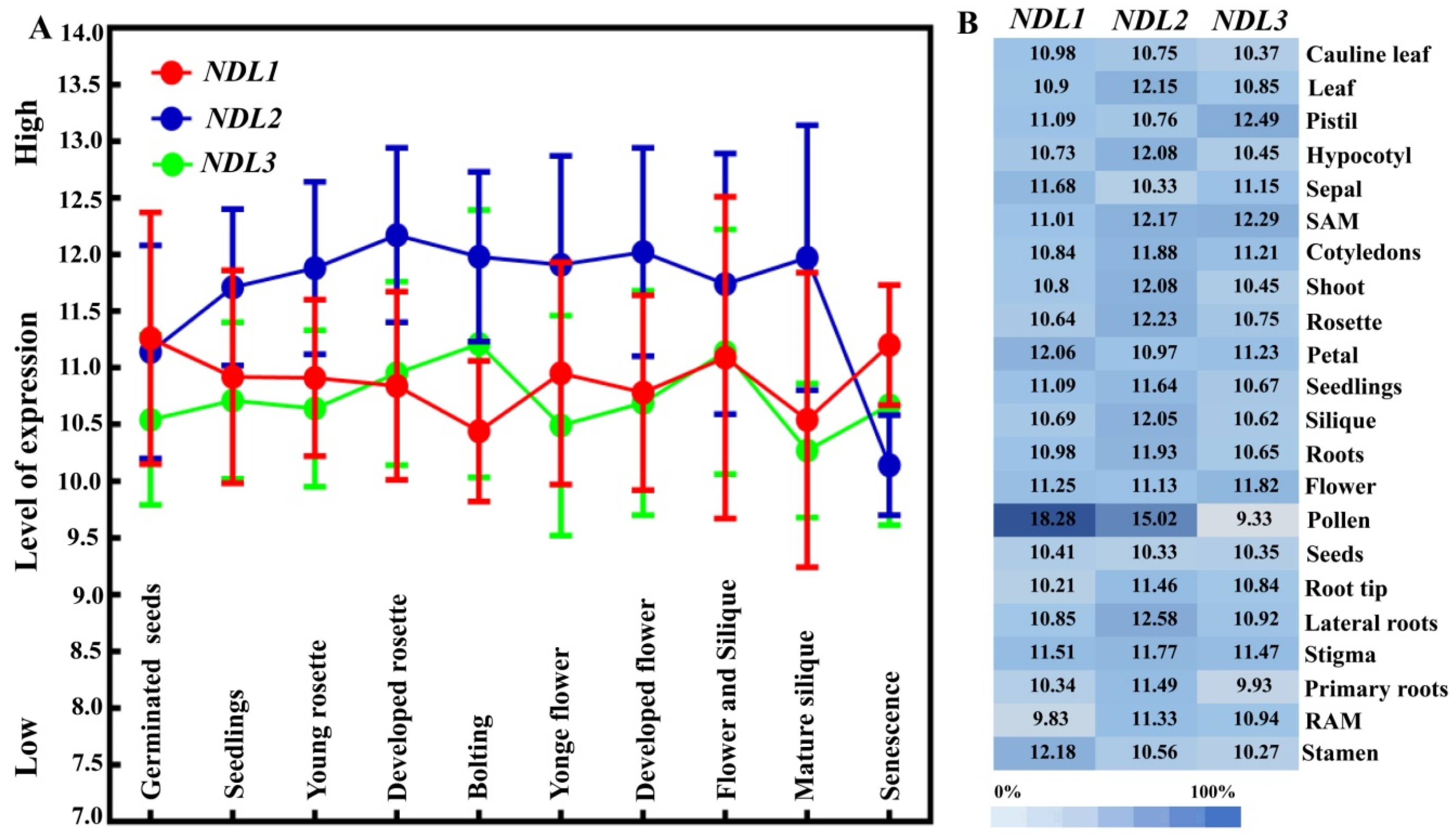
2.2. Comparative In Silico Expression Analysis of NDL Members

Comparative expression analysis of NDL1, NDL2, and NDL3 has been performed using genevestigator microarray data. All three NDL members showed expression across all developmental stages, NDL2 showed expression little higher than NDL1 and NDL3 (Figure 2A). Microarray analysis in different plant tissues revealed that NDL genes exhibit differential expression pattern across various organs. NDL1 and NDL2 showed highest expression in pollen while NDL3 showed highest expression in carpel and shoot apex (Figure 2B). Since, the role of NDLs has been already established in auxin transport during root development [14]. We closely looked at the expression levels of the NDLs members in different parts of roots and found that expression level of all the members were comparable, in primary roots NDL1 and NDL2 is comparatively little higher than NDL3. Overall expression pattern revealed ubiquitous expression of the gene family which indicates important function of the family members during plant growth and developmental responses (Figure 2B).

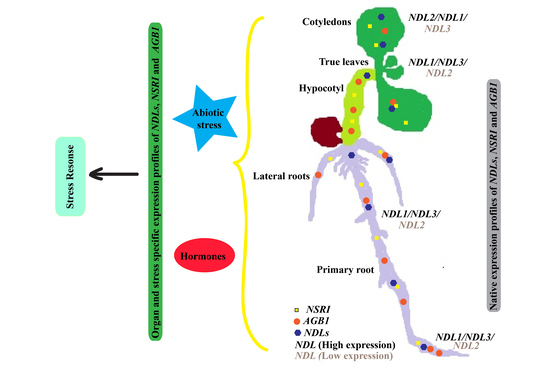
2.3. Comparative In Vivo Expression Analysis of NDLs during Early Stages of the Plant Growth

In-vivo expression analysis of all three NDL members showed that the localization pattern of GUS is quite unique for each member. During the initial stages of plant growth (4–12 day-old seedlings) pNDL1-NDL1-GUS showed localization in cotyledonary leaves, true leaves, various parts of primary and lateral roots including elongation zone and root apical meristem (RAM) (Figure 3A–C and Table 3). pNDL2-GUS showed GUS expression in cotyledonary leaves, true leaves, hypocotyl and maturation zone of primary root, but strikingly missing from root tip and in lateral roots (Figure 3G–I and Table 3). pNDL3-GUS expression was missing in cotyledonary leaves, present in newly emerging true leaves, present in the RAM of the primary root and in lateral roots (Figure 3M–O and Table 3).

2.4. Comparative In Vivo Expression Analysis of NDLs in Absence of AGB1

Previously, we have found that presence of AGB1 doesn’t affect transcriptional levels of NDL1 but necessary to maintain the post translational steady state level of NDL1 [14]. Since all three NDL members had shown physical interaction with AGB1 in Y2H we hypothesized that NDL2 and NDL3 might require its partner, AGB1, either to regulate expression levels or for post-translational stability similar to NDL1. In order to test this speculation, expression of pNDL2-GUS and pNDL3-GUS was studied in agb1-2 background.
In agb1-2 background pNDL1-NDL1-GUS localization was absent from cotyledonary leaves (Figure 3D–F and Table 3) but present in true leaves. In case of roots the localization was absent from both primary and lateral roots RAM (Figure 3D,E inset) whereas present in other parts of roots similar to Col-0 background. In case of pNDL2-GUS and pNDL3-GUS localization pattern of the GUS was same in agb1-2 mutant background as compared to wild type Col-0. (Figure 3J–L for NDL2, Figure 3P–R for NDL3 and Table 3). These finding indicate that expression levels of both NDL2 and NDL3 are also unaffected by the absence of AGB1 similar to previously reported for NDL1.

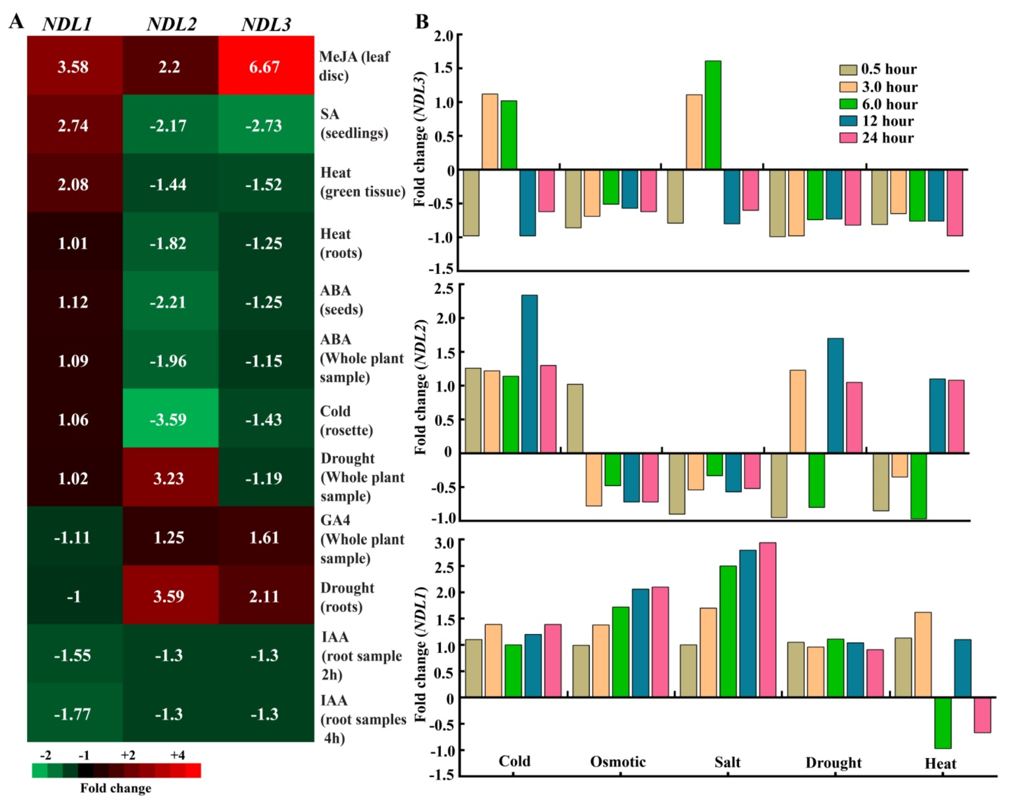
2.5. In Silico Expression Analysis of NDLs under Various Stress and Hormonal Treatments

Previous study has confirmed that NDLs play role in auxin transport during lateral root formation [14]. Correlation of gene expression and responses of morpho-physiological traits under severe water-deficient conditions indicate substantial alteration in expression levels of AtNDL1 during drought stress responses using tanscriptomic analysis [15]. In order to find out if independent NDL members play independent specific roles or have combined redundant functions during abiotic stress exposure and hormonal treatments, we have analyzed their comparative expression profiles from publically available databases. (Genevestigator and eFP browser).
Genevestigator Microarray data revealed that after auxin (IAA) treatment, expression levels of NDL1 decreased to nearly 1.77 fold, expression of NDL2 and NDL3 showed a similar trend, decrease of around 1.3 fold. (Figure 4A). In presence of ABA, NDL2 showed a decrease (2.21 fold) in expression level, as compared to NDL1 and NDL3 which showed an upregulation up to ~1.12 fold and down regulation of nearly 1.25 fold respectively. Unlike NDL2 and NDL3, NDL1 showed slight decrease (1.1 fold) in its expression in response to GA (Figure 4A).
All NDL members also showed a response toward various abiotic stresses (heat, cold and drought). In case of heat treatment in green tissue NDL1 showed increase (2.08 fold) as compared to NDL2 and NDL3 which showed a decrease in expression (1.44 and 1.52 fold respectively) in case of roots also similar trend was there. Even for cold treatment NDL1 showed positive regulation (1.06 fold up) while down regulation was observed for NDL2 and NDL3 (3.59 and 1.43 fold respectively). Under drought stress all members have positive response, NDL2 showed upregulation in expression (3.59 fold) followed by NDL3 and NDL1 (increase of 2.11 and 1.60 fold respectively, Figure 4A). The observed high expression of NDL2 gene in drought stress thereby could possibly be linked to the fact that multiple ABA-responsive factor binding elements are present in promoter sequence of NDL2.
To find out optimal time point for the in vivo expression analysis we also performed comparative in silico expression analysis of NDLs at different time points during various abiotic stress treatments using eFP browser. For NDL1 maximum transcript levels compared to no-treatment control were detected at 24 h. In case of NDL2 expression, levels fluctuated during different stress treatments and variation was found at different time points but overall maximum levels were observed at 24 h. A trend similar to NDL2 was observed for NDL3 expression during different treatments and time points (Figure 4B)

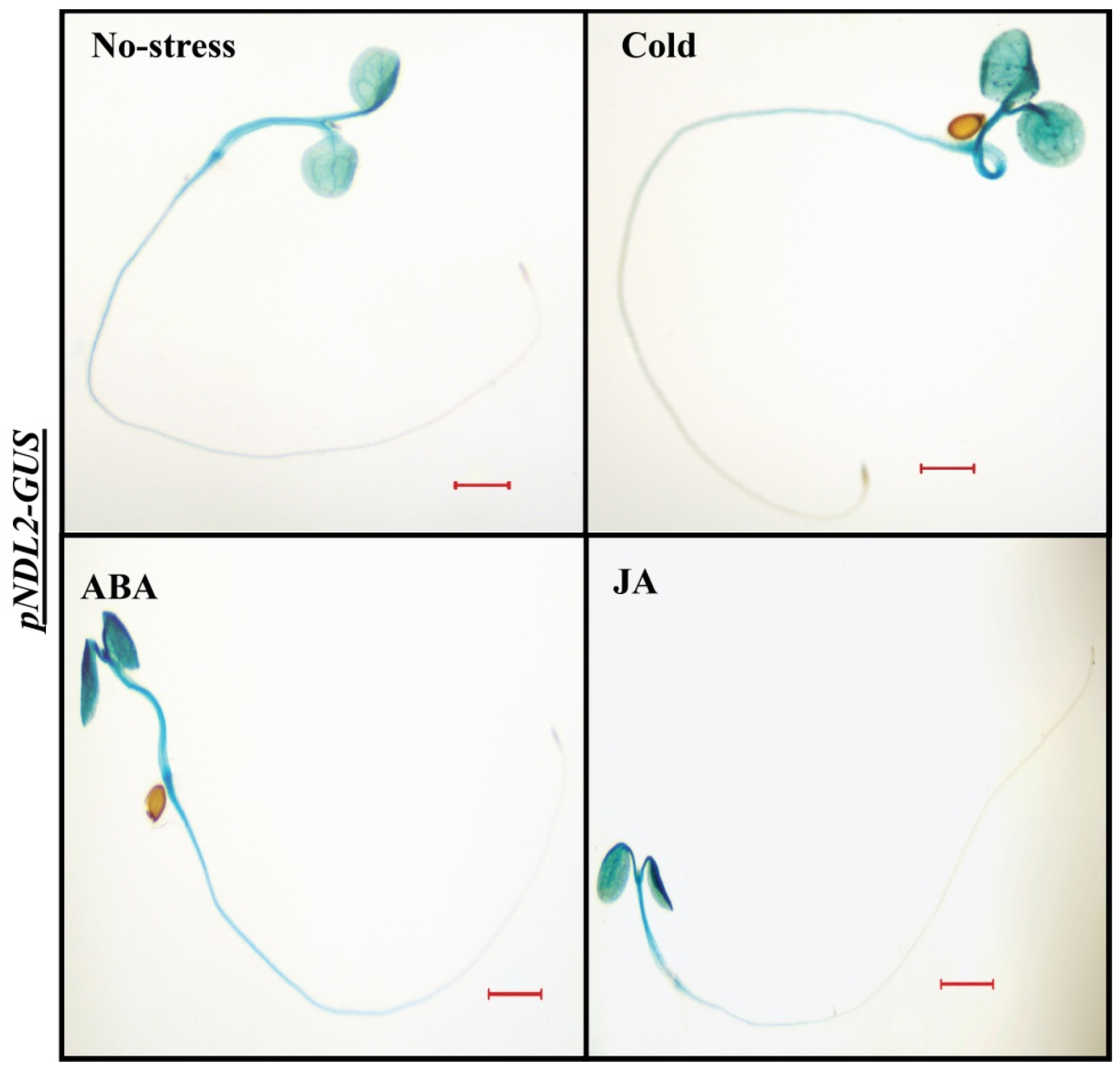
2.6. In Vivo Expression Analysis under Abiotic Stresses and Hormonal Treatments

In order to find out maximum expression levels for all three NDLs promoter GUS/β-glucuronidase activity was tested using 4-Methylumbelliferyl-β-D-glucuronide (MUG) assay for two time points (6 h and 24 h) in Col-0 background, no significant difference was found between expression levels of NDLs and two time points. (Supplementary Figure S1). Based on the in vivo GUS/β-glucuronidase activity levels and in silico analysis of the expression profiles of all three NDL members, treatment with various hormones and abiotic stresses were designed. Six-day-old seedlings were subjected to 24 h treatment with various hormones (ABA, GA, IAA, JA, SA, and BAP) and abiotic stresses (cold, heat, Mannitol, PEG and salt) followed by measurement of GUS activity either histochemically or by fluorometry.
pNDL1-NDL1-GUS, showed increase in the GUS staining intensity when treated with cold, IAA (auxin), JA, Salicyclic acid (SA), BAP (cytokinin), and Poly ethylene glycol (PEG) and almost no staining observed with heat treatment (Figure 5). GUS staining remained unaffected after treatment with ABA (Supplementary Figure S2), GA (Supplementary Figure S3) and mannitol (Supplementary Figure S4). In case of salt treatment, no difference was found in root but hypocotyle showed increase in the GUS staining intensity (Supplementary Figure S5) compared to no stress treatment control.
pNDL2-GUS showed increased levels of GUS staining in the cotyledons and hypocotyl after treatment with ABA, cold and JA (Figure 6), primary root showed no expression similar to no treatment control. BAP (Supplementary Figure S2), IAA, GA, heat (Supplementary Figure S3), Mannitol and PEG (Supplementary Figure S4) and SA (Supplementary Figure S5) and other treatments had no effect on expression level as no difference was observed compared to no stress treatment control.
In case of pNDL3-GUS, no difference in the GUS staining was detected when seedlings were treated with ABA, BAP, cold (Supplementary Figure S2), IAA, GA, JA, heat (Supplementary Figure S3), salt and SA (Supplementary Figure S5). Mannitol and PEG treatment resulted in decreased level of pNDL3-GUS expression in RAM and hypocotyl in comparison to no treatment control which showed diminished staining at both the places. GUS staining was absent in the rest of the seedlings, same as in case of no treatment control of six-day old seedling (Figure 7 and Supplementary Figure S4).
Results indicate that all the three family members’ show independent response when encountering various stress/hormonal treatments. Both NDL1 and NDL3 after treatment with Mannitol and PEG showed difference in GUS intensity indicates that both the members are working when plants encounter with drought and osmotic stress. Although both the members have altered expression in same stress mechanism of functioning seems opposite as NDL1 showed up regulation after PEG treatment while NDL3 showed down regulation when treated with PEG (compare Figure 5B and Figure 7), while NDL2 expression remains unaffected by both treatments in tested plant stage (Supplementary Figure S4).

2.7. In Vivo Expression Analysis of NDLs in Absence of AGB1 under Abiotic Stress and Hormonal Treatments

Previous study has reported that expression levels of NDL1 were not affected but protein steady state levels were affected by AGB1 [14]. In order to find out role of AGB1 on expression levels of NDL2 and NDL3 their expression patterns were studied in agb1-2 mutant. We found that expression of NDL2 and NDL3 were not affected in agb1-2 mutant and stays same as in wild type Col-0 background (Figure 3). In order to find-out whether AGB1 has any role on expression during abiotic stress or hormonal treatments expression pattern was observed for all the three members in absence of AGB1 after abiotic (cold, heat, Mannitol, salt and PEG), and hormonal (ABA, GA, IAA, JA, SA and BAP) treatments.
pNDL1-NDL1-GUS showed increased GUS intensity in primary root of the seedlings compared to no treatment control for GA, Mannitol, PEG and JA while no staining was detected in the primary root after heat treatment (Figure 8A,B). MUG assay was also performed and significantly increased GUS activity was observed only for PEG and Mannitol treatment in comparison to no-treatment control seedlings (Figure 8C). GUS staining intensity remains unaffected for ABA, BAP, cold (Supplementary Figure S2), IAA (Supplementary Figure S3), salt and SA (Supplementary Figure S5) treatments. Response of ABA and salt for pNDL1-NDL1-GUS remained similar in both the background (in wild type Col-0 as well as in agb1-2 mutant) meaning presence of AGB1 don’t make any difference on expression, while for cold, IAA, SA and BAP upregulation was detected in the cotyledons and in primary root in comparison to no treatment control seedlings in case of wild type
Col-0 background while remains unaffected in agb1-2 mutant background meaning presence of AGB1 is vital for NDL1 stability during cold, IAA, SA and BAP treatment, indicating stress specific role of NDL1-AGB1 module.
The expression analysis for pNDL2-GUS in terms of GUS staining was also observed in absence of AGB1. Seedlings were treated with ABA, cold, IAA, GA, heat, JA, Mannitol, PEG, salt, SA and BAP. No difference in GUS staining was detected in agb1-2 mutant background compared to no treatment control (Supplementary Figures S2–S5) suggesting no role/requirement of AGB1 for NDL2 expression during treatment in agb1-2 background. In case of cold, ABA and JA treatment upregulation of pNDL2-GUS was detected in the cotyledons in wild type Col-0 background (Figure 6) but in agb1-2 background this effect was not present (Supplementary Figures S2–S5) meaning AGB1 presence is required during cold, ABA and JA treatments for NDL2 expression upregulation.
In case of pNDL3-GUS, no difference in expression was detected in for ABA, BAP, Cold (Supplementary Figure S2), IAA, GA, JA, heat (Supplementary Figure S3), salt, SA (Supplementary Figure S5) in comparison to no treatment control. For all these treatments results were similar in both the background in wild type as well as in agb1-2 mutant background. Very interestingly after treatment with PEG and Mannitol in agb1-2 mutant background intensity of GUS staining showed increase in the RAM which is opposite effect compared to the Col-0 background where a decline in hypocotyl and almost absence of expression was observed in RAM (Figure 7). This means differential and opposite role of AGB1 is operating for regulating NDL3 expression under normal and dehydration stress condition (Figure 9). Presence of AGB1 is needed under stress condition to downregulate NDL3 expression, which might need to be downregulated under dehydration stress.

3. Discussion

Comparative analysis for all three members of NDL gene family revealed their role in many common and unique functions. In-silico analysis revealed that all three members share many common transcription factor binding sites (Table 1). Presence of MYC binding factors postulate evolutionary conserved regulatory mechanisms of downregulation similar to animal systems for AtNDL family. Regulatory regions of all the three members contain motifs for stresses (biotic and abiotic), development, and for hormonal responses. MYB recognition sites like MYB1AT and MYB1LEPR are present in all the NDL members. MYB1AT is a MYB recognition site which is also present in the promoter of RD22 which is ABA-induced and drought-responsive gene in Arabidopsis [18]. Another dehydration-responsive motif ABRELATERD1 is also present in the NDL family members, this motif has been reported in the up-stream region of the ERD1 (early response to dehydration 1). Promoters with ERD1 motif are involved in response to ABA and also show significant upregulation under water stress [20]. Both AtMYC and AtMYB are known to function as transcriptional activators in abscisic acid signaling presence of these motifs in combination with dehydration-responsive motifs in NDL family members indicates their role in ABA-dependent early response to dehydration.
In the present study, we have only analysed early stage of plant growth but Rymaszewski et al., 2017 has analysed plant growth starting from early stages till reproductive stage. They found positive co-relation of morpho-physiological traits like projected rosette area, and increased transpiration rate under low water-deficient conditions and expression levels for NDL1. They also speculate that these acclimations may be driven by largely stress hormone ABA. In another study Du et al., 2018 [21] has also found that during low water treatment conditions plants shifts for drought escape mechanism and leads to early flowering to complete their life cycle. Activation of the flowering genes are both ABA-dependent and ABA-independent [21]. All these finding indicates the complex network between stress, hormones and developmental responses.
Interestingly both AGB1 and GPA1 both are interacting partners of NDL members [14] are also interacting partners of one of the MYB (AT3G24120) in G protein interactome using in Y2H [3].
ANAERO1CONSENSUS motif is indicative of involvement of the gene family during anaerobic conditions. MYCCONSENSUSAT regulates transcription of CBF/DREB1 genes during cold. Presence MYC recognition sites had been postulated to regulate the gene transcription by a basic bHLH transcription activator during cold stress [22]. MYCCONSENSUSAT sites are present mainly at the genes which have more expression in seed and the response to cold is ABA-mediated [23]. MYCCONSENSUSAT motif was also detected in the promoter of the gene which showed response for JA. WBOXATNPR1 is a disease related motif that is established as a site for binding SA-induced WRKY TFs [23]. Presence of a P1BS and SURECOREATSULTR11 motif indicative of the involvement of the family members for nutrient availability. P1BS motifs are associated with phosphate starvation response, while SURECOREATSULTR11 are found in the genes involved during sulfur deficiency responses (SULTR1; 1) [24,25].
All the three NDL members contain various hormone-responsive transcription factors binding sites. Responses of MYCCONSENSUSAT and ABRELATERD1 are ABA-dependent during desiccation and cold responses. MYCCONSENSUSAT motif also present in the genes which are involved in JA-mediated defense responses. Cytokinin response regulator ARR1AT and SA-responsive WBOXATNPR1 is also present commonly in all the three family members. Light-responsive motifs GT1CONSENSUS and SORLIP1AT are also common in all the members. GT1CONSENSUS a light-responsive motif, also showed tissue-specific localization, it is present in the up-stream region of the right regulated genes which are highly expressed in leaf [23]. Other than these common shared regulatory elements unique TFs binding sites to NDL1, NDL2 and NDL3 were also observed (Table 2). NDL1 showed presence of exclusive binding sites for E2F, which targets cell cycle-regulated expression. E2F controls cell cycle by the regulation of the transcription of the genes that are involved during cell cycle and DNA replication [26,27] Arabidopsis E2F are classified as classical E2F proteins (E2Fa-c) and atypical E2F proteins (E2Fd-f) [28]. In G-protein interactome database NDL1 shows interaction with CKS2 (cell cycle regulatory subunit) and cyclin-dependent kinase G1 hinting specific function during cell cycle. Previously it is established that sucrose and D-glucose enhances the stability of NDL1 protein [14] which goes in accordance with presence of SBOXATRBCS, important for sugar and ABA responses. Role of NDL1 during primary and lateral root growth and development is already established presence of RHERPATEXPA7, a root hair specific cis-element needs further probing to study role of NDL1 in root hair development.
Likewise, NDL2 strikingly shows presence of various kinds of ABA-responsive elements like ABREATCONSENSUS, which play role during ABA signaling and responsible for abiotic stress tolerance [29], ABREATRD22 and ACGTABREMOTIFA2OSEM. ACGTABREMOTIFA2OSEM motif present in the promoter of rice OsEm gene, which is regulated by seed specific transcription factor and ABA-responsive [30]. ACGTABREMOTIFA2OSEM motif also found in maturing seeds and acts as a binding site for bZIP TFs ABI5 [31]. Those genes which are highly expressed in mature seeds of Arabidopsis are found to be enriched with ABRE motif [32]. A single copy of ABRE is not sufficient for ABA-mediated responses. Multiple copy number of ABRE along with a coupling element forms the active complex for ABA-mediated responses [33]. Along with ABA-responsive binding sites several other sites like, GA and JA-responsive sites GADOWNAT and T/GBOXATPIN2 are also present in the promoter region of NDL2. GADOWNAT is common sequence found in genes which downregulates after GA treatments. GADOWNAT is shown to be identical as ABRE [34], also hints the regulation of gene under ABA responses. T/GBOXATPIN2 is a wounding response motif, found in the promoter of JA-responsive genes [35]. As per the in-silico analysis presence of different kinds of ABA response factors and seed specific transcription factors which are again ABA-dependent for their responses. Since our in vivo expression analysis proved absence of the NDL2 expression in the roots. NDL2 members might be specifically involved during seed germination and growth responses.
The NDL3 promoter showed presence of -ABRERATCAL, an early stress-induced motif related to ABRE and found in the up-stream region of Ca2+ ion-responsive genes, CGCGBOXAT is a Ca2+-dependent Calmodulin binding motif is also present both indicative of involvement of NDL3 during calcium and ABA-mediated stress adaptations [36,37,38]. GCCCORE has been found in the promoter region of the several pathogen-responsive genes showed JA-dependent defense responses [39]. HDZIPIIIAT involved in vascular differentiation and patterning [40] and required for polar auxin transport in shoot [41], NDLs combined role is already established for polar auxin transport in roots [14] Increased HDZIPIIIAT activity leads to formation of extra cotyledons [42], a phenotype somewhat similar to tricot phenotype of ndl microRNA downregulated lines [16]. While downregulation of HDZIPIIIAT showed the loss of cotyledons formation [42,43]. All these findings indicate that NDL3 might be a Ca2+-responsive gene during stress signaling. NDL3 also involved in similar functions like NDL1 as both share similar in vivo localization during early stages of plant growth
The combinatorial effect of unique and common transcription factors present in NDL gene family essentially suggests that NDL1, NDL2, and NDL3 do share a basic common regulatory mechanism of downregulation by MYC/MYB. Our in vivo analysis also shows that during different stages of life cycle, and when plant encounter specific stress conditions differential regulation of all NDL members is possible and this could be attributed to the unique transcription factor binding sites present in each gene respectively.
Initial developmental study for all three members of the family showed that they are very specific about their expression pattern. In wild type background, NDL1 was expressed in cotyledonary leaves, true leaves, primary and lateral roots. Functional characterization of NDL1 has been done in detail and it has been found that NDL1 is required for primary root and shoot meristem initiation growth and lateral root/shoot branching [14,16]. For NDL2 the expression was detected in cotyledonary leaves, true leaves and in maturation zone of primary root, no expression was detected in RAM and in lateral roots (Figure 3) indicating function in different organs compared to NDL1 and NDL3, which share overlapping expression zones. Presence of GA and cold-responsive elements, along with different kind of ABA-responsive factors in NDL2 promoter is suggestive of its involvement during seed germination and early growth as during low temperature ABA biosynthesis and GA catabolism is up regulated that lead to seed dormancy [44].
In silico and in vivo expression analysis in case of NDL3 was detected in newly emerging true leaves and at primary and lateral root tips (Figure 3). NDL3 show response during drought stress and expression pattern is again quite similar with NDL1 response (Figure 7; Figure 9) meaning, both the members share overlapping expression patterns in vivo and are playing role in similar physiological processes.
Previously it has been found that in agb1 mutant NDL1 expression levels are of wild type level meaning NDL1 transcript is unaffected by the AGB1 (Figure 5D) [14] Similar to NDL1 study we found that in vivo expression levels/patterns of NDL2 and NDL3 remains similar to Col-0 levels even in agb1-2 mutant, meaning AGB1 does not affect the transcript levels of NDLs.
NDL1 protein stability in young primary root needs presence of AGB1, as NDL1 undergoes proteosomal degradation in absence of AGB1 [14], effect of AGB1 on stability and localization of NDL2 and NDL3 is still pending and needs to be characterized.
The effect of different hormones and abiotic stress responses also shows that along with the common response of NDL1 and NDL3 during osmotic/drought stress the different family members show specific responses for various treatments. NDL1 localization is upregulated during cold, IAA, JA, SA and cytokinin treatment and downregulated after heat stress (Figure 5A), meaning presence of AGB1 is essential for NDL1 stability during cold, IAA, JA, SA and BAP treatment, indicating stress specific role of NDL1-AGB1 module.
Although NDL1 and NDL3 share similar expression domains, NDL3 expression remains unaffected by cold, IAA, JA, SA, cytokinin and heat treatments (Supplementary Figures S2–S5) meaning differential specificity in their functions.
Various ABA binding sites are present in the promoter region of NDL2 and in accordance after ABA treatment (also for cold and JA) the NDL2 expression shows upregulation in Col-0 background but did not showed any difference in agb1-2 mutant background indicating presence of AGB1 is essential for NDL2 expression upregulation during these treatments.
In case of NDL3 expression upon treatment with Mannitol and dehydration stress it was found that AGB1 is negatively regulating NDL3 expression under normal and dehydration stress condition (Figure 9). Presence of AGB1 is needed under stress condition to downregulate NDL3 expression, which may be downregulated under dehydration stress.
Although various common TFs have been found in the promoter analysis but all the three members did not show the response for each of them. Every members of the family behaves differentially even though they share the common regulatory motifs. This happens because the family members also showed differential expression pattern and response to various stresses and hormones could be specific for particular developmental stage, our analysis is limited for initial growth stage of the plant. Previously it was found that AGB1 protein is needed for NDL1 stability and here we also found that the protein stability was affected in mutant background. In addition, AGB1 was also confirmed to be involved in drought stress [45]. NDL1 is also predicted to be a stress marker for drought [15]. Our in-vivo study proves that during the combined function out of three members two of them (NDL1 and NDL3) are involved during drought stress response. NDL1 is acting as a general abiotic stress responder during heat, cold, IAA, JA, SA and in cytokinin responses as protein steady state levels show upregulation after all these treatments, while NDL3 expression remains unaffected by all these treatments, meaning limited and specific role. NDL2 doesn’t show any alteration in expression after Mannitol and PEG treatments but showed significant upregulation for ABA, cold, and JA treatment.
In summary dehydration stress (Mannitol and PEG treatments) caused increase in steady state protein levels of NDL1 in both wild type Col-0 as well as in agb1-2 mutant background. Whereas NDL3 showed downregulation of expression in wild type Col-0 background while upregulation in agb1-2 mutant background in RAM. Expression levels of pNDL2-GUS were affected in AGB1-dependent manner during cold, ABA and JA treatment. These results strongly support the direct involvement of NDL1 and NDL3 during osmotic/drought stress responses and NDL2 might be playing ABA-dependent indirect role. AGB1 is also playing role in differential regulation of expression of these NDLs members during different treatments by affecting protein stability (seen in case of NDL1) or transcript levels (as seen in case of NDL3 expression) in stress specific manner.

4. Material and Methods

4.1. Plant Material and Growth Conditions

Arabidopsis Col-0 ecotype was used in the present study. Agrobacterium (strain GV3101)-mediated floral dip method [46] was used to generate transcriptional fusion transgenic (pNDL2, 3-GUS) in wild type Col-0 as well as in the agb1-2 mutant background. pNDL1-NDL1-GUS translational fusion lines were taken from previous study [14]. Transformed seeds were selected on ½ MS medium (Himedia, Mumbai, India) containing 25 mg/L Hygromycin (Duchefa, Amsterdam, Netherland), resistant plants were moved to the soil and grown to maturity in a growth room with a photoperiod 16h light/8h dark at 22 °C, and the light intensity of 100 μmolm−2s−1. Three independent single insertion T3 homozygous lines were obtained and used for developmental and stress treatments. For developmental study, seeds were grown vertically for, 4-day, 8-day and 12-day respectively followed by in vivo GUS assay. For stress treatment and fluorometric analysis six day old seedlings were used.

4.2. Isolation and Cloning of NDL2 and NDL3 Promoters

Genomic DNA was isolated with minor modifications from the Arabidopsis thaliana Col-0 using Doyle method [47]. Primers listed in Supplementary Table S1 were used to amplify the promoter region of NDL2 and NDL3. Amplified fragments were then cloned into a pENTR/D-TOPO entry vector (Invitrogen, Massachusetts, United States) gateway, followed by pGWB3 destination vectors) with C-terminus GUS reporter. For NDL1 previously published lines were used [14].

4.3. In-Silico Analysis

Sequence information and location of NDL1, NDL2 and NDL3 was retrieved from https://www.arabidopsis.org/. In silico analysis was done using online programs PLANTPAN 2.0 [48]. For the expression pattern of NDL genes in different stages of development in different tissues, and time points data from the free version of the GENEVESTIGATOR online portal (https://www.genevestigator.com/gv/plant.jsp) and eFP browser (http://bar.utoronto.ca/efp/cgi-bin/efpWeb.cgi) was used for analysis.

4.4. GUS Staining Assay

Three independent lines for pNDL1-NDL1-GUS and published pNDL1-NDL1-GUS lines were germinated and grown on ½ MS media for 4-day, 8-day and 12-days. Gus staining on the seedlings was done using Jefferson method [49].

4.5. Fluorometric GUS Assay

For quantitatively GUS activity. 0.5 g of seedlings (6-day old) were harvested and frozen into liquid N2 in 1.5 mL micro centrifuge tube, followed by grinding and protein extraction in extraction buffer (50 mM sodium phosphate buffer, pH 7.0, 10 mM EDTA, 0.1% Triton X-100, and 10 mM β-mercaptoethanol). The homogenate was centrifuged at 10,000 g for 20 min at 4 °C. Supernatant was collected and assayed for protein concentration using Bradford method [50]. Five microgram of protein was added to GUS assay buffer (1X = 50 mM sodium phosphate buffer, pH 7.0, 10 mM EDTA, 0.1% Triton X-100, and 10 mM β-mercaptoethanol containing 2 mM MUG (Himedia, Mumbai, India) total volume was made up to 500 μL using ddH2O. Samples were incubated at 37 °C for one hour followed termination of the reaction using 400 μL of 0.2 M Na2CO3. For quantitative GUS analysis duplicate samples were assayed. The reaction product 4-methylumbelliferon (MU) was detected fluorometrically at excitation and emission of 365 nm and 455 nm respectively [51] using Tecan Spark multimode micro plate reader. GUS activity was expressed in nmol MU min−1 μg−1 protein.

4.6. Hormone and Abiotic Stress Treatments

Seeds were stratified and grown vertically on ½ MS media. Six day old seedlings were subjected to 24 h treatment of sodium chloride (NaCl; 150 mM), Mannitol (300 mM), cold (4 °C), heat (37 °C), abscisic acid (ABA; 20 μM), Indole-3-acetic acid (IAA; 10 μM), salicylic acid (SA; 10 μM), methyl jasmonate (JA; 10 μM), polyethylene glycol (PEG 6000, 20%) and Gibberellic acid (GA; 20 μM) in liquid MS. NaCl, Mannitol and PEG were obtained from Himedia (Himedia, Mumbai, India) all the other hormones and fine chemicals were obtained from Sigma (Sigma, Missouri, United States).

4.7. Accession Numbers

Sequence data from the article can be found from https://www.arabidopsis.org/using accession number: AT5G56750 (NDL1), AT5G11790 (NDL2), AT2G19620 (NDL3) and AT4G34460 (AGB1). Chromosomal locations of the respective genes are as follows: NDL1: Chromosome 5 (22957629-22960916), NDL2: Chromosome 5 (3799408-3803216), NDL3: Chromosome 2 (8485991-8488963), AGB1: Chromosome 4 (16477031-16479620), Regulator of G protein signaling protein-RGS1: Chromosome 3 (9532613-9535629), ANNEXIN-ANN1: Chromosome 1 (13225168-13227239), Sodium and Lithium-Tolerant 1-SLTI: Chromosome 2 (15761295-15763451), Arabidopsis Ribosomal Protein -RS27A: Chromosome 3 (22611521-22612905), O-Acetylserine (Thiol) Lyase -OAS-TL: Chromosome 4 (8517960-8520596).

5. Conclusions

Activation of specific AGB1-NDL module interaction might be stress specific and hormone regulated, specificity of action is further added in different stages of growth and development due to differential expression patterns of NDL members. NDL1 protein stability in young primary root needs presence of AGB1, as NDL1 undergoes proteosomal degradation in absence of AGB1 [14]. Though AGB1 physically interacts with all NDLs it might differentially regulate their stability and hence their function during different stresses, effect of AGB1 on protein stability of NDL2 and NDL3 is still pending and needs to be characterized. Interestingly, Yeast 2 Hybrid (Y2H) confirmed NDL1 interactors includes-Annexin 1 (ANNAT1-has role in drought stress), Sodium and Lithium-Tolerant 1 (SLT1 involved in salt stress), Lesion Stimulating Disease 1 (LSD1 regulates cold stress), O-Acetylserine (Thiol) Lyase (OAS-TL) Isoform A1 (OASA1 role in cadmium tolerance) and Arabidopsis Ribosomal Protein S27 (ARS27A involved in genotoxic stress) [3]. These interactions speculate that NDL proteins might plays role in stress signaling in the form of multimeric complexes with these stress effectors and other G protein core components (RGS1 and AGB1) in stress specific manner.
All of these findings together including regulatory elements in silico and in vivo expression profiling indicate that NDL family members along with AGB1 play–key differential roles in different organs during different stages of plant growth and developmental. Collectively, our data suggest that NDL-AGB1 modules are abiotic stress and hormone treatment-responsive and could be used as stress markers. In long run they could be potential candidates for crop improvement strategies (Figure 10).

Supplementary Materials

Author Contributions

Conceptualization, Y.M.; methodology, Y.M. and A.K.; validation, A.K.; formal analysis, A.K.; investigation, A.K.; resources, Y.M.; data curation, A.K.; writing—original draft preparation, A.K.; writing—review and editing, Y.M.; visualization, A.K.; supervision, Y.M.; project administration, Y.M.; funding acquisition, Y.M.

Funding

Work in the Mudgil’s Lab is supported by grants from the DST-SERB (EMR/2016/002780), DBT (BT/PR20657/BPA/118/206/2016) and Delhi University R&D and DU-DST PURSE grant. A.K is supported by JRF/SRF fellowship from CSIR.

Acknowledgments

We thank Swati Singh (Delhi University, Botany Department) for cloning of NDL2 and NDL3 promoter in Entry Vector and Diwakar Bhardwaj (Delhi University, Botany Department) for his help in generating T3 transgenic lines used in the study. We would also like to extend our thanks to Aniket (Delhi University, Ambedkar Centre for Biomedical Research) for his help with fluorometer.

Conflicts of Interest

The authors declare no conflict of interest.

References

  1. Colaneri, A.C.; Tunc-Ozdemir, M.; Huang, J.P.; Jones, A.M. Growth attenuation under saline stress is mediated by the heterotrimeric G protein complex. BMC Plant Biol. 2014, 14, 129. [Google Scholar] [CrossRef] [PubMed]
  2. Ming, C.H.; Xu, D.B.; Fang, G.N.; Wang, E.H.; Gao, S.Q.; Xu, Z.S.; Li, L.C.; Zhang, X.H.; Miin, D.H. G-protein β subunit AGB1 positively regulates salt stress tolerance in Arabidopsis. J. Integr. Agric. 2015, 14, 314–325. [Google Scholar]
  3. Klopffleisch, K.; Phan, N.; Augustin, K.; Bayne, R.S.; Booker, K.S.; Botella, J.R.; Carpita, N.C.; Carr, T.; Chen, J.G.; Cooke, T.R.; et al. Arabidopsis G-protein interactome reveals connections to cell wall carbohydrates and morphogenesis. Mol. Syst. Biol. 2011, 7, 532. [Google Scholar] [CrossRef] [PubMed]
  4. Kalaydjieva, L.; Gresham, D.; Gooding, R.; Heather, L.; Baas, F.; De Jonge, R.; Blechschmidt, K.; Angelicheva, D.; Chandler, D.; Worsley, P.; et al. N-myc downstream-regulated gene 1 is mutated in hereditary motor and sensory neuropathy–Lom. Am. J. Hum. Genet. 2000, 67, 47–58. [Google Scholar] [CrossRef] [PubMed]
  5. Yan, S.F.; Lu, J.; Zou, Y.S.; Soh-Won, J.; Cohen, D.M.; Buttrick, P.M.; Cooper, D.R.; Steinberg, S.F.; Mackman, N.; Pinsky, D.J.; et al. Hypoxia-associated induction of early growth response-1 gene expression. J. Biol. Chem. 1999, 274, 15030–15040. [Google Scholar] [CrossRef] [PubMed]
  6. Salnikow, K.; Su, W.; Blagosklonny, M.V.; Costa, M. Carcinogenic metals induce hypoxia-inducible factor-stimulated transcription by reactive oxygen species-independent mechanism. Cancer Res. 2000, 60, 3375–3378. [Google Scholar] [PubMed]
  7. Lachat, P.; Shaw, P.; Gebhard, S.; Van Belzen, N.; Chaubert, P.; Bosman, F.T. Expression of NDRG1, a differentiation-related gene, in human tissues. Histochem. Cell Biol. 2002, 118, 399–408. [Google Scholar] [CrossRef] [PubMed]
  8. Salnikow, K.; Kluz, T.; Costa, M.; Piquemal, D.; Demidenko, Z.N.; Xie, K.; Blagosklonny, M.V. The regulation of hypoxic genes by calcium involves c-Jun/AP-1, which cooperates with hypoxia-inducible factor 1 in response to hypoxia. Mol. Cell. Boil. 2002, 22, 1734–1741. [Google Scholar] [CrossRef] [PubMed]
  9. Wu, D.; Zhau, H.E.; Huang, W.C.; Iqbal, S.; Habib, F.K.; Sartor, O.; Cvitanovic, L.; Marshall, F.F.; Xu, Z.; Chung, L.W.K. cAMP-responsive element-binding protein regulates vascular endothelial growth factor expression: Implication in human prostate cancer bone metastasis. Oncogene 2007, 26, 5070–5077. [Google Scholar] [CrossRef] [PubMed]
  10. Zhou, D.; Salnikow, K.; Costa, M. Cap43, a novel gene specifically induced by Ni2+ compounds. Cancer Res. 1998, 58, 2182–2189. [Google Scholar] [PubMed]
  11. Krauter-Canham, R.; Bronner, R.; Evrard, J.L.; Hahne, G.; Friedt, W.; Steinmetz, A. A transmitting tissue-and pollen-expressed protein from sunflower with sequence similarity to the human RTP protein. Plant Sci. 1997, 129, 191–202. [Google Scholar] [CrossRef]
  12. Lazarescu, E.; Friedt, W.; Horn, R.; Steinmetz, A. Expression analysis of the sunflower SF21 gene family reveals multiple alternative and organ-specific splicing of transcripts. Gene 2006, 374, 77–86. [Google Scholar] [CrossRef] [PubMed]
  13. Lazarescu, E.; Friedt, W.; Steinmetz, A. Organ-specific alternatively spliced transcript isoforms of the sunflower SF21C gene. Plant Cell Rep. 2010, 29, 673–683. [Google Scholar] [CrossRef] [PubMed]
  14. Mudgil, Y.; Uhrig, J.F.; Zhou, J.; Temple, B.; Jiang, K.; Jones, A.M. Arabidopsis N-MYC DOWNREGULATED-LIKE1, a positive regulator of auxin transport in a G protein–mediated pathway. Plant Cell. 2009, 21, 3591–3609. [Google Scholar] [CrossRef] [PubMed]
  15. Rymaszewski, W.; Vile, D.; Bediee, A.; Dauzat, M.; Luchaire, N.; Kamrowska, D.; Granier, C.; Hennig, J. Stress-related gene expression reflects morphophysiological responses to water deficit. Plant Physiol. 2017, 174, 1913–1930. [Google Scholar] [CrossRef] [PubMed]
  16. Mudgil, Y.; Ghawana, S.; Jones, A.M. N-MYC down-regulated-like proteins regulate meristem initiation by modulating auxin transport and MAX2 expression. PLoS ONE 2013, 8, e77863. [Google Scholar] [CrossRef] [PubMed]
  17. Luscher, B.; Eisenman, R.N. New light on Myc and Myb. Part II. Myb. Genes Dev. 1990, 4, 2235–2241. [Google Scholar] [CrossRef] [PubMed]
  18. Abe, H.; Urao, T.; Ito, T.; Seki, M.; Shinozaki, K.; Yamaguchi-Shinozaki, K. Arabidopsis AtMYC2 (bHLH) and AtMYB2 (MYB) function as transcriptional activators in abscisic acid signaling. Plant Cell. 2003, 15, 63–78. [Google Scholar] [CrossRef]
  19. Yamaguchi-Shinozaki, K.; Shinozaki, K. Organization of cis-acting regulatory elements in osmotic-and cold-stress-responsive promoters. Trends Plant Sci. 2005, 10, 88–94. [Google Scholar] [CrossRef]
  20. Simpson, S.D.; Nakashima, K.; Narusaka, Y.; Seki, M.; Shinozaki, K.; Yamaguchi-Shinozaki, K. Two different novel cis-acting elements of erd1, a clpA homologous Arabidopsis gene function in induction by dehydration stress and dark-induced senescence. Plant J. 2003, 33, 259–270. [Google Scholar] [CrossRef]
  21. Du, H.; Huang, F.; Wu, N.; Li, X.; Hu, H.; Xiong, L. Integrative regulation of drought escape through ABA-dependent and-independent pathways in rice. Mol. Plant. 2018, 4, 584–597. [Google Scholar] [CrossRef]
  22. Chinnusamy, V.; Ohta, M.; Kanrar, S.; Lee, B.H.; Hong, X.; Agarwal, M.; Zhu, J.K. ICE1: A regulator of cold-induced transcriptome and freezing tolerance in Arabidopsis. Genes Dev. 2003, 17, 1043–1054. [Google Scholar] [CrossRef]
  23. Wang, C.; Wang, Y.; Pan, Q.; Chen, S.; Feng, C.; Hai, J.; Li, H. Comparison of Trihelix transcription factors between wheat and Brachypodiumdistachyon at genome-wide. BMC Genom. 2019, 20, 142. [Google Scholar] [CrossRef]
  24. Rouached, H.; Secco, D.; Arpat, B.; Poirier, Y. The transcription factor PHR1 plays a key role in the regulation of sulfate shoot-to-root flux upon phosphate starvation in Arabidopsis. BMC Plant Biol. 2011, 11, 19. [Google Scholar] [CrossRef]
  25. Sobkowiak, L.; Bielewicz, D.; Małecka, E.; Jakobsen, I.; Albrechtsen, M.; Szweykowska-Kulinska, Z.; Pacak, A.M. The role of the P1BS element containing promoter-driven genes in Pi transport and homeostasis in plants. Front. Plant Sci. 2012, 3, 58. [Google Scholar] [CrossRef]
  26. Helin, K. Regulation of cell proliferation by the E2F transcription factors. Curr. Opin. Genet. Dev. 1998, 8, 28–35. [Google Scholar] [CrossRef]
  27. Lammens, T.; Li, J.; Leone, G.; De Veylder, L. Atypical E2Fs: New players in the E2F transcription factor family. Trends Cell Biol. 2009, 19, 111–118. [Google Scholar] [CrossRef]
  28. De Veylder, L.; Beeckman, T.; Inze, D. The ins and outs of the plant cell cycle. Nat. Rev. Mol. Cell Biol. 2007, 8, 655. [Google Scholar] [CrossRef]
  29. Narusaka, Y.; Nakashima, K.; Shinwari, Z.K.; Sakuma, Y.; Furihata, T.; Abe, H.; Narusaka, M.; Shinozaki, K.; Yamaguchi-Shinozaki, K. Interaction between two cis-acting elements, ABRE and DRE, in ABA-dependent expression of Arabidopsis rd29A gene in response to dehydration and high-salinity stresses. Plant J. 2003, 34, 137–148. [Google Scholar] [CrossRef]
  30. Hattori, T.; Totsuka, M.; Hobo, T.; Kagaya, Y.; Yamamoto-Toyoda, A. Experimentally determined sequence requirement of ACGT-containing abscisic acid response element. Plant Cell Physiol. 2002, 43, 136–140. [Google Scholar] [CrossRef]
  31. Yang, X.; Yang, Y.N.; Xue, L.J.; Zou, M.J.; Liu, J.Y.; Chen, F.; Xue, H.W. Rice ABI5-Like1 regulates abscisic acid and auxin responses by affecting the expression of ABRE-containing genes. Plant Physiol. 2011, 156, 1397–1409. [Google Scholar] [CrossRef]
  32. Nakabayashi, K.; Okamoto, M.; Koshiba, T.; Kamiya, Y.; Nambara, E. Genome-wide profiling of stored mRNA in Arabidopsis thaliana seed germination: Epigenetic and genetic regulation of transcription in seed. Plant J. 2005, 41, 697–709. [Google Scholar] [CrossRef]
  33. Skriver, K.; Olsen, F.L.; Rogers, J.C.; Mundy, J. Cis-acting DNA elements responsive to gibberellin and its antagonist abscisic acid. Proc. Natl. Acad. Sci. USA 1991, 88, 7266–7270. [Google Scholar] [CrossRef]
  34. Ogawa, M.; Hanada, A.; Yamauchi, Y.; Kuwahara, A.; Kamiya, Y.; Yamaguchi, S. Gibberellin biosynthesis and response during Arabidopsis seed germination. Plant Cell. 2003, 15, 1591–1604. [Google Scholar] [CrossRef]
  35. Boter, M.; Ruiz-Rivero, O.; Abdeen, A.; Prat, S. Conserved MYC transcription factors play a key role in jasmonate signaling both in tomato and Arabidopsis. Genes Dev. 2004, 18, 1577–1591. [Google Scholar] [CrossRef]
  36. Kaplan, B.; Davydov, O.; Knight, H.; Galon, Y.; Knight, M.R.; Fluhr, R.; Fromm, H. Rapid transcriptome changes induced by cytosolic Ca2+ transients reveal ABRE-related sequences as Ca2+-responsive cis-elements in Arabidopsis. Plant Cell. 2006, 18, 2733–2748. [Google Scholar] [CrossRef]
  37. Yang, T.; Poovaiah, B.W. A calmodulin-binding/CGCG box DNA-binding protein family involved in multiple signaling pathways in plants. J. Biol. Chem. 2002, 277, 45049–45058. [Google Scholar] [CrossRef]
  38. Du, L.; Ali, G.S.; Simons, K.A.; Hou, J.; Yang, T.; Reddy, A.S.; Poovaiah, B.W. Ca2+/calmodulin regulates salicylic-acid-mediated plant immunity. Nature 2009, 457, 1154–1158. [Google Scholar] [CrossRef]
  39. Brown, R.L.; Kazan, K.; McGrath, K.C.; Maclean, D.J.; Manners, J.M.A. Role for the GCC-box in jasmonate-mediated activation of the PDF1.2 gene of Arabidopsis. Plant Physiol. 2003, 132, 1020–1032. [Google Scholar] [CrossRef]
  40. Yang, J.H.; Wang, H. Molecular mechanisms for vascular development and secondary cell wall formation. Front. Plant Sci. 2016, 7, 356. [Google Scholar] [CrossRef]
  41. Huang, T.; Harrar, Y.; Lin, C.; Reinhart, B.; Newell, N.R.; Talavera-Rauh, F.; Hokin, S.A.; Barton, M.K.; Kerstetter, R.A. Arabidopsis KANADI1 acts as a transcriptional repressor by interacting with a specific cis-element and regulates auxin biosynthesis, transport, and signaling in opposition to HD-ZIPIII factors. Plant Cell. 2014, 26, 246–262. [Google Scholar] [CrossRef]
  42. Emery, J.F.; Floyd, S.K.; Alvarez, J.; Eshed, Y.; Hawker, N.P.; Izhaki, A.; Baum, S.F.; Bowman, J.L. Radial patterning of Arabidopsis shoots by class III HD-ZIP and KANADI genes. Curr. Biol. 2003, 13, 1768–1774. [Google Scholar] [CrossRef]
  43. Izhaki, A.; Bowman, J.L. KANADI and class III HD-Zip gene families regulate embryo patterning and modulate auxin flow during embryogenesis in Arabidopsis. Plant Cell. 2007, 19, 495–508. [Google Scholar] [CrossRef]
  44. Footitt, S.; Douterelo-Soler, I.; Clay, H.; Finch-Savage, W.E. Dormancy cycling in Arabidopsis seeds is controlled by seasonally distinct hormone-signaling pathways. Proc. Natl. Acad. Sci. USA 2011, 108, 20236–20241. [Google Scholar] [CrossRef]
  45. Xu, D.B.; Chen, M.; Ma, Y.N.; Xu, Z.S.; Li, L.C.; Chen, Y.F.; Ma, Y.Z.A. G-protein β subunit, AGB1, negatively regulates the ABA response and drought tolerance by down-regulating AtMPK6-related pathway in Arabidopsis. PLoS ONE 2015, 10, e0116385. [Google Scholar] [CrossRef]
  46. Clough, S.J.; Bent, A.F. Floral dip: A simplified method for Agrobacterium-mediated transformation of Arabidopsis thaliana. Plant J. 1998, 16, 735–743. [Google Scholar] [CrossRef]
  47. Doyle, J.J.; Doyle, J.L. Isolation of plant DNA from fresh tissue. Focus 1990, 12, 39–40. [Google Scholar]
  48. Chang, W.C.; Lee, T.Y.; Huang, H.D.; Huang, H.Y.; Pan, R.L. PlantPAN: Plant promoter analysis navigator, for identifying combinatorial cis-regulatory elements with distance constraint in plant gene groups. BMC Genom. 2008, 9, 561. [Google Scholar] [CrossRef]
  49. Jefferson, R.A.; Kavanagh, T.A.; Bevan, M.W. GUS fusions: Beta-glucuronidase as a sensitive and versatile gene fusion marker in higher plants. EMBO J. 1987, 6, 3901–3907. [Google Scholar] [CrossRef]
  50. Bradford, M.M. ARapid and sensitive method for the quantitation of microgram quantities of protein utilizing the principle of protein-dye binding. Anal. Biochem. 1976, 72, 248–254. [Google Scholar] [CrossRef]
  51. Ren, Y.; Zhao, J. Functional analysis of the rice metallothionein gene OsMT2b promoter in transgenic Arabidopsis plants and rice germinated embryos. Plant Sci. 2009, 176, 528–538. [Google Scholar] [CrossRef]
Figure 1. Mapping of the Arabidopsis N-MYC Downregulated Like Proteins (AtNDL) gene family and putative abiotic stress interacting partners, NDLs stress related interactors (NSRI) on Arabidopsis genome along with the stress related elements in the promoter region of AtNDL gene family. (A) Physical Map of AGI at TAIR using Chromosome Map tool to indicate Chromosomal location of NDLs (NDL1, NDL2, and NDL3) and their stress related interactors in Arabidopsis. The names of the genes are shown to the right of each chromosome and location of genes is indicating their absolute location on respective chromosome. RGS1-Regulator of G protein signaling, ANN1-ANNEXIN, SLTI-Sodium and Lithium-Tolerant 1, RS27A- Arabidopsis Ribosomal Protein, OAS-TL-O-Acetylserine (Thiol) Lyase. (B) In silico analysis of the promoter region of AtNDL gene family promoter regions analyzed to get information about common cis-regulatory elements present on each of the member. (C) Specific cis-regulatory elements present on each AtNDL member. Different colors are used to indicate for each cis-regulatory element.
Ijms 20 04736 g001
Figure 2. In silico expression analysis of NDL gene family. (A) GENEVESTIGATOR microarray expression analysis of NDL1, NDL2 and NDL3 during different developmental stages of plant growth. (B) In silico microarray analysis showing tissue-specific expression of NDL1, NDL2, and NDL3 using GENEVESTIGATOR.
Ijms 20 04736 g002
Figure 3. In planta expression analysis of NDL gene family during different developmental stagesin presence and absence of AGB1: (AC, GI and MO). Histochemical GUS staining of pNDL1-NDL1-GUS, pNDL2-GUS and pNDL3-GUS seedlings in wild type Col-0 background. (DF, JL and PR) Histochemical GUS staining of pNDL1-NDL1-GUS, pNDL2-GUS and pNDL3-GUS seedlings in agb1-2 mutant background. Histochemical GUS analysis in transgenic Arabidopsis plants during different stages of development was carried out 4 day, 8 day, and 12 day old seedlings were analyzed. GUS expression is under the control of NDL2 and NDL3 promoter and translational fusion in case of NDL1, Scale Bar = 0.2 μM.
Ijms 20 04736 g003
Figure 4. In silico expression analysis of NDL gene family during abiotic stress and hormonal treatment using genevestigator and eFP browser: (A). Comparative in-silico expression analysis of NDL1, NDL2, and NDL3 using GENEVESTIGATOR microarray data, under different abiotic stress and hormonal treatments (B). In silico analysis of NDL1, NDL2, and NDL3 during abiotic stress treatments at different time points (0.5, 3, 6, 12, and 24 h) comparative expression is shown as fold change compared to no stress control.
Ijms 20 04736 g004
Figure 5. In vivo histochemical GUS activity for NDL1 in wild type background in response to different abiotic stresses and hormones: (A). Histochemical analysis of GUS activity for NDL1 in wild type background in response to different abiotic stress and hormones. Changes in intensity of GUS staining were detected with Heat, Cold, IA, JA, SA and BAP. GUS staining was done in seedlings that were six days old and 24 h of stress treatment followed. (B) Histochemical GUS staining of 6-D-old pNDL1-NDL1-GUS seedlings in wild type Col-0 background. Increased levels of GUS staining intensity detected with PEG at root tip (inset). Result shown is representative of three independent biological replicates (n ≥ 10 in each experiment). Scale Bar = 0.2 μM for seedlings and 0.02 µM for inset RAM.
Ijms 20 04736 g005
Figure 6. In vivo histochemical analysis of GUS activity for NDL2 in wild type Col-0 background in response to different abiotic stresses and hormones: Histochemical GUS staining of 6-D-old transgenic Arabidopsis seedlings (pNDL2-GUS) in wild type Col-0 background. Increased intensity levels of GUS staining detected with ABA, cold, and JA. Result shown is representative of three independent biological replicates (n ≥ 10 in each experiment). Scale Bar = 0.2 μM.
Ijms 20 04736 g006
Figure 7. In vivo histochemical analysis of GUS activity for NDL3 in wild type Col-0 background in response to different abiotic stresses and hormones: (A). Histochemical GUS staining of 6-D-old transgenic Arabidopsis seedlings (pNDL3-GUS) in wild type Col-0 background. Changes in intensity of GUS staining detected with PEG and Mannitol in RAM. (B) and in hypocotyl part of the seedlings. Result shown is representative of three independent biological replicates (n ≥ 10 in each experiment). Scale Bar = 0.2μM for seedlings and 0.02 μM for RAM.
Ijms 20 04736 g007
Figure 8. In vivo histochemical GUS staining of 6-day old transgenic Arabidopsis seedlings (pNDL1-NDL1-GUS) in agb1-2 mutant background: Histochemical GUS staining was done in transgenic Arabidopsis seedlings that were six days old treatment was given for 24 h. (A) Changes in GUS intensity levels were detected with GA, heat and JA. (B). Increased GUS staining detected for Mannitol and PEG at the cell division and elongation part of the primary root. (C) Abiotic stress treatments followed by fluorometric MUG assay was done for quantitative estimation of stress response. Significantly higher GUS activity was obtained for Mannitol and PEG treatment compared to No-stress control indicative of NDL1 involvement during osmotic and drought responses (Students t-test = * p value < 0.05, Error bars represent SD). Result shown is representative of three independent biological replicates (n ≥ 10 in each experiment). Scale Bar = 0.2 μM.
Ijms 20 04736 g008
Figure 9. In vivo histochemical GUS staining of 6-day old transgenic Arabidopsis seedlings (pNDL3-GUS) in agb1-2 mutant background: (A). Increased levels of GUS staining intensity were detected in RAM compare to almost no staining in no treatment control with Mannitol and PEG treatment. (B) Histochemical GUS staining was analysed in whole seedling also. ne Six days old Arabidopsis transgenic seedlings were subjected to treatment for 24 h. Result shown is representative of three independent biological replicates (n ≥ 10 in each experiment). Scale Bar = 0.2 μM for seedlings and 0.02 μM for RAM.
Ijms 20 04736 g009
Figure 10. AtNDLs responses to various abiotic and hormonal treatments in presence and absence of AGB1: (A) Diagrammatic representation of AtNDLs in wild type background in response to various abiotic and hormonal treatments. (B) AtNDLs responses to various abiotic and hormonal treatments in agb1-2 mutant background. The green lines indicates that the gene was up regulated while the red lines indicates the downregulation of gene after the respective treatments.
Ijms 20 04736 g010
Table 1. List of common transcription factor (TF) binding sites in NDL1, NDL2, and NDL3 promoter.
S.NO.TFsFunction
1MYB1ATDehydration-responsive elements
2ARR1ATCytokinin response regulators
3GT1CONSENSUSSalicylic acid-responsive elements, Light-responsive elements
4MYCCONSENSUSATdehydration-responsive
5SURECOREATSULTR11SURE contains auxin response factor (ARF) binding sequence
6L1BOXATPDF1MYB binding motif
7WBOXATNPR1Salicyclic acid-responsive elements
8MYBCOREATCYCB1Cyclin B1-responsive elements
9ABRELATERD1ABA-responsive elements
10ANAERO1CONSENSUSAnaerobic-responsive elements
11WBBOXPCWRKY1Pathogenesis-related elements
12LEAFYATAGTarget sequence of LEAFY
13P1BSPhosphate starvation-responsive elements
14SORLIP1ATLight-responsive elements
15MYB1LEPRDefence-related elements
Table 2. List of specific TFs binding sites in promoter region of NDL1, NDL2, and NDL3.
S.NO.TFsFunction
NDL1E2FATCell cycle-responsive elements
RHERPATEXPA7Root hair specific-cis elements
SBOXATRBCSSugar and ABA-responsive elements
NDL2ABREATCONSENSUSABA-responsive elements
ABREATRD22ABA-responsive elements
ABRERATCALCa2+-responsive elements
ACGTABREMOTIFA2OSEMABA-responsive elements
GADOWNATGA-responsive elements
MYBATRD22Dehydration-responsive elements
SITEIIATCYTCResponsible for oxidative phosphorylation
T/GBOXATPIN2JA-responsive elements
UP1ATMSDUpregulation after main stem decapitation
NDL3ABRERATCALCa2+-responsive elements
AGL2ATCONSENSUSAGAMOUS-LIKE 2
CGCGBOXATCa2+/Calmodulin response elements
GCCCOREPathogen-responsive elements
Table 3. AtNDLs localization during various stages of development and after abiotic and hormonal treatments.
Col-0agb1-2
StageOrganTissueAtNDL1AtNDL2AtNDL3AtNDL1AtNDL2AtNDL3
8-day old seedlingsPRRT++++++
CDZ++++++++
EZ++++
MZ++++++
LR ++++++++
Hypocotyl ++++
Cotyledons ++++++
Leaves ++++++++++++
6-day old seedlings (Cold)PRRT++++++
CDZ++++++++
EZ++++
MZ+++++
Hypocotyl ++++++++
Cotyledons ++++++++++
6-day old seedlings (Heat)PRRT++++
CDZ++++
EZ
MZ++++
Hypocotyl +++++
Cotyledons +++++
6-day old seedlings (Mannitol)PRRT++++++
CDZ++++++
EZ+
MZ++++
Hypocotyl ++++++
Cotyledons ++++++++
6-day old seedlings (PEG)PRRT++++++
CDZ++++++++
EZ+++
MZ++++
Hypocotyl +++++
Cotyledons +++++++
6-day old seedlings (Salt)PRRT++++++
CDZ++++++++
EZ+++
MZ+++++
Hypocotyl ++++++++
Cotyledons +++++++
6-day old seedlings (ABA)PRRT++++++
CDZ++++++++
EZ++++
MZ++++++
Hypocotyl ++++
Cotyledons +++++++++
6-day old seedlings (GA)PRRT++++++
CDZ++++++++
EZ+++++
MZ++++++
Hypocotyl ++++++
Cotyledons +++++++
6-day old seedlings (IAA)PRRT++++++
CDZ+++++++++
EZ+++++
MZ++++++
Hypocotyl +++++++
Cotyledons ++++++++
6-day old seedlings (JA)PRRT+++++++++
CDZ+++++++++
EZ+++++
MZ+++++++
Hypocotyl ++++++
Cotyledons +++++++
6-day old seedlings (SA)PRRT++++++
CDZ+++++++
EZ++
MZ++++
Hypocotyl ++++++
Cotyledons ++++++++
6-day old seedlings (BAP) PRRT++++++
CDZ++++++++
EZ++++
MZ+++++++
Hypocotyl ++++
Cotyledons +++++++++
6-day old seedlings (Control) PRRT++++++
CDZ++++++++
EZ++++
MZ++++++
Hypocotyl ++++++
Cotyledons ++++++++
PR = Primary Root, LR = Lateral Root, CDZ = Cell division Zone, EZ = Elongation Zone, MZ = Maturation Zone, Root tip = RT. (-) No GUS staining, (++) basal staining level in each genotype without any treatment, (+++) increased level of staining, (+) decreased levels of staining compared to basal genotypic levels.

