Article

A Novel Effector Protein of Apple Proliferation Phytoplasma Disrupts Cell Integrity of Nicotiana spp. Protoplasts

1
Applied Genomics and Molecular Biology, Laimburg Research Centre, 39040 Auer/Ora (BZ), Italy
2
Jasmonate Function & Mycorrhiza, Leibniz Institute of Plant Biochemistry, 06120 Halle, Germany
*
Author to whom correspondence should be addressed.
Int. J. Mol. Sci. 2019, 20(18), 4613; https://doi.org/10.3390/ijms20184613
Submission received: 7 September 2019 / Revised: 13 September 2019 / Accepted: 14 September 2019 / Published: 18 September 2019
(This article belongs to the Special Issue Microbial Virulence Factors)

Abstract

:
Effector proteins play an important role in the virulence of plant pathogens such as phytoplasma, which are the causative agents of hundreds of different plant diseases. The plant hosts comprise economically relevant crops such as apples (Malus × domestica), which can be infected by ‘Candidatus Phytoplasma mali’ (P. mali), a highly genetically dynamic plant pathogen. As the result of the genetic and functional analyses in this study, a new putative P. mali effector protein was revealed. The so-called “Protein in Malus Expressed 2” (PME2), which is expressed in apples during P. mali infection but not in the insect vector, shows regional genetic differences. In a heterologous expression assay using Nicotiana benthamiana and Nicotiana occidentalis mesophyll protoplasts, translocation of both PME2 variants in the cell nucleus was observed. Overexpression of the effector protein affected cell integrity in Nicotiana spp. protoplasts, indicating a potential role of this protein in pathogenic virulence. Interestingly, the two genetic variants of PME2 differ regarding their potential to manipulate cell integrity. However, the exact function of PME2 during disease manifestation and symptom development remains to be further elucidated. Aside from the first description of the function of a novel effector of P. mali, the results of this study underline the necessity for a more comprehensive description and understanding of the genetic diversity of P. mali as an indispensable basis for a functional understanding of apple proliferation disease.

Graphical Abstract

1. Introduction

Phytoplasma are small, biotrophic bacteria that cause hundreds of different plant diseases and are involved in their infection cycle not only in plant hosts, but also in insect vectors. ’Candidatus Phytoplasma mali’ (P. mali), the causal agent of apple proliferation (AP) disease, has caused significant economic losses in apple production in Northern Italy (one of Europe’s main production areas) in the last decades [1]. Phytoplasma are obligate plant and insect symbionts that exhibit a biphasic life cycle comprising reproduction in certain phloem-feeding insects as well as in plants [2,3]. Within their plant host, phytoplasma colonize the phloem. By ingestion of phloem sap, insect vectors acquire the phytoplasma, with the colonization of those insects enabling the transmission of the pathogen between host plants [3,4]. Although several concepts of phytoplasma effector biology were able to be unraveled for the ’Candidatus Phytoplasma asteris’ strain Aster Yellow Witches’ Broom (AY-WB) in the model plant Arabidopsis thaliana [5,6,7,8,9,10,11,12], the understanding of effector-driven changes induced by P. mali remain limited. Genetic and functional homologues of AY-WB phytoplasma protein SAP11 could be identified in P. mali [10,13]. Recently a novel effector was described that exhibits E3 Ubiquitin ligase function and affects the plant’s basal defense [14]. Furthermore, the immunodominant membrane protein Imp of P. mali was shown not to be involved in symptom development but is considered to play a role during plant colonization [15]. A role of phytoplasmal HflB proteases and an AAA+ ATPase in AP virulence has been hypothesized but not yet clarified [16,17,18]. P. mali encodes genes for a Sec-dependent protein secretion system, whereas genes encoding components of other secretion systems, such as the type three secretion system, are mainly lacking [19,20]. Secreted phytoplasma proteins may directly interact with cellular host components and thus manipulate the cell’s metabolism [3]. Potential effector proteins may thus be identified by the presence of a characteristic N-terminal secretion signal.
The aim of this study was to characterize the function of the phytoplasmal “Protein in Malus Expressed 2” (PME2) from P. mali that exhibits genetic features indicating that it acts as an effector protein in plants. To unravel PME2s potential role as an effector, this study analyzed (1) whether it is genetically conserved; (2) whether it is expressed during infection; (3) where it is translocated within the plant cell; and (4) if it induces morphological changes within the expressing plant cells.
To address these questions, we analyzed the expression of PME2 in P. mali-infected Malus × domestica leaf and root tissue, and in infected Cacopsylla picta (i.e., insect species transmitting P. mali). In infected Malus × domestica we found two distinct genetic variants of pme2. In addition, heterologous overexpression of PME2 in mesophyll protoplasts of Nicotiana spp. was used to gain insights into the subcellular localization of PME2 as well as its effects on plant cell integrity. These data were complemented by the expression of PME2 in yeast. With the data presented here, the first steps into unraveling the molecular mechanism of PME2 function were taken, but further experiments in the future will be indispensable.

2. Results

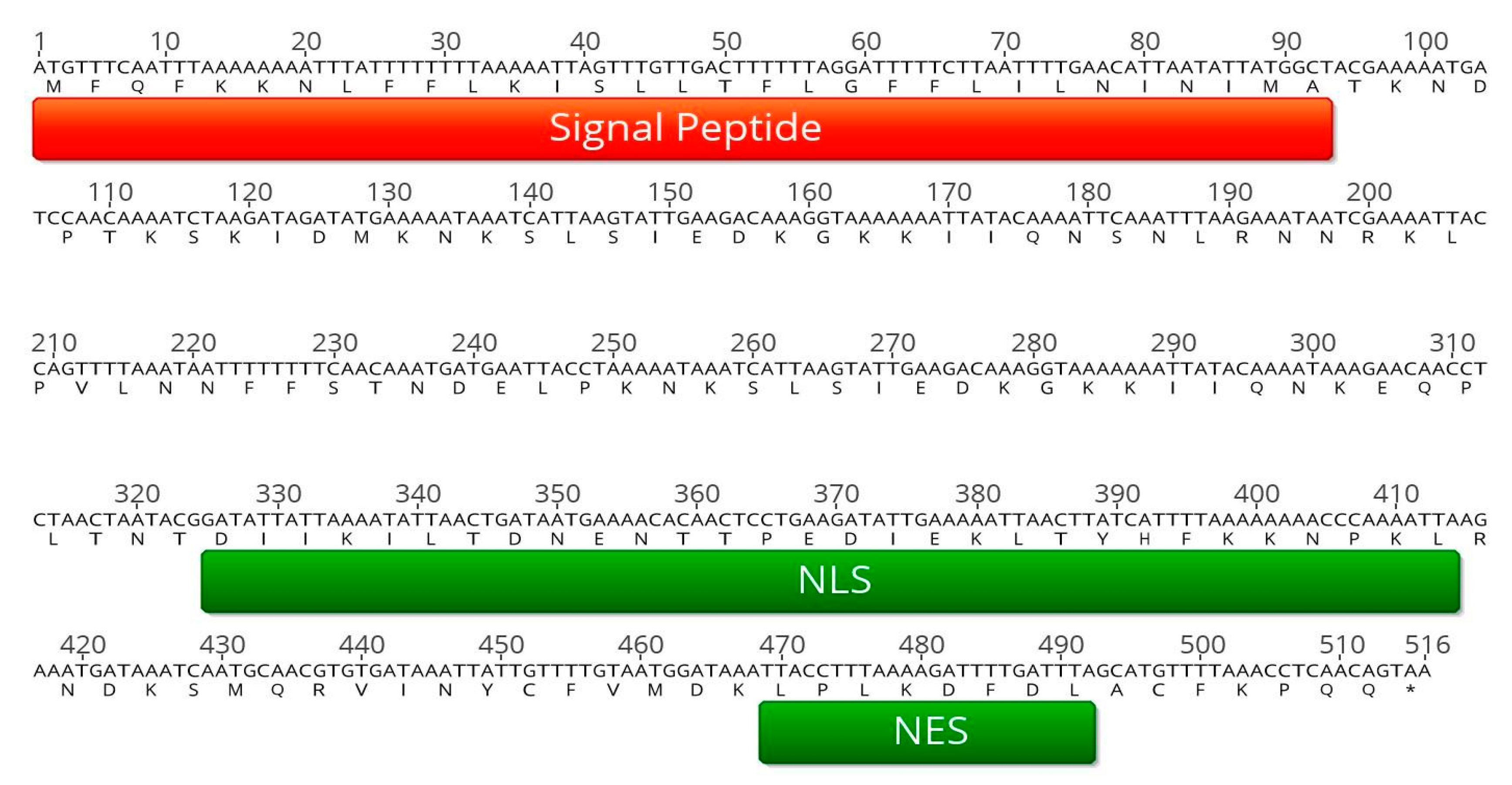
2.1. In Silico Analysis of PME2 Indicates Effector Potential

Bioinformatic analysis of conserved hypothetical proteins encoded in the P. mali genome [19] revealed that CAP18323.1, encoded by the gene atp_00136, contains interesting features that might confer effector function. Neural networks and hidden Markov prediction models (Transmembrane Helices Hidden Markov Model; TMHMM) were applied to analyze CAP18323.1 for the presence of a signal peptide and the presence of transmembrane regions (SignalP v. 3.0 [21], TMHMM [22]). Since phytoplasma phylogenetically belong to Gram-positive bacteria [3], a prediction algorithm trained on this bacterial group was applied. The N-terminal amino acid-stretch 1–31 contains a signal peptide that is supposed to confer Sec-dependent secretion of the protein (Figure 1). Further transmembrane regions were not predicted, indicating that CAP18323.1 is not inserted in a membrane. Upon translation, N-terminal signal peptides are cleaved [23]. At the C-terminal part of CAP18323.1 an importin α/β-dependent nuclear localization site (NLS) and a nuclear export signal (NES) were predicted [24,25]. The absence of transmembrane regions in the mature protein, the predicted localization in the plant cytoplasma or the nucleus (WoLF PSORT, [26]), and the small size of about 16 kDa (Analysis Tool on the ExPASy Server, [27]) indicate that CAP18323.1 may exhibit an effector function (Figure 1).

2.2. Atp_00136 (Pme2) is Expressed in P. Mali-Infected Malus × Domestica but not in the Insect Vector C. Picta

Subsequently, it was analyzed whether atp_00136 was expressed in apple trees infected with P. mali. Leaf and root samples of P. mali-infected and non-infected Malus × domestica cv. “Golden Delicious” trees were taken in May and October. Expression of atp_00136 was analyzed with atp_00136-specific primers and Malus × domestica cDNA derived from mRNA. Expression of atp_00136 was confirmed in P. mali-infected leaf and root tissue by the detection of distinct amplicons at the expected size in the respective samples (Figure 2).
Using quantitative PCR (qPCR) the expression levels of atp_00136 and P. mali in the samples were quantified. The results show that atp_00136 is only expressed in tissue colonized by P. mali (Table 1). Since identified expressed genes were named in a chronological manner, atp_00136 was named “Protein in Malus Expressed 2” (pme2) based on the general recommendations for bacterial gene nomenclature [28].
To analyze if pme2 was expressed in the transmitting insect vectors during infection, three P. mali-infected C. picta individuals were analyzed for the expression of the potential effector. In the RNA/cDNA of all infected individuals, P. mali-specific transcripts of the ribosomal protein rpl22 were detected, but expression of pme2 was not detectable.

2.3. Genetic Variability of Pme2

Cloned amplicon sequencing revealed that the prevalent variant of pme2 from infected trees in South Tyrol (North-East Italy) differs compared to the pme2 sequence of the P. mali AT strain from Germany [19]. In a total of 20 samples from naturally infected apple trees in the regions Burggraviato and Val Venosta, a prevalent, conserved sequence of pme2 was identified (pme2ST; accession number MN224214). This conserved variant exhibits a single nucleotide polymorphism (SNP) in the sequence stretch before the NLS, and two SNPs within and one SNP after the NLS compared to the AT strain (Figure 3). All four SNPs in the pme2ST variant lead to nonsynonymous missense substitutions at the protein level as compared to the pme2 sequence published previously [19] (pme2AT). The NLS of pme2ST has a slightly higher prediction score than the NLS of pme2AT. The most striking difference between pme2AT and pme2ST is a stretch of 120 bp in pme2ST which is absent in pme2AT. This stretch is a partial duplication of a fragment also present in pme2AT (Figure 3). In three Malus × domestica samples, a very sporadic sequence of pme2 could be detected that did not contain the pme2ST characteristic sequence duplication but showed strong sequence similarity to pme2AT. The sporadic sequence contains six SNPs at positions 218 (A > T), 220 (A > G), 322 (A > G), 331 (A > C), 344 (C < T), and 427 (T > G) that lead to nonsynonymous missense mutations (accession number MN224215) compared to pme2AT. However, in the trees in which these very sporadic pme2 sequences were found, pme2ST could also be detected, indicating the presence of a mixed population of different P. mali strains.

2.4. PME2ST and PME2AT Translocate to the Nucleus of Nicotiana spp. Protoplasts

To identify the subcellular localization of the PME2 protein in the plant cell, mesophyll protoplasts of Nicotiana occidentalis and N. benthamiana were transformed, with expression vectors coding for PME2AT and PME2ST tagged with GFP or mCherry-fluorescent protein to allow subcellular tracking. The N-terminal signal part was not considered for these studies, since it is removed from the processed, mature CAP18323.1 protein. N. occidentalis and N. benthamiana can be infected with P. mali. Upon infection, both Nicotiana species show disease-specific symptoms and are thus appropriate model plants for P. mali effector studies [15,29]. Confocal microscopy analysis revealed that overexpressed PME2AT and PME2ST are translocated to the nucleus of Nicotiana spp. protoplasts. This translocation was independent of the used tag and Nicotiana species (Figure 4 and Figures S1–S3). The in vivo results therefore confirm the in silico prediction that PME2ST and PME2AT are translocated to the nucleus of potential host cells.
A leaf infiltration assay using Agrobacterium strain EHA105 transformed with PME2 encoding expression vectors did not result in detectable expression or phenotypic alterations of either PME2AT or PME2ST in both Nicotiana species. Nonetheless, positive controls expressing the fluorophore tag only and leaves infiltrated with the P. mali SAP11-like effector protein ATP_00189 [13] as control showed strong signals (Figure S5), indicating that PME2 expression might be somehow blocked or is immediately degraded by the plant.

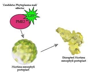
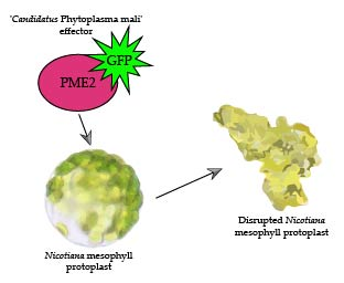
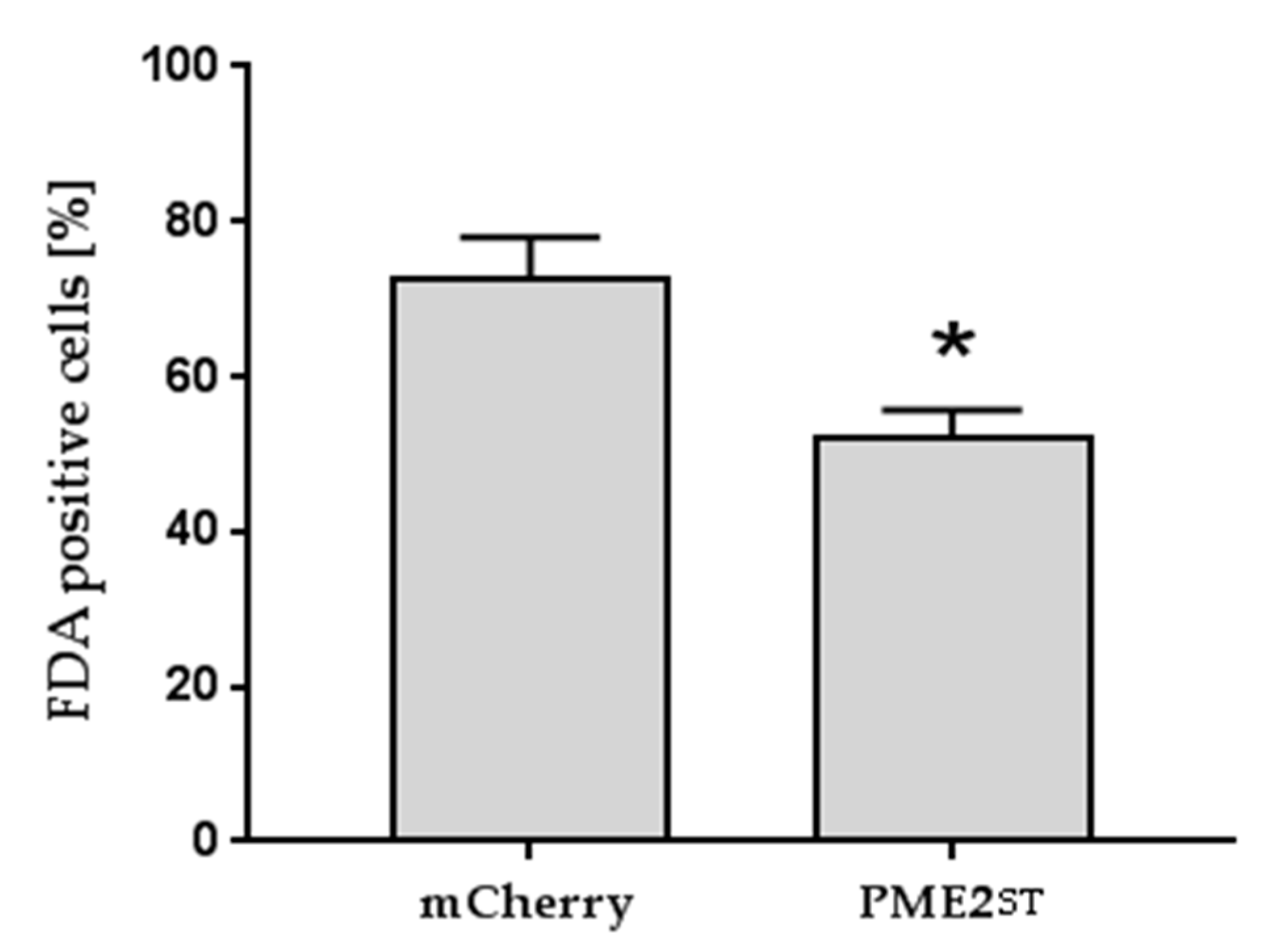
2.5. PME2ST but not PME2AT Affect Cell Integrity of Nicotiana spp. Protoplasts

Protoplasts transformed with the PME2ST expression vector often showed shrinkage, and only about 50% of the N. benthamiana protoplasts were viable 20 h post-transformation compared to the transformation control expressing the fluorophore only or a GFP with NLS (Figure 5a). The shrunk cells lysed and only the remaining cell debris was microscopically detectable (Figure 4). The effect on protoplast integrity was observed in protoplasts expressing PME2ST:GFP and PME2ST:mCherry, and thus was independent of the fluorophore used as a tag for microscopic analyses. Similar results were obtained using N. occidentalis as the heterologous PME2ST expression system. The mCherry-tagged PME2ST induced a weak but significant reduction of viability in N. occidentalis protoplasts (Figure 5b). The GFP-tagged PME2ST showed the same tendency but to a stronger extent, i.e., it reduced cell viability by about 50%, which is similar to the effect seen in N. benthamiana protoplasts. Cell viability stain with fluorescein diacetate (FDA) showed similar results, i.e., that N. benthamiana protoplasts transformed with the PME2ST-expressing vector showed a significantly reduced viability (Figure 6). Shrunk cells were positive for propidium iodide (PI) staining (Figure S4), indicating that these cells were dead.
Interestingly, PME2AT did not have an effect on protoplast integrity in N. benthamiana nor in N. occidentalis protoplasts (Figure 5).

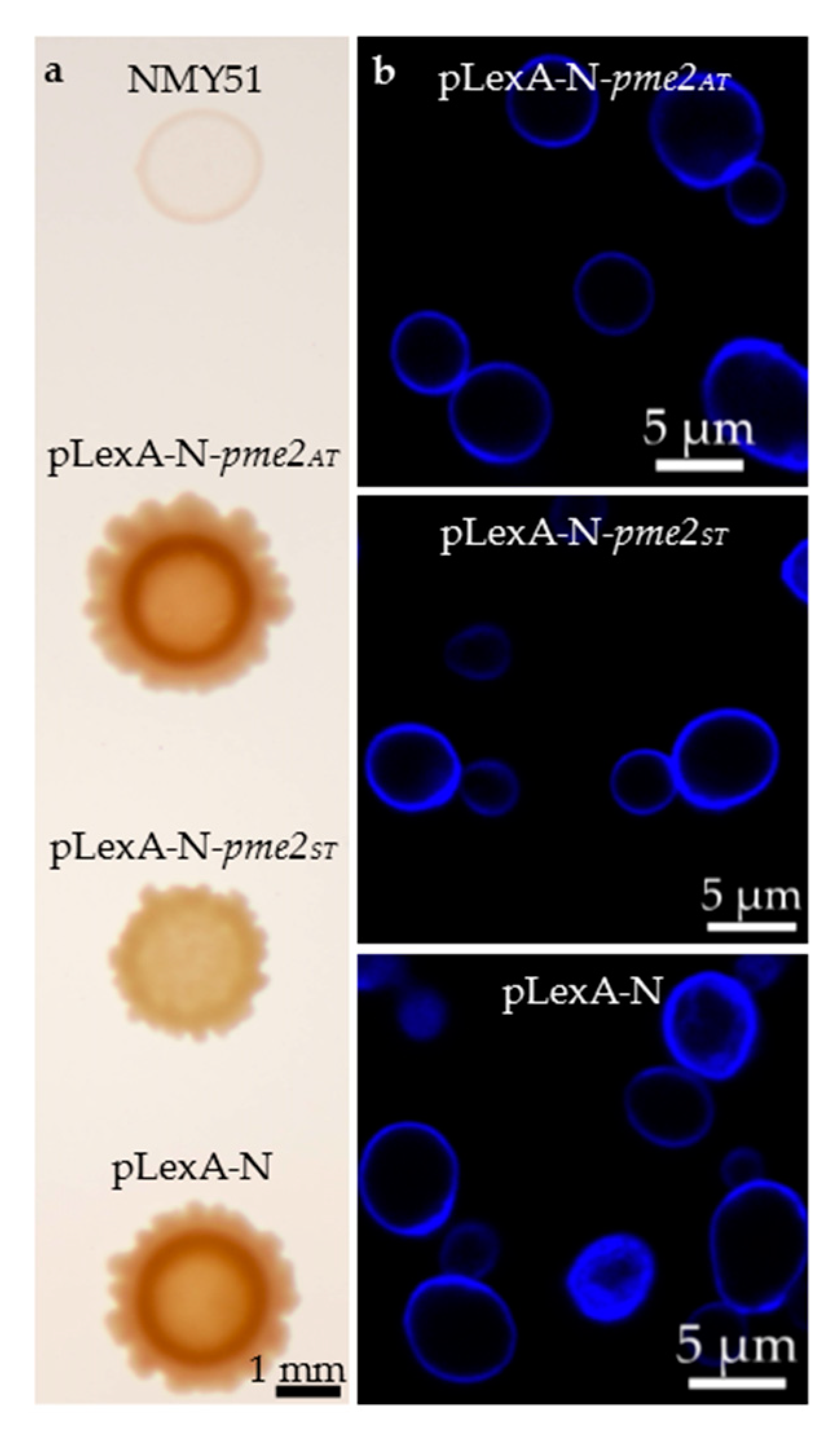
2.6. A Yeast Two-Hybrid Screen Was Unsuitable for the Elucidation of PME2ST Function

Upon expression of PME2ST, the yeast reporter strain Saccharomyces cerevisiae NMY51 showed several macroscopic aberrations in colony growth (Figure 7a). However, at the microscopic level when visualizing the yeast cell wall with calcofluor white, no phenotypic differences between yeast cells expressing PME2AT, PME2ST, and empty bait vector pLexA-N could be detected (Figure 7b). Considering the effect of mere PME2ST expression on growth of the yeast reporter strain, the relevance of any identified interaction in a yeast two-hybrid screen remains highly questionable and the assay was therefore not performed.

3. Discussion

The results of this study show that PME2ST (a variant of CAP18323.1 previously annotated as “conserved hypothetical protein”) affects plant cell integrity. Based on our findings and the definition that effectors are secreted pathogen proteins altering host-cell structure and function [30], we propose defining PME2 as a phytoplasmal effector. Interestingly, two different variants of PME2 were identified and both variants translocate to the nucleus of plant cells, but only the newly described regional variant PME2ST subsequently affects protoplast integrity. The small size of about (at a maximum) 21 kDa (PME2ST: 21 kDa and PME2AT: 16 kDa; both considering the mature protein without the signal peptide) indicates that PME2 can be translocated from the phloem and target adjacent tissues or be distributed systemically in the plant [3]. Subcellular localization using microscopy requires the use of fluorescent tags that are attached to the protein of interest. Tagging can affect subcellular localization of the protein; however, we used two different tags (GFP and mCherry) to analyze whether tagging influences the target localization. In cells expressing the tag only, a localization of the fluorescent signal in the cytoplasm could be observed. PME2 was localized only in the nucleus and since no signal was visible in other cell compartments, it can be assumed that the observed localization is effector-mediated (see also [31]). Only protoplasts transformed with PME2ST showed significant cell disruption as indirectly quantified by counting the remaining viable cells and FDA staining of the protoplasts. Shrunk cells were positive for the PI stain but did not show a GFP signal. The lack of the GFP-signal might be caused by a disruption of the nucleus, protein degradation, and/or leakage of the signal into the surrounding medium. Cells expressing PME2ST were intact, indicating that the effector either exhibits a dose-dependent or delayed effect on cell integrity.
Both variants of PME2 contain an N-terminal signal peptide, a nuclear localization signal (NLS), and a nuclear export signal (NES). It is a common feature of nuclear proteins to contain both NLS and NES and these signals coordinate the translocation of the protein between nucleus and cytoplasm [32]. Nuclear targeting of proteins containing a classical NLS is mediated by the importin α/β heterodimer through NLS-dependent binding to the importin α subunit and importin β–mediated attachment to the nuclear pore complex [33,34]. The SNPs in the NLS region of PME2ST lead to a (slightly) higher sequence-based NLS prediction; thus, the differences might show a stronger translocation to the nucleus. The NES signal (which indicates that shuttling of PME2 between nucleus and cytoplasm might occur) is the same in both variants. Even though nucleocytoplasmic distribution is predicted, PME2 was only detected in the nucleus. Many proteins containing NLS and NES appear to be localized in the nucleus because the rate of import to the nucleus is higher than the rate of export to the cytoplasm [35]. It remains thus unclear if PME2 is strictly limited to the nucleus or if a constant shuttling between nucleus and cytosol occurs.
Bacterial effectors that translocate to the nucleus, the so-called nuclear effectors, can affect master switches of the host immune machinery or alter host transcription to the benefit of the pathogen [31]. Effectors from different phytoplasma species target plant–host transcription factors or affect gene expression on the transcriptional level to alter the host metabolism to their own benefit [4,12,13,36,37]. However, none of these effectors have yet been reported to exhibit such detrimental effects during in planta expression. The effector protein BR1 of the phloem colonizing squash leaf curl geminivirus shuttles between the cytoplasm and the nucleus of protoplasts [38]. Upon binding to the second movement protein BL1, BR1 shuttles to the cytoplasma [39] and the concerted action between BR1 and BL1 mediates cell-to-cell movement of the virus within the phloem and to adjacent cells [35,38,40,41]. To unravel BR1 function it was necessary to identify its interaction partner, a general approach to investigate effector function. Yeast two-hybrid (Y2H) screens have been successfully applied to determine phytoplasmal effector targets on the molecular level [10,13]. These screens allow the screening of a protein of interest (effector) against a library containing hundreds of thousands of different potential interaction partners of a certain host species [42,43]. Successful interaction is monitored by a genetic reporter system that complements certain auxotrophies in the recombinant yeast reporter strain. However, a Y2H with PME2ST is not suitable since PME2ST expression strongly affected the Y2H yeast reporter strain. This effect on yeast cells further supports the finding that PME2ST exhibits a strong effect not only on plant, but also on yeast cells, even though the latter do not have relevance as phytoplasma host cells. Since PME2ST exhibits such a strong effect on the expressing host and non-host cells, alternative approaches must be applied to unravel its molecular function. Nicotiana spp. leaf infiltration assays with recombinant Agrobacterium strains expressing PME2 failed. It remains furthermore elusive as to whether PME2 exhibits effects on the host plant phenotype. Considering the PME2ST effects on protoplasts it can be assumed that a systemic overexpression would lead to overwhelming deleterious effects in transgenic plants that express this effector. The results show that PME2 is expressed in roots and leaves of infected Malus × domestica, but not in infected individuals of its insect vector C. picta, underlining the hypothesis that PME2 plays a role as an effector protein in plant cells. However, it needs further clarification if expression is fine-tuned in a tempo-spatial manner in the plant host.
A neatly coordinated and local expression during infection might have very local effects and might not lead to cell disruption as seen in heterologous overexpression experiments. It is hypothesized that phytoplasma are able to degrade plant cell walls or generate holes in plant cell membranes to expedite cell-to-cell effector translocation [4]. Infection with P. mali induces cytochemical modifications and injuries of the affected phloem cells [44,45]. It is speculated that plasma membrane integrity is affected by until-now unknown P. mali effector(s) and that plasma membrane disruption is involved in the observed phloem damage induced by virulent P. mali strains [45]. However, since molecular indications are lacking, interpretation of the mode of PME2 action remains speculative. Subsequent approaches to analyze PME2 function should comprise assays that do not depend on functional living cells.
Since PME2 is translocated to the nucleus it is possible that it directly targets the host DNA by mimicking DNA regulatory elements, such as transcription factors or repressors. Some plant pathogen effectors bind host DNA and thus modulate gene expression [46,47]. An example of these effectors are TAL effectors of the plant pathogen Xanthomonas. TAL effectors bind promoter elements and regulate plant host expression to the benefit of the pathogen [48,49,50,51]. The effector AvrBs3 of Xanthomonas translocates to the nucleus where it acts as a transcription factor and affects the size of mesophyll cells [52]. Bioinformatic prediction and sequence comparison did not indicate that PME2 has similarity with currently known transcription factors or other gene expression regulating factors in plants.
Both P. mali strains from which the two different PME2 variants were derived cause infection and typical disease symptoms in Malus × domestica. Thus, the effect of PME2ST on cell integrity seems to be dispensable for infection and symptom development but might affect strain virulence. However, a direct comparison between the two strains regarding their virulence is missing. It might also be possible that another effector of P. mali strain AT (unknown at the time of this research), mimics and thus complements the function that PME2AT is lacking.
Some P. mali strains strongly differ regarding their virulence potential in Malus × domestica and several studies addressed the genetic identification of virulence factors or certain genetic determinants that account for these differences [17,18,53,54,55]. Since phytoplasma cannot be genetically manipulated, determining the importance of an effector during infection often involves tortuous experimental paths. In this study we provide the first characterization of the P. mali effector PME2 and its effect on cells of potential plant hosts. We report an interesting difference between two variants of PME2 that occur in Italy and Germany, claiming that further full genomic sequence analysis is required to better understand how P. mali manipulates its host on the molecular level.

4. Materials and Methods

4.1. Verification of Pme2 Expression in Malus × Domestica and C. Picta

For the verification of pme2 expression by PCR in infected apple root and leaf samples RNA was extracted from the plant tissue as described in [13]. Extracted RNA was subjected to DNase treatment using DNAfree Turbo reagent (Ambion, Austin, TX, USA) and cDNA synthesis was performed using the SuperScript™ VILO™ cDNA Synthesis Kit (Invitrogen, Waltham, MA, USA). The generated cDNA was diluted 1:200 in nuclease free water and cDNA integrity was checked in all samples by performing a control PCR targeting the house-keeping gene transcript putative tip41-like family (transcript identifier: Mdo.1349) using the primers 5’-ACATGCCGGAGATGGTGTTTGG-3’ (forward) and 5’-ACTTCCAGAGTACGGCGTTGTG-3’ (reverse). Contamination with genomic DNA was checked by performing a PCR with primers amplifying a fragment within the non-coding region trnL of chloroplast DNA using the primers B49317 and A49855 [56]. No DNA contamination was detected in any of the cDNA samples, and the amplification of the putative tip41-like transcript fragment was positive, thus confirming the integrity of the generated cDNA. PCR reactions to verify pme2 expression were set up in a total reaction volume of 10 µL, using 2 µL of diluted cDNA (1:200) as template, 0.05 µL GoTaq® DNA Polymerase (Promega, Madison, WI, USA), 2 µL of 5X Green GoTaq® Reaction Buffer (Promega, Madison, WI, USA), 0.2 µL dNTP-mix (40 mM), 1 µL of forward primer ATP00136_forw_EcoRI (10 µM, 5’-CCCCCCGAATTCATGTTTCAATTTAAAAAAAATTTA-3’), and 1 µL of reverse primer ATP00136_rev_SalI (10 µM, 5’-CCCCCCGTCGACATTATTACTGTTGAGGTTTAA-3’). Cycling conditions were applied as follows: 95 °C for 5 min followed by 40 cycles of 95 °C for 1 min, 44.9 °C for 1 min, 72 °C for 1 min, and a final elongation step at 72 °C for 5 min. PCR products were visualized on 1% agarose gel. Additionally, pme2 expression level was detected by qPCR based on SYBR-Green chemistry using the primer pair ATP00136_GW_fwd (5’-CACCATGACGAAAAATGATCCAACAAA-3’)/ATP00136_nostopp_rev (5’-CTGTTGAGGTTTAAAACAT-3’) in a total reaction volume of 20 µL using 4.0 µL of diluted cDNA (1:200) as a template together with 10.0 µL 2× SYBR FAST qPCR Kit Master Mix (Kapa Biosystems/αmann-La Roche, Basel, Switzerland), 1.0 µL of each primer (10 µM), and 4.0 µL of nuclease free water. qPCR conditions were as follows: an initial denaturation step at 95 °C for 20 s followed by 34 cycles of 95 °C for 3 s and 60 °C for 30 s and a melting curve ramp from 65 to 95 °C, at increments of 0.5 °C every 5 s (CFX384 Touch Real-Time PCR Detection System; BioRad, Hercules, CA, USA). Data analysis was performed using the CFX ManagerTM software (BioRad, Hercules, CA, USA).
To control whether pme2 is expressed in infected individuals of the insect vector C. picta, RNA of six potentially infected and two uninfected F1 individuals was extracted with the ZR Tissue & Insect RNA MicroPrepTM kit (ZymoResearch, Irvine, CA, USA) according to the manufacturer’s instructions. Extracted RNA was subjected to DNase treatment using DNAfree Turbo reagent (Ambion, Austin, TX, USA) and RNA integrity was controlled with an RNA ScreenTape on a TapeStation 2200 (both Agilent, Santa Clara, CA, USA). cDNA was synthesized with the iScriptTM cDNA Synthesis Kit (BioRad, Hercules, CA, USA). Together with the cDNA synthesis a control was performed lacking the reverse transcriptase (-RT). Here, 2 µL of diluted cDNA (1:200) were used as template in a total qPCR reaction volume of 10 µL, together with 5 µL 2× SYBR FAST qPCR Kit Master Mix (Kapa Biosystems/Hoffmann-La Roche, Basel, Switzerland), 2 µL of nuclease free water, and 0.5 µL of forward and reverse primer (10 µM). The primer combination qPSY-WG-F and qPSY-WG-R, targeting the species-specific wingless gene [57], was used to determine cDNA integrity. P. mali infection was detected in three of the six individuals with primer pair rpAP15f-mod and rpAP15r3, targeting the ribosomal protein gene rpl22 [58]. Pme2 expression was checked with primer pair ATP00136_GW_fwd and ATP00136_nostopp_rev using the same qPCR conditions as described for the qPCR detection in Malus × domestica leaf samples.

4.2. Amplification, Subcloning, and Sequencing of atp_00136

DNA was purified from leaves from P. mali infected Malus x domestica cv Golden Delicious trees (10 trees from Burggraviato and 10 trees from Val Venosta) using the DNeasy Plant Mini kit (Qiagen, Hilden, Germany) following the manufacturer’s instructions. DNA was diluted 1:10 in water and 2 µL template were used in a total PCR reaction volume of 50 µL as follows: atp_00136 was amplified using 0.02 U/µL Phusion High-Fidelity DNA Polymerase (Thermo Fisher Scientific, Waltham, MA, USA) using HF-buffer supplied by the manufacturer, 400 µM dNTPs, and 0.5 µM of each primer (forward: 5’-CCCCCCGAATTCATGTTTCAATTTAAAAAAAATTTA-3’; reverse: 5’-CCCCCCGTCGACATTATTACTGTTGAGGTTTAA-3’). DNA was denatured at 98 °C for 30 s followed by 30 cycles of denaturation for 10 s at 98 °C, amplification for 30 s at 49.3 °C, and elongation at 72 °C for 30 s. The PCR was finalized by a terminal elongation step at 72 °C for 5 min. The PCR product was purified using the Illustra GFX PCR DNA and Gel Band Purification Kit (GE Healthcare, Chicago, IL, USA) and 1 µg of purified PCR product was digested with 4 U EcoRI and SalI following the manufacturer’s instructions (Thermo Fisher Scientific, Waltham, MA, USA), ligated into equally digested pUC19 using T4-Ligase (Thermo Fisher Scientific, Waltham, MA, USA) and transformed into MegaX DH10B™ T1R cells (Life Technologies, Carlsbad, CA, USA). At least five clones from each tree were sequenced with pUC19 specific primers (GATC Biotech, Constance, Germany) and analyzed to see different variants of the gene indicating a mixed infection.

4.3. Subcloning of Pme2 into GreenGate Expression Vectors

The genes pme2ST and pme2AT were subcloned into the GreenGate-entry module pGGC000 [59] using the primer pair ATP00136pP_CBsaI_fw (5’-AACAGGTCTCAGGCTCCATGACGAAAAATGATCCAACAAA-3’) and ATP00136pP_DBsaI_rv (5’-AACAGGTCTCACTGACTGTTGAGGTTTAAAACAT-3’). Using different components from the GreenGate-kit plant, transformation constructs coding for pme2AT-linker-GFP or pme2AT-linker-mCherry and pme2ST-linker-GFP or pme2ST-linker-mCherry, driven by the 35S promoter and flanked at the 3’-end by the RBCS terminator, including kanamycin as the plant resistance marker, were designed. The following modules were assembled by GreenGate reaction in a total volume of 15 µL: 150 ng pGGA004 (35S), 150 ng pGGB003 (B-dummy), 150 ng pGGC000-pme2AT or pGGC00-pme2ST, 150 ng pGGD001 (linker-GFP) or pGGD003 (linker-mCherry), 150 ng pGGE001 (RBCS), 150 ng pGGF007 (pNOS:KanR:tNOS), and 100 ng pGGZ001 (empty destination vector). Subsequently, 1.5 µL 10× CutSmart Buffer (New England Biolab, Ipswich, MA, USA), 1.5 µL ATP (10 mM), 1.0 µL T4 DNA Ligase (5 u/µL) (Thermo Fisher Scientific, Waltham, MA, USA), and 1.0 µL BsaI-HF®v2 (20,000 u/mL) (New England Biolab, Ipswich, MA, USA) were added to the module mixture, and 30 cycles of 2 min at 37 °C and 2 min at 16 °C each, followed by 50 °C for 5 min and 80 °C for 5 min were performed. Subsequently, 5 µL of the reaction mixture were used for heat-shock transformation of ccdB-sensitive One Shot® TOP10 chemically competent Escherichia coli (Invitrogen, Carlsbad, CA, USA). For the assembly of positive controls, the modules pGGC012 (GFP-NLS) or pGGC014 (GFP) or pGGC015 (mCherry) were used instead of the above mentioned pGGC000 modules. The correct assembly of the plant transformation constructs was confirmed by sequencing. Plasmid-DNA for protoplast transformation was obtained as described elsewhere [60], using the NucleoSnap® Plasmid Midi preparation kit (Macherey-Nagel, Düren, Germany) and PEG precipitation.

4.4. Protoplast Isolation and Transformation

Protoplasts of N. benthamiana and N. occidentalis were isolated from four- to five-week-old plants, cultivated under long photoperiod conditions (16 h/8 h, 24 °C/22 °C, 70% rH) and transformed as described in [60] using 10 µg plasmid-DNA per 20,000 protoplasts. After 18 h, at least 100 protoplasts of each transformation were checked for the occurrence of GFP or mCherry-fluorescence using a confocal laser scanning microscope (LSM800, Zeiss, Oberkochen, Germany) with an excitation wavelength of 488 nm for GFP and 561 nm for mCherry. The detection wavelength of GFP was set between 410 nm and 575 nm and of mCherry between 575 nm and 650 nm. Autofluorescence of chlorophyll was detected between 650 nm and 700 nm. After 20 h the number of intact protoplasts/mL was determined by counting in a Fuchs-Rosenthal chamber. Protoplast transformation and viability determination was repeated independently four times.
Only experiments in which at least 20% of the protoplasts in the control setup were viable after transformation were considered for further evaluation. Significant outliers were removed from the data set using the GraphPad QuickCalcs Outlier calculator online tool (https://www.graphpad.com/quickcalcs/Grubbs1.cfm; status of information 16th September 2019). Greisser Greenhouse correction on raw data and one-way-ANOVA with a Tukey Posttest were performed to analyze statistical differences between groups (GraphPad Prism 7.01., GraphPad Software, San Diego, CA, USA). To allow a better visual comparison, data were normalized to each respective control, which was set to 1.
Additionally, protoplast viability was visualized by propidium iodide (PI) and counted by fluorescein diacetate (FDA) staining in three independent repetitions. For the first, 20 µL of protoplasts transformed with GFP tagged expression vectors were mixed with 20 µL of PI solution (10 µg/mL PI in 0.65 M mannitol). FDA staining was done according to [61] using 20 µL of protoplasts transformed with either mCherry tagged PME2AT expression vectors or a vector expressing only mCherry and 20 µL of FDA solution (0.1 mg/mL FDA in 0.65 M mannitol). Fluorescence of PI, mCherry, and GFP was recorded using a LSM800 confocal laser scanning microscope (Zeiss, Oberkochen, Germany) with excitation and detection wavelengths for GFP and mCherry as described above and for PI excitation at 561 nm and detection between 560 nm and 640 nm.

4.5. Nicotiana spp. Leaf Infiltration

For subcellular localization of PME2, the two GreenGate expression vectors, as well as GFP and GFP-NLS expression vectors as positive controls, were subcloned by electroporation into Agrobacterium tumefaciens strain EHA105. As an additional control, we subcloned a GreenGate expression vector expressing the SAP11-like P. mali effector protein ATP_00189 [13] with an N-terminal fused GFP tag into A. tumefaciens strain EHA105. The transgenic A. tumefaciens clones were cultured for 2 days at 28 °C in liquid selective LB medium. Subsequently, 0.5 OD/mL were resuspended in infiltration medium (10 mM MgCl2, 10 mM MES, 200 µM acetosyringone, pH 5.7) and infiltrated with a blunt syringe into leaves from four- to five-week-old N. occidentalis and N. benthamiana. Fluorescence was recorded after 48 h and 72 h using the confocal laser scanning microscope (LSM800, Zeiss, Oberkochen, Germany) with excitation for GFP at 488 nm and detection between 410 nm and 546 nm and excitation for mCherry at 561 nm and detection between 562 and 624 nm.

4.6. Expression in Yeast

For a potential Y2H, pme2AT and pme2ST were subcloned into bait-vector pLexA-N as described in [13,62] with primer pair ATP00136_forw_EcoRI/ATP00136_rev_SalI. The bait-plasmids pLexA-N-pme2ST and pLexA-N-pme2AT were transformed into S. cerevisiae strain NMY51. Growth aberrations of yeast colonies on selective SD-trp plates were observed and recorded by photographing.
For calcofluor white staining, yeast cells were grown overnight in SD-trp liquid media. Subsequently, 2 mL of the overnight culture were centrifuged, supernatant removed, and the cells resuspended in clear phosphate-buffered saline (PBS) buffer. Then, 10 µL of a 5 mM calcofluor white solution (Biotium, Fremont, California) were added to the cell suspension and incubated for 20 min at room temperature. The yeast cell wall was visualized by a confocal laser scanning microscope (LSM800, Zeiss, Oberkochen, Germany) with excitation at 405 nm and detection wavelength between 400 nm and 560 nm.

5. Conclusions

In this study we identified and characterized the novel P. mali effector protein PME2. This effector contains an NLS and an NES sequence and translocates to the nucleus of N. benthamiana mesophyll protoplasts. Two naturally occurring genetic variants of PME2, namely PME2ST and PME2AT, differ regarding their ability to induce cellular modifications in yeast and plant cells. When overexpressed, the variant PME2ST affects yeast growth and reduces the viability of Nicotiana spp. mesophyll protoplasts. These findings indicate that PME2 might play a role for P. mali virulence in plants. Despite the similarities between both PME2 variants, this effect was not observed in yeast or protoplasts expressing PME2AT. The results of our study show for the first time that a phytoplasmal effector causes detrimental effects when overexpressed in protoplasts.

Supplementary Materials

Supplementary materials can be found at https://www.mdpi.com/1422-0067/20/18/4613/s1.

Author Contributions

Conceptualization, K.J. and K.S.; methodology, K.J., C.M., K.S., H.S., and B.H.; validation, C.M. and K.J.; formal analysis, C.M., K.J., and C.K.; investigation, C.M. and K.J.; resources, K.J., T.L., and K.S.; data curation, C.M. and C.K.; writing—original draft preparation, C.M. and K.J.; writing—review and editing, C.M., H.S., B.H., C.K., K.S., T.L., and K.J.; visualization, C.M. and K.J.; supervision, K.J., T.L., and K.S.; project administration, K.J., T.L., and K.S.; funding acquisition, K.J., K.S., and T.L.

Funding

This research was funded by the APPL2.0 and APPLIII project within the Framework agreement in the field of invasive species in fruit growing and major pathologies (PROT. VZL_BZ 09.05.2018 0002552) and was co-funded by the Autonomous Province of Bozen/Bolzano, Italy and the South Tyrolean Apple Consortium.

Acknowledgments

We would like to thank Andreas Putti and Laura Russo of the Food Microbiology Laboratory at the Laimburg Research Centre for helpful and interesting discussions about yeast biology. Furthermore, we would like to thank Mirko Moser from the Fondazione Edmund Mach for discussing phytoplasma sequence data, Vicky Oberkofler for lab assistance, and Amy Kadison for her English proofreading.

Conflicts of Interest

The authors declare no conflict of interest.

Abbreviations

APApple proliferation
AY-WBAster Yellow Witches’ Broom
C. pictaCacopsylla picta
FDAFluorescein diacetate
GFPGreen fluorescent protein
N.Nicotiana
NESNuclear export signal
NLSNuclear localization signal
PIPropidium iodide
P. maliCandidatus Phytoplasma mali
PME2Protein in Malus Expressed 2
qPCRquantitative PCR
SNPSingle nucleotide polymorphism
Y2HYeast two-hybrid

References

  1. Strauss, E. Phytoplasma research begins to bloom. Science 2009, 325, 388–390. [Google Scholar] [CrossRef]
  2. Christensen, N.M.; Axelsen, K.B.; Nicolaisen, M.; Schulz, A. Phytoplasmas and their interactions with hosts. Trends Plant Sci. 2005, 10, 526–535. [Google Scholar] [CrossRef] [PubMed]
  3. Hogenhout, S.A.; Oshima, K.; Ammar, E.-D.; Kakizawa, S.; Kingdom, H.N.; Namba, S. Phytoplasmas: Bacteria that manipulate plants and insects. Mol. Plant Pathol. 2008, 9, 403–423. [Google Scholar] [CrossRef] [PubMed]
  4. Sugio, A.; MacLean, A.M.; Kingdom, H.N.; Grieve, V.M.; Manimekalai, R.; Hogenhout, S.A. Diverse targets of phytoplasma effectors: From plant development to defense against insects. Annu. Rev. Phytopathol. 2011, 49, 175–195. [Google Scholar] [CrossRef] [PubMed]
  5. Bai, X.; Correa, V.R.; Toruño, T.Y.; Ammar, E.-D.; Kamoun, S.; Hogenhout, S.A. AY-WB phytoplasma secretes a protein that targets plant cell nuclei. Mol. Plant Microbe Interact. 2009, 22, 18–30. [Google Scholar] [CrossRef]
  6. Lu, Y.-T.; Cheng, K.-T.; Jiang, S.-Y.; Yang, J.-Y. Post-translational cleavage and self-interaction of the phytoplasma effector SAP11. Plant Signal. Behav. 2014, 9, e28991. [Google Scholar] [CrossRef] [PubMed] [Green Version]
  7. Lu, Y.-T.; Li, M.-Y.; Cheng, K.-T.; Tan, C.M.; Su, L.-W.; Lin, W.-Y.; Shih, H.-T.; Chiou, T.-J.; Yang, J.-Y. Transgenic plants that express the phytoplasma effector SAP11 show altered phosphate starvation and defense responses. Plant Physiol. 2014, 164, 1456–1469. [Google Scholar] [CrossRef]
  8. Sugawara, K.; Honma, Y.; Komatsu, K.; Himeno, M.; Oshima, K.; Namba, S. The alteration of plant morphology by small peptides released from the proteolytic processing of the bacterial peptide TENGU. Plant Physiol. 2013, 162, 2005–2014. [Google Scholar] [CrossRef]
  9. MacLean, A.M.; Sugio, A.; Makarova, O.V.; Findlay, K.C.; Grieve, V.M.; Tóth, R.; Nicolaisen, M.; Hogenhout, S.A. Phytoplasma effector SAP54 induces indeterminate leaf-like flower development in Arabidopsis plants. Plant Physiol. 2011, 157, 831–841. [Google Scholar] [CrossRef]
  10. Sugio, A.; Kingdom, H.N.; MacLean, A.M.; Grieve, V.M.; Hogenhout, S.A. Phytoplasma protein effector SAP11 enhances insect vector reproduction by manipulating plant development and defense hormone biosynthesis. Proc. Natl. Acad. Sci. USA 2011, 108, E1254–E1263. [Google Scholar] [CrossRef] [Green Version]
  11. Tomkins, M.; Kliot, A.; Marée, A.F.; Hogenhout, S.A. A multi-layered mechanistic modelling approach to understand how effector genes extend beyond phytoplasma to modulate plant hosts, insect vectors and the environment. Curr. Opin. Plant Biol. 2018, 44, 39–48. [Google Scholar] [CrossRef] [PubMed]
  12. MacLean, A.M.; Orlovskis, Z.; Kowitwanich, K.; Zdziarska, A.M.; Angenent, G.C.; Immink, R.G.H.; Hogenhout, S.A. Phytoplasma effector SAP54 hijacks plant reproduction by degrading MADS-box proteins and promotes insect colonization in a RAD23-dependent manner. PLoS Biol. 2014, 12, e1001835. [Google Scholar] [CrossRef] [PubMed]
  13. Janik, K.; Mithöfer, A.; Raffeiner, M.; Stellmach, H.; Hause, B.; Schlink, K.; Mithofer, A. An effector of apple proliferation phytoplasma targets TCP transcription factors-a generalized virulence strategy of phytoplasma? Mol. Plant Pathol. 2017, 18, 435–442. [Google Scholar] [CrossRef] [PubMed]
  14. Strohmayer, A.; Moser, M.; Si-Ammour, A.; Krczal, G.; Boonrod, K. ’Candidatus Phytoplasma mali’ genome encodes a protein that functions as a E3 Ubiquitin Ligase and could inhibit plant basal defense. Mol. Plant Microbe Interact. 2019. [Google Scholar] [CrossRef] [PubMed]
  15. Boonrod, K.; Munteanu, B.; Jarausch, B.; Jarausch, W.; Krczal, G. An immunodominant membrane protein (Imp) of ’Candidatus Phytoplasma mali’ binds to plant actin. Mol. Plant Microbe Interact. 2012, 25, 889–895. [Google Scholar] [CrossRef] [PubMed]
  16. Seemüller, E.; Kampmann, M.; Kiss, E.; Schneider, B. HflB gene-based phytopathogenic classification of ’Candidatus Phytoplasma mali’ strains and evidence that strain composition determines virulence in multiply infected apple trees. Mol. Plant Microbe Interact. 2011, 24, 1258–1266. [Google Scholar] [CrossRef]
  17. Schneider, B.; Sule, S.; Jelkmann, W.; Seemüller, E. Suppression of aggressive strains of ’Candidatus phytoplasma mali’ by mild strains in Catharanthus roseus and Nicotiana occidentalis and indication of similar action in apple trees. Phytopathology 2014, 104, 453–461. [Google Scholar] [CrossRef] [PubMed]
  18. Seemüller, E.; Zikeli, K.; Furch, A.C.U.; Wensing, A.; Jelkmann, W. Virulence of ‘Candidatus Phytoplasma mali’ strains is closely linked to conserved substitutions in AAA+ ATPase AP460 and their supposed effect on enzyme function. Eur. J. Plant Pathol. 2017, 86, 141. [Google Scholar] [CrossRef]
  19. Kube, M.; Schneider, B.; Kuhl, H.; Dandekar, T.; Heitmann, K.; Migdoll, A.M.; Reinhardt, R.; Seemüller, E. The linear chromosome of the plant-pathogenic mycoplasma ’Candidatus Phytoplasma mali’. BMC Genomics 2008, 9, 306. [Google Scholar] [CrossRef]
  20. Kube, M.; Mitrovic, J.; Duduk, B.; Rabus, R.; Seemüller, E. Current view on phytoplasma genomes and encoded metabolism. Scientific World J. 2012, 2012, 185942. [Google Scholar] [CrossRef]
  21. Bendtsen, J.D.; Nielsen, H.; von Heijne, G.; Brunak, S. Improved prediction of signal peptides: SignalP 3.0. J. Mol. Biol. 2004, 340, 783–795. [Google Scholar] [CrossRef] [PubMed]
  22. Krogh, A.; Larsson, B.; von Heijne, G.; Sonnhammer, E.L. Predicting transmembrane protein topology with a hidden Markov model: Application to complete genomes. J. Mol. Biol. 2001, 305, 567–580. [Google Scholar] [CrossRef] [PubMed]
  23. Beckwith, J. The Sec-dependent pathway. Res. Microbiol. 2013, 164, 497–504. [Google Scholar] [CrossRef] [PubMed] [Green Version]
  24. Kosugi, S.; Hasebe, M.; Tomita, M.; Yanagawa, H. Systematic identification of cell cycle-dependent yeast nucleocytoplasmic shuttling proteins by prediction of composite motifs. Proc. Natl. Acad. Sci. USA 2009, 106, 10171–10176. [Google Scholar] [CrossRef] [PubMed] [Green Version]
  25. La Cour, T.; Kiemer, L.; Mølgaard, A.; Gupta, R.; Skriver, K.; Brunak, S. Analysis and prediction of leucine-rich nuclear export signals. Protein Eng. Des. Sel. 2004, 17, 527–536. [Google Scholar] [CrossRef] [PubMed] [Green Version]
  26. Horton, P.; Park, K.-J.; Obayashi, T.; Fujita, N.; Harada, H.; Adams-Collier, C.J.; Nakai, K. WoLF PSORT: Protein localization predictor. Nucleic Acids Res. 2007, 35, W585–W587. [Google Scholar] [CrossRef] [PubMed]
  27. Gasteiger, E.; Hoogland, C.; Gattiker, A.; Duvaud, S.; Wilkins, M.R.; Appel, R.D.; Bairoch, A. Protein identification and analysis tools on the ExPASy server. In The Proteomics Protocols Handbook; Walker, J.M., Ed.; Humana Press: Totowa, NJ, USA, 2005; pp. 571–607. ISBN 978-1-59259-890-8. [Google Scholar]
  28. Journal of Bacteriology. Nomenclature: Genetic Nomenclature. Available online: https://jb.asm.org/content/nomenclature (accessed on 16 September 2019).
  29. Luge, T.; Kube, M.; Freiwald, A.; Meierhofer, D.; Seemüller, E.; Sauer, S. Transcriptomics assisted proteomic analysis of Nicotiana occidentalis infected by Candidatus Phytoplasma mali strain AT. Proteomics 2014, 14, 1882–1889. [Google Scholar] [CrossRef] [PubMed]
  30. Hogenhout, S.A.; Van der Hoorn, R.A.L.; Terauchi, R.; Kamoun, S. Emerging concepts in effector biology of plant-associated organisms. Mol. Plant Microbe Interact. 2009, 22, 115–122. [Google Scholar] [CrossRef] [PubMed]
  31. Chaudhari, P.; Ahmed, B.; Joly, D.L.; Germain, H. Effector biology during biotrophic invasion of plant cells. Virulence 2014, 5, 703–709. [Google Scholar] [CrossRef] [PubMed] [Green Version]
  32. Fu, X.; Liang, C.; Li, F.; Wang, L.; Wu, X.; Lu, A.; Xiao, G.; Zhang, G. The rules and functions of nucleocytoplasmic shuttling proteins. Int. J. Mol. Sci. 2018, 19, 1445. [Google Scholar] [CrossRef] [PubMed]
  33. Corbett, A.H.; Silver, P.A. Nucleocytoplasmic transport of macromolecules. Microbiol. Mol. Biol. Rev. 1997, 61, 193–211. [Google Scholar] [PubMed]
  34. Görlich, D.; Dabrowski, M.; Bischoff, F.R.; Kutay, U.; Bork, P.; Hartmann, E.; Prehn, S.; Izaurralde, E. A novel class of RanGTP binding proteins. J. Cell Biol 1997, 138, 65–80. [Google Scholar] [CrossRef] [PubMed]
  35. Ward, B.; Medville, R.; Lazarowitz, S.-G.; Turgeon, R. The geminivirus BL1 movement protein is associated with endoplasmic reticulum-derived tubules in developing phloem cells. J. Virol. 1997, 71, 3726–3733. [Google Scholar] [PubMed]
  36. Minato, N.; Himeno, M.; Hoshi, A.; Maejima, K.; Komatsu, K.; Takebayashi, Y.; Kasahara, H.; Yusa, A.; Yamaji, Y.; Oshima, K.; et al. The phytoplasmal virulence factor TENGU causes plant sterility by downregulating of the jasmonic acid and auxin pathways. Sci. Rep. 2014, 4, 7399. [Google Scholar] [CrossRef] [PubMed] [Green Version]
  37. Kitazawa, Y.; Iwabuchi, N.; Himeno, M.; Sasano, M.; Koinuma, H.; Nijo, T.; Tomomitsu, T.; Yoshida, T.; Okano, Y.; Yoshikawa, N.; et al. Phytoplasma-conserved phyllogen proteins induce phyllody across the Plantae by degrading floral MADS domain proteins. J. Exp. Bot. 2017, 68, 2799–2811. [Google Scholar] [CrossRef] [PubMed]
  38. Noueiry, A.O.; Lucas, W.J.; Gilbertson, R.L. Two proteins of a plant DNA virus coordinate nuclear and plasmodesmal transport. Cell 1994, 76, 925–932. [Google Scholar] [CrossRef]
  39. Sanderfoot, A.A.; Ingham, D.J.; Lazarowitz, S.-G. A viral movement protein as a nuclear shuttle: The geminivirus BR1 movement protein contains domains essential for interaction with BL1 and nuclear localization. Plant Physiol. 1996, 110, 23–33. [Google Scholar] [CrossRef] [PubMed]
  40. Sanderfoot, A.A.; Lazarowitz, S.-G. Cooperation in viral movement: The geminivirus BL1 movement protein interacts with BR1 and redirects it from the nucleus to the cell periphery. The Plant Cell 1995, 7, 1185–1194. [Google Scholar] [CrossRef] [PubMed]
  41. Sanderfoot, A.A.; Lazarowitz, S.-G. Getting it together in plant virus movement: Cooperative interactions between bipartite geminivirus movement proteins. Trends Cell Biol. 1996, 6, 353–358. [Google Scholar] [CrossRef]
  42. Fields, S.; Song, O.-K. A novel genetic system to detect protein–protein interactions. Nature 1989, 340, 245–246. [Google Scholar] [CrossRef]
  43. Brückner, A.; Polge, C.; Lentze, N.; Auerbach, D.; Schlattner, U. Yeast two-hybrid, a powerful tool for systems biology. Int. J. Mol. Sci. 2009, 10, 2763–2788. [Google Scholar] [CrossRef] [PubMed]
  44. Musetti, R.; Paolacci, A.; Ciaffi, M.; Tanzarella, O.A.; Polizzotto, R.; Tubaro, F.; Mizzau, M.; Ermacora, P.; Badiani, M.; Osler, R. Phloem cytochemical modification and gene expression following the recovery of apple plants from apple proliferation disease. Phytopathology 2010, 100, 390–399. [Google Scholar] [CrossRef] [PubMed]
  45. Zimmermann, M.R.; Schneider, B.; Mithöfer, A.; Reichelt, M.; Seemüller, E.; Furch, A.C.U. Implications of Candidatus Phytoplasma mali infection on phloem function of apple trees. Endocytobiosis Cell Res. 2015, 26, 67–75. [Google Scholar]
  46. Kay, S.; Hahn, S.; Marois, E.; Wieduwild, R.; Bonas, U. Detailed analysis of the DNA recognition motifs of the Xanthomonas type III effectors AvrBs3 and AvrBs3Deltarep16. Plant J. 2009, 59, 859–871. [Google Scholar] [CrossRef] [PubMed]
  47. Win, J.; Chaparro-Garcia, A.; Belhaj, K.; Saunders, D.G.O.; Yoshida, K.; Dong, S.; Schornack, S.; Zipfel, C.; Robatzek, S.; Hogenhout, S.A.; et al. Effector biology of plant-associated organisms: Concepts and perspectives. Cold Spring Harb. Symp. Quant. Biol. 2012, 77, 235–247. [Google Scholar] [CrossRef] [PubMed]
  48. Duan, Y.P.; Castañeda, A.; Zhao, G.; Erdos, G.; Gabriel, D.W. Expression of a single, host-specific, bacterial pathogenicity gene in plant cells elicits division, enlargement, and cell death. Mol. Plant Microbe Interact. 1999, 12, 556–560. [Google Scholar] [CrossRef]
  49. Boch, J.; Scholze, H.; Schornack, S.; Landgraf, A.; Hahn, S.; Kay, S.; Lahaye, T.; Nickstadt, A.; Bonas, U. Breaking the code of DNA binding specificity of TAL-type III effectors. Science 2009, 326, 1509–1512. [Google Scholar] [CrossRef]
  50. Domingues, M.N.; de Souza, T.A.; Cernadas, R.A.; de Oliveira, M.L.P.; Docena, C.; Farah, C.S.; Benedetti, C.E. The Xanthomonas citri effector protein PthA interacts with citrus proteins involved in nuclear transport, protein folding and ubiquitination associated with DNA repair. Mol. Plant Pathol. 2010, 11, 663–675. [Google Scholar] [CrossRef]
  51. de Souza, T.A.; Soprano, A.S.; de Lira, N.P.V.; Quaresma, A.J.C.; Pauletti, B.-A.; Paes Leme, A.-F.; Benedetti, C.-E. The TAL effector PthA4 interacts with nuclear factors involved in RNA-dependent processes including a HMG protein that selectively binds poly(U) RNA. PLoS ONE 2012, 7, e32305. [Google Scholar] [CrossRef]
  52. Kay, S.; Hahn, S.; Marois, E.; Hause, G.; Bonas, U. A bacterial effector acts as a plant transcription factor and induces a cell size regulator. Science 2007, 318, 648–651. [Google Scholar] [CrossRef]
  53. Seemüller, E.; Schneider, B. Differences in virulence and genomic features of strains of ’Candidatus Phytoplasma mali’, the apple proliferation agent. Phytopathology 2007, 97, 964–970. [Google Scholar] [CrossRef] [PubMed]
  54. Seemüller, E.; Kampmann, M.; Kiss, E.; Schneider, B. Molecular differentiation of severe and mild strains of ‘Candidatus Phytoplasma mali’ and evidence that their interaction in multiply infected trees determines disease severity. Bull. Insectology 2011, 64, 163–164. [Google Scholar]
  55. Rid, M.; Mesca, C.; Ayasse, M.; Gross, J. Apple proliferation phytoplasma influences the pattern of plant volatiles emitted depending on pathogen virulence. Front. Ecol. Evol. 2016, 3, 271. [Google Scholar] [CrossRef]
  56. Taberlet, P.; Gielly, L.; Pautou, G.; Bouvet, J. Universal primers for amplification of three non-coding regions of chloroplast DNA. Plant Mol. Biol. 1991, 17, 1105–1109. [Google Scholar] [CrossRef] [PubMed]
  57. Mittelberger, C.; Obkircher, L.; Oettl, S.; Oppedisano, T.; Pedrazzoli, F.; Panassiti, B.; Kerschbamer, C.; Anfora, G.; Janik, K. The insect vector Cacopsylla picta vertically transmits the bacterium ‘Candidatus Phytoplasma mali’ to its progeny. Plant Pathol 2017, 66, 1015–1021. [Google Scholar] [CrossRef]
  58. Monti, M.; Martini, M.; Tedeschi, R. EvaGreen Real-time PCR protocol for specific ’Candidatus Phytoplasma mali’ detection and quantification in insects. Mol. Cell. Probes 2013, 27, 129–136. [Google Scholar] [CrossRef]
  59. Lampropoulos, A.; Sutikovic, Z.; Wenzl, C.; Maegele, I.; Lohmann, J.U.; Forner, J. GreenGate - A novel, versatile, and efficient cloning system for plant transgenesis. PLoS ONE 2013, 8, e83043. [Google Scholar] [CrossRef]
  60. Janik, K.; Stellmach, H.; Mittelberger, C.; Hause, B. Characterization of phytoplasmal effector protein interaction with proteinaceous plant host targets using bimolecular fluorescence complementation (BiFC). In Phytoplasmas: Methods and Protocols; Musetti, R., Pagliari, L., Eds.; Humana Press: New York, NY, USA, 2019; pp. 321–331. ISBN 978-1-4939-8837-2. [Google Scholar]
  61. Heslop-Harrison, J.; Heslop-Harrison, Y. Evaluation of pollen viability by enzymatically induced fluorescence; intracellular hydrolysis of fluorescein diacetate. Stain Technol. 1970, 45, 115–120. [Google Scholar] [CrossRef]
  62. Janik, K.; Schlink, K. Unravelling the function of a bacterial effector from a non-cultivable plant pathogen using a yeast two-hybrid screen. J. Vis. Exp. 2017, 119, e55150. [Google Scholar] [CrossRef]
Figure 1. Results of the in silico analysis. Sequence analysis of atp_00136 revealed the presence of an N-terminal signal peptide (indicated in red), as well as a nuclear localization signal (NLS), and a C-terminal nuclear export signal (NES), both indicated in green. Graphs were generated with Geneious Prime 2018 version 11.1.4.
Ijms 20 04613 g001
Figure 2. Expression of pme2 (CAP18323.1) in ’Candidatus Phytoplasma mali’ (P. mali)-infected Malus × domestica. Transcripts of pme2 were detected by PCR using cDNA from Malus × domestica infected with P. mali. A discrete band of the size indicative for the pme2 transcript was detected in P. mali-infected (Pm+) but not in non-infected (Pm–) leaves and roots harvested in October. DNA derived from an infected Malus × domestica served as a positive control (pc) and water as the non-template control (ntc).
Ijms 20 04613 g002
Figure 3. Sequence comparison of PME2ST and PME2AT. The protein variants (a) PME2AT and (b) PME2ST contain the same N-terminal signal peptide sequence (red). PME2ST (b) contains a duplicated amino acid stretch (the replicative sequences 1 and 2; marked in blue) of a partial sequence also present in PME2AT (a). Both variants show slight differences in and directly before the nuclear localization signal sequences (NLS, green). The nuclear export signal sequence (NES, green) is identical in both protein variants. Amino acid differences of PME2ST to the PME2AT variant are shown in black, whereas similarities are shown in grey. Graphs were generated with Geneious Prime 2018 version 11.1.4.
Ijms 20 04613 g003
Figure 4. PME2ST and PME2AT are translocated to the nucleus of mesophyll protoplasts. Mesophyll protoplasts of Nicotiana benthamiana were transformed with the plasmid pGGZ001 encoding C-terminal GFP-tagged PME2ST (first column), PME2AT (second column), GFP N-terminally fused to a NLS sequence (third column), or GFP only as a control for nuclear localization (fourth column). Expression of the transgenes was under the control of a 35S promoter. The upper panel shows autofluorescence of chloroplasts (Chl), the second panel the signal derived from the GFP, and the third panel the bright field image and the last panel an overlay of all images (merged). Microscopic analysis was performed with a Zeiss LSM 800. Corresponding images after expression of mCherry-tagged PME2 and of use of Nicotiana occidentalis mesophyll protoplasts are presented in Figures S1–S3. Bars represent 20 µm.
Ijms 20 04613 g004
Figure 5. PME2ST overexpression reduces viability of (a) N. benthamiana and (b) N. occidentalis mesophyll protoplasts. For each assay, 20,000 mesophyll protoplasts were transformed with the plasmid pGGZ001 encoding PME2ST, PME2AT (tagged with GFP or mCherry), the GFP-tagged control for nuclear localization (NLS), or the mCherry tag (tag only) and viable protoplasts were counted. Overexpression of the transgenes was under the control of a 35S promoter. Data represent the mean viability +/– SE of 3–4 independent experiments. The respective control (NLS or tag only) was set at 1 to allow comparison between different experiments. Differences between the groups were determined applying a one way-ANOVA analysis. Significant differences (p < 0.05) between groups are indicated with an asterisk (*).
Ijms 20 04613 g005
Figure 6. PME2ST overexpression reduces viability of N. benthamiana mesophyll protoplasts. Mesophyll protoplasts were transformed with the plasmid pGGZ001 encoding mCherry-tagged PME2ST (PMEST) or the mCherry tag only (mCherry) and stained with fluorescein diacetate (FDA) to detect viable cells. Data represent the mean percentage of FDA-positive stained cells +/– SE (n = 3). The statistical difference between the two groups was determined by using a Student’s t-test and is indicated with an asterisk (*p < 0.05).
Ijms 20 04613 g006
Figure 7. PME2ST overexpression in the yeast reporter strain NMY51 leads to macroscopic aberrations. Saccharomyces cerevisiae strain NMY51 was transformed with the yeast two-hybrid (Y2H) bait vector pLexA-N, which encodes tryptophan auxotrophy, expressing PME2AT, PME2ST, or the empty vector only, and drop-plated onto SD-trp plates. In comparison to the empty pLexA-N vector and the vector expressing PMEAT, colonies expressing PMEST showed reduced growth and remained white (a). Yeast cells stained with calcofluor white did not show any phenotypic differences on single-cell level (b). Calcofluor white fluorescence was visualized on a confocal microscope (LSM 800, Carl Zeiss AG, Oberkochen, Germany).
Ijms 20 04613 g007
Table 1. Detection of atp_00136 in cDNA samples from infected and non-infected leaf tissue from May and October 2011. In May phytoplasma were only detectable in the roots but not in the leaves. atp_00136 was only detectable in P. mali-infected and colonized tissue. Cq values are given as the mean value of three repeated qPCR runs.
MonthStatusPoolcDNA Integrity (tip41)Phytoplasma (16S)atp_00136
Maynon-infected126.38N/AN/A
226.38N/AN/A
Octnon-infected326.58N/AN/A
226.59N/AN/A
326.58N/AN/A
Mayinfected126.56N/AN/A
226.53N/AN/A
326.61N/AN/A
Octinfected126.7123.6728.00
226.4423.1827.66
326.4823.3428.34

