Article

Cytosolic Copper Binding by a Bacterial Storage Protein and Interplay with Copper Efflux

Institute for Cell and Molecular Biosciences, Newcastle University, Newcastle upon Tyne NE2 4HH, UK
*
Author to whom correspondence should be addressed.
Int. J. Mol. Sci. 2019, 20(17), 4144; https://doi.org/10.3390/ijms20174144
Submission received: 22 June 2019 / Revised: 6 August 2019 / Accepted: 12 August 2019 / Published: 25 August 2019

Abstract

:
Escherichia coli has a well-characterized copper (Cu) transporting ATPase (CopA) that removes this potentially toxic metal ion from the cytosol. Growth of the strain lacking CopA (ΔcopA) is inhibited above 0.5 mM Cu, whilst a similar effect does not occur in wild type (WT) E. coli until over 2.5 mM Cu. Limited expression of CopA can restore growth to WT levels in ΔcopA E. coli in the presence of Cu. To study the influence of a bacterial cytosolic Cu storage protein (Csp3) on how E. coli handles Cu, the protein from Bacillus subtilis (BsCsp3) has been overexpressed in the WT and ΔcopA strains. BsCsp3 can protect both strains from Cu toxicity, promoting growth at up to ~1.5 and ~3.5 mM Cu, respectively. Higher levels of Csp3 expression are needed to provide resistance to Cu toxicity in ΔcopA E. coli. At 1.5 mM Cu, BsCsp3 purified from ΔcopA E. coli binds up to approximately four equivalents of Cu(I) per monomer. A similar number of Cu(I) equivalents can be bound by BsCsp3 purified from WT E. coli also grown at 1.5 mM Cu, a concentration that does not cause toxicity in this strain. Much lower amounts of BsCsp3 are produced in WT E. coli grown in the presence of 3.4 mM Cu, but the protein still counteracts toxicity and is almost half loaded with Cu(I). Csp3s can protect E. coli from Cu toxicity by sequestering cuprous ions in the cytosol. This appears to include an ability to acquire and withhold Cu(I) from the main efflux system in a heterologous host.

1. Introduction

A novel family of proteins (the Csps) that can bind and store large quantities of copper (Cu), mainly via Cys residues pointing into the cores of their four-helix bundles, have been discovered in the methanotroph Methylosinus trichosporium OB3b [1,2,3,4,5]. There are three Csps in this methane-oxidizing bacterium, with MtCsp1 and MtCsp2 predicted to be secreted from the cytosol via the twin-arginine translocase (Tat) system [6], i.e., folded and potentially with Cu(I) bound, whilst MtCsp3 is cytosolic. The MtCsp1 gene is upregulated upon increasing Cu in a similar manner to genes encoding subunits of the enzyme it stores Cu for [1,4]; the particulate (membrane-bound) methane monooxygenase [7]. Csps are found in approximately 40% of methanotrophs, and are present in a range of other bacteria, with the most widespread and abundant being those that lack a signal peptide, i.e., Csp3s [1,2,4].
The safe handling of Cu is particularly problematic in the reducing cytosol as Cu(I) can be highly toxic if not carefully regulated [8,9]. For many years this has been associated with reactive oxygen species formation, but more recently it has become clear that a major mechanism of toxicity is driven by the ability of Cu(I) to readily displace, or bind at locations for, the native cofactor in other metalloproteins, probably driven by its higher affinities for these sites [10]. The primary target of mismetallation appears to be enzymes with exposed Fe-S clusters [10,11], and Cu is also able to block the assembly of these cofactors [11,12,13]. It has been suggested that bacteria evolved to deal with the potential toxicity of Cu, and particularly Cu(I), by not using enzymes requiring this metal ion in the cytosol [14,15,16]. The prevalence of Csp3s in bacteria [2,4] indicates that cytosolic Cu-handling and use in these organisms may be more complex than originally thought.
Bacteria possess systems to help regulate cytosolic Cu concentrations. The best characterized, and most widespread, involves the Cu-exporting P-type ATPase CopA [15,17,18,19,20,21,22,23,24,25,26], which can be accompanied by the cytosolic Cu metallochaperone CopZ [15,22,24,27,28]. The copZ gene is absent in Escherichia coli [21], but it has recently been shown that a very closely related protein (called CopA(Z)) can be generated by programmed ribosomal frameshifting of the copA gene using the first of its two cognate metal-binding domains [26]. The Cu-regulated transcription of CopA typically involves either CueR (previously called CopR) [14,17,29], CsoR [30,31], or CopY [22,32,33]. In E. coli the regulator is CueR, which also controls the expression of CueO [29]; a multi-Cu oxidase exported into the periplasm that has cuprous oxidase activity [21,34].
Studies in Streptomyces lividans have shown that its Csp3 gene is upregulated in 400 μM Cu [35], and the protein has been suggested to provide additional protection against Cu toxicity [36]. In Streptomyces coelicolor the Csp3 gene is downregulated in germinating spores that were obtained in 80 μM Cu [37]. The M. trichosporium OB3b [7] and Pseudomonas aeruginosa [38] Csp3s do not respond to elevated Cu levels. It therefore seems unlikely that cytosolic Csp3s perform a role solely in protecting against Cu toxicity. Rather, the safe storage of Cu(I) for target enzymes, as demonstrated for the exported MtCsp1 [1], potentially also including delivery of the metal ion, would appear to be their most likely function, implying a previously unidentified requirement for Cu in the cytosol. To store Cu(I) in this compartment Csp3s need to be able to compete with, and maintain their store of Cu(I) in the presence of, other homeostasis proteins, including efflux systems. Metalloproteins that are secreted via the Tat system are largely thought to bind their cofactor prior to export [6,16], and therefore acquiring Cu(I) in the presence of cytosolic detoxification systems could also be important for Csp1s. However, CueO is Tat exported, but only in a partially folded state, and is unable to acquire Cu in the cytosol of E. coli in the absence of its signal peptide, even when overexpressed under conditions of Cu stress [39].
Previous work in our lab has shown that overexpression of the Csp3 from Bacillus subtilis (BsCsp3) in the E. coli strain lacking CopA (ΔcopA), and therefore also CopA(Z), facilitates growth in Cu [2]. Herein, we further investigate how Csp3s prevent Cu toxicity in E. coli, including analyzing their ability to bind Cu(I) in the cytosol, which is relevant for the functions these proteins perform. The chosen heterologous host (E. coli) is ideal for these experiments as it has a well-characterized Cu homeostasis system that does not include a Csp, and its efflux of this metal ion by CopA has been extensively studied [15,17,18,19,20,21,25,26]. Using both wild type (WT) and ΔcopA E. coli, we have further investigated the cytosolic Cu(I)-binding ability of BsCsp3, which is a tetramer of four-helix bundles that can accommodate up to ~80 Cu(I) ions [2]. This has involved analyzing the influence on growth at increasing Cu concentrations when overexpressing BsCsp3 (and also MtCsp3) in the cytosol. Furthermore, BsCsp3 has been purified from ΔcopA and WT E. coli grown at carefully selected Cu concentrations to quantify the amount of Cu(I) bound. The results obtained provide an important functional insight into Csp3s and Cu availability in the cytosol of E. coli under different Cu stress conditions.

2. Results

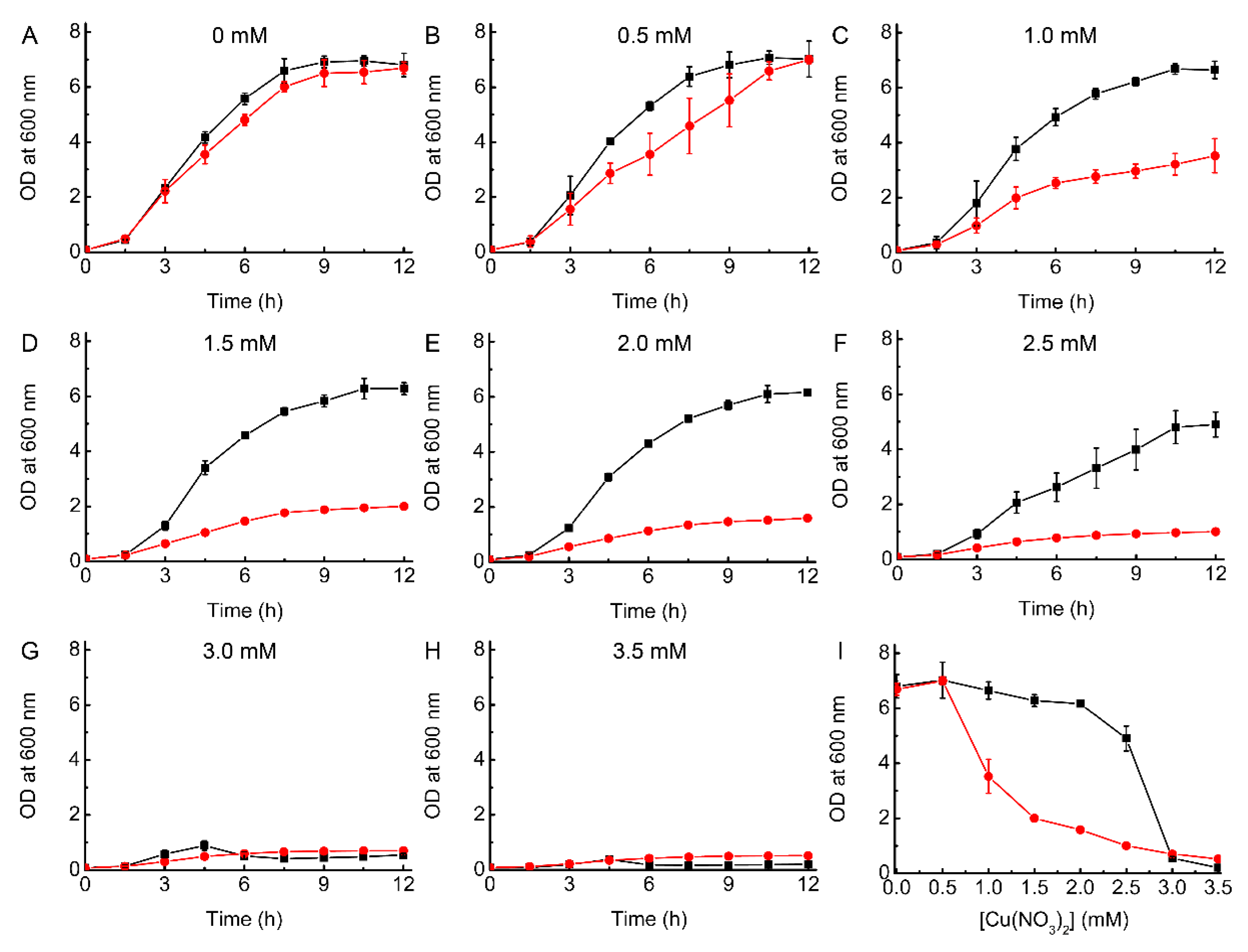
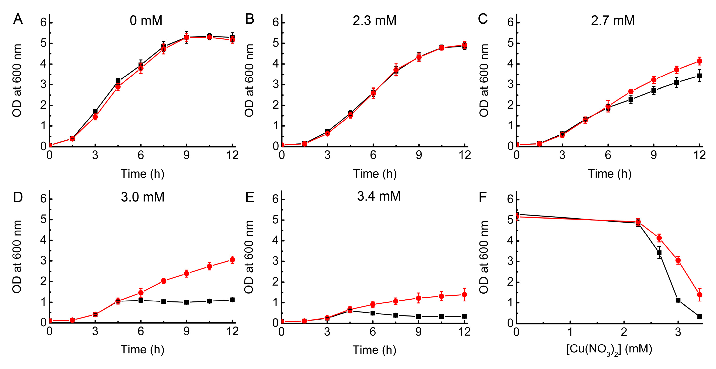
2.1. Growth of WT and ΔcopA E. coli as a Function of Cu Concentration

The addition of Cu has a limited effect on the growth of WT E. coli in LB media up to a concentration of 2.5 mM (Figure 1), with the OD at 12 h changing from ~6.8 without Cu to ~4.9. However, by 3.0 mM Cu there is a dramatic decrease in growth, and the OD after 12 h only reaches ~0.5 (Figure 1G). Previous studies, also in LB media, have found similar significant effects on WT E. coli growth at this Cu concentration [19,20], whilst in other work ~3 mM Cu had little influence and higher concentrations were needed to cause toxicity [18,40,41]. These differences are probably related to the actual Cu concentration in the media, and because of the large effect on growth over a narrow Cu concentration range. We have therefore ensured that stock Cu(II) solutions were carefully quantified prior to use (see Materials and Methods).
Removal of the Cu pump in ΔcopA E. coli increases Cu sensitivity (Figure 1), with 1.0 mM resulting in an OD at 12 h of ~3.5 compared to ~6.7 in the absence of added Cu (i.e., ~50% inhibition of growth). This effect is even greater when 1.5 to 2.5 mM Cu is present in the media, with the cells experiencing much more severe toxicity (OD values after 12 h of ~1.0 to ~2.0). However, growth is not completely inhibited until 3.0 mM Cu, as is also the case for WT E. coli. The data for ΔcopA E. coli is also relatively consistent with previous reports [18,19,40,41], although the detailed influence of Cu concentration on growth does vary (again probably due to differences in the actual Cu concentrations used). For example, in one case, significant growth is reported at 4 mM Cu [40], whilst in another, cells failed to grow at 2.0 mM [41]. Our data indicate that very high Cu concentrations can be tolerated to some extent by E. coli, even in the absence of CopA, potentially due to other available mechanisms to remove the metal from the cell such as the Cus system [21].

2.2. Complementation Studies of ΔcopA E. coli

The overexpression of CopA in ΔcopA E. coli induced by the addition of 0.2% l-arabinose results in similar growth at increasing Cu concentrations as for ΔcopA transformed with pBAD33 (plasmid) alone (Figure 2A and Figure S1), and also for this strain with no plasmid (Figure 1). However, if l-arabinose is not added complementation is achieved (Figure 2B,C, Figures S2 and S3), and ΔcopA E. coli transformed with pBAD33_copA grows in a similar manner to the WT strain beyond 2 mM Cu. Overall, this is consistent with previously reported data [18,40], when the caveat about variations in Cu concentrations between studies is considered. That complementation of ΔcopA E. coli does not require induction was noted previously [18], and suggested to be due to sufficient protein being produced under these conditions to remove Cu (in the absence of an inducer some expression of CopA is expected [42]). However, the inability to complement the ΔcopA strain in the presence of l-arabinose has not been reported. It appears that overexpression (with inducer) fails to produce a sufficient amount of an active form of this large and complex membrane protein [23].

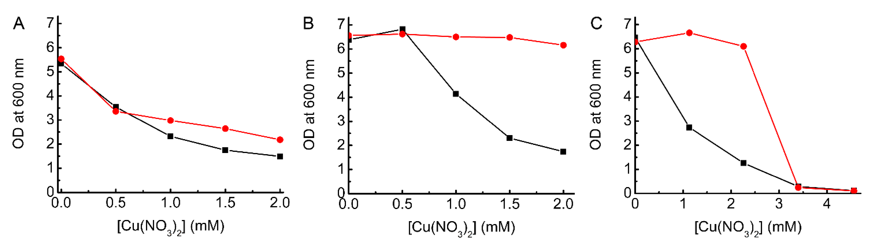
2.3. Testing the Ability of BsCsp3 to Confer Resistance to Cu Toxicity in E. coli

Cytosolic overexpression of BsCsp3 in E. coli using the same plasmid as for CopA enhances the growth of both the ΔcopA and WT strains in the presence of toxic levels of Cu. BsCsp3 improves the growth of ΔcopA E. coli at 0.5 to 1.5 mM Cu (Figure 3A and Figure S4, these growth curves have been shown previously [2], but have not been discussed in any detail), but not as effectively as in the complemented strain, particularly beyond 1 mM Cu (Figure 2B,C). Relatively high expression of BsCsp3 at 0.5 and 1.0 mM Cu (Figure S5 and Table S1) enables growth to continue beyond 4.5 h (Figure S4), which does not happen in plasmid-only controls, reaching ODs at 12 h of ~4.6 and ~4.4, respectively (Figure 3A). These are very similar to the OD obtained in the absence of added Cu (~4.3), whilst the value for the plasmid-only control remains at ~2.1 from 4.5–12 h at 1 mM Cu (i.e., BsCsp3 gives a ~2.1-fold improvement in growth at this Cu concentration). At 1.5 mM Cu, the overexpression of BsCsp3 is almost two-fold lower than at 1.0 mM in ΔcopA E. coli (Figure S5 and Table S1), yet the protein can still minimize toxicity with the OD at 12 h increasing from ~1.8 in plasmid-only controls to ~3.0 (1.7-fold enhanced growth).
The overexpression of BsCsp3 also provides a benefit to growth in WT E. coli when Cu becomes toxic, which occurs above 2.8 mM (Figure S6). The most significant advantage was found at 3.4 mM Cu (Figure 3B) with an OD after 12 h of ~2.3 compared to ~0.5 in plasmid-only controls. This is despite the low expression levels of BsCsp3 in the WT strain at this Cu concentration (Figure S7 and Table S2).
Cells producing BsCsp3 acquire more Cu than plasmid-only controls (Figure 4), and the influence of overexpression on preventing Cu-induced toxicity generally corresponds with increased intracellular Cu concentration. The largest difference in Cu accumulation (~2.2-fold) for ΔcopA E. coli is found at 1.5 mM added Cu (Figure 4A). In WT E. coli Cu levels are approximately two- to three-fold greater than plasmid-only controls (Figure 4B) in cells overexpressing BsCsp3 at non-toxic Cu concentrations (1.1 to 2.3 mM). At 3.4 mM Cu, which has a dramatic effect on cell growth (Figure 1 and Figure 3B), a similar increase in acquired Cu (~3-fold) is observed when expressing the protein (Figure 4B). For plasmid-only controls, the Cu concentrations are greater for ΔcopA E. coli than in the WT strain for cells grown in the presence of comparable amounts of Cu (Figure 4), consistent with limited data that is available in the literature (for example, see reference [43]).

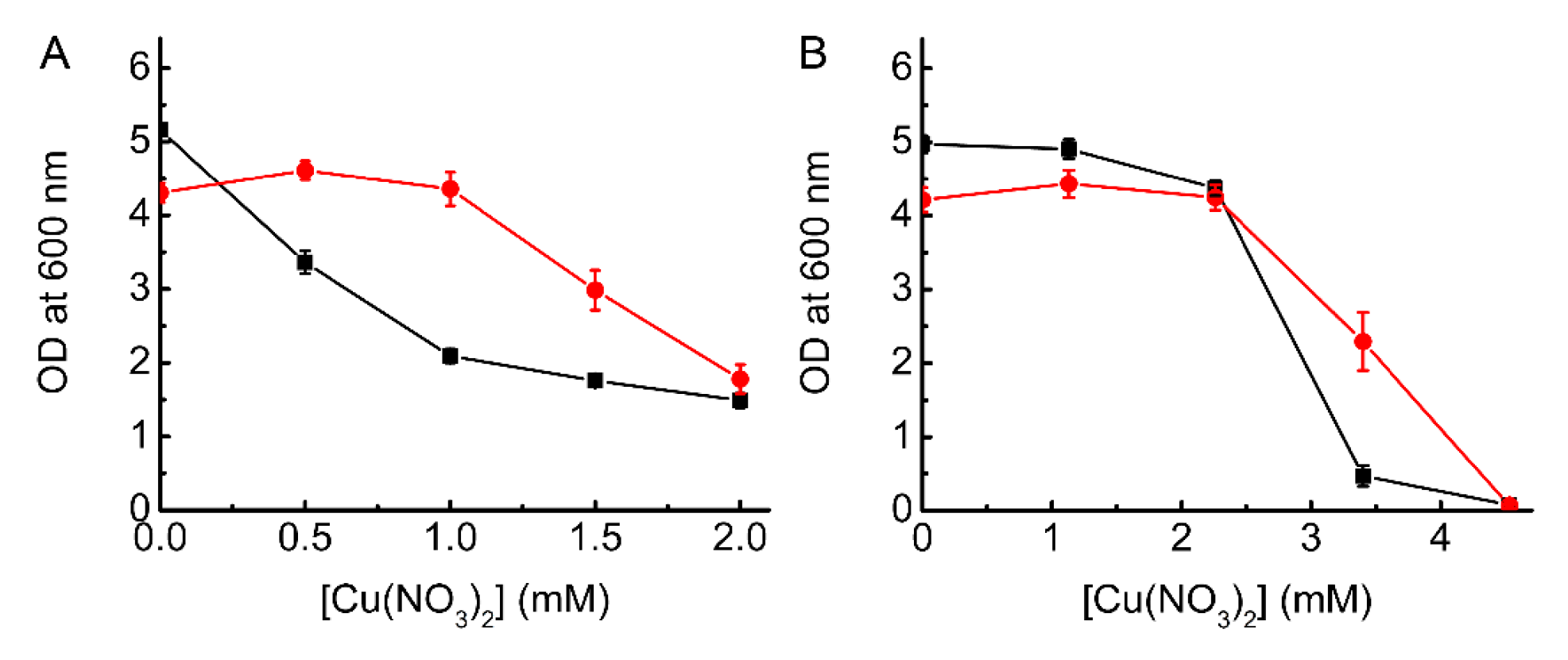
2.4. Testing the Ability of MtCsp3 to Confer Resistance to Cu Toxicity in E. coli

The overexpression of MtCsp3 (the Csp3 from M. trichosporium OB3b) does not benefit the growth of ΔcopA E. coli upon increasing Cu concentration (Figure S8). The levels of MtCsp3 produced in ΔcopA E. coli are lower than those of BsCsp3, and decrease more significantly as Cu is added (Figures S5 and S9 and Tables S1 and S3). However, MtCsp3 facilitates the growth of WT E. coli in a similar Cu concentration range as found for BsCsp3 (Figure 5), and is expressed at a comparable level (Figures S7 and S10 and Tables S2 and S4).

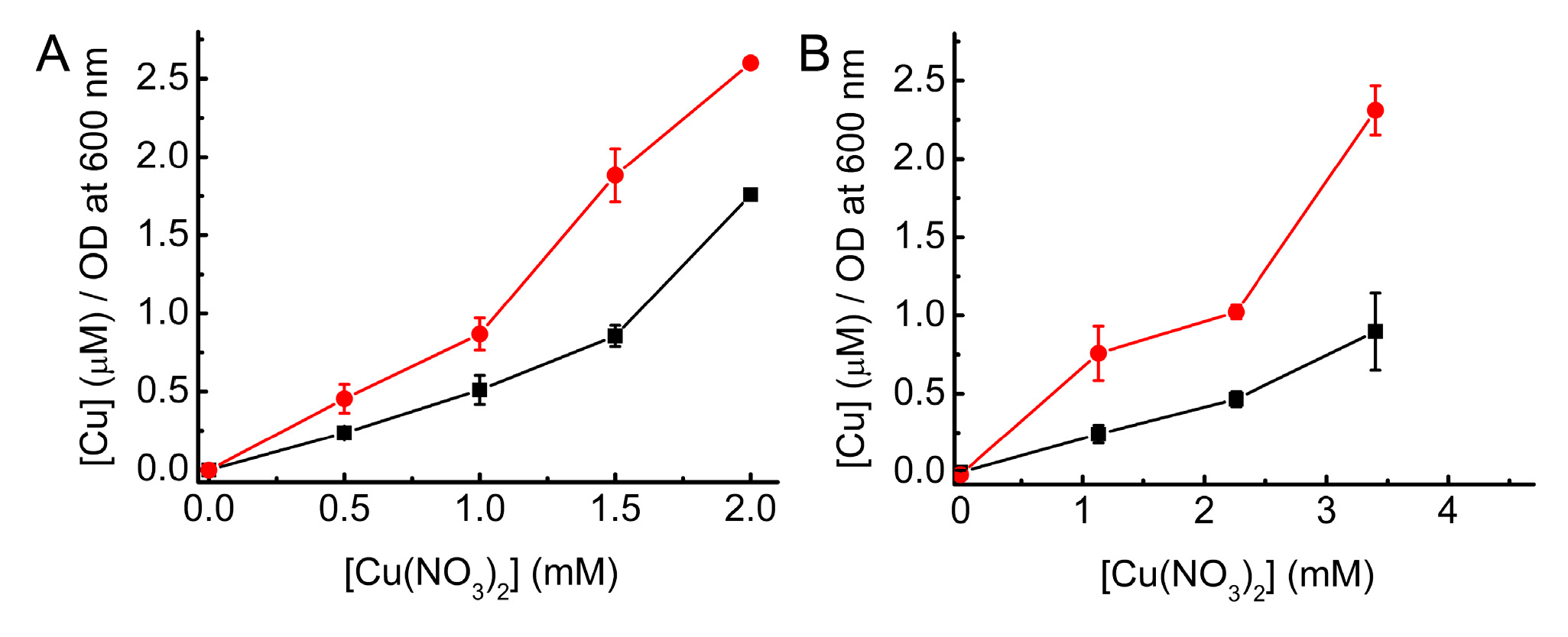
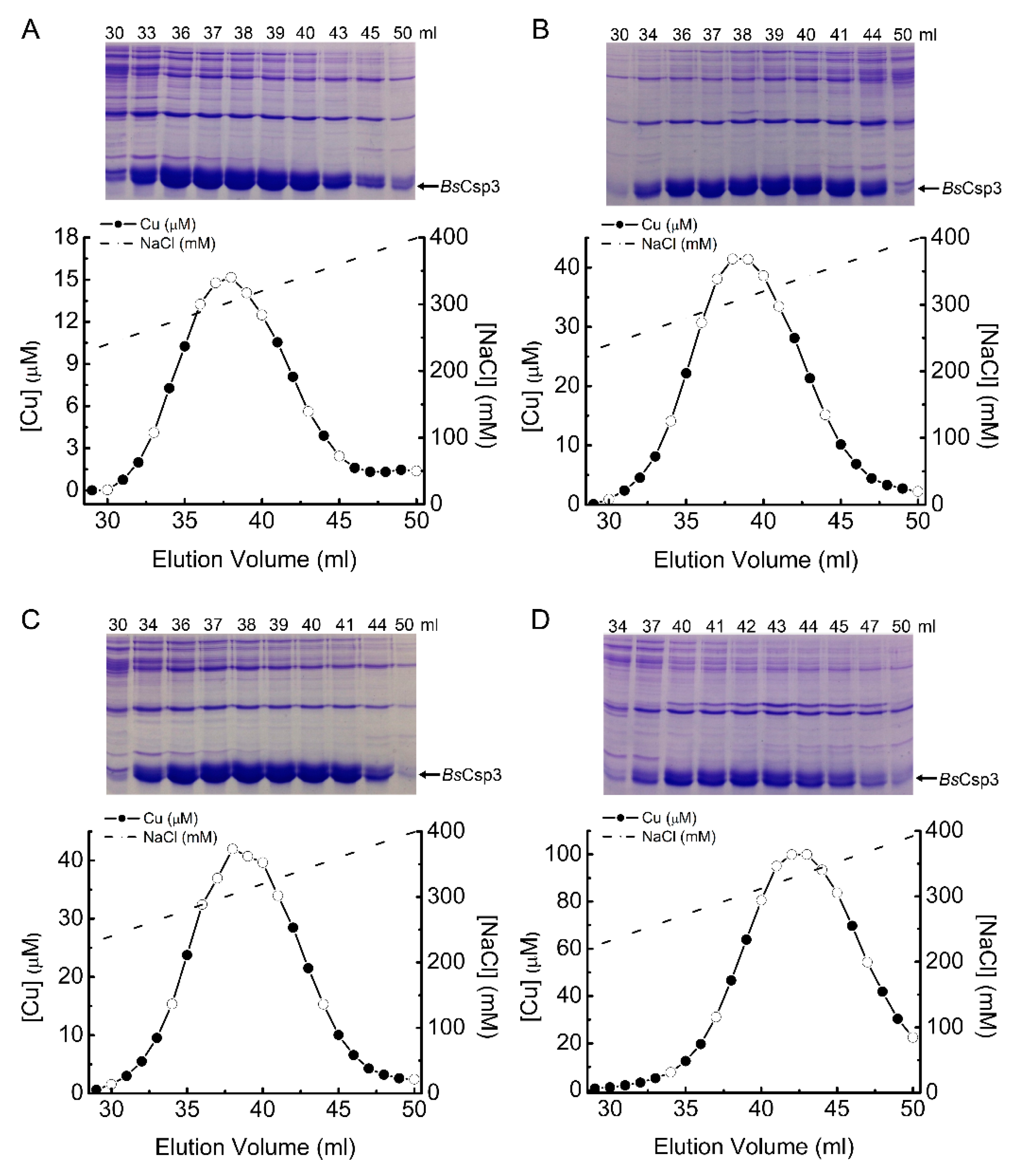
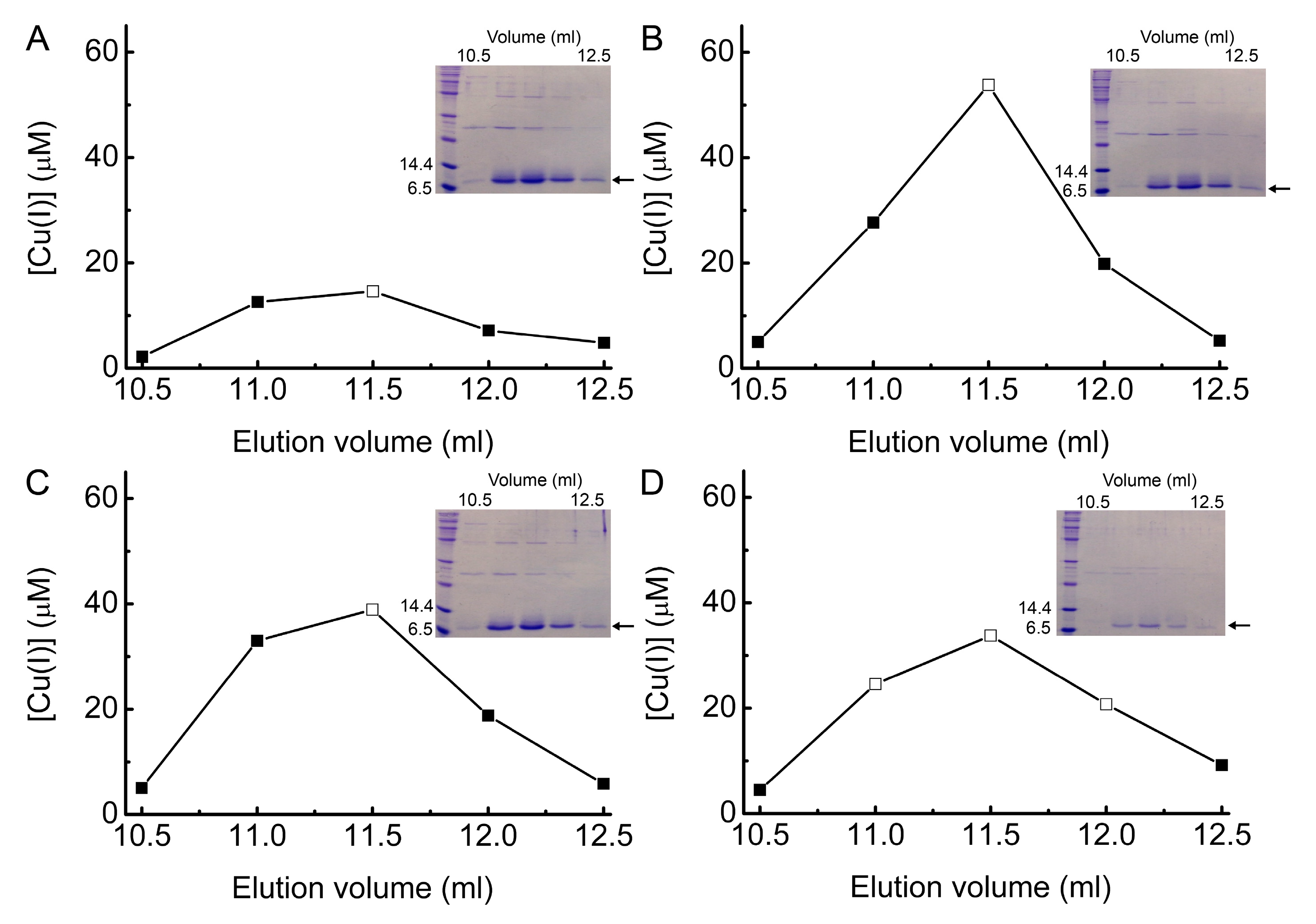
2.5. Investigating Intracellular Cu Binding by BsCsp3 in E. coli

Experiments were carried out to determine if the Cu accumulated by ΔcopA and WT E. coli is bound to BsCsp3. Cell-free extracts from the ΔcopA strain overexpressing the protein at 1.0 and 1.5 mM Cu (Figure 6A,B), and WT E. coli grown at 1.5 and 3.4 mM Cu (Figure 6C,D), were resolved by anion-exchange chromatography, and fractions analyzed for Cu by atomic absorption spectroscopy (AAS) and protein content by SDS-PAGE. In all cases, the Cu peak that elutes at ~270 to 350 mM NaCl matches the intensity profile of the BsCsp3 SDS-PAGE band, and the large amount of Cu in these fractions must be mainly bound to this protein.
Gel-filtration chromatography (Figure 7 and Figure S11) was used to further purify BsCsp3 present in anion-exchange fractions possessing the highest Cu concentrations from both ΔcopA and WT E. coli. In all cases, the variation in Cu(I) content of the fractions for the main peak that eluted from the gel-filtration column matches the intensity profile of the BsCsp3 band in SDS-PAGE analyses of these fractions (Figure 7 and Figure S11). Furthermore, this peak elutes at a very similar position to the purified BsCsp3 tetramer [2], particularly when the absorbance at 240 nm against elution volume plots are considered (Figure S12). Fractions possessing large amounts of Cu were concentrated, and their purity checked by SDS-PAGE (Figure S13). The protein and Cu(I) (Figure S14) concentrations of these samples were determined and are shown in Table 1 and Figure S5. Being more selective about which fractions are combined and analyzed has a significant influence on the measured Cu(I) occupancy for BsCsp3, but does not alter the relative amounts of Cu(I)-bound protein from the two E. coli strains grown at the two different Cu concentrations.

3. Discussion

There are two major outcomes form this study. Firstly, the overexpression of BsCsp3 allows E. coli to grow at Cu concentrations not normally possible. This is true for both WT E. coli (MtCsp3 also aids the growth of WT at similar Cu concentrations) and for the strain lacking the main Cu-efflux pump (ΔcopA E. coli). Secondly, in cells expressing BsCsp3, this protein can bind a significant amount of Cu(I). Taken together, these two observations suggest that the intracellular Csp3s can rescue cells from toxic concentrations of Cu(I) by sequestering excess cuprous ions and thereby preventing them from taking part in harmful interactions with cellular machinery. BsCsp3 can also acquire Cu(I) at concentrations that are not toxic in WT E. coli.
The large amount of BsCsp3 produced in ΔcopA E. coli at 1 mM Cu (Figure S5 and Table S1) can overcome the toxicity caused in its absence (Figure 3A and Figure S4), yet the purified protein binds only approximately one equivalent of Cu(I) (Table 1). MtCsp3 expression is lower in ΔcopA E. coli (Figure S9 and Table S3) and does not provide any protection against Cu toxicity (Figure S8). This is consistent with the sites that can cause toxicity in ΔcopA E. coli at 1 mM Cu having Cu(I) affinities tighter than those of Csp3s (average values of (1−2) × 1017 M−1 have been measured for Csp3s [2]), with the ability of BsCsp3 to acquire Cu(I) being driven by the large amount of protein present. The locations of all sites that can result in bacterial Cu toxicity remain to be determined, although Fe-S cluster-containing proteins are currently the main target identified [8,9,10,11,12,13], and these would be expected to be able to bind Cu(I) tightly. Some Cu(I) may reside in cytosolic pools bound by a highly-abundant species such as glutathione (GSH) that has been implicated in Cu(I) handling in E. coli [41], although a role in toxicity is currently unclear [8,9]. The abundance of GSH in E. coli [44,45], along with its affinity for Cu(I) [46], is consistent with a Csp3 being able to compete with it for cuprous ions.
At 1.5 mM Cu, BsCsp3 is still able to counteract toxicity in ΔcopA E. coli (Figure 3A), even though expression levels are almost two-fold lower than at 1 mM (Figure S5 and Table S1). There is a sizable increase in the amount of Cu/cell at 1.5 mM in this strain, both in the absence and presence of BsCsp3 (Figure 4A), and the purified protein can now bind up to approximately four equivalents of Cu(I) (Table 1). This increase in intracellular Cu(I) has resulted in a larger number of sites that cause toxicity, and possibly also increased levels of Cu(I) in cytosolic pools, which are more accessible to BsCsp3 (higher occupancy despite lower expression), and some of the additional sites must therefore have lower Cu(I) affinities. BsCsp3 appears to be able to remove Cu(I) from different cytosolic sites in ΔcopA E. coli promoting growth. At 2 mM the intracellular Cu concentration increases further in ΔcopA E. coli (Figure 4A), and the expression of BsCsp3 is significantly lower (Figure S5 and Table S1). As a result, the protein is no longer able to provide a growth advantage compared to plasmid-only controls (Figure 3A).
The overexpression of BsCsp3 in WT gives rise to lower Cu accumulation than in ΔcopA E. coli (Figure 4 and Table S6), but the levels are higher (~2–3-fold) compared to plasmid-only controls (Figure 4B). However, unlike the drastic effect on growth observed in ΔcopA E. coli, WT cells show no sign of experiencing toxicity in the 1.1 to 2.3 mM Cu concentration range (Figure 1 and Figure 3B). At 1.5 mM Cu, BsCsp3 expression levels in WT are double those in ΔcopA E. coli (Figures S5 and S7 and Tables S1 and S2), and the protein still acquires up to approximately four equivalents of Cu(I) (Table 1). Under these conditions, cytosolic Cu(I) is presumably being adequately handled by the efflux system (no obvious signs of toxicity). Thus, BsCsp3 must be acquiring Cu(I) from different locations in WT than in ΔcopA E. coli, with CopA being the most likely source.
In WT E. coli, overexpressing either BsCsp3 (Figure 3B) or MtCsp3 (Figure 5F) provides a benefit to growth in the relatively narrow Cu concentration window that causes toxicity, with the greatest advantage observed above approximately 2.5 mM. At these Cu concentrations, the WT strain (Figure 1), and plasmid-only control cells (Figure 3B and Figure 5), start to exhibit signs of Cu stress probably due to the efflux system becoming saturated. Although the expression levels of BsCsp3 and MtCsp3 are low at 3.4 mM Cu (Figures S7 and S10 and Tables S2 and S4), both proteins provide protection against Cu toxicity, enabling cells to reach an OD after 12 h that is approximately four- to five-fold higher than plasmid-only controls (Figure 3B and Figure 5).
BsCsp3 purified from WT E. coli grown at 3.4 mM Cu can be ~50% occupied (it proved difficult to purify MtCsp3, so the number of Cu(I) equivalents bound could not be determined). Saturation of the efflux system at this Cu concentration results in Cu(I) binding at sites causing toxicity. Despite their relatively low expression levels, Csp3s can acquire Cu(I) from these sites helping to prevent their harmful effects. The quantity of Cu(I) acquired by low amounts of BsCsp3 (Table 1 and Table S5) indicates that in WT E. coli under toxicity-causing conditions the protein obtains Cu(I) from an additional source, or sources, than at 1.5 mM Cu (no toxicity), and also to those in ΔcopA E. coli. This behavior would appear to be consistent with the proposed ability of Csp3s to act as a secondary system for buffering cellular Cu [36]. However, such functionality would be expected to require significant Cu-dependent upregulation, which is not observed in the majority of bacteria tested to date [7,35,37,38].
Another Cu(I)-binding four-helix bundle has been found to provide protection against Cu toxicity in E. coli [47]. This is not a naturally occurring protein, but was generated via a de novo design approach using a library of DNA sequences coding for proteins with this fold. The library was transformed into E. coli, with selection based on the ability to grow in an otherwise toxic Cu concentration. A protein, ConK, was found that can bind up to seven Cu(I) ions primarily via His residues, albeit with a relatively low (μM) affinity. The overexpression of ConK allowed WT E. coli (BW25113) to grow at up to 2.2 mM Cu (OD values were only measured after 20 h, and there are again uncertainties about the actual Cu concentrations used). A variant of ConK in which three of the His residues were mutated, two to Tyr and the other to Leu, enabled growth at 7 mM Cu. Overexpression of the WT protein also promoted growth at similar Cu concentrations in a strain lacking CopA and CueO, as well as the Cus [21] detoxification system. Unexpectedly, intracellular Cu levels were lowered in WT and mutant cells expressing ConK, and a mechanism that combats Cu toxicity by assisting in removal from the cytosol has been proposed [47]. How this is achieved is unknown, but could involve the transfer of Cu(I) to CopA in WT E. coli, which would be favored by the low affinity of ConK for Cu(I). The much tighter Cu(I) affinities of Csp3s will enable them to acquire Cu(I) from this efflux system.

4. Materials and Methods

4.1. Analysis of WT and the copA Deletion Strains of E. coli BW25113 and the Influence of BsCsp3 Overexpression

WT E. coli BW25113 (Coli Genetic Stock Centre, Yale University, New Haven, CT, USA, CGSC number 7636) and the strain with the copA gene inactivated through allelic replacement (herein called ΔcopA; CGSG number 8625) were obtained from the CGSC library [48]. The difference between these strains was verified by PCR with the following primers; 5′-CCGATTTTTTAATCTTTACGGAC-3′ and 5′-GCGTCTTATCAGGCCTACAAACCTG-3′ designed to hybridize 100 bp upstream and downstream of the copA gene, giving PCR fragments of 2754 and 1571 bp, respectively (Figure S15). The copA gene was amplified from genomic DNA using the primers 5′-GTCCCCGCCCATATGTCACAAACTATCGACC-3′ (forward, NdeI restriction site in bold) and 5′-GGTCGTGCCCCATGGTTATTCCTTCGGTTTAAACCG-3′ (reverse, NcoI restriction site in bold, stop codon underlined). The obtained fragment was digested and cloned into the NdeI and NcoI sites of pET29a, verified by sequencing, and sub-cloned into the XbaI and HindIII sites of pBAD33 to give pBAD33_copA. The Bscsp3 gene was sub-cloned from pET29a [2] into the XbaI and HindIII sites of pBAD33 to give pBAD33_Bscsp3.
Cultures were grown (agitation at 250 rpm) in liquid LB medium at 37 °C, and kanamycin (50 µg/mL, Formedium, Hunstanton, UK) and chloramphenicol (30 µg/mL, Merck KGaA, Darmstadt, Germany) were added as required. Overnight cultures were diluted 100-fold into LB and LB plus Cu(NO3)2 (0.5 to 4.6 mM) without antibiotics, either in the presence or absence of 0.2% l-arabinose, and the OD value at 600 nm was measured at regular intervals. For the addition of Cu, a 500 mM stock of Cu(II) nitrate trihydrate (Merck KGaA) was prepared in 100 mL of MilliQ water (Millipore Ltd., Watford, UK). After autoclaving, the Cu concentration was quantified by AAS using standards ranging from 0.2 to 1.8 ppm Cu in 2% nitric acid [49]. When cells were needed for Cu quantification and for analyzing protein expression by SDS-PAGE, ~35 and ~5 mL respectively, of a culture (50 mL in a 250 mL Erlenmeyer flask) grown for 12 h was collected by centrifugation at 5000× g for 10 min (4 °C) and frozen at −30 °C. Thawed cells for Cu quantification were washed twice with 20 mL of 20 mM Tris pH 8.0 plus 10 mM EDTA and 100 mM NaCl, and then with 20 mL of 20 mM Tris pH 8.0 plus 100 mM NaCl. The cells were digested in 200 μL of 65% nitric acid (Ultrapur, Merck KGaA) for up to three days at room temperature, centrifuged at 12,000× g for 10 min, diluted in MilliQ water to give a final nitric acid concentration of 2%, and analyzed for Cu by AAS. For SDS-PAGE analysis, thawed cells were resuspended in 3 mL of 20 mM Tris pH 8.5 and sonicated. Typically, 24 μL of this sample was taken to test for total protein expression, and the same volume was removed from the supernatant after centrifugation at 12,000× g for 10 min. The concentrations of BsCsp3 and MtCsp3 in images of SDS-PAGE gels were quantified with ImageJ using pure protein standards (18.6 μM for BsCsp3 and 15.0 μM for MtCsp3) that were run on the same gel.

4.2. Purification and Cu(I)-Binding Stoichiometry of BsCsp3

For the purification of BsCsp3, 500 mL cultures were grown in 2 L Erlenmeyer flasks and harvested after growth for 12 h, during which the OD was monitored (Figure S16 and Table S6), and the resulting pellets were stored at −30 °C. Thawed cells were washed twice with 20 mL of 20 mM Tris pH 8.0 plus 10 mM EDTA and 100 mM NaCl, and then with 20 mL of 20 mM Tris pH 8.0 plus 100 mM NaCl. Washed cells were resuspended in 20 mM Tris pH 8.0, sonicated, and centrifuged at 40,000× g for 30 min. The supernatant was diluted five-fold with 20 mM Tris pH 8.0 and loaded onto a HiTrap Q FF anion-exchange column (1 mL, GE Healthcare Life Sciences, Little Chalfont, UK) equilibrated in the same buffer. Proteins were eluted with a linear NaCl gradient (0 to 400 mM NaCl, total volume 50 mL). The BsCsp3-containing fractions were analyzed for Cu by AAS, and protein content by SDS-PAGE. Fractions containing high concentrations of Cu were further purified on a Superdex 75 10/300 GL column (GE Healthcare Life Sciences), equilibrated in 20 mM Hepes pH 7.5 plus 200 mM NaCl. The Cu(I) concentration in eluted 500 μL fractions was quantified under anaerobic conditions using the high affinity chromophoric Cu(I) ligand bathcuproine disulfonate (BCS, Merck KGaA) [1,2,49,50,51]. Oxygen-free solutions were prepared in an anaerobic chamber (Belle Technology, Weymouth, UK, [O2] << 2 ppm), and 50 μL of each fraction was added using a gastight syringe to a 1000 μL solution of BCS (~2.5 mM) in 20 mM Hepes pH 7.5 plus 200 mM NaCl in the presence of guanidine hydrochloride (final concentration ~6.5 M) in a sealed anaerobic cuvette. The formation of [Cu(BCS)2]3− was monitored by UV-Vis spectrophotometry at 483 nm (ε = 12,500 M−1cm−1), typically for 2 h [1,2,49,50,51]. The protein content of these fractions was analyzed by SDS-PAGE.
To determine the number of Cu(I) equivalents bound to BsCsp3, 500 μL fractions from the Superdex 75 column containing high concentrations of Cu(I)-BsCsp3 were analyzed. Fractions eluting around 11.5 mL were typically chosen, and for most conditions higher occupancies were obtained using a single fraction, although the inclusion of more fractions has less of an effect in the case of samples from WT E. coli grown at 3.4 mM Cu. Fractions were concentrated under anaerobic conditions to 150 μL, using a Vivaspin 500 centrifugal concentrator (10 kDa molecular weight cut-off, Sartorius AG., Göttingen, Germany). The protein concentration was determined with a Bradford assay corrected using a Bradford:DTNB ratio (the 5,5′-dithiobis(2-nitrobenzoic acid) (DTNB) assay for thiols has routinely been used to measure the concentration of Csps) of 1.31 for purified apo-BsCsp3 [1,2,49]. The Cu(I) concentration was determined by adding 50 μL (for some samples 12 μL was used as precipitation occurred with 50 μL) of the protein (0.46–4.81 μM) to BCS as described above.

5. Conclusions

The data obtained indicate that sites resulting in cellular toxicity with different Cu(I) affinities can be occupied in the cytosol of E. coli. Csp3s are able to compete with these, but their effectiveness can require high-level expression. The binding of Cu(I) at higher-affinity toxicity-causing sites (or bound to high-abundance species) requires greater concentrations of the apo-Csp3 to drive equilibria toward Cu(I)-Csp3. This observation, albeit in a heterologous system, provides important insight into Cu(I)-trafficking pathways. For example, as well as considering their affinities [52], the cellular concentrations of proteins need to be taken into account when assessing directional Cu (and other metal-ion) transfer within cells, as discussed previously [53]. Cytosolic Csp3s can acquire Cu(I) from the efflux system, as demonstrated by the isolation of Cu(I)-BsCsp3 from WT E. coli at a Cu concentration that has no effect on growth.
The highest Cu(I) occupancy is observed for BsCsp3 purified from WT E. coli experiencing Cu toxicity, and despite very low expression levels, a significant benefit to growth is provided. Under these conditions, whether Cu(I) is being acquired directly from an overloaded efflux system or from alternative sites that can cause toxicity, and have lower Cu(I) affinities to those occupied in ΔcopA E. coli, or both, is difficult to determine. The Cu(I) removal kinetics are slow for all Csp3s studied to date [2], which should help these proteins hold onto Cu(I) they acquire until it is needed. Cu(I) removal is much faster from Csp1s, which may bind cuprous ions in the cytosol prior to Tat export [1,6]. However, CueO from E. coli does not bind Cu(I) when overexpressed with its Tat signal peptide removed, including in LB plus 1.5 mM Cu [39]. The behavior of this Cu-enzyme is very different from that of a cytosolic Cu storage protein (Csp3), which we show can acquire considerable amounts of Cu(I) in this compartment of E. coli under very similar conditions. Our studies therefore also provide important information about the Cu(I)-binding capabilities of Csp3s within the cytosol of a bacterium.

Supplementary Materials

Supplementary materials can be found at https://www.mdpi.com/1422-0067/20/17/4144/s1.

Author Contributions

Conceptualization, J.L. and C.D.; methodology, J.L. and C.D.; formal analysis, J.L.; investigation, J.L.; data curation, J.L. and C.D.; writing—original draft preparation, C.D.; writing—review and editing, J.L. and C.D.; supervision, C.D.; project administration, C.D.; funding acquisition, C.D.

Funding

This research received no external funding.

Acknowledgments

We thank Newcastle University for an Overseas Research Scholarship (ORS) award to J.L. and a reviewer for help with the Discussion section of this paper.

Conflicts of Interest

The authors declare no conflict of interest.

References

  1. Vita, N.; Platsaki, S.; Baslé, A.; Allen, S.J.; Paterson, N.G.; Crombie, A.T.; Murrell, J.C.; Waldron, K.J.; Dennison, C. A four-helix bundle stores copper for methane oxidation. Nature 2015, 525, 140–143. [Google Scholar] [CrossRef] [PubMed] [Green Version]
  2. Vita, N.; Landolfi, G.; Baslé, A.; Platsaki, S.; Lee, J.; Waldron, K.J.; Dennison, C. Bacterial cytosolic proteins with a high capacity for Cu(I) that protect against copper toxicity. Sci. Rep. 2016, 6, 39065. [Google Scholar] [CrossRef] [PubMed] [Green Version]
  3. Baslé, A.; Platsaki, S.; Dennison, C. Visualizing biological copper storage: The importance of thiolate-coordinated tetranuclear clusters. Angew. Chem. Int. Ed. 2017, 56, 8697–8700. [Google Scholar] [CrossRef] [PubMed]
  4. Dennison, C.; David, S.; Lee, J. Bacterial copper storage proteins. J. Biol. Chem. 2018, 293, 4616–4627. [Google Scholar] [CrossRef] [PubMed] [Green Version]
  5. Dennison, C. The coordination chemistry of copper uptake and storage for methane oxidation. Chem. Eur. J. 2019, 25, 74–86. [Google Scholar] [CrossRef] [PubMed]
  6. Palmer, T.; Berks, B.C. The twin-arginine translocation (Tat) protein export pathway. Nat. Rev. Microbiol. 2012, 10, 483–496. [Google Scholar] [CrossRef] [PubMed]
  7. Gu, W.; Semrau, J.D. Copper and cerium-regulated gene expression in Methylosinus trichosporium OB3b. Appl. Microbiol. Biotechnol. 2017, 101, 8499–8516. [Google Scholar] [CrossRef] [PubMed]
  8. Dupont, C.L.; Grass, G.; Rensing, C. Copper toxicity and the origin of bacterial resistance-new insights and applications. Metallomics 2011, 3, 1109–1118. [Google Scholar] [CrossRef]
  9. Solioz, M. Copper toxicity. In Copper and Bacteria—Evolution, Homeostasis and Toxicity; Springer International Publishing: Cham, Switzerland, 2018. [Google Scholar]
  10. Macomber, L.; Imlay, J.A. The iron-sulfur clusters of dehydratases are primary intracellular targets of copper toxicity. Proc. Natl. Acad. Sci. USA 2009, 106, 8344–8349. [Google Scholar] [CrossRef] [Green Version]
  11. Fung, D.K.C.; Lau, W.Y.; Chan, W.T.; Yan, A. Copper efflux is induced during anaerobic amino acid limitation in Escherichia coli to protect iron-sulfur cluster enzymes and biogenesis. J. Bacteriol. 2013, 195, 4556–4568. [Google Scholar] [CrossRef]
  12. Chillappagari, S.; Seubert, A.; Trip, H.; Kruipers, O.P.; Marahiel, M.A.; Miethke, M. Copper stress affects iron homeostasis by destabilizing iron-sulfur cluster formation in Bacillus subtilis. J. Bacteriol. 2010, 192, 2512–2524. [Google Scholar] [CrossRef] [PubMed]
  13. Tan, G.; Cheng, Z.; Pang, Y.; Landry, A.P.; Li, J.; Ding, H. Copper binding in IscA inhibits iron-sulfur cluster assembly in Escherichia coli. Mol. Microbiol. 2014, 93, 629–644. [Google Scholar] [CrossRef] [PubMed]
  14. Changela, A.; Chen, K.; Xue, Y.; Holschen, J.; Outten, C.E.; O’Halloran, T.V.; Mondragón, A. Molecular basis of metal-ion selectivity and zeptomolar sensitivity by CueR. Science 2003, 301, 1383–1387. [Google Scholar] [CrossRef] [PubMed]
  15. Rensing, C.; McDevitt, S.F. The copper metallome in prokaryotic cells. Met. Ions Life Sci. 2013, 12, 417–450. [Google Scholar] [PubMed]
  16. Tottey, S.; Waldron, K.J.; Firbank, S.J.; Reale, B.; Bessant, C.; Sato, K.; Cheek, T.R.; Gray, J.; Banfield, M.J.; Dennison, C.; et al. Protein-folding location can regulate manganese-binding versus copper-or zinc-binding. Nature 2008, 455, 1138–1142. [Google Scholar] [CrossRef] [PubMed]
  17. Petersen, C.; Møller, L.B. Control of copper homeostasis in Escherichia coli by a P-type ATPase, CopA, and a MerR-like transcriptional activator, CopR. Gene 2000, 261, 289–298. [Google Scholar] [CrossRef]
  18. Rensing, C.; Fan, B.; Sharma, R.; Mitra, B.; Rosen, B.P. CopA: An Escherichia coli Cu(I)-translocating P-type ATPase. Proc. Natl. Acad. Sci. USA 2000, 97, 652–656. [Google Scholar] [CrossRef]
  19. Fan, B.; Grass, G.; Rensing, C.; Rosen, B.P. Escherichia coli CopA N-terminal Cys(X)2Cys motifs are not required for copper resistance or transport. Biochem. Biophys. Res. Commun. 2001, 286, 414–418. [Google Scholar] [CrossRef]
  20. Fan, B.; Rosen, B.P. Biochemical characterization of CopA, the Escherichia coli Cu(I)-translocating P-type ATPase. J. Biol. Chem. 2002, 277, 46987–46992. [Google Scholar] [CrossRef]
  21. Rensing, C.; Grass, G. Escherichia coli mechanisms of copper homeostasis in a changing environment. FEMS Microbiol. Rev. 2003, 27, 197–213. [Google Scholar] [CrossRef]
  22. Solioz, M.; Abicht, H.K.; Mermod, M.; Mancini, S. Response of gram-positive bacteria to copper stress. J. Biol. Inorg. Chem. 2010, 15, 3–14. [Google Scholar] [CrossRef] [PubMed]
  23. Gourdon, P.; Liu, X.; Skjørringe, T.; Morth, J.P.; Møller, L.B.; Pedersen, B.P.; Nissen, P. Crystal structure of a copper-transporting PIB-type ATPase. Nature 2011, 475, 59–64. [Google Scholar] [CrossRef] [PubMed]
  24. Argüello, J.M.; Raimunda, D.; Padilla-Benavides, T. Mechanisms of copper homeostasis in bacteria. Front. Cell. Infect. Microbiol. 2013, 3, 73. [Google Scholar] [CrossRef] [PubMed] [Green Version]
  25. Drees, S.L.; Beyer, D.F.; Lenders-Lomscher, C.; Lübben, M. Distinct functions of serial metal-binding domains in the Escherichia coli P1B-ATPase CopA. Mol. Microbiol. 2015, 97, 423–438. [Google Scholar] [CrossRef] [PubMed]
  26. Meydan, S.; Klepacki, D.; Karthikeyan, S.; Margus, T.; Thomas, P.; Jones, J.E.; Khan, Y.; Briggs, J.; Dinman, J.D.; Vázquez-Laslop, N.; et al. Programmed ribosomal frameshifting generates a copper transporter and a copper chaperone from the same gene. Mol. Cell 2017, 65, 207–219. [Google Scholar] [CrossRef] [PubMed]
  27. Cobine, P.; Wickramasinghe, W.A.; Harrison, M.D.; Weber, T.; Solioz, M.; Dameron, C.T. The Enterococcus hirae copper chaperone CopZ delivers copper(I) to the CopY repressor. FEBS Lett. 1999, 445, 27–30. [Google Scholar] [CrossRef]
  28. Multhaup, G.; Strausak, D.; Bissig, K.D.; Solioz, M. Interaction of the CopZ copper chaperone with the CopA copper ATPase of Enterococcus hirae assessed by surface plasmon resonance. Biochem. Biophys. Res. Commun. 2001, 288, 172–177. [Google Scholar] [CrossRef]
  29. Outten, F.W.; Outten, C.E.; Hale, J.A.; O’Halloran, T.V. Transcriptional activation of an Escherichia coli copper efflux regulon by the chromosomal MerR homologue, CueR. J. Biol. Chem. 2000, 275, 31024–31029. [Google Scholar] [CrossRef]
  30. Smaldone, G.T.; Helmann, J.D. CsoR regulates the copper efflux operon copZA in Bacillus subtilis. Microbiology 2007, 153, 4123–4128. [Google Scholar] [CrossRef]
  31. Liu, T.; Ramesh, A.; Ma, Z.; Ward, S.K.; Zhang, L.; George, G.N.; Talaat, A.M.; Sacchettini, J.C.; Giedroc, D.P. CsoR is a novel Mycobacterium tuberculosis copper-sensing transcriptional regulator. Nat. Chem. Biol. 2007, 3, 60–68. [Google Scholar] [CrossRef]
  32. Odermatt, A.; Solioz, M. Two trans-acting metalloregulatory proteins controlling expression of the copper-ATPases of Enterococcus hirae. J. Biol. Chem. 1995, 270, 4349–4354. [Google Scholar] [CrossRef] [PubMed]
  33. Strausak, D.; Solioz, M. CopY is a copper-inducible repressor of the Enterococcus hirae copper ATPases. J. Biol. Chem. 1997, 272, 8932–8986. [Google Scholar] [CrossRef] [PubMed]
  34. Singh, S.K.; Grass, G.; Rensing, C.; Montfort, W.R. Cuprous oxidase activity of CueO from Escherichia coli. J. Bacteriol. 2004, 186, 7815–7817. [Google Scholar] [CrossRef] [PubMed]
  35. Dwarakanath, S.; Chaplin, A.K.; Hough, M.A.; Rigali, S.; Vijgenboom, E.; Worrall, J.A. Response to copper stress in Streptomyces lividans extends beyond genes under direct control of a copper-sensitive operon repressor protein (CsoR). J. Biol. Chem. 2012, 287, 17833–17847. [Google Scholar] [CrossRef] [PubMed]
  36. Straw, M.L.; Chaplin, A.K.; Hough, M.A.; Paps, J.; Bavro, V.N.; Wilson, M.T.; Vijgenboom, E.; Worrall, J.A.R. A cytosolic copper storage protein provides a second level of copper tolerance in Streptomyces lividans. Metallomics 2018, 10, 180–193. [Google Scholar] [CrossRef]
  37. González-Quiñónez, N.; Corte-Rodríguez, M.; Álvarez-Fernández-García, R.; Rioseras, B.; López-García, M.T.; Fernández-García, G.; Montes-Bayón, M.; Manteca, A.; Yagüe, P. Cytosolic copper is a major modulator of germination, development and secondary metabolism in Streptomyces coelicolor. Sci. Rep. 2019, 9, 4214. [Google Scholar] [CrossRef] [PubMed]
  38. Quintana, J.; Novoa-Aponte, L.; Argüello, J.M. Copper homeostasis networks in the bacterium Pseudomonas aeruginosa. J. Biol. Chem. 2017, 292, 15691–15704. [Google Scholar] [CrossRef]
  39. Stolle, P.; Hou, B.; Brüser, T. The Tat substrate CueO is transported in an incomplete folding state. J. Biol. Chem. 2016, 291, 13520–13528. [Google Scholar] [CrossRef]
  40. González-Guerrero, M.; Raimunda, D.; Cheng, X.; Argüello, J.M. Distinct functional roles of homologous Cu+ efflux ATPases in Pseudomonas aeruginosa. Mol. Microbiol. 2010, 78, 1246–1258. [Google Scholar] [CrossRef]
  41. Helbig, K.; Bleuel, C.; Krauss, G.J.; Nies, D.H. Glutathione and transition-metal homeostasis in Escherichia coli. J. Bacteriol. 2008, 190, 5431–5438. [Google Scholar] [CrossRef]
  42. Bowers, L.M.; Lapoint, K.; Anthony, L.; Pluciennik, A.; Filutowicz, M. Bacterial expression system with tightly regulated gene expression and plasmid copy number. Gene 2004, 340, 11–18. [Google Scholar] [CrossRef] [PubMed]
  43. Young, C.A.; Gordon, L.D.; Fang, Z.; Holder, R.C.; Reid, S.D. Copper tolerance and characterization of a copper-responsive operon, copYAZ, in an M1T1 clinical strain of Streptococcus pyogenes. J. Bacteriol. 2015, 197, 2580–2592. [Google Scholar] [CrossRef] [PubMed]
  44. Smirnova, G.V.; Muzyka, N.G.; Glukhovchenko, N.; Oktyabrsky, O.N. Effects of menadione and hydrogen peroxide on glutathione status in growing Escherichia coli. Free Radic. Biol. Med. 2000, 28, 1009–1016. [Google Scholar] [CrossRef]
  45. Newton, G.L.; Rawat, M.; La Clair, J.J.; Jothivasan, V.K.; Budiarto, T.; Hamilton, C.J.; Claiborne, A.; Helmann, J.D.; Fahey, R.C. Bacillithiol is an antioxidant thiol produced in Bacilli. Nat. Chem. Biol. 2009, 5, 625–627. [Google Scholar] [CrossRef] [PubMed]
  46. Morgan, M.T.; Hguyen, L.A.H.; Hancock, H.L.; Fahrni, C.J. Glutathione limits aquacopper(I) to sub-femtomolar concentrations through cooperative assembly of a tetranuclear cluster. J. Biol. Chem. 2017, 292, 21558–21567. [Google Scholar] [CrossRef] [PubMed] [Green Version]
  47. Hoegler, K.J.; Hecht, M.H. A de novo protein confers copper resistance in Escherichia coli. Protein Sci. 2016, 25, 1249–1259. [Google Scholar] [CrossRef] [PubMed]
  48. Baba, T.; Ara, T.; Hasegawa, M.; Takai, Y.; Okumura, Y.; Baba, M.; Datsenko, K.A.; Tomita, M.; Wanner, B.L.; Mori, H. Construction of Escherichia coli K-12 in-frame, single-gene knockout mutants: The Keio collection. Mol. Syst. Biol. 2006, 2, 1–11. [Google Scholar] [CrossRef]
  49. Badarau, A.; Firbank, S.J.; McCarthy, A.A.; Banfield, M.J.; Dennison, C. Visualizing the metal-binding versatility of copper trafficking sites. Biochemistry 2010, 49, 7798–7810. [Google Scholar] [CrossRef] [PubMed]
  50. Badarau, A.; Dennison, C. Copper trafficking mechanism of CXXC-containing domains: Insight from the pH-dependence of their Cu(I) affinities. J. Am. Chem. Soc. 2011, 133, 2983–2988. [Google Scholar] [CrossRef] [PubMed]
  51. Allen, S.; Badarau, A.; Dennison, C. Cu(I) affinities of the domain 1 and 3 sites in the human metallochaperone for Cu, Zn-superoxide dismutase. Biochemistry 2012, 51, 1439–1448. [Google Scholar] [CrossRef]
  52. Banci, L.; Bertini, I.; Ciofi-Baffoni, S.; Kozyreva, T.; Zovo, K.; Palumaa, P. Affinity gradients drive copper to cellular destinations. Nature 2010, 465, 645–648. [Google Scholar] [CrossRef] [PubMed]
  53. Badarau, A.; Dennison, C. Thermodynamics of copper and zinc distribution in the cyanobacterium Synechocystis PCC 6803. Proc. Natl. Acad. Sci. USA 2011, 108, 13007–13012. [Google Scholar] [CrossRef] [PubMed]
Figure 1. Comparison of Cu resistance in WT and ΔcopA E. coli. Growth (37 °C) of WT (black squares) and ΔcopA (red circles) E. coli in LB media plus 0 (A), 0.5 (B), 1.0 (C), 1.5 (D), 2.0 (E), 2.5 (F), 3.0 (G), and 3.5 (H) mM Cu(NO3)2. Also shown (I) is a comparison of growth after 12 h. The OD values are averages from two (A), (B) and (G), and three (C) to (F), independent growth experiments (standard deviations are shown), whilst the experiment shown in (H) was performed once.
Ijms 20 04144 g001
Figure 2. Complementation studies of ΔcopA E. coli. Growth after 12 h (37 °C) of ΔcopA E. coli plus pBAD33_copA (red circles) and pBAD33 (black squares) in LB media in the presence (A) and absence (B,C) of 0.2% l-arabinose at increasing concentrations of added Cu(NO3)2 (all single replicates and the data shown in (C) were acquired over a larger Cu(NO3)2 concentration range). Growth curves are shown in Figures S1–S3.
Ijms 20 04144 g002
Figure 3. The influence of BsCsp3 overexpression on the growth of ΔcopA and WT E. coli in Cu. Growth (37 °C) after 12 h for ΔcopA (A) and WT (B) E. coli plus pBAD33_Bscsp3 (red circles) and pBAD33 (black squares) in LB media plus 0.2% l-arabinose in the presence of increasing concentrations of added Cu(NO3)2. Average OD values and standard deviations from three independent experiments are shown, and the growth curves are available in Figures S4 and S6, respectively.
Ijms 20 04144 g003
Figure 4. The influence of overexpressing BsCsp3 on Cu levels in E. coli. (A) The Cu concentration in ΔcopA E. coli plus pBAD33_Bscsp3 (red circles) compared to plasmid-only controls (black squares) grown for 12 h in 0, 0.5, 1.0, 1.5 and 2.0 mM Cu(NO3)2. (B) Results for WT E. coli plus pBAD33_Bscsp3 (red circles) and plasmid-only controls (black squares) grown for 12 h in 0, 1.1, 2.3, and 3.4 mM Cu(NO3)2 (Cu was not quantified in cells at 4.5 mM Cu(NO3)2 due to the lack of significant growth). Average values and standard deviations from three independent experiments are shown, apart from at 2.0 mM in (A) where the amount of Cu was measured once.
Ijms 20 04144 g004
Figure 5. The influence of MtCsp3 overexpression on the growth of WT E. coli in Cu. Growth (37 °C) of WT E. coli plus pBAD33_Mtcsp3 (red circles) and pBAD33 (black squares) in LB media plus 0.2% l-arabinose in the presence of 0 (A), 2.3 (B), 2.7 (C), 3.0 (D), and 3.4 (E) mM Cu(NO3)2. A comparison of growth after 12 h is given in (F). Average OD values and standard deviations from three independent experiments are shown.
Ijms 20 04144 g005
Figure 6. Comparison of Cu and protein contents in cell-free extracts of E. coli overexpressing BsCsp3. The quantification of Cu in fractions that elute between approximately 230–400 mM NaCl from a HiTrap Q anion-exchange column in 20 mM Tris pH 8.0 when BsCsp3 is purified from ΔcopA E. coli grown in 1.0 (A) and 1.5 (B) mM Cu(NO3)2, and from WT E. coli grown in 1.5 (C) and 3.4 (D) mM Cu(NO3)2 are shown. Those fractions whose Cu concentration is indicated with open circles were analyzed by SDS-PAGE, with the elution volume above the lane.
Ijms 20 04144 g006
Figure 7. Gel-filtration chromatography of BsCsp3-containing anion-exchange fractions. Plots of Cu(I) concentration against elution volume when the anion-exchange fractions that eluted at 37 and 39 mL respectively, from cell-free extracts of ΔcopA E. coli overexpressing BsCsp3 grown in 1.0 (A) and 1.5 (B) mM Cu(NO3)2 were analyzed by gel-filtration chromatography. Also shown are the gel-filtration analyses of the anion-exchange fractions eluting at 39 and 43 mL when overexpressing BsCsp3 in WT E. coli grown in 1.5 (C) and 3.4 (D) mM Cu(NO3)2, respectively. Insets show SDS-PAGE gels confirming the main protein component in these fractions is BsCsp3 (indicated by an arrow), and open squares identify which fractions were analyzed.
Ijms 20 04144 g007
Table 1. The number of Cu(I) equivalents bound by BsCsp3 from the two E. coli strains grown at different Cu concentrations 1.
E. coli Strain and Added Cu(NO3)2 Concentration[Cu(I)] (µM)[BsCsp3] (µM)[Cu(I)]/[BsCsp3] 2
ΔcopA in 1.0 mM Cu(NO3)250.945.71.1
ΔcopA in 1.5 mM Cu(NO3)216239.14.1
WT in 1.5 mM Cu(NO3)216638.74.3
WT in 3.4 mM Cu(NO3)218920.29.4 3
1 The values shown are the Cu(I) and protein concentrations for BsCsp3 purified by gel-filtration chromatography (Figure 7). 2 Lower Cu(I) occupancies, but with the same overall pattern in the two strains grown at the different Cu(NO3)2 concentrations as above, are obtained when more fractions eluting from the gel-filtration column are combined and concentrated for another anion-exchange fraction that was analyzed (see Figure S11 and Table S5). 3 The protein concentration is possibly overestimated due to the lower purity of this sample (see Figure S13A), and the Cu(I) occupancy of BsCsp3 could therefore be higher than the value quoted.

