Review

Peroxisomes and Innate Immunity: Antiviral Response and Beyond

Institute of Biomedicine (iBiMED) & Department of Medical Sciences, University of Aveiro, 3810-193 Aveiro, Portugal
*
Author to whom correspondence should be addressed.
Int. J. Mol. Sci. 2019, 20(15), 3795; https://doi.org/10.3390/ijms20153795
Submission received: 29 June 2019 / Revised: 31 July 2019 / Accepted: 1 August 2019 / Published: 3 August 2019
(This article belongs to the Special Issue Peroxisomes under the Spotlight: Collaboration is the Way to Go)

Abstract

:
Peroxisomes are ubiquitous organelles with well-defined functions in lipid and reactive oxygen species metabolism, having a significant impact on a large number of important diseases. Growing evidence points to them, in concert with mitochondria, as important players within the antiviral response. In this review we summarize and discuss the recent findings concerning the relevance of peroxisomes within innate immunity. We not only emphasize their importance as platforms for cellular antiviral signaling but also review the current information concerning their role in the control of bacterial infections. We furthermore review the recent data that pinpoints peroxisomes as regulators of inflammatory processes.

1. Introduction

Peroxisomes are dynamic, multifunctional, and ubiquitous organelles present in almost all eukaryotic cells [1]. They are bound by a single lipid membrane that surrounds a granular matrix, and their shape and size can alter in response to environmental stimuli [2,3,4]. Peroxisomes are crucial metabolic organelles that play important roles in lipid and reactive oxygen species (ROS) metabolism [5,6]. However, their functions are dependent on cell type, tissue, and organism. Peroxisomes interact functionally and morphologically with other organelles, such as the endoplasmic reticulum, mitochondria, or lipid droplets [7].
Peroxisomal dysfunctions have been linked to severe metabolic disorders in humans [4,8]. In recent years, however, it became clearer that peroxisomes also assume important nonmetabolic roles in diseases such as aging, cancer, or neurodegenerative disorders [8,9,10,11,12,13,14], as well as protective functions within the innate immune response [15,16,17].
The innate immune system is responsible for identifying threats and initiating a sequence of responses that allow the elimination of potentially infectious pathogens [18]. It involves the recognition of the pathogen by the infected cell and the production of chemical factors that lead to the recruitment of immune cells to the site of infection. It will ultimately activate the adaptive immune system and stimulate inflammation to promote healing and hamper the spread of infection [19].
In this review, we summarize and discuss the role of peroxisomes within innate immunity. We not only highlight their importance for the cellular antiviral response but also discuss different reports that demonstrate their role in the control of infections by other microbes. We furthermore review the role of peroxisomes as regulators of inflammatory processes.

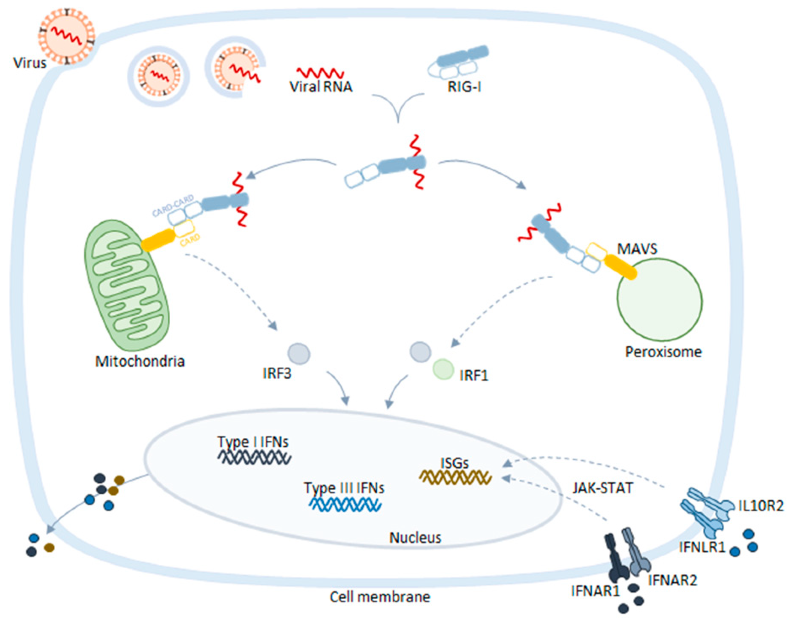
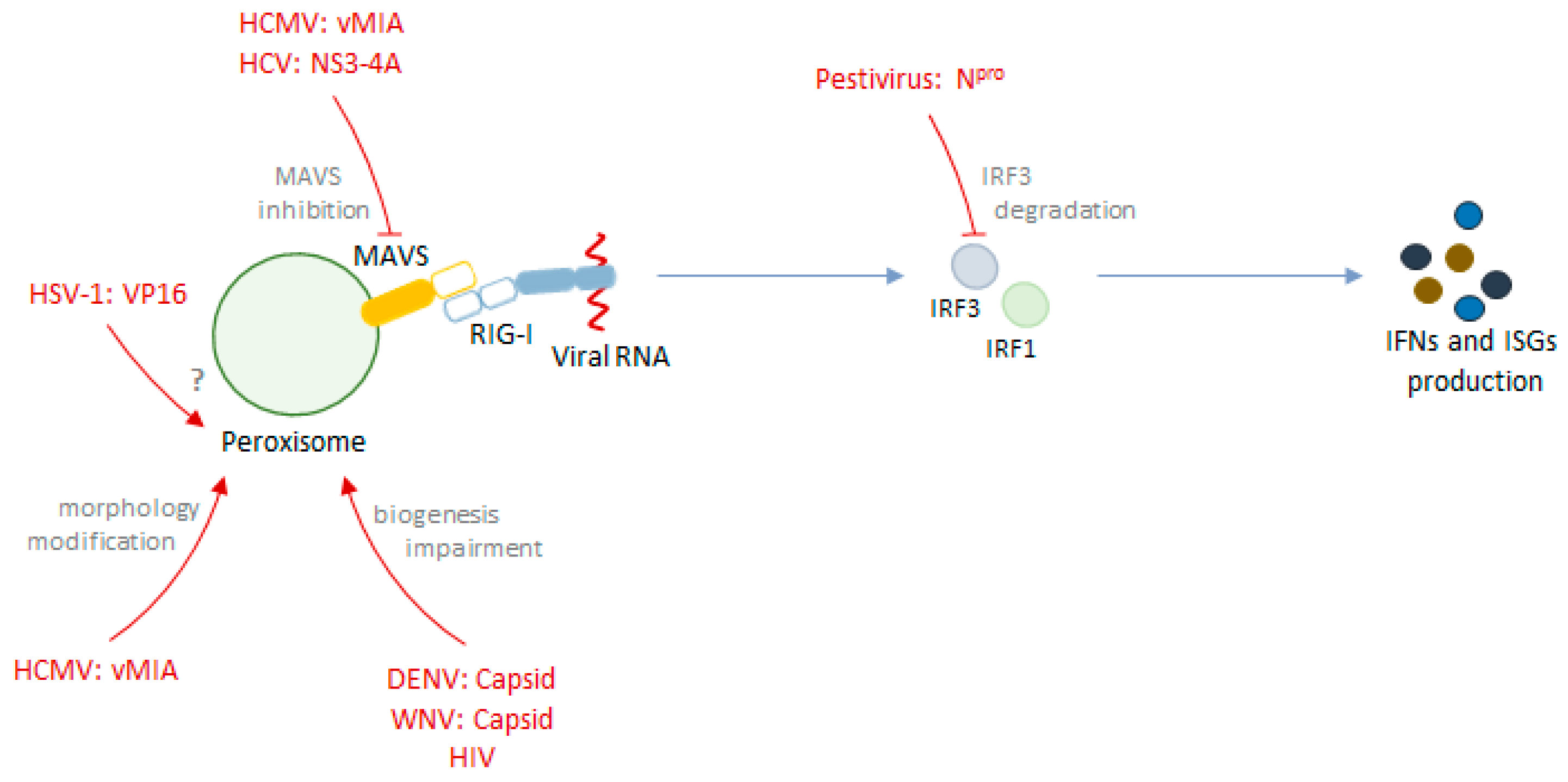
2. Peroxisomes as Platforms for Cellular Antiviral Responses

Peroxisomes harbor the essential adaptor transmembrane protein of the retinoic-inducible gene-I (RIG-I)-like receptors (RLR) signaling, the mitochondrial antiviral signaling protein (MAVS) [20,21] (Figure 1). Upon infection, viral RNA is released into the cytosol where it is sensed by the cytosolic receptors RIG-I and/or melanoma differentiation-associated gene-5 (MDA5) [22,23,24,25]. Upon activation, these receptors travel to peroxisomes, mitochondria, or mitochondria-associated membranes (MAMs) to activate MAVS [20,21,26,27,28,29,30], through interaction via their caspase activation and recruitment domains (CARDs). This interaction induces a conformational change on MAVS, leading to the formation of resistant prion fiber-like active aggregates [31] and the subsequent amplification of downstream signaling, culminating with the production of interferons (IFNs) and IFN-stimulated genes (ISGs) that function as direct antiviral effectors [32] (Figure 1).
Dixit et al. described key differences, concerning the signaling kinetics as well as the end products, between peroxisomal and mitochondrial antiviral signaling pathways. The authors observed that peroxisomal MAVS signaling induces a rapid, but transient, type I IFN-independent expression of ISGs, while mitochondrial MAVS signaling responds with a later type I IFN-dependent and long-lasting induction of defense factors, with autocrine and paracrine effects [20]. The authors, however, discuss that these kinetic differences may be cell specific, since they were not observed in macrophages [20]. Importantly, the cooperation between peroxisomal and mitochondrial MAVS seems to be essential for a potent induction of ISGs and type I IFNs expression. When analyzing the MAVS downstream signaling, the authors further demonstrated that, although tumor necrosis factor receptor-associated factor (TRAF) 3, TRAF6, and IFN regulatory factor 3 (IRF3) were required for the signaling from both organelles, IRF1 seems to be specifically activated by peroxisomal MAVS. Further details concerning the peroxisomal signaling pathway as well as the mechanisms that drive the specific activation of IRF1 are yet to be disclosed. Dixit et al. have also found that the previously described negative regulator of MAVS, nucleotide-binding oligomerization domain-like receptor X1 (NLRX1) that is exclusively located in the mitochondria [33], does not restrict the signal from peroxisomal MAVS [20]. In a subsequent study, the same group reported that the peroxisome-dependent pathway, in addition to the induction of ISGs production, also promotes the expression of type III IFNs [34], a class of IFNs that has tissue-specific roles in antiviral immunity [35]. Moreover, they demonstrated that type III IFNs can be stimulated by a diversity of viruses, and identified peroxisomes as the signaling platforms from which their expression is driven, complementing the type I IFNs solely induced upon mitochondrial signaling [34].
The MAVS-specific signaling from distinct organelles was more recently contested by another group [21]. They have reported that the activation of MAVS in either the peroxisomes or mitochondria induces the expression of both type I and type III IFNs, in similar levels. Moreover, they suggest that the absence of peroxisomes does not affect the capacity of cells to mount an effective antiviral response. These contradictory results may be due to the distinct experimental setups, cell lines, and methodologies used, but should certainly be clarified in the near future. Nevertheless, the fact that distinct viruses have developed specific strategies to target and evade the peroxisomal antiviral signaling (discussed in the next section of this review) certainly highlights the significance of this organelle in the context of the cellular antiviral immune response. Furthermore, the specific metabolic and morphological differences between peroxisomes and mitochondria are likely to be responsible for particular differences between these signaling mechanisms, such as distinct interactors of MAVS or adaptations to the different virus life cycles.

Viral Evasion of the Peroxisome-Dependent Antiviral Response

The important role of peroxisomes as signaling platforms in RLR antiviral immunity is supported by numerous studies that report the specific evasion of peroxisome-dependent signaling by different viruses. It has been demonstrated that the human cytomegalovirus (a virus with a slow replication cycle that has developed highly sophisticated immune evasion strategies [36]) specifically highjacks the transport machinery of the peroxisomal membrane protein in order to transport its own protein, viral mitochondrial-inhibitor of apoptosis (vMIA), to this organelle [37]. At peroxisomes, vMIA interacts with MAVS and inhibits peroxisome-dependent antiviral signaling. vMIA has previously been found to induce mitochondrial fragmentation and, consequently, inhibit mitochondria-dependent signaling [38]. Importantly, although peroxisomes also fragment in the presence of this protein, it was shown that this morphology change is not essential for vMIA’s inhibition of the signaling from this organelle [37].
Different groups have also demonstrated that the hepatitis C virus protein complex NS3-4A localizes at peroxisomes, cleaving MAVS at the organelle’s surface, and impairs the production of ISGs, as it had previously been shown for mitochondria and MAMs [21,28,30,39].
Dengue and West Nile viruses were also shown to impair peroxisome biogenesis and dampen the early innate immune signaling from peroxisomes, through PEX19 sequestration by their capsid proteins [40].
Herpes simplex virus 1 was also observed to evade the peroxisomal MAVS-dependent signaling through the viral protein VP16, via a mechanism that has not yet been unveiled [41].
Additionally, Npro from pestiviruses was reported to localize at peroxisomes, alongside with IRF3 and ubiquitin, inducing IRF3 degradation and inhibiting the downstream antiviral signaling [42].
Table 1 summarizes the above-mentioned strategies of evasion of the peroxisome-dependent antiviral response by different viruses.
Some other viruses have been described to interfere with the peroxisome-dependent antiviral signaling, although specific mechanisms of evasion have not yet been disclosed. Upon human immunodeficiency virus (HIV) infection, secondary structured HIV-derived RNA was detected at peroxisomes and induced IRF1 and IRF3 activation, as well as NF-кB, with apparently low expression of type I and III IFNs however [43]. Additionally, it was described that HIV infection upregulates miRNAs that target essential genes required for peroxisomal biogenesis. While MAVS was one of the targets of these miRNAs, no further studies to understand if HIV modulated the peroxisome-dependent antiviral signaling were performed. Curiously, the transfection of miRNAs that target PEX genes led to an increase of the mRNA levels of several innate immunity genes [44]. Hepatitis B virus was also described to induce NF-кB due to the targeting of its protein HBx to peroxisomes [45].
Figure 2 summarizes the above-mentioned mechanisms of interplay between different viruses and the peroxisome-dependent antiviral response.

3. Peroxisomes and the Antimicrobial Immune Response: Beyond Viral Restriction

While the role of peroxisomes in innate immunity gained more visibility with the discovery of the localization of MAVS at this organelle, peroxisomes had already been implicated in other innate immunity processes. In 1979, Eguchi et al. proposed that, during phagocytosis in rat peritoneal macrophages, peroxisomes relocated to regions juxtaposed to phagosomes in order to discharge catalase [46]. Catalase, a peroxisomal enzyme with bactericidal activity in the presence of hydrogen peroxide, had been previously identified in the phagocytic vesicle fraction of lysed alveolar macrophages [47]. Moreover, it has been shown that phagocytosis induction increases peroxisome numbers [46].
Later, a study with Drosophila and animal cells with impaired PEX5 and PEX7 revealed that peroxisomes are essential for the eradication of microbial infections. The impaired cells were incapable to react to microbial pathogens, presenting defects in immune signaling and reduced viability [48]. Both Drosophila and murine macrophages have shown compromised phagocytosis due to defects on actin organization, as well as lysosome formation and/or maturation, which was shown to be associated with the accumulation of ROS andreactive nitrogen species (RNS). Moreover, treatment of macrophages with peroxisome-derived lipids enhanced the capacity of macrophages to engulf bacteria [48]. Similarly, Facciotti et al. demonstrated that the same type of lipids is essential for the maturation of invariant natural killer T (NKT) cells in the thymus [49].
More recently, Di Cara et al., using Drosophila as an animal model, revealed that peroxisomes are essential platforms in the maintenance of enteric health and the functionality of the gut–microbe interface, efficiently coordinating different mechanisms such as stress, metabolic and immunity signaling pathways. Moreover, impaired metabolic signaling led to an increase of autophagy-induced epithelial cell death, and the reduced immune response led to a decrease in the reactivity to a subsequent immune challenge and early death [50].
Additionally, Odendall et al. have shown that, in the context of infection with Listeria monocytogenes (a bacteria that is signaled by the RIG-I/MAVS pathway and induces mitochondrial disruption [51,52]), peroxisomal MAVS has a dominant role in the coordination of an IFN response, since the expression of peroxisomal MAVS strongly potentiates the production of type I and type III IFNs in Jeg3 trophoblasts [34].
These results provide clear evidence that peroxisomes are not only essential for antiviral immunity but also for the elimination of other microbes such as bacteria.

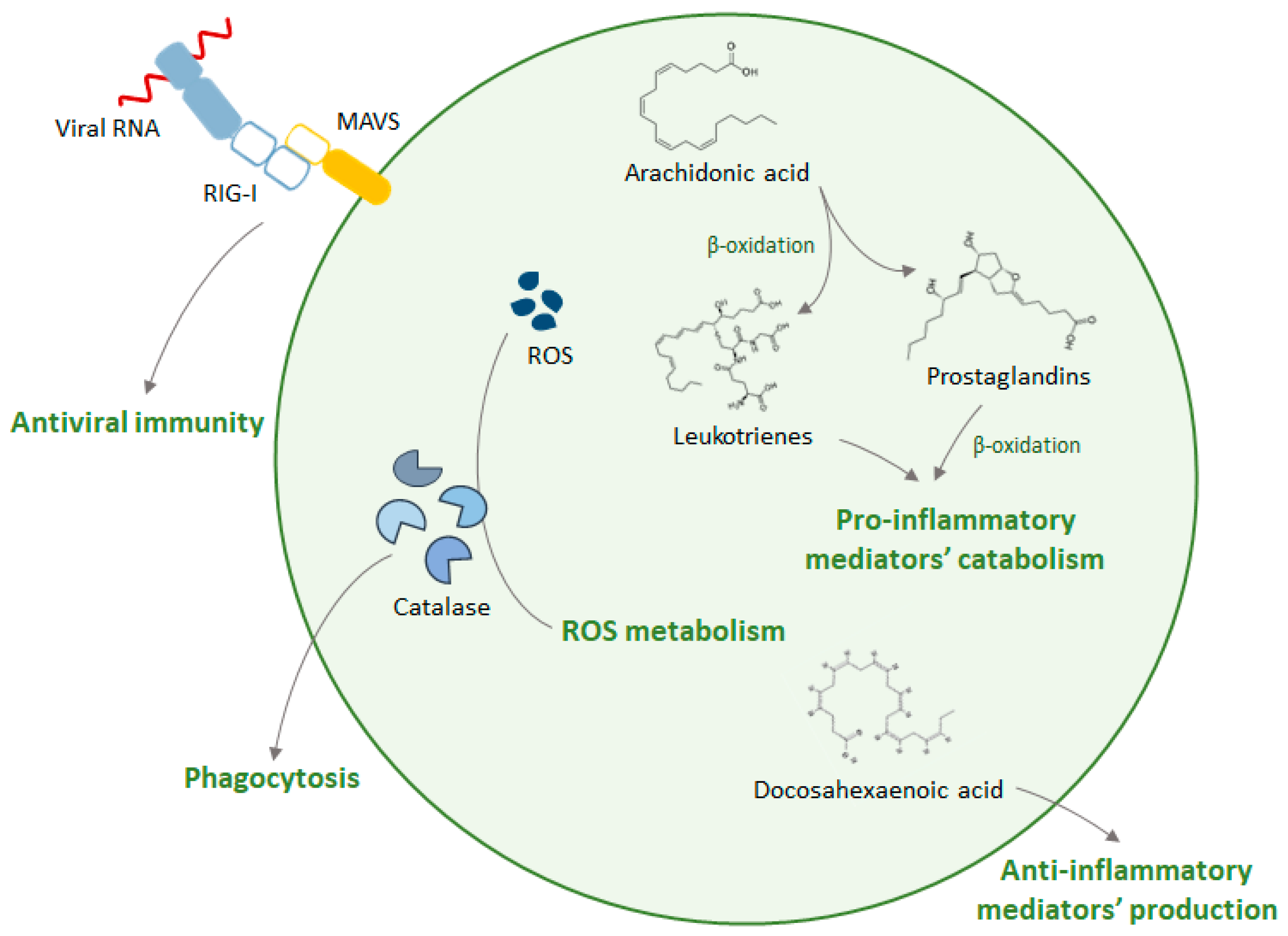
4. Peroxisomes and Inflammation

Inflammation comprises different mechanisms that allow the host to respond to infections and tissue damage, promoting pathogen destruction and wound healing. Upon infection, receptors of the innate immune system activate the production of a variety of proinflammatory mediators. These components, in turn, elicit a local inflammatory exudate, which consists of plasma proteins and leukocytes. At these sites, direct contact with pathogens or cytokines activates neutrophils, culminating with the release of toxic content of neutrophil granules, such as ROS and RNS, which have an unspecific targeting towards pathogens and the host (reviewed in [53]).
Peroxisomes’ role on the elimination of ROS and RNS species establishes a connection between this organelle and inflammation, since catalase and peroxiredoxins, besides neutralizing ROS generated during β-oxidation of lipids, are also essential for maintaining cellular redox homeostasis [17,54,55]. It was shown that tumor necrosis factor-alpha (TNF-α), a proinflammatory cytokine rapidly released upon infection or trauma [56], suppresses peroxisomal β-oxidation in rat hepatocytes and downregulates the expression of mRNAs encoding for peroxisomal proteins such as catalase and acyl-CoA oxidase [57,58].
Peroxisomes were also shown to metabolize leukotrienes and prostaglandins, important modulators of inflammation [59,60,61]. The inactivation of these proinflammatory lipids through β-oxidation produces metabolites that can act as resolution mediators of inflammation [17,61]. Supporting this, Vijayan et al. have also shown that the induction of peroxisomal proliferation in macrophages dampens lipopolysaccharide (LPS)-induced proinflammatory cytokines, while the disruption of their function has the opposite effect, leading to a hyper-induction of these cytokines. Furthermore, they suggest that the up-regulation of peroxisomal proliferation may serve as an auto-regulatory mechanism in macrophages, which renders protection against uncontrolled activation. With this, peroxisomes may act as late-phase inflammation suppressors at the post-translational level, to self-regulate inflammatory macrophages [62].
The loss of peroxisomal functions has also been associated with an intensification of the inflammatory response that can be explained by an accumulation of arachidonic acid metabolites, observed in different models of pathology [63]. For example, it was suggested that loss of peroxisomal β-oxidation from non-neural cells (e.g., microglia and/or infiltrating monocytes) worsens the inflammatory state of the brain [64,65].

5. Conclusions and Future Perspectives on the Role of Peroxisomes in Innate Immunity

Peroxisomes are no longer considered mere metabolic organelles, and they are now widely recognized as signaling hubs and protective organelles with significant physiological functions and impacts on many important human diseases. Their emergence as regulators of the innate immune response against viral infections has raised the interest in this organelle, and a growing body of evidence demonstrates that different viruses have developed specific mechanisms to counteract the peroxisome-dependent antiviral response. However, the specificities of this organelle’s dynamics that influence these immune responses needs to be further clarified. Moreover, it is also unknown whether inter-organelle interactions are involved in the establishment of antiviral signaling pathways. Finally, it remains unclear whether other antiviral signaling pathways (besides the RIG-I/MAVS network) may also operate from peroxisomes. Further studies may reveal peroxisome- dependent host mechanisms that can be exploited not only to the discovery of specific viral combat strategies but also to the potential development of broad-spectrum antiviral therapeutics.
As discussed above and summarized in Figure 3, the involvement of peroxisomes in innate immune mechanisms goes beyond the antiviral response, as they have also been shown to coordinate antimicrobial defenses against bacteria and act as anti-inflammatory platforms.
A further understanding of the mechanisms involved in the role of peroxisomes in innate immunity and inflammation may not only disclose new targets for antiviral and/or antibacterial therapy, but it may also prove beneficial for therapeutic interventions in chronic inflammatory disorders.

Funding

This work was supported by the Portuguese Foundation for Science and Technology (FCT): PTDC/BIA-CEL/31378/2017 (POCI-01-0145-FEDER-031378), CEECIND/03747/2017, SFRH/BD/137851/2018, UID/BIM/04501/2013, POCI-01-0145-FEDER-007628 under the scope of the Operational Program “Competitiveness and internationalization”, in its FEDER/FNR component.

Conflicts of Interest

The authors declare no conflict of interest.

References

  1. Islinger, M.; Voelkl, A.; Fahimi, H.D.; Schrader, M. The peroxisome: an update on mysteries 2.0. Histochem. Cell Biol. 2018, 150, 443–471. [Google Scholar] [CrossRef] [PubMed] [Green Version]
  2. Mast, F.D.; Fagarasanu, A.; Knoblach, B.; Rachubinski, R.A. Peroxisome Biogenesis: Something Old, Something New, Something Borrowed. Physiology 2010, 25, 347–356. [Google Scholar] [CrossRef] [PubMed] [Green Version]
  3. Smith, J.J.; Aitchison, J.D. Peroxisomes take shape. Nat. Rev. Mol. Cell Biol. 2013, 14, 803–817. [Google Scholar] [CrossRef] [PubMed] [Green Version]
  4. Ribeiro, D.; Castro, I.; Fahimi, H.D.; Schrader, M. Peroxisome morphology in pathology. Histol. Histopathol. 2012, 27, 661–676. [Google Scholar] [CrossRef] [PubMed]
  5. Schrader, M.; Costello, J.L.; Godinho, L.F.; Azadi, A.S.; Islinger, M. Proliferation and fission of peroxisomes—An update. Biochim. Biophys. Acta Mol. Cell Res. 2016, 1863, 971–983. [Google Scholar] [CrossRef] [PubMed]
  6. Lodhi, I.J.; Semenkovich, C.F. Peroxisomes: A Nexus for Lipid Metabolism and Cellular Signaling. Cell Metab. 2014, 19, 380–392. [Google Scholar] [CrossRef] [Green Version]
  7. Schrader, M.; Grille, S.; Fahimi, H.D.; Islinger, M. Peroxisome Interactions and Cross-Talk with Other Subcellular Compartments in Animal Cells. In Peroxisomes and their Key Role in Cellular Signaling and Metabolism; del Rio, L.A., Ed.; Springer Science & Business Media: Dordrecht, The Netherlands, 2013; pp. 1–23. [Google Scholar]
  8. Wanders, R.J.A.A. Metabolic functions of peroxisomes in health and disease. Biochimie 2014, 98, 36–44. [Google Scholar] [CrossRef] [PubMed]
  9. Cipolla, C.M.; Lodhi, I.J. Peroxisomal Dysfunction in Age-Related Diseases. Trends Endocrinol. Metab. 2017, 28, 297–308. [Google Scholar] [CrossRef] [Green Version]
  10. Dorninger, F.; Forss-Petter, S.; Berger, J. From peroxisomal disorders to common neurodegenerative diseases—The role of ether phospholipids in the nervous system. FEBS Lett. 2017, 591, 2761–2788. [Google Scholar] [CrossRef]
  11. Scherz-Shouval, R.; Elazar, Z. Regulation of autophagy by ROS: physiology and pathology. Trends Biochem. Sci. 2011, 36, 30–38. [Google Scholar] [CrossRef]
  12. Klouwer, F.C.C.; Berendse, K.; Ferdinandusse, S.; Wanders, R.J.A.; Engelen, M.; Poll-The, B.T. Zellweger spectrum disorders: clinical overview and management approach. Orphanet J. Rare Dis. 2015, 10, 151. [Google Scholar] [CrossRef]
  13. Valença, I.; Pértega-Gomes, N.; Vizcaino, J.R.; Henrique, R.M.; Lopes, C.; Baltazar, F.; Ribeiro, D. Localization of MCT2 at peroxisomes is associated with malignant transformation in prostate cancer. J. Cell. Mol. Med. 2015, 19, 723–733. [Google Scholar] [CrossRef] [PubMed]
  14. Dahabieh, M.S.; Di Pietro, E.; Jangal, M.; Goncalves, C.; Witcher, M.; Braverman, N.E.; del Rincón, S.V. Peroxisomes and cancer: The role of a metabolic specialist in a disease of aberrant metabolism. Biochim. Biophys. Acta Rev. Cancer 2018, 1870, 103–121. [Google Scholar] [CrossRef] [PubMed]
  15. Dixit, E.; Kagan, J.C. Intracellular Pathogen Detection by RIG-I-Like Receptors. In Advances in immunology; Elsevier Inc.: Cambridge, MA, USA, 2013; Volume 117, pp. 99–125. ISBN 9780124105249. [Google Scholar]
  16. Arciello, M.; Gori, M.; Balsano, C. Mitochondrial dysfunctions and altered metals homeostasis: new weapons to counteract HCV-related oxidative stress. Oxid. Med. Cell. Longev. 2013, 2013, 971024. [Google Scholar] [CrossRef] [PubMed]
  17. Terlecky, S.R.; Terlecky, L.J.; Giordano, C.R.; Vazquez-Carrera, M. Peroxisomes, oxidative stress, and inflammation. World J. Biol. Chem. 2012, 3, 93–97. [Google Scholar] [CrossRef] [PubMed]
  18. Janeway, C.A. Approaching the Asymptote? Evolution and Revolution in Immunology. Cold Spring Harb. Symp. Quant. Biol. 1989, 54, 1–13. [Google Scholar] [CrossRef]
  19. Palm, N.W.; Medzhitov, R. Pattern recognition receptors and control of adaptive immunity. Immunol. Rev. 2009, 227, 221–233. [Google Scholar] [CrossRef]
  20. Dixit, E.; Boulant, S.; Zhang, Y.; Lee, A.S.Y.; Odendall, C.; Shum, B.; Hacohen, N.; Chen, Z.J.; Whelan, S.P.; Fransen, M.; et al. Peroxisomes are signaling platforms for antiviral innate immunity. Cell 2010, 141, 668–681. [Google Scholar] [CrossRef]
  21. Bender, S.; Reuter, A.; Eberle, F.; Einhorn, E.; Binder, M.; Bartenschlager, R. Activation of Type I and III Interferon Response by Mitochondrial and Peroxisomal MAVS and Inhibition by Hepatitis C Virus. PLOS Pathog. 2015, 11, e1005264. [Google Scholar] [CrossRef]
  22. Ablasser, A.; Bauernfeind, F.; Hartmann, G.; Latz, E.; Fitzgerald, K.A.; Hornung, V. RIG-I-dependent sensing of poly(dA:dT) through the induction of an RNA polymerase III–transcribed RNA intermediate. Nat. Immunol. 2009, 10, 1065–1072. [Google Scholar] [CrossRef]
  23. Yoneyama, M.; Kikuchi, M.; Natsukawa, T.; Shinobu, N.; Imaizumi, T.; Miyagishi, M.; Taira, K.; Akira, S.; Fujita, T. The RNA helicase RIG-I has an essential function in double-stranded RNA-induced innate antiviral responses. Nat. Immunol. 2004, 5, 730–737. [Google Scholar] [CrossRef] [PubMed]
  24. Pichlmair, A.; Schulz, O.; Tan, C.-P.; Rehwinkel, J.; Kato, H.; Takeuchi, O.; Akira, S.; Way, M.; Schiavo, G.; Reis e Sousa, C. Activation of MDA5 requires higher-order RNA structures generated during virus infection. J. Virol. 2009, 83, 10761–10769. [Google Scholar] [CrossRef] [PubMed]
  25. Kato, H.; Takeuchi, O.; Mikamo-Satoh, E.; Hirai, R.; Kawai, T.; Matsushita, K.; Hiiragi, A.; Dermody, T.S.; Fujita, T.; Akira, S. Length-dependent recognition of double-stranded ribonucleic acids by retinoic acid-inducible gene-I and melanoma differentiation-associated gene 5. J. Exp. Med. 2008, 205, 1601–1610. [Google Scholar] [CrossRef] [PubMed]
  26. Seth, R.B.; Sun, L.; Ea, C.-K.; Chen, Z.J. Identification and Characterization of MAVS, a Mitochondrial Antiviral Signaling Protein that Activates NF-κB and IRF3. Cell 2005, 122, 669–682. [Google Scholar] [CrossRef] [PubMed]
  27. Xu, L.-G.; Wang, Y.-Y.; Han, K.-J.; Li, L.-Y.; Zhai, Z.; Shu, H.-B. VISA is an adapter protein required for virus-triggered IFN-beta signaling. Mol. Cell 2005, 19, 727–740. [Google Scholar] [CrossRef] [PubMed]
  28. Meylan, E.; Curran, J.; Hofmann, K.; Moradpour, D.; Binder, M.; Bartenschlager, R.; Tschopp, J. Cardif is an adaptor protein in the RIG-I antiviral pathway and is targeted by hepatitis C virus. Nature 2005, 437, 1167–1172. [Google Scholar] [CrossRef] [PubMed]
  29. Kawai, T.; Takahashi, K.; Sato, S.; Coban, C.; Kumar, H.; Kato, H.; Ishii, K.J.; Takeuchi, O.; Akira, S. IPS-1, an adaptor triggering RIG-I- and Mda5-mediated type I interferon induction. Nat. Immunol. 2005, 6, 981–988. [Google Scholar] [CrossRef]
  30. Horner, S.M.; Liu, H.M.; Park, H.S.; Briley, J.; Gale, M. Mitochondial-associated endoplasmic reticulum membranes (MAM) form innate immune synapses and are targeted by hepatitis C virus. Proc. Natl. Acad. Sci. USA 2011, 108, 14590–14595. [Google Scholar] [CrossRef]
  31. Hou, F.; Sun, L.; Zheng, H.; Skaug, B.; Jiang, Q.-X.; Chen, Z.J. MAVS forms functional prion-like aggregates to activate and propagate antiviral innate immune response. Cell 2011, 146, 448–461. [Google Scholar] [CrossRef]
  32. Kell, A.M.; Gale, M. RIG-I in RNA virus recognition. Virology 2015, 479, 110–121. [Google Scholar] [CrossRef]
  33. Moore, C.B.; Ting, J.P.Y.Y. Regulation of Mitochondrial Antiviral Signaling Pathways. Immunity 2008, 28, 735–739. [Google Scholar] [CrossRef] [PubMed] [Green Version]
  34. Odendall, C.; Dixit, E.; Stavru, F.; Bierne, H.; Franz, K.M.; Durbin, A.F.; Boulant, S.; Gehrke, L.; Cossart, P.; Kagan, J.C. Diverse intracellular pathogens activate type III interferon expression from peroxisomes. Nat. Immunol. 2014, 15, 717–726. [Google Scholar] [CrossRef] [PubMed]
  35. Donnelly, R.P.; Kotenko, S.V. Interferon-lambda: a new addition to an old family. J. Interferon Cytokine Res. 2010, 30, 555–564. [Google Scholar] [CrossRef] [PubMed]
  36. Marques, M.; Ferreira, A.R.; Ribeiro, D. The Interplay between Human Cytomegalovirus and Pathogen Recognition Receptor Signaling. Viruses 2018, 10, 514. [Google Scholar] [CrossRef] [PubMed]
  37. Magalhães, A.C.; Ferreira, A.R.; Gomes, S.; Vieira, M.; Gouveia, A.; Valença, I.; Islinger, M.; Nascimento, R.; Schrader, M.; Kagan, J.C.; et al. Peroxisomes are platforms for cytomegalovirus’ evasion from the cellular immune response. Sci. Rep. 2016, 6, 26028. [Google Scholar] [CrossRef] [PubMed]
  38. Castanier, C.; Garcin, D.; Vazquez, A.; Arnoult, D.; Ablasser, A.; Bauernfeind, F.; Hartmann, G.; Latz, E.; Fitzgerald, K.; Hornung, V.; et al. Mitochondrial dynamics regulate the RIG-I-like receptor antiviral pathway. EMBO Rep. 2010, 11, 133–138. [Google Scholar] [CrossRef]
  39. Ferreira, A.R.; Magalhães, A.C.; Camões, F.; Gouveia, A.; Vieira, M.; Kagan, J.C.; Ribeiro, D. Hepatitis C virus NS3-4A inhibits the peroxisomal MAVS-dependent antiviral signalling response. J. Cell. Mol. Med. 2016, 20, 750–757. [Google Scholar] [CrossRef]
  40. You, J.; Hou, S.; Malik-Soni, N.; Xu, Z.; Kumar, A.; Rachubinski, R.A.; Frappier, L.; Hobman, T.C. Flavivirus infection impairs peroxisome biogenesis and early anti-viral signaling. J. Virol. 2015, 89, 12349–12361. [Google Scholar] [CrossRef]
  41. Zheng, C.; Su, C. Herpes simplex virus 1 infection dampens the immediate early antiviral innate immunity signaling from peroxisomes by tegument protein VP16. Virol. J. 2017, 14, 1–8. [Google Scholar] [CrossRef] [Green Version]
  42. Jefferson, M.; Whelband, M.; Mohorianu, I.; Powell, P.P. The pestivirus N terminal protease Npro redistributes to mitochondria and peroxisomes suggesting new sites for regulation of IRF3 by Npro. PLoS ONE 2014, 9, e88838. [Google Scholar] [CrossRef]
  43. Berg, R.K.; Melchjorsen, J.; Rintahaka, J.; Diget, E.; Søby, S.; Horan, K.A.; Gorelick, R.J.; Matikainen, S.; Larsen, C.S.; Ostergaard, L.; et al. Genomic HIV RNA induces innate immune responses through RIG-I-dependent sensing of secondary-structured RNA. PLoS ONE 2012, 7, 1–10. [Google Scholar] [CrossRef] [PubMed]
  44. Xu, Z.; Asahchop, E.L.; Branton, W.G.; Gelman, B.B.; Power, C.; Hobman, T.C. MicroRNAs upregulated during HIV infection target peroxisome biogenesis factors: Implications for virus biology, disease mechanisms and neuropathology. PLoS Pathog. 2017, 13, e1006360. [Google Scholar] [CrossRef] [PubMed]
  45. Han, J.-M.M.; Kang, J.-A.A.; Han, M.-H.H.; Chung, K.-H.H.; Lee, C.-R.R.; Song, W.-K.K.; Jun, Y.; Park, S.-G.G. peroxisome-localized hepatitis Bx protein increases the invasion property of hepatocellular carcinoma cells. Arch. Virol. 2014, 159, 2549–2557. [Google Scholar] [CrossRef] [PubMed]
  46. Eguchi, M.; Sannes, P.L.; Spicer, S.S. Peroxisomes of rat peritoneal macrophages during phagocytosis. Am. J. Pathol. 1979, 95, 281–294. [Google Scholar] [PubMed]
  47. Stossel, T.P.; Mason, R.J.; Pollard, T.D.; Vaughan, M. Isolation and Properties of Phagocytic Vesicles II. ALVEOLAR MACROPHAGES. J. Clin. Investig. 1972, 51, 604–614. [Google Scholar] [CrossRef] [PubMed]
  48. Di Cara, F.; Sheshachalam, A.; Braverman, N.E.; Rachubinski, R.A.; Simmonds, A.J. Peroxisome-Mediated Metabolism Is Required for Immune Response to Microbial Infection. Immunity 2017, 47, 93–106. [Google Scholar] [CrossRef] [PubMed]
  49. Facciotti, F.; Ramanjaneyulu, G.S.; Lepore, M.; Sansano, S.; Cavallari, M.; Kistowska, M.; Forss-Petter, S.; Ni, G.; Colone, A.; Singhal, A.; et al. Peroxisome-derived lipids are self antigens that stimulate invariant natural killer T cells in the thymus. Nat. Immunol. 2012, 13, 474–480. [Google Scholar] [CrossRef]
  50. Di Cara, F.; Bülow, M.H.; Simmonds, A.J.; Rachubinski, R.A. Dysfunctional peroxisomes compromise gut structure and host defense by increased cell death and Tor-dependent autophagy. Mol. Biol. Cell 2018, 29, 2766–2783. [Google Scholar] [CrossRef]
  51. Hagmann, C.A.; Herzner, A.M.; Abdullah, Z.; Zillinger, T.; Jakobs, C.; Schuberth, C.; Coch, C.; Higgins, P.G.; Wisplinghoff, H.; Barchet, W.; et al. RIG-I Detects Triphosphorylated RNA of Listeria monocytogenes during Infection in Non-Immune Cells. PloS ONE 2013, 8, e62872. [Google Scholar] [CrossRef]
  52. Stavru, F.; Bouillaud, F.; Sartori, A.; Ricquier, D.; Cossart, P. Listeria monocytogenes transiently alters mitochondrial dynamics during infection. Proc. Natl. Acad. Sci. USA 2011, 108, 3612–3617. [Google Scholar] [CrossRef]
  53. Medzhitov, R. Origin and physiological roles of inflammation. Nature 2008, 454, 428–435. [Google Scholar] [CrossRef] [PubMed]
  54. Sandalio, L.M.; Rodríguez-Serrano, M.; Romero-Puertas, M.C.; del Río, L.A. Role of Peroxisomes as a Source of Reactive Oxygen Species (ROS) Signaling Molecules. In Peroxisomes and their Key Role in Cellular Signaling and Metabolism; del Rio, L.A., Ed.; Springer Science & Business Media: Dordrecht, The Netherlands, 2013; pp. 231–255. [Google Scholar]
  55. Fransen, M.; Nordgren, M.; Wang, B.; Apanasets, O. Role of peroxisomes in ROS/RNS-metabolism: Implications for human disease. Biochim. Biophys. Acta Mol. Basis Dis. 2012, 1822, 1363–1373. [Google Scholar] [CrossRef]
  56. Parameswaran, N.; Patial, S. Tumor necrosis factor-α signaling in macrophages. Crit. Rev. Eukaryot. Gene Expr. 2010, 20, 87–103. [Google Scholar] [CrossRef] [PubMed]
  57. Beier, K.; Völkl, A.; Fahimi, H.D. Suppression of peroxisomal lipid β-oxidation enzymes by TNF-α. FEBS Lett. 1992, 310, 273–276. [Google Scholar] [CrossRef]
  58. Beier, K.; Völkl, A.; Fahimi, H.D. TNF-α downregulates the peroxisome proliferator activated receptor-α and the mRNAs encoding peroxisomal proteins in rat liver. FEBS Lett. 1997, 412, 385–387. [Google Scholar] [CrossRef]
  59. Jedlitschky, G.; Mayatepek, E.; Keppler, D. Peroxisomal leukotriene degradation: biochemical and clinical implications. Adv. Enzyme Regul. 1993, 33, 181–194. [Google Scholar] [CrossRef]
  60. Diczfalusy, U.; Kase, B.F.; Alexson, S.E.; Björkhem, I. Metabolism of prostaglandin F2 alpha in Zellweger syndrome. Peroxisomal beta-oxidation is a major importance for in vivo degradation of prostaglandins in humans. J. Clin. Investig. 1991, 88, 978–984. [Google Scholar] [CrossRef]
  61. Wanders, R.J.A. Peroxisomes in human health and disease: metabolic pathways, metabolite transport, interplay with other organelles and signal transduction. In Peroxisomes and their Key Role in Cellular Signaling and Metabolism; del Río, L.A., Ed.; Springer Science & Business Media: Dordrecht, The Netherlands, 2013; pp. 23–44. [Google Scholar]
  62. Vijayan, V.; Srinu, T.; Karnati, S.; Garikapati, V.; Linke, M.; Kamalyan, L.; Mali, S.R.; Sudan, K.; Kollas, A.; Schmid, T.; et al. A New Immunomodulatory Role for Peroxisomes in Macrophages Activated by the TLR4 Ligand Lipopolysaccharide. J. Immunol. 2017, 198, 2414–2425. [Google Scholar] [CrossRef] [Green Version]
  63. Schrader, M.; Fahimi, H.D. Peroxisomes and oxidative stress. Biochim. Biophys. Acta 2006, 1763, 1755–1766. [Google Scholar] [CrossRef] [Green Version]
  64. Bottelbergs, A.; Verheijden, S.; Van Veldhoven, P.P.; Just, W.; Devos, R.; Baes, M. Peroxisome deficiency but not the defect in ether lipid synthesis causes activation of the innate immune system and axonal loss in the central nervous system. J. Neuroinflamm. 2012, 9, 61. [Google Scholar] [CrossRef]
  65. Verheijden, S.; Beckers, L.; Casazza, A.; Butovsky, O.; Mazzone, M.; Baes, M. Identification of a chronic non-neurodegenerative microglia activation state in a mouse model of peroxisomal β-oxidation deficiency. Glia 2015, 63, 1606–1620. [Google Scholar] [CrossRef] [PubMed]
Figure 1. Mitochondrial antiviral signaling protein (MAVS)-dependent antiviral signaling pathway. Upon infection, viral RNA is released into the cytosol where it is sensed by retinoic-inducible gene-I (RIG-I) and/or melanoma differentiation-associated gene-5 (MDA5). These receptors travel to peroxisomes and mitochondria to activate MAVS, inducing a downstream signaling cascade that culminates with the production of type I interferons (IFNs), type III IFNs, and IFN-stimulated genes (ISGs). Once secreted, IFNs bind to specific receptors on the cell surface, activating the janus kinase/signal transducers and activators of transcription (JAK-STAT) pathway and generating an amplifying loop that results in the accumulation of different classes of ISGs. The conjugation of these responses leads to the restriction of viral replication and spreading to neighboring cells. IFNAR—interferon alfa/beta receptor complex; IFNLR—interferon lambda receptor complex; and IL10R2—interleukin-10 receptor 2.
Ijms 20 03795 g001
Figure 2. Schematic representation of the interplay between different viruses and the peroxisome-dependent antiviral signaling.
Ijms 20 03795 g002
Figure 3. Schematic representation of the peroxisomal functions within innate immunity. Peroxisomes play an important role in antiviral defense through the RIG-I/MAVS-dependent signaling. Additionally, peroxisomes function as anti-inflammatory platforms as they metabolize and produce, respectively, proinflammatory and anti-inflammatory mediators. Moreover, peroxisomes discharge catalase into phagosomes, which is essential for reactive oxygen species (ROS) metabolism during inflammation.
Ijms 20 03795 g003
Table 1. Viral evasion strategies that target peroxisome-dependent antiviral signaling.
VirusViral ProteinMechanismCell TypeRef.
Human cytomegalovirusviral mitochondrial-inhibitor of apoptosis (vMIA)Interaction with MAVSMEFs[37]
Hepatitis C virusNS3-4ACleavage of MAVSMEFs, Huh7, A549, HEK293T[21,39]
Herpes simplex virus 1VP16UnknownHEK293, MEFs, HEK293T[41]
Dengue virus and West Nile virusCapsidPeroxisome biogenesis impairmentA549, HEK293T[40]
PestivirusesNproInduction of IRF3 degradationMEFs[42]

