Review

Cardiac Insulin Resistance in Heart Failure: The Role of Mitochondrial Dynamics

Internal Medicine III, Hamamatsu University School of Medicine, 1-20-1 Handayama, Higashi-ku, Hamamatsu 431-3192, Japan
*
Author to whom correspondence should be addressed.
Int. J. Mol. Sci. 2019, 20(14), 3552; https://doi.org/10.3390/ijms20143552
Submission received: 31 May 2019 / Revised: 12 July 2019 / Accepted: 18 July 2019 / Published: 20 July 2019

Abstract

:
Heart failure (HF) frequently coexists with conditions associated with glucose insufficiency, such as insulin resistance and type 2 diabetes mellitus (T2DM), and patients with T2DM have a significantly high incidence of HF. These two closely related diseases cannot be separated on the basis of their treatment. Some antidiabetic drugs failed to improve cardiac outcomes in T2DM patients, despite lowering glucose levels sufficiently. This may be, at least in part, due to a lack of understanding of cardiac insulin resistance. Basic investigations have revealed the significant contribution of cardiac insulin resistance to the pathogenesis and progression of HF; however, there is no clinical evidence of the definition or treatment of cardiac insulin resistance. Mitochondrial dynamics play an important role in cardiac insulin resistance and HF because they maintain cellular homeostasis through energy production, cell survival, and cell proliferation. The innovation of diagnostic tools and/or treatment targeting mitochondrial dynamics is assumed to improve not only the insulin sensitivity of the myocardium and cardiac metabolism, but also the cardiac contraction function. In this review, we summarized the current knowledge on the correlation between cardiac insulin resistance and progression of HF, and discussed the role of mitochondrial dynamics on the pathogenesis of cardiac insulin resistance and HF. We further discuss the possibility of mitochondria-targeted intervention to improve cardiac metabolism and HF.

1. Introduction

Heart failure (HF) is a condition in which the myocardium is unable to meet the whole body’s demand for blood and oxygen and is caused by complex and multiple factors. Because of its high morbidity and mortality, HF is becoming one of the most critical health hazards around the world. An estimated global prevalence of HF may exceed 26 million, and the number of new patients with HF is expanding [1]. To minimize the increasing medical costs among patients with HF, further efforts are required to shorten the hospitalization period and prevent HF recurrence. For that purpose, more effective medicines and/or treatments toward HF are required to innovate both bench and bedside research.
HF frequently coexists with conditions associated with glucose insufficiency, such as insulin resistance or type 2 diabetes mellitus (T2DM), and T2DM patients have a higher incidence of HF than normal subjects. However, the intensive glucose lowering therapy cannot always improve the prognosis of T2DM patients [2], and some antidiabetic drugs failed to improve cardiac outcomes in T2DM patients, despite sufficiently lowering glucose levels [2]. This may be due to a lack of understanding and/or knowledge of cardiac insulin resistance. Although cardiac insulin resistance is considered to develop independently of systemic insulin resistance, the alterations due to systemic insulin resistance, such as overnutrition, oxidative stress, neurohormonal imbalance and increased cytokines significantly contribute to the promotion of cardiac insulin resistance [3]. In this basic research field, there is obvious evidence that cardiac insulin resistance significantly contributes to the pathogenesis and progression of HF [4]. However, currently, neither the definition nor the treatment of cardiac insulin resistance in patients with HF and/or T2DM has been provided.
Mitochondria are organelles that can alter their morphology and location according to mitochondrial dynamics (fusion and fission). Mitochondrial fusion, which is regulated by mitofusin 1 (Mfn1), mitofusin 2 (Mfn2), and optic atrophy-1 (OPA1), supports the intraorganellar exchange of proteins, substrates, and mitochondrial DNA to enhance the stability of the mitochondria. Emerging evidence suggests that an altered mitochondrial morphology (elongated or fragmented) is correlated with the pathophysiology of cardiac diseases and metabolic insufficiency [5]. Accordingly, the development of diagnostic and/or therapeutic tools targeting aberrant mitochondrial dynamics may lead to a preferable result for cardiac insulin resistance, which may contribute to a better prognosis of HF.
In this review, we summarize the current knowledge regarding the relationship between cardiac insulin resistance and the dysregulation of mitochondrial dynamics in patients with HF and discuss the possibility of a mitochondria-targeted diagnosis and interventions in cardiac insulin resistance.

2. Correlation between HF and Glucose Insufficiency

2.1. Common Coexistence of T2DM in HF Patients

Regardless of the phenotypes (HF with reduced ejection fraction: HFrEF, HF with mid-range ejection fraction: HFmrEF, or HF with preserved ejection fraction: HFpEF), HF is well known to coexist with T2DM [6]. The population-based cohort study (Reykjavik study held during 1967–1997) [7] revealed that the prevalence of HF in T2DM patients was significantly higher (12%, odds ratio was 2.8, 95% CI 2.2–3.6) than those with normal glucose levels (3.2%). Previous major clinical trials reported that the prevalence of HF in T2DM patients is approximately 10% to 30% [6]. Conversely, HF is also well associated with T2DM [6], although the prevalence of T2DM in HF markedly differs by region. The previous major clinical trials on chronic HF indicated that the prevalence of T2DM was approximately 30%, irrespective of the HF phenotypes.
This common coexistence may be responsible for their incidences. In the Kaiser permanent study, a significant higher incidence of T2DM was seen in patients with HF than those without HF (13.6% vs. 9.2%) over five years of observation [8]. In addition, a Danish nationwide cohort study suggested that a significant number of HF patients (8%) developed T2DM over a three-year follow up [9]. The severity of HF is also associated with a risk of T2DM development. Previous clinical trials clearly revealed that the higher the severity of HF, as indicated by the New York Heart Association class, the higher the risk of T2DM development [10,11]. A population-based investigation, which studied 1.9 million T2DM patients (without cardiovascular events), recently demonstrated the increased incidence of HF (14.1%), which is more frequent than vascular events such as stroke and myocardial infarction [12]. Higher HbA1c levels in T2DM patients were associated with a higher incidence of HF and hospitalization than those with lower HbA1c levels [13,14].
Furthermore, while HF affects the severity and/or risk of T2DM, the reverse is also true. The comorbidity of patients with HF and T2DM have been reported to worsen HF symptoms than those without T2DM, despite having similar ejection fractions as shown by echocardiograms [15,16]. T2DM also increased the all-cause mortality in patients with HF and readmission with HF [16,17]. In addition, higher HbA1c levels in patients with HF is associated with higher mortality and cardiovascular hospitalization [6]. The CHARM (candesartan in heart failure—assessment of mortality and morbidity) trial revealed that a 1% increase in HbA1c elevated the risk of cardiovascular mortality (hazard ratio 1.1) in patients with T2DM and HF (both HFrEF and HFpEF) [18]. In addition, the GISSI (gruppo Italiano per lo studio della sopravvivenza nell’infarto miocardico; Italian group for the study of the survival of Myocardial Infarction)-HF study indicated that a higher HbA1c was an independent predictor of the composite outcome of mortality and cardiovascular hospitalization [19].
Thus, HF and T2DM coexist frequently and T2DM affects not only new-onset HF, but also morbidity associated with HF.

2.2. Antidiabetic Agents and HF Outcome in T2DM Patients

Although insulin resistance and T2DM are strong risk factors for HF, intensive glycemic control using several types of anti-diabetes drugs is known to increase the risk of hospitalization for HF [2]. Therefore, new anti-diabetes drugs must be subjected to evaluation for their cardiovascular safety in clinical trials before approval.
Treating the impaired insulin sensitivity seems to be effective against both T2DM and HF; however, the outcome differs depending on the type of insulin-sensitizing agent [20,21]. The use of thiazolidinediones (peroxisome proliferator-activated receptor-gamma; PPAR-χ agonists) and both rosiglitazone [21] and pioglitazone [22] led to more HF hospitalizations than the use of a placebo [21,23] according to a randomized clinical trial, because PPAR-χ agonists may enhance renal sodium retention [24], which results in worsening HF. In contrast to PPAR-χ agonists, another insulin-sensitizing agent, metformin, has been recommended as a first-line treatment for patients with T2DM who have cardiovascular risks [25,26]. Although no randomized clinical trials on administering metformin for T2DM patients with HF are currently available, large observational studies revealed that metformin was associated with a lower risk of hospitalization for HF [27].
Glucagon-like peptide-1 (GLP-1) receptor agonists enhance insulin secretion and sensitivity. Although GLP-1 receptor agonists expected the cardioprotective effects due to the abundant expression of GLP-1 receptor in human myocardium, clinical trials revealed no improvements in HF hospitalization [6], whereas improvements in cardiac contraction [28] and a significant reduction in cardiovascular death were seen [29].
Recent investigations have revealed that sodium–glucose cotransporter 2 inhibitors (SGLT2i) significantly reduced the risk of HF hospitalization in T2DM patients [30,31,32]. The precise mechanisms underlying the beneficial effects of SGLT2i in patients with HF have not been fully elucidated; however, it is considered that multiple extra-myocardial factors, such as osmotic diuresis, blood pressure reduction, sympathetic nerve suppression, and renal protection by SGLT2i are involved [33], rather than direct effects of the failing myocardium, as SGLT2 is not expressed in the myocardium, even under pathophysiological conditions [34].
Thus, because not all anti-diabetic agents have reduced the risk of HF, it seems that lowering glucose or insulin sensitization cannot simply reduce the risk of HF in T2DM patients.

3. Cardiac Metabolic Deficiency under HF

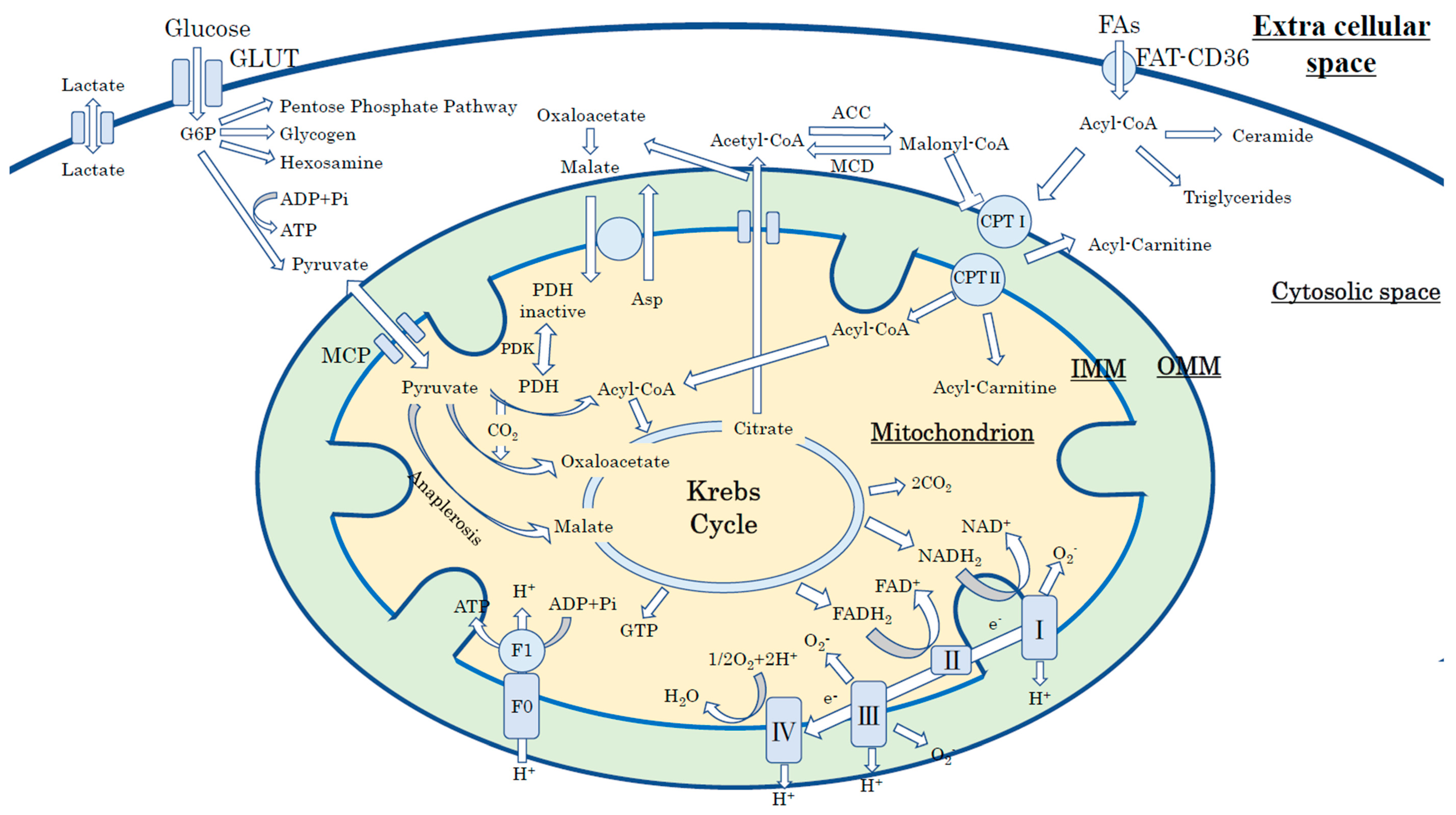
3.1. Cardiac Energy Metabolism in the Physiological Condition

To maintain a continuous contraction, human hearts are required to produce a high amount of energy phosphate; 3.5–5 kg/day adenosine triphosphate (ATP) is produced [35] from various substrate such as fatty acids (FAs), carbohydrates (glucose and lactate), ketones, and amino acids. In the physiological condition, fatty acids are major substrates that provide approximately 40% to 60% of the total energy, whereas glucose contributes less than 25% to myocardial energy production [36]. However, hearts can rapidly alter their substrate use depending on the hormonal status, workload of the heart, and circulating substrate condition on the basis of metabolic flexibility [35].
Glucose enters cardiac myocytes through glucose transporter 1 (GLUT1) and 4 (GLUT4); GLUT1 is insulin-independent and located on the cellular membrane, and GLUT4 is translocated to the cellular membrane in an insulin-dependent manner. Intracellular glucose is initially phosphorylated by hexokinase to glucose-6-phosphate (G6P), which is a substrate of glycolysis, to produce pyruvate, NADH, and 2ATP. Pyruvate that derives from glycolysis is either converted to lactate via lactate dehydrogenase (LDH) or transferred to the mitochondria (via the mitochondria pyruvate carrier: MPC) and oxidized by the Krebs cycle (Figure 1) [36].
Human hearts prefer to utilize long-chain fatty acids that bind to albumin and/or are released from triacylglycerol contained in chylomicrons or very-low-density lipoproteins (VLDL) [35]. Circulating free fatty acids (FAs) enter cardiac myocytes through transport proteins (fatty acid translocase; FAT-CD36; plasma membrane fatty acid-binding protein (FABPpm), and fatty acid transport proteins 1 and 6 (FATP1 and FATP6)) or by passive diffusion [36,37]. Cardiac FA uptake via FAT-CD36 is stimulated by insulin or AMP-activated kinase (AMPK) [37]. After entering into the cytosol, FAs are esterified into long-chain fatty acyl CoA [38] and directly shuttled to the mitochondria to start β-oxidation. The long-chain fatty acyl CoA is converted to long-chain acylcarnitine by carnitine palmitoyltransferase isomers-I (CPT-I) located in the outer mitochondrial membrane (OMM) and then converted back to long-chain fatty acyl CoA by palmitoyltransferase isomers II (CPT-II) located in the inner mitochondrial membrane (IMM) [35,36].

3.2. Cardiac Insulin Resistance and Impaired Cardiac Metabolic Flexibility under HF

HF is a syndrome in which hearts fail to respond to systemic demands for many etiological reasons, and cardiac energy production is generally impaired regardless of the type and/or stage of HF. However, the exact energy metabolic profile of HF remains controversial, because there is still a discrepancy regarding the substrate preference where the oxidation rate of substrates (fatty acid, glucose, and ketone body) varies by model and/or stage of heart failure [36].
It is generally believed that a failing heart, in which oxygen is deficient, alters the main source of energy where the heart is less dependent on FA oxidation and switches to glycolysis as its main source of energy. Indeed, under pathophysiological conditions such as HF, glycolysis and glucose oxidation support the myocardial energy demand, as a small amount of ATP is obtained by substrate-level phosphorylation via glycolysis and may be important for the maintenance of intracellular ionic homeostasis even in a reduced O2 environment. In addition, impaired mitochondrial TCA (tricarboxylic acid cycle) activity and decreased enzyme transcription involved in FA oxidation have been reported under various types of HF, such as a pressure-overload and a rapid pacing-induced HF model [39].
In contrast to FA oxidation in HF, glucose metabolism is varied by its etiology and stage of HF [40]. In fact, cardiac glucose uptake varies with experimental models; e.g., glucose uptake was decreased in the HF model by aortic constriction, whereas increased in the HF model by Dahl salt-sensitive rat [40]. Emerging bodies of evidence have revealed a strong relationship between HF and insulin resistance [41]. Because both glucose and fatty acid metabolism in the heart are tightly controlled by insulin signaling, insulin resistance plays a critical role in the pathogenesis of HF. Indeed, an impairment in insulin signaling and the development of insulin resistance, which frequently proceed cardiac dysfunction in heart failure, are major determinant factors of HF progression [42]. Insulin resistance and/or T2DM increases the delivery of FAs to the myocardium. Previous investigations showed increased myocardium lipid accumulation in T2DM patients by cardiac biopsy [43] and cardiac magnetic resonance imaging (MRI) [44], and the accumulation of lipid metabolites such as triacylglycerol, ceramide, and diacylglycerol—which can induce further metabolic deficiency and/or apoptosis—is toxic to cardiac myocytes [36,37,38]. In addition, it is well known that cardiac metabolic flexibility is impaired [45,46] in some cardiac diseases, which consequently reduces myocardial ATP production [47] and cardiac function.

3.3. Molecular Mechanism of Impaired Insulin Signaling under HF

In HF, the compensatory hyper-adrenergic state can increase the circulating plasma FAs, which are derived from adipose tissue (Figure 2). The excess circulating FAs inhibit glucose uptake by impaired insulin signaling in skeletal muscle (and cardiomyocytes) and, thereby, increase blood glucose by insulin resistance. The increased circulating FAs also lead to excessive ROS generation and oxygen waste through activated beta-oxidation in the mitochondria, which leads to further adverse effects for preexisting impaired cardiac contractility. Thus, these metabolic alterations (such as increased circulating FAs, hyperglycemia, and hyperinsulinemia) induced by the compensatory hyperadrenergic state in HF further deteriorate cardiac function and should be corrected via metabolic intervention.

4. The Role of Mitochondrial Dynamics in Cardiac Insulin Resistance and HF

4.1. Mitochondrial Energy Production in a Normal Heart

Under physiological conditions, the mitochondria produces most of the ATP (>95%) that supports the intracellular energy demands of cardiac muscle through oxidative phosphorylation (OXPHOS) in the inner mitochondrial membrane (IMM) in a group of proteins known as the electron transport chain (ETC) [35]. In OXPHOS, ATP synthesis (F1-F0 ATPase) essentially depends on a potent electrical and proton gradient across the mitochondrial membrane (mitochondrial membrane potential; ΔΨm = −180 ~ −200 mV).
Because mitochondrial Ca2+ plays an important role in ATP synthesis [48], mitochondrial Ca2+ regulation is important for various cellular physiological processes. Classically, mitochondria accumulate Ca2+ through the mitochondrial Ca2+ uniporter by primarily using the electrical gradient (ΔΨm) [49]. To respond to the beat-to-beat base energy demand, mitochondrial Ca2+ is mobilized from the sarcoplasmic reticulum (SR) [50] through a physical coupling between the SR to the mitochondria [51]. Mitochondrial Ca2+ efflux in the heart is mainly conducted by the mitochondrial Na+/Ca2+ exchanger (NCLX) and by the mitochondrial permeability transition pore (mPTP) [49,52].
Mitochondria are known as the major organelles that produce reactive oxygen species (ROS) through mitochondrial ETC activity. However, myocardial ROS remains at a low level under the physiological conditions, because of effective mitochondrial antioxidant systems, such as manganese superoxide dismutase (MnSOD), catalase, and glutathione peroxidase (GPx) [53].

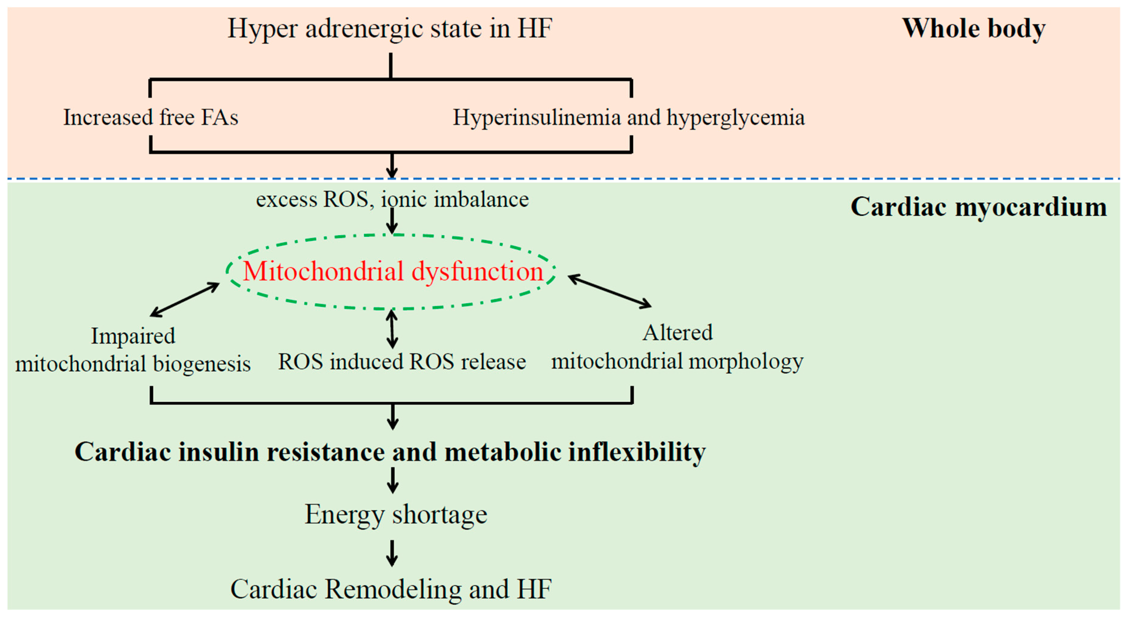
4.2. Mitochondrial Dysfunction Induces Cardiac Insulin Resistance in Patients with HF

Emerging bodies of evidence have revealed that mitochondrial dysfunction is a key player in pathogenesis in insulin resistance and heart failure [54,55]. In patients with HF, mitochondrial dysfunction induces excess ROS production, reduced mitochondrial membrane potential, impaired ETC complex activity, and abnormal mitochondrial biogenesis, which thereby results in decreased ATP synthesis [47,56] and cardiac remodeling [55] (Figure 2). Mitochondrial ROS elevation is promoted through a self-amplifying loop called ROS-induced ROS release (RIRR); that is, the initial mitochondrial ROS elevation can induce the consequent mitochondrial ROS burst. As RIRR is associated with the mitochondrial permeability transition pore (mPTP) opening and consequent ΔΨm dissipation [52,57], the myocardial ROS burst during the reperfusion phase might result from RIRR.
In patients with insulin resistance and T2DM, the reduced mitochondrial OXPHOS—due to mitochondrial dysfunction—promotes excess intracellular lipid metabolite accumulation (such as fatty acyl CoAs and diacylglycerol) in the skeletal muscle [58]. The intracellular lipid metabolite accumulation activates serine/threonine kinases, such as protein kinase C (PKC-θ rodents, PKC-β and -δ humans) [59,60], which in turn produce phosphorylate serine residues on IRS-1, inhibit insulin-induced PI 3-kinase activity, and lead to reduced AKT (protein kinase B) 2 activity after insulin stimulation. Impaired AKT2 activity suppresses insulin-mediated GLUT4 translocation to the plasma membrane, which consequently compromises insulin-mediated glucose uptake.
Thus, mitochondrial dysfunction plays a key role in the pathogenesis of HF and insulin resistance, and targeted treatment to the mitochondria has been considered to improve HF and insulin resistance.

4.3. Dysregulation of Mitochondrial Dynamics in HF

To respond to the change of cellular demand, mitochondria can alter their morphology through fusion and fission events (so-called mitochondrial dynamics), and the dysregulation of mitochondrial dynamics is known to be correlated with the pathogenesis of cardiac diseases [5]. Mitochondrial fusion, which is regulated by mitofusin 1 (Mfn1), mitofusin 2 (Mfn2), and optic atrophy-1 (OPA1), supports the inter-organellar exchange of proteins, substrates, and mitochondrial DNA to enhance the stability of mitochondria [61]. Mitochondrial fission, which is regulated by mitochondrial fission proteins, such as dynamin-related protein-1 (DRP1), mitochondrial fission factor (MFF) [62], and fission 1 homologue protein (Fis1) [63], increases the number and capacity of mitochondria during cell division. In addition, mitochondrial fission also facilitates the control of mitochondrial quality by mitophagy, which removes damaged mitochondria through lysosomal autopsy.
Evidence suggests that the alteration in mitochondrial morphology correlates with the pathophysiology of insulin resistance and heart failure [64,65]. In the failing myocardium, mitochondrial fission was apparent, and the association of both mitochondrial dynamic proteins (decrease in fusion proteins and/or increase in fission proteins) with a failing heart was reported [66,67]. In addition, it has been reported that small mitochondria were frequently seen in the skeletal muscle in obese and T2DM subjects [68,69] as well. On the other hand, chronic oxidative stress is known to play the most crucial role for the pathogenesis of insulin resistance [70]. We have reported that DRP1 and ROS have a mutual enhancing relationship, in which DRP1 enhances ROS generation, and vice versa [71]. Although the underlining mechanism remains elusive, the increase in DRP1 and mitochondrial fragmentation are interlinked with ROS. This reciprocal augmentation of DRP1 and ROS could be a key player which induces mitochondrial dysfunction and the ultimate fate of myocardial insulin resistance.

5. Cardiac Insulin Resistance in Patients with HF

5.1. Diagnosis of Cardiac Insulin Resistance in Patients with HF and DM

Despite this significant contribution of cardiac insulin resistance in the basic investigations of HF, there is no current available definition and/or diagnosis criteria of cardiac insulin resistance. Insulin resistance is clinically defined as a condition of impaired insulin sensitivity to glucose uptake and/or utilization. Systemic (whole body) insulin resistance has been defined by the “homeostasis model assessment for insulin resistance (HOMA-IR: normal; <1.6, insulin resistance; >2.5)”, which is calculated as follows: fasting plasma glucose (mmol/L) × fasting serum insulin (IU/L)/405 [72]. However, currently, neither relevant clinical diagnostic criteria nor clear reference values of cardiac insulin resistance have been established.
The 18F-fluorodeoxyglucose positron emission tomography (FDG-PET) is known to be a clinically extant method to assess myocardial glucose uptake. The insufficiency of cardiac glucose uptake has been investigated using the hyper-insulinemic/euglycemic clamp method, which was developed by the American Society of Nuclear Cardiology and can indicate maximal myocardial glucose uptake [73]. However, so far, FDG-PET remains in the relative comparison (with normal subjects and/or among disease groups) [74] and fails to provide any clear cutoff values to diagnose cardiac insulin resistance. The assessment of cardiac insulin resistance may require at least two independent points of myocardial FDG uptake, i.e., fasting (6–12 h fasting to minimize the heterogeneity of FDG accumulation) and maximal glucose uptake (by glucose and, if feasible, insulin loading). Because the bioactive half-life of 18F-fluorodeoxyglucose (18F-FDG) is approximately 110 min [75], multiple consecutive scans are technically impossible and single scans are applied even in the evaluation of coronary artery disease (myocardial ischemia). Thus, the development of a glucose tracer that enables temporal assessment (fasting and glucose with insulin loading) and spatial dynamic FDG-PET is desired to assess cardiac insulin resistance.
Cardiac biopsies and the investigation of insulin signaling molecules, such as insulin receptor substrate 1 (IRS1), IRS1-associated PI3K (IRS1-PI3K), and GLUT4, may be alternative and direct diagnostic methods to evaluate myocardial insulin resistance [76]. The hyper-phosphorylation of IRS1 and/or IRS1-PI3K reduced the expression of GLUT4 in the myocardium, which may reflect cardiac insulin resistance. However, this also remains in relative comparison with normal subjects. In addition, Cook et al. reported that glucose utility was reduced both in patients with T2DM and LV dysfunction, whereas IRS1-PI3K was unexpectedly elevated, and concluded that behaviors of insulin signaling molecules in cardiac insulin resistance are different from those of whole body insulin resistance [76]. Because degraded mitochondrial morphology frequently accompanies myocardial metabolic disorders in patients with HF [64,65], it seems possible that the assessment of mitochondrial morphology and/or size in electron microscopy may at least partially support a diagnosis of cardiac insulin resistance and metabolic disorder. Further investigations are required to reveal the relationship between aberrant mitochondrial morphology and cardiac insulin resistance in a clinical study.

5.2. Innovation of Treatments to Improve Cardiac Insulin Resistance

Although an improvement in cardiac insulin resistance is anticipated to afford preferable effects on HF, clinically established treatment toward cardiac insulin resistance is not currently available. Evidence on the validated anti-diabetic agents may give us some tips on developing new medication for cardiac insulin resistance. As mentioned above, there is a discrepancy regarding the cardiac outcome of insulin-sensitizing agents (see Section 2.2). Because thiazolidinediones, which are considered a contraindication for patients with HF [21,23], are PPAR-χ agonists, they can activate the uptake and oxidation of FAs (simultaneously with glucose) and further enforce FA uptake and oxidation in a failing heart. The enhancement of FA metabolism under HF, which may exceed the capacity of impaired mitochondrial respiration, can lead to the accumulation of lipid metabolites [37], accelerate dysregulated mitochondrial dynamics [71], which enhance cardiac insulin resistance, and myocardium energy shortage, and thereby deteriorate HF [65,77]. In contrast, metformin—which has been recommended as a first-line treatment to T2DM patients who have cardiovascular risks [25,26]—enhances energy expenditure, suppresses hepatic gluconeogenesis, and induces insulin sensitization through AMP-activated protein kinase (AMPK) activation. Activated AMPK can also suppress the mitochondrial fission protein of DRP1, which can resist lipotoxicity-induced mitochondrial dysfunction [77], and ameliorate cardiac insulin resistance.
Mdivi-1 is a selective DRP1 (mitochondrial fission protein) inhibitor. A number of investigations have reported the preferable effects of mdiivi1 on ischemia/reperfusion injury [5] and cardiac hypertrophy in animal models [78]. Little is known about the effect of mdivi1 on T2DM and HF. Furthermore, clinically relevant evidence has been less reported. Recently, Ishikawa et al. formulated poly nanoparticles containing mdivi-1, which could be delivered to the cytosol and mitochondria, and exhibited better cardiac protection against ischemia reperfusion injury than mdivi1 alone through the inhibition of mitochondrial outer membrane permeabilization (MOMP) [79]. On the other hand, there are some reports that suspect a selective inhibitory effect of mdivi-1 on Drp1. Bordt et al. showed that mdivi-1 promotes mitochondrial complex I-dependent O2 consumption and produces reverse electron transfer-mediated ROS with an independent manner of mitochondrial elongation, and concluded that mdivi-1 does not selectively inhibit Drp1 but contributes to mitochondrial morphological alteration and cellular protection by reversible Complex I inhibition and mitochondrial ROS suppression [80]. Elamipretide is a mitochondria-targeting agent, which selectively acts on mitochondrial ECT to improve the efficacy of electron transport. Although there are fewer human clinical trials, a number of animal investigations have revealed promising results on HF [81]. Thus, currently, there is no clinically available treatment targeting mitochondria or mitochondrial dynamics. Further investigations are required to reveal the effects of mitochondrial targeting agents on cardiac insulin resistance and HF.

6. Summary

HF frequently coexists with glucose insufficiency such as T2DM and insulin resistance. However, some antidiabetic drugs failed to improve cardiac outcomes in T2DM patients, although glucose levels are sufficiently lowered to cause an effect. This may, at least in part, suggest a lack of understanding of cardiac insulin resistance, as despite the significant contribution of cardiac insulin resistance to the pathogenesis and progression of HF in basic research, there is no information on the definition or treatment of cardiac insulin resistance from a clinical study.
Because mitochondrial dynamics play an important role in the pathogenesis of HF and insulin resistance, the development of a mitochondria-targeted diagnosis and/or treatment may afford preferable outcomes for patients with HF. Further investigations are encouraged to reveal the relationship between mitochondrial dynamics and cardiac insulin resistance in HF.

Funding

This work was supported by JSPS KAKENHI Grant Numbers JP16K09428 (to M.S.).

Conflicts of Interest

The authors declare no conflict of interest. The funders had no role in the design of the study; in the collection, analyses, or interpretation of data; in the writing of the manuscript, or in the decision to publish the results.

Abbreviations

T2DMType 2 diabetes mellitus
HFHeart failure
ATPAdenosine triphosphate
FDG-PET18F-2-deoxyglucose positron emission tomography
HFrEFHF with a reduced ejection fraction
HFpEFHF with a preserved ejection fraction
HFmrEFHF with a mid-range ejection fraction
mPTPMitochondrial permeability transition pore
GLUT1/4Glucose transporter 1/4
OXPHOSOxidative phosphorylation
ROSReactive oxygen species
FAsFatty acids
Mfn1/2Mitofusin 1/2
OPA1Optic atrophy-1
DRP1Dynamin-related protein-1
MFFMitochondrial fission factor
Fis1Mitochondrial fission protein 1
PPAR-χPeroxisome proliferator-activated receptor-gamma
GLP-1Glucagon-like peptide-1
MOMPMitochondrial outer membrane permeabilization

References

  1. Savarese, G.; Lund, L.H. Global Public Health Burden of Heart Failure. Card. Fail. Rev. 2017, 3, 7–11. [Google Scholar] [CrossRef]
  2. Udell, J.A.; Cavender, M.A.; Bhatt, D.L.; Chatterjee, S.; Farkouh, M.E.; Scirica, B.M. Glucose-lowering drugs or strategies and cardiovascular outcomes in patients with or at risk for type 2 diabetes: A meta-analysis of randomised controlled trials. Lancet Diabetes Endocrinol. 2015, 3, 356–366. [Google Scholar] [CrossRef]
  3. Aroor, A.R.; Mandavia, C.H.; Sowers, J.R. Insulin resistance and heart failure: Molecular mechanisms. Heart Fail. Clin. 2012, 8, 609–617. [Google Scholar] [CrossRef]
  4. Zhang, L.; Jaswal, J.S.; Ussher, J.R.; Sankaralingam, S.; Wagg, C.; Zaugg, M.; Lopaschuk, G.D. Cardiac insulin-resistance and decreased mitochondrial energy production precede the development of systolic heart failure after pressure-overload hypertrophy. Circ. Heart Fail. 2013, 6, 1039–1048. [Google Scholar] [CrossRef]
  5. Ong, S.B.; Hausenloy, D.J. Mitochondrial morphology and cardiovascular disease. Cardiovasc. Res. 2010, 88, 16–29. [Google Scholar] [CrossRef] [Green Version]
  6. Seferovic, P.M.; Petrie, M.C.; Filippatos, G.S.; Anker, S.D.; Rosano, G.; Bauersachs, J.; Paulus, W.J.; Komajda, M.; Cosentino, F.; de Boer, R.A.; et al. Type 2 diabetes mellitus and heart failure: A position statement from the Heart Failure Association of the European Society of Cardiology. Eur. J. Heart Fail. 2018, 20, 853–872. [Google Scholar] [CrossRef] [PubMed]
  7. Thrainsdottir, I.S.; Aspelund, T.; Thorgeirsson, G.; Gudnason, V.; Hardarson, T.; Malmberg, K.; Sigurdsson, G.; Ryden, L. The association between glucose abnormalities and heart failure in the population-based Reykjavik study. Diabetes Care 2005, 28, 612–616. [Google Scholar] [CrossRef]
  8. Nichols, G.A.; Hillier, T.A.; Erbey, J.R.; Brown, J.B. Congestive heart failure in type 2 diabetes: Prevalence, incidence, and risk factors. Diabetes Care 2001, 24, 1614–1619. [Google Scholar] [CrossRef]
  9. Demant, M.N.; Gislason, G.H.; Kober, L.; Vaag, A.; Torp-Pedersen, C.; Andersson, C. Association of heart failure severity with risk of diabetes: A Danish nationwide cohort study. Diabetologia 2014, 57, 1595–1600. [Google Scholar] [CrossRef] [PubMed]
  10. Amato, L.; Paolisso, G.; Cacciatore, F.; Ferrara, N.; Ferrara, P.; Canonico, S.; Varricchio, M.; Rengo, F. Congestive heart failure predicts the development of non-insulin-dependent diabetes mellitus in the elderly. The Osservatorio Geriatrico Regione Campania Group. Diabetes Metab. 1997, 23, 213–218. [Google Scholar]
  11. Tenenbaum, A.; Motro, M.; Fisman, E.Z.; Leor, J.; Freimark, D.; Boyko, V.; Mandelzweig, L.; Adler, Y.; Sherer, Y.; Behar, S. Functional class in patients with heart failure is associated with the development of diabetes. Am. J. Med. 2003, 114, 271–275. [Google Scholar] [CrossRef]
  12. Shah, A.D.; Langenberg, C.; Rapsomaniki, E.; Denaxas, S.; Pujades-Rodriguez, M.; Gale, C.P.; Deanfield, J.; Smeeth, L.; Timmis, A.; Hemingway, H. Type 2 diabetes and incidence of cardiovascular diseases: A cohort study in 1.9 million people. Lancet Diabetes Endocrinol. 2015, 3, 105–113. [Google Scholar] [CrossRef]
  13. Pazin-Filho, A.; Kottgen, A.; Bertoni, A.G.; Russell, S.D.; Selvin, E.; Rosamond, W.D.; Coresh, J. HbA 1c as a risk factor for heart failure in persons with diabetes: The Atherosclerosis Risk in Communities (ARIC) study. Diabetologia 2008, 51, 2197–2204. [Google Scholar] [CrossRef]
  14. Kishimoto, I.; Makino, H.; Ohata, Y.; Tamanaha, T.; Tochiya, M.; Kada, A.; Ishihara, M.; Anzai, T.; Shimizu, W.; Yasuda, S.; et al. Hemoglobin A1c predicts heart failure hospitalization independent of baseline cardiac function or B-type natriuretic peptide level. Diabetes Res. Clin. Pract. 2014, 104, 257–265. [Google Scholar] [CrossRef]
  15. Suskin, N.; McKelvie, R.S.; Burns, R.J.; Latini, R.; Pericak, D.; Probstfield, J.; Rouleau, J.L.; Sigouin, C.; Solymoss, C.B.; Tsuyuki, R.; et al. Glucose and insulin abnormalities relate to functional capacity in patients with congestive heart failure. Eur. Heart J. 2000, 21, 1368–1375. [Google Scholar] [CrossRef] [Green Version]
  16. Kristensen, S.L.; Preiss, D.; Jhund, P.S.; Squire, I.; Cardoso, J.S.; Merkely, B.; Martinez, F.; Starling, R.C.; Desai, A.S.; Lefkowitz, M.P.; et al. Risk Related to Pre-Diabetes Mellitus and Diabetes Mellitus in Heart Failure With Reduced Ejection Fraction: Insights From Prospective Comparison of ARNI With ACEI to Determine Impact on Global Mortality and Morbidity in Heart Failure Trial. Circ. Heart Fail. 2016, 9, e002560. [Google Scholar] [CrossRef]
  17. MacDonald, M.R.; Petrie, M.C.; Varyani, F.; Ostergren, J.; Michelson, E.L.; Young, J.B.; Solomon, S.D.; Granger, C.B.; Swedberg, K.; Yusuf, S.; et al. Impact of diabetes on outcomes in patients with low and preserved ejection fraction heart failure: An analysis of the Candesartan in Heart failure: Assessment of Reduction in Mortality and morbidity (CHARM) programme. Eur. Heart J. 2008, 29, 1377–1385. [Google Scholar] [CrossRef] [Green Version]
  18. Gerstein, H.C.; Swedberg, K.; Carlsson, J.; McMurray, J.J.; Michelson, E.L.; Olofsson, B.; Pfeffer, M.A.; Yusuf, S.; CHARM Program Investigators. The hemoglobin A1c level as a progressive risk factor for cardiovascular death, hospitalization for heart failure, or death in patients with chronic heart failure: An analysis of the Candesartan in Heart failure: Assessment of Reduction in Mortality and Morbidity (CHARM) program. Arch. Intern. Med. 2008, 168, 1699–1704. [Google Scholar]
  19. Dauriz, M.; Targher, G.; Temporelli, P.L.; Lucci, D.; Gonzini, L.; Nicolosi, G.L.; Marchioli, R.; Tognoni, G.; Latini, R.; Cosmi, F.; et al. Prognostic Impact of Diabetes and Prediabetes on Survival Outcomes in Patients With Chronic Heart Failure: A Post-Hoc Analysis of the GISSI-HF (Gruppo Italiano per lo Studio della Sopravvivenza nella Insufficienza Cardiaca-Heart Failure) Trial. J. Am. Heart Assoc. 2017, 6, e005156. [Google Scholar] [CrossRef]
  20. Eurich, D.T.; Weir, D.L.; Majumdar, S.R.; Tsuyuki, R.T.; Johnson, J.A.; Tjosvold, L.; Vanderloo, S.E.; McAlister, F.A. Comparative safety and effectiveness of metformin in patients with diabetes mellitus and heart failure: Systematic review of observational studies involving 34,000 patients. Circ. Heart Fail. 2013, 6, 395–402. [Google Scholar] [CrossRef]
  21. Komajda, M.; McMurray, J.J.; Beck-Nielsen, H.; Gomis, R.; Hanefeld, M.; Pocock, S.J.; Curtis, P.S.; Jones, N.P.; Home, P.D. Heart failure events with rosiglitazone in type 2 diabetes: Data from the RECORD clinical trial. Eur. Heart J. 2010, 31, 824–831. [Google Scholar] [CrossRef] [PubMed]
  22. Lago, R.M.; Singh, P.P.; Nesto, R.W. Congestive heart failure and cardiovascular death in patients with prediabetes and type 2 diabetes given thiazolidinediones: A meta-analysis of randomised clinical trials. Lancet 2007, 370, 1129–1136. [Google Scholar] [CrossRef]
  23. Kolwicz, S.C., Jr.; Purohit, S.; Tian, R. Cardiac metabolism and its interactions with contraction, growth, and survival of cardiomyocytes. Circ. Res. 2013, 113, 603–616. [Google Scholar] [CrossRef] [PubMed]
  24. Horita, S.; Nakamura, M.; Satoh, N.; Suzuki, M.; Seki, G. Thiazolidinediones and Edema: Recent Advances in the Pathogenesis of Thiazolidinediones-Induced Renal Sodium Retention. PPAR Res. 2015, 2015, 646423. [Google Scholar] [CrossRef] [PubMed]
  25. Shah, D.D.; Fonarow, G.C.; Horwich, T.B. Metformin therapy and outcomes in patients with advanced systolic heart failure and diabetes. J. Card. Fail. 2010, 16, 200–206. [Google Scholar] [CrossRef]
  26. Garber, A.J.; Abrahamson, M.J.; Barzilay, J.I.; Blonde, L.; Bloomgarden, Z.T.; Bush, M.A.; Dagogo-Jack, S.; DeFronzo, R.A.; Einhorn, D.; Fonseca, V.A.; et al. Consensus Statement by the American Association of Clinical Endocrinologists and American College of Endocrinology on the Comprehensive Type 2 Diabetes Management Algorithm - 2017 Executive Summary. Endocr. Pract. 2017, 23, 207–238. [Google Scholar] [CrossRef]
  27. MacDonald, M.R.; Petrie, M.C.; Hawkins, N.M.; Petrie, J.R.; Fisher, M.; McKelvie, R.; Aguilar, D.; Krum, H.; McMurray, J.J. Diabetes, left ventricular systolic dysfunction, and chronic heart failure. Eur. Heart J. 2008, 29, 1224–1240. [Google Scholar] [CrossRef]
  28. Arturi, F.; Succurro, E.; Miceli, S.; Cloro, C.; Ruffo, M.; Maio, R.; Perticone, M.; Sesti, G.; Perticone, F. Liraglutide improves cardiac function in patients with type 2 diabetes and chronic heart failure. Endocrine 2017, 57, 464–473. [Google Scholar] [CrossRef]
  29. Marso, S.P.; Daniels, G.H.; Brown-Frandsen, K.; Kristensen, P.; Mann, J.F.; Nauck, M.A.; Nissen, S.E.; Pocock, S.; Poulter, N.R.; Ravn, L.S.; et al. Liraglutide and Cardiovascular Outcomes in Type 2 Diabetes. N. Engl. J. Med. 2016, 375, 311–322. [Google Scholar] [CrossRef] [Green Version]
  30. Zinman, B.; Wanner, C.; Lachin, J.M.; Fitchett, D.; Bluhmki, E.; Hantel, S.; Mattheus, M.; Devins, T.; Johansen, O.E.; Woerle, H.J.; et al. Empagliflozin, Cardiovascular Outcomes, and Mortality in Type 2 Diabetes. N. Engl. J. Med. 2015, 373, 2117–2128. [Google Scholar] [CrossRef]
  31. Neal, B.; Perkovic, V.; Mahaffey, K.W.; de Zeeuw, D.; Fulcher, G.; Erondu, N.; Shaw, W.; Law, G.; Desai, M.; Matthews, D.R.; et al. Canagliflozin and Cardiovascular and Renal Events in Type 2 Diabetes. N. Engl. J. Med. 2017, 377, 644–657. [Google Scholar] [CrossRef] [PubMed]
  32. Wiviott, S.D.; Raz, I.; Bonaca, M.P.; Mosenzon, O.; Kato, E.T.; Cahn, A.; Silverman, M.G.; Zelniker, T.A.; Kuder, J.F.; Murphy, S.A.; et al. Dapagliflozin and Cardiovascular Outcomes in Type 2 Diabetes. N. Engl. J. Med. 2019, 380, 347–357. [Google Scholar] [CrossRef] [PubMed]
  33. Sano, M. Hemodynamic Effects of Sodium-Glucose Cotransporter 2 Inhibitors. J. Clin. Med. Res. 2017, 9, 457–460. [Google Scholar] [CrossRef] [PubMed] [Green Version]
  34. Bertero, E.; Prates Roma, L.; Ameri, P.; Maack, C. Cardiac effects of SGLT2 inhibitors: The sodium hypothesis. Cardiovasc. Res. 2018, 114, 12–18. [Google Scholar] [CrossRef] [PubMed]
  35. Lopaschuk, G.D.; Ussher, J.R.; Folmes, C.D.; Jaswal, J.S.; Stanley, W.C. Myocardial fatty acid metabolism in health and disease. Physiol. Rev. 2010, 90, 207–258. [Google Scholar] [CrossRef]
  36. Karwi, Q.G.; Uddin, G.M.; Ho, K.L.; Lopaschuk, G.D. Loss of Metabolic Flexibility in the Failing Heart. Front. Cardiovasc. Med. 2018, 5, 68. [Google Scholar] [CrossRef] [PubMed] [Green Version]
  37. Goldberg, I.J.; Trent, C.M.; Schulze, P.C. Lipid metabolism and toxicity in the heart. Cell Metab. 2012, 15, 805–812. [Google Scholar] [CrossRef]
  38. Chiu, H.C.; Kovacs, A.; Ford, D.A.; Hsu, F.F.; Garcia, R.; Herrero, P.; Saffitz, J.E.; Schaffer, J.E. A novel mouse model of lipotoxic cardiomyopathy. J. Clin. Investig. 2001, 107, 813–822. [Google Scholar] [CrossRef] [Green Version]
  39. Abdurrachim, D.; Luiken, J.J.; Nicolay, K.; Glatz, J.F.; Prompers, J.J.; Nabben, M. Good and bad consequences of altered fatty acid metabolism in heart failure: Evidence from mouse models. Cardiovasc. Res. 2015, 106, 194–205. [Google Scholar] [CrossRef]
  40. Doenst, T.; Nguyen, T.D.; Abel, E.D. Cardiac metabolism in heart failure: Implications beyond ATP production. Circ. Res. 2013, 113, 709–724. [Google Scholar] [CrossRef]
  41. Rutter, M.K.; Parise, H.; Benjamin, E.J.; Levy, D.; Larson, M.G.; Meigs, J.B.; Nesto, R.W.; Wilson, P.W.; Vasan, R.S. Impact of glucose intolerance and insulin resistance on cardiac structure and function: Sex-related differences in the Framingham Heart Study. Circulation 2003, 107, 448–454. [Google Scholar] [CrossRef]
  42. Ingelsson, E.; Sundstrom, J.; Arnlov, J.; Zethelius, B.; Lind, L. Insulin resistance and risk of congestive heart failure. JAMA 2005, 294, 334–341. [Google Scholar] [CrossRef]
  43. Regan, T.J.; Lyons, M.M.; Ahmed, S.S.; Levinson, G.E.; Oldewurtel, H.A.; Ahmad, M.R.; Haider, B. Evidence for cardiomyopathy in familial diabetes mellitus. J. Clin. Investig. 1977, 60, 884–899. [Google Scholar] [CrossRef]
  44. McGavock, J.M.; Lingvay, I.; Zib, I.; Tillery, T.; Salas, N.; Unger, R.; Levine, B.D.; Raskin, P.; Victor, R.G.; Szczepaniak, L.S. Cardiac steatosis in diabetes mellitus: A 1H-magnetic resonance spectroscopy study. Circulation 2007, 116, 1170–1175. [Google Scholar] [CrossRef]
  45. Chandler, M.P.; Kerner, J.; Huang, H.; Vazquez, E.; Reszko, A.; Martini, W.Z.; Hoppel, C.L.; Imai, M.; Rastogi, S.; Sabbah, H.N.; et al. Moderate severity heart failure does not involve a downregulation of myocardial fatty acid oxidation. Am. J. Physiol. Heart Circ. Physiol. 2004, 287, H1538–H1543. [Google Scholar] [CrossRef]
  46. O’Donnell, J.M.; Fields, A.D.; Sorokina, N.; Lewandowski, E.D. The absence of endogenous lipid oxidation in early stage heart failure exposes limits in lipid storage and turnover. J. Mol. Cell. Cardiol. 2008, 44, 315–322. [Google Scholar] [CrossRef] [Green Version]
  47. Neubauer, S.; Horn, M.; Cramer, M.; Harre, K.; Newell, J.B.; Peters, W.; Pabst, T.; Ertl, G.; Hahn, D.; Ingwall, J.S.; et al. Myocardial phosphocreatine-to-ATP ratio is a predictor of mortality in patients with dilated cardiomyopathy. Circulation 1997, 96, 2190–2196. [Google Scholar] [CrossRef]
  48. Bers, D.M. Excitation-Contraction Coupling and Cardiac Contractile Force, 2nd ed.; Kluwer Academic Publishers: Dordrecht, The Netherlands; Boston, MA, USA, 2001; p. xxiv. 427p. [Google Scholar]
  49. Saotome, M.; Katoh, H.; Satoh, H.; Nagasaka, S.; Yoshihara, S.; Terada, H.; Hayashi, H. Mitochondrial membrane potential modulates regulation of mitochondrial Ca2+ in rat ventricular myocytes. Am. J. Physiol. Heart Circ. Physiol. 2005, 288, H1820–H1828. [Google Scholar] [CrossRef]
  50. Pacher, P.; Thomas, A.P.; Hajnoczky, G. Ca2+ marks: Miniature calcium signals in single mitochondria driven by ryanodine receptors. Proc. Natl. Acad. Sci. USA 2002, 99, 2380–2385. [Google Scholar] [CrossRef]
  51. Eisner, V.; Csordas, G.; Hajnoczky, G. Interactions between sarco-endoplasmic reticulum and mitochondria in cardiac and skeletal muscle–pivotal roles in Ca2+ and reactive oxygen species signaling. J. Cell Sci. 2013, 126 Pt 14, 2965–2978. [Google Scholar] [CrossRef]
  52. Saotome, M.; Katoh, H.; Yaguchi, Y.; Tanaka, T.; Urushida, T.; Satoh, H.; Hayashi, H. Transient opening of mitochondrial permeability transition pore by reactive oxygen species protects myocardium from ischemia-reperfusion injury. Am. J. Physiol. Heart Circ. Physiol. 2009, 296, H1125–H1132. [Google Scholar] [CrossRef] [Green Version]
  53. Raedschelders, K.; Ansley, D.M.; Chen, D.D. The cellular and molecular origin of reactive oxygen species generation during myocardial ischemia and reperfusion. Pharmacol. Ther. 2012, 133, 230–255. [Google Scholar] [CrossRef]
  54. Kumar, A.A.; Kelly, D.P.; Chirinos, J.A. Mitochondrial Dysfunction in Heart Failure With Preserved Ejection Fraction. Circulation 2019, 139, 1435–1450. [Google Scholar] [CrossRef]
  55. Kiyuna, L.A.; Albuquerque, R.P.E.; Chen, C.H.; Mochly-Rosen, D.; Ferreira, J.C.B. Targeting mitochondrial dysfunction and oxidative stress in heart failure: Challenges and opportunities. Free Radic. Biol. Med. 2018, 129, 155–168. [Google Scholar] [CrossRef]
  56. Neubauer, S. The failing heart--an engine out of fuel. N. Engl. J. Med. 2007, 356, 1140–1151. [Google Scholar] [CrossRef]
  57. Zorov, D.B.; Filburn, C.R.; Klotz, L.O.; Zweier, J.L.; Sollott, S.J. Reactive oxygen species (ROS)-induced ROS release: A new phenomenon accompanying induction of the mitochondrial permeability transition in cardiac myocytes. J. Exp. Med. 2000, 192, 1001–1014. [Google Scholar] [CrossRef]
  58. Petersen, K.F.; Dufour, S.; Befroy, D.; Garcia, R.; Shulman, G.I. Impaired mitochondrial activity in the insulin-resistant offspring of patients with type 2 diabetes. N. Engl. J. Med. 2004, 350, 664–671. [Google Scholar] [CrossRef]
  59. Kim, J.A.; Wei, Y.; Sowers, J.R. Role of mitochondrial dysfunction in insulin resistance. Circ. Res. 2008, 102, 401–414. [Google Scholar] [CrossRef]
  60. Turner, N.; Heilbronn, L.K. Is mitochondrial dysfunction a cause of insulin resistance? Trends Endocrinol. Metab. 2008, 19, 324–330. [Google Scholar] [CrossRef]
  61. Youle, R.J.; van der Bliek, A.M. Mitochondrial fission, fusion, and stress. Science 2012, 337, 1062–1065. [Google Scholar] [CrossRef]
  62. Otera, H.; Wang, C.; Cleland, M.M.; Setoguchi, K.; Yokota, S.; Youle, R.J.; Mihara, K. Mff is an essential factor for mitochondrial recruitment of Drp1 during mitochondrial fission in mammalian cells. J. Cell Biol. 2010, 191, 1141–1158. [Google Scholar] [CrossRef] [Green Version]
  63. Benard, G.; Karbowski, M. Mitochondrial fusion and division: Regulation and role in cell viability. Semin. Cell Dev. Biol. 2009, 20, 365–374. [Google Scholar] [CrossRef] [Green Version]
  64. Givvimani, S.; Pushpakumar, S.B.; Metreveli, N.; Veeranki, S.; Kundu, S.; Tyagi, S.C. Role of mitochondrial fission and fusion in cardiomyocyte contractility. Int. J. Cardiol. 2015, 187, 325–333. [Google Scholar] [CrossRef] [Green Version]
  65. Dorn, G., II. Mitochondrial fission/fusion and cardiomyopathy. Curr. Opin. Genet. Dev. 2016, 38, 38–44. [Google Scholar] [Green Version]
  66. Chen, L.; Gong, Q.; Stice, J.P.; Knowlton, A.A. Mitochondrial OPA1, apoptosis, and heart failure. Cardiovasc. Res. 2009, 84, 91–99. [Google Scholar] [CrossRef] [Green Version]
  67. Javadov, S.; Rajapurohitam, V.; Kilic, A.; Hunter, J.C.; Zeidan, A.; Said Faruq, N.; Escobales, N.; Karmazyn, M. Expression of mitochondrial fusion-fission proteins during post-infarction remodeling: The effect of NHE-1 inhibition. Basic Res. Cardiol. 2011, 106, 99–109. [Google Scholar] [CrossRef]
  68. Zorzano, A.; Liesa, M.; Palacin, M. Role of mitochondrial dynamics proteins in the pathophysiology of obesity and type 2 diabetes. Int. J. Biochem. Cell Biol. 2009, 41, 1846–1854. [Google Scholar] [CrossRef]
  69. Zorzano, A.; Liesa, M.; Palacin, M. Mitochondrial dynamics as a bridge between mitochondrial dysfunction and insulin resistance. Arch. Physiol. Biochem. 2009, 115, 1–12. [Google Scholar] [CrossRef]
  70. Boudina, S.; Bugger, H.; Sena, S.; O’Neill, B.T.; Zaha, V.G.; Ilkun, O.; Wright, J.J.; Mazumder, P.K.; Palfreyman, E.; Tidwell, T.J.; et al. Contribution of impaired myocardial insulin signaling to mitochondrial dysfunction and oxidative stress in the heart. Circulation 2009, 119, 1272–1283. [Google Scholar] [CrossRef]
  71. Watanabe, T.; Saotome, M.; Nobuhara, M.; Sakamoto, A.; Urushida, T.; Katoh, H.; Satoh, H.; Funaki, M.; Hayashi, H. Roles of mitochondrial fragmentation and reactive oxygen species in mitochondrial dysfunction and myocardial insulin resistance. Exp. Cell Res. 2014, 323, 314–325. [Google Scholar] [CrossRef] [Green Version]
  72. Bonora, E.; Targher, G.; Alberiche, M.; Bonadonna, R.C.; Saggiani, F.; Zenere, M.B.; Monauni, T.; Muggeo, M. Homeostasis model assessment closely mirrors the glucose clamp technique in the assessment of insulin sensitivity: Studies in subjects with various degrees of glucose tolerance and insulin sensitivity. Diabetes Care 2000, 23, 57–63. [Google Scholar] [CrossRef]
  73. Bacharach, S.L.; Bax, J.J.; Case, J.; Delbeke, D.; Kurdziel, K.A.; Martin, W.H.; Patterson, R.E. PET myocardial glucose metabolism and perfusion imaging: Part 1-Guidelines for data acquisition and patient preparation. J. Nucl. Cardiol. 2003, 10, 543–556. [Google Scholar] [CrossRef]
  74. Fallavollita, J.A.; Luisi, A.J., Jr.; Yun, E.; deKemp, R.A.; Canty, J.M., Jr. An abbreviated hyperinsulinemic-euglycemic clamp results in similar myocardial glucose utilization in both diabetic and non-diabetic patients with ischemic cardiomyopathy. J. Nucl. Cardiol. 2010, 17, 637–645. [Google Scholar] [CrossRef]
  75. Knuuti, M.J.; Nuutila, P.; Ruotsalainen, U.; Saraste, M.; Harkonen, R.; Ahonen, A.; Teras, M.; Haaparanta, M.; Wegelius, U.; Haapanen, A.; et al. Euglycemic hyperinsulinemic clamp and oral glucose load in stimulating myocardial glucose utilization during positron emission tomography. J. Nucl. Med. 1992, 33, 1255–1262. [Google Scholar]
  76. Cook, S.A.; Varela-Carver, A.; Mongillo, M.; Kleinert, C.; Khan, M.T.; Leccisotti, L.; Strickland, N.; Matsui, T.; Das, S.; Rosenzweig, A.; et al. Abnormal myocardial insulin signalling in type 2 diabetes and left-ventricular dysfunction. Eur. Heart J. 2010, 31, 100–111. [Google Scholar] [CrossRef]
  77. Sakamoto, A.; Saotome, M.; Hasan, P.; Satoh, T.; Ohtani, H.; Urushida, T.; Katoh, H.; Satoh, H.; Hayashi, H. Eicosapentaenoic acid ameliorates palmitate-induced lipotoxicity via the AMP kinase/dynamin-related protein-1 signaling pathway in differentiated H9c2 myocytes. Exp. Cell Res. 2017, 351, 109–120. [Google Scholar] [CrossRef] [Green Version]
  78. Hasan, P.; Saotome, M.; Ikoma, T.; Iguchi, K.; Kawasaki, H.; Iwashita, T.; Hayashi, H.; Maekawa, Y. Mitochondrial fission protein, dynamin-related protein 1, contributes to the promotion of hypertensive cardiac hypertrophy and fibrosis in Dahl-salt sensitive rats. J. Mol. Cell. Cardiol. 2018, 121, 103–106. [Google Scholar] [CrossRef]
  79. Ishikita, A.; Matoba, T.; Ikeda, G.; Koga, J.; Mao, Y.; Nakano, K.; Takeuchi, O.; Sadoshima, J.; Egashira, K. Nanoparticle-Mediated Delivery of Mitochondrial Division Inhibitor 1 to the Myocardium Protects the Heart From Ischemia-Reperfusion Injury Through Inhibition of Mitochondria Outer Membrane Permeabilization: A New Therapeutic Modality for Acute Myocardial Infarction. J. Am. Heart Assoc. 2016, 5, e003872. [Google Scholar]
  80. Bordt, E.A.; Clerc, P.; Roelofs, B.A.; Saladino, A.J.; Tretter, L.; Adam-Vizi, V.; Cherok, E.; Khalil, A.; Yadava, N.; Ge, S.X.; et al. The Putative Drp1 Inhibitor mdivi-1 Is a Reversible Mitochondrial Complex I Inhibitor that Modulates Reactive Oxygen Species. Dev. Cell 2017, 40, 583–594.e6. [Google Scholar] [CrossRef] [Green Version]
  81. Steggall, A.; Mordi, I.R.; Lang, C.C. Targeting Metabolic Modulation and Mitochondrial Dysfunction in the Treatment of Heart Failure. Diseases 2017, 5, 14. [Google Scholar] [CrossRef]
Figure 1. Cardiac energy metabolism in the physiological condition. FAs, fatty acids; GLUT, glucose transporter; FAT-CD36, fatty acid translocase; G6P, glucose-6-phosphate; MPC, mitochondria; OMM, outer mitochondrial membrane; IMM, inner mitochondrial membrane; CPT-I/II, carnitine palmitoyltransferase isomers-I/II.
Ijms 20 03552 g001
Figure 2. The role of mitochondrial dysfunction in the pathogenesis of cardiac insulin resistance and HF.
Ijms 20 03552 g002

