Article

Physiologically Relevant Alternative Carbon Sources Modulate Biofilm Formation, Cell Wall Architecture, and the Stress and Antifungal Resistance of Candida glabrata

1
Department of Medical Microbiology and Parasitology, Faculty of Medicine and Health Sciences, Universiti Putra Malaysia, Serdang 43400 UPM, Selangor, Malaysia
2
Department of Pathology, Faculty of Medicine and Health Sciences, Universiti Putra Malaysia, Serdang 43400 UPM, Selangor, Malaysia
3
Department of Biomedical Sciences, Faculty of Medicine and Health Sciences, Universiti Putra Malaysia, Serdang 43400 UPM, Selangor, Malaysia
4
Infectomics Cluster, Advanced Medical and Dental Institute, Universiti Sains Malaysia, Kepala Batas 13200, Pulau Pinang, Malaysia
5
MRC Centre for Medical Mycology at the University of Aberdeen, Institute of Medical Sciences, Foresterhill, Aberdeen AB25 2ZD, UK
*
Author to whom correspondence should be addressed.
Int. J. Mol. Sci. 2019, 20(13), 3172; https://doi.org/10.3390/ijms20133172
Submission received: 12 April 2019 / Revised: 21 May 2019 / Accepted: 29 May 2019 / Published: 28 June 2019
(This article belongs to the Section Molecular Microbiology)

Abstract

:
Flexibility in carbon metabolism is pivotal for the survival and propagation of many human fungal pathogens within host niches. Indeed, flexible carbon assimilation enhances pathogenicity and affects the immunogenicity of Candida albicans. Over the last decade, Candida glabrata has emerged as one of the most common and problematic causes of invasive candidiasis. Despite this, the links between carbon metabolism, fitness, and pathogenicity in C. glabrata are largely unexplored. Therefore, this study has investigated the impact of alternative carbon metabolism on the fitness and pathogenic attributes of C. glabrata. We confirm our previous observation that growth on carbon sources other than glucose, namely acetate, lactate, ethanol, or oleate, attenuates both the planktonic and biofilm growth of C. glabrata, but that biofilms are not significantly affected by growth on glycerol. We extend this by showing that C. glabrata cells grown on these alternative carbon sources undergo cell wall remodeling, which reduces the thickness of their β-glucan and chitin inner layer while increasing their outer mannan layer. Furthermore, alternative carbon sources modulated the oxidative stress resistance of C. glabrata as well as the resistance of C. glabrata to an antifungal drug. In short, key fitness and pathogenic attributes of C. glabrata are shown to be dependent on carbon source. This reaffirms the perspective that the nature of the carbon sources available within specific host niches is crucial for C. glabrata pathogenicity during infection.

1. Introduction

Nutrient assimilation is essential for the growth of all living organisms from microbes to complex multicellular organisms. Without appropriate nutritional resources, pathogens are unable to proliferate and colonise human tissues to establish infections [1]. The diverse microenvironments in the human host are both dynamic and complex in that they often contain mixtures of different carbon sources, the concentrations of which change over time [2]. In addition, the host immune system actively deprives invading pathogens of specific nutrients such as essential micronutrients and amino acids [3,4,5,6]. Successful human pathogens respond by activating robust nutrient scavenging mechanisms to ensure their survival and propagation within the host [7,8,9,10].
Candida species are amongst the most common cause of hospital-acquired systemic mycoses. The spectrum of disease caused by Candida species ranges from superficial infections like oral thrush and vulvovaginal candidiasis (VVC) to lethal invasive infections like invasive candidiasis. Candida albicans is the predominant causative agent of life-threatening systemic candidiasis. Yet there is an alarming increase in the incidence of candidiasis caused by Candida glabrata and other non-C. albicans Candida (NCAC) species [11] due in part to the intrinsic resistance of these species to azole antifungal drugs [12,13].
Candida cells colonising different anatomical sites encounter microenvironments with different carbon source availabilities. Glucose might be an efficient carbon and energy source for the growth of microorganisms in vitro and be present in the bloodstream, but it is scarce in host niches such as the lower gastrointestinal tract or inside macrophages. Indeed, Candida species such as C. albicans and C. glabrata display nutrient starvation responses following phagocytosis by macrophages [3,7,9,14]. For this reason, efficient metabolic adaptation is integral to the pathogenicity of Candida species as well as classical virulence factors such as cellular morphogenesis, phenotypic switching, secreted aspartyl proteinases, adhesins, invasins, and biofilm formation [15,16]. Genes encoding enzymes involved in gluconeogenesis, the glyoxylate cycle and fatty acid β-oxidation, are induced following phagocytosis, suggesting that the microenvironment inside macrophages is deficient in glucose and that the engulfed Candida cell must switch to alternative carbon sources. Also, the colon is deficient in glucose, meaning that lactate assimilation is essential for the propagation of C. glabrata cells in the gastrointestinal tract [9]. C. glabrata is also able to metabolise acetic acid produced by lactic acid bacteria in the vagina [17].
Adaptation to different carbon sources affects other virulence-related properties. For example, C. albicans cells that metabolise lactate have a thinner cell wall with a reduced β-glucan and chitin inner layer when compared with glucose-grown control cells [2]. These changes in cell wall architecture in lactate-grown cells enhances the resistance of C. albicans to multiple antifungal drugs as well as osmotic and cell wall stresses. Furthermore, C. albicans cells exposed to lactate are less visible to innate immune cells than control untreated cells [18,19]. Moreover, lactate-grown cells are more efficient in killing the host’s macrophages. In contrast, C. glabrata cells that assimilate acetate are more susceptible to fluconazole, albeit in the presence of glucose, generate less robust biofilms, and are more susceptible to macrophage killing [20].
Despite the recent increase in the incidence of candidiasis caused by C. glabrata [11,21], relatively little is known about the physiological behaviour of C. glabrata in the host microenvironments. Therefore, in this study we have compared the effects of glucose and alternative carbon utilisation on the growth, biofilm formation, cell wall architecture, stress response, and antifungal susceptibility of C. glabrata.

2. Results

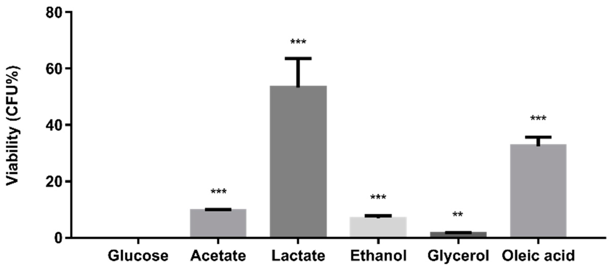
2.1. Alternative Carbon Sources Affect the Planktonic and Biofilm Growth of C. glabrata

As a starting point, the planktonic growth of C. glabrata was compared on glucose and alternative carbon sources using a plate-based microtiter growth assay. C. glabrata grew on all the carbon sources tested in this study, including glucose, acetate, lactate, ethanol, glycerol, and oleic acid (Figure 1a), thereby recapitulating our previous observations [22]. Predictably, the planktonic behaviour of all cultures was similar during the first 4 h of incubation. On glucose, C. glabrata entered exponential phase after 4 h whereas, on alternative carbon sources, the C. glabrata grown remained in the lag phase for longer (over 6 h). Growth was strong on glucose (doubling time, Td ~ 2 h) and glycerol (Td ~ 6 h), medium on lactate (Td ~ 8 h), and less strong on oleic acid (Td ~ 10 h), ethanol (Td ~ 10 h) and acetate (Td ~ 14 h). Although C. glabrata utilises a variety of alternative carbon sources, we infer it has a preference for glycerol and lactate over the other alternative carbon sources tested.
Next, we compared the biofilm growth of C. glabrata on the same carbon sources (Figure 1b). With the exception of glycerol, biofilm growth was reduced significantly on the alternative carbon sources tested when compared with the glucose control. Substantial reductions in biofilm growth were observed for C. glabrata on acetate (52.4%), lactate (30.3%), ethanol (26.9%), and oleic acid (27.3%), thereby confirming our previous report [23]. The ultrastructure of these biofilms was examined by SEM (Figure 2). The C. glabrata biofilms comprised only blastoconidia: no filamentous cells were observed. The aggregates of C. glabrata cells were smaller in biofilms grown in alternative carbon sources, consistent with the reduced growth on these carbon sources.

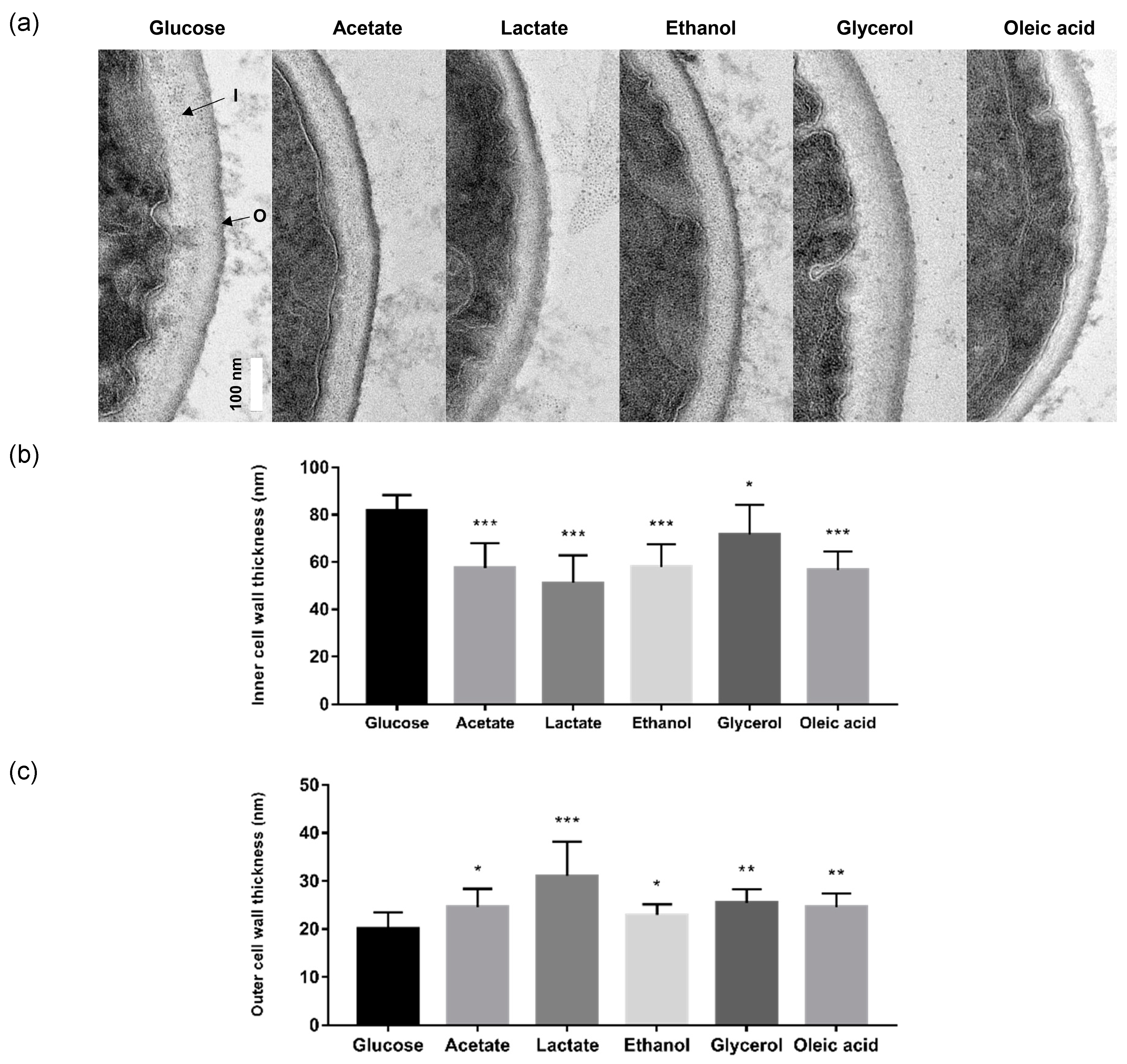
2.2. Alternative Carbon Sources Influence C. glabrata Cell Wall Architecture

The effect of alternative carbon sources on the cell wall architecture of C. glabrata was investigated by TEM. This revealed significant differences in cell wall architecture and thickness depending on the carbon source (Figure 3a). The thickness of the inner cell wall, which comprises largely β-glucan and chitin, was reduced significantly following growth on acetate (57.42 ± 10.55 nm), lactate (51.09 ± 11.80 nm), ethanol (57.94 ± 9.48 nm), and oleic acid (56.72 ± 7.73 nm) when compared with the glucose control (81.70 ± 6.68 nm) (Figure 3b). The effect on the inner cell wall was less dramatic for glycerol (71.73 ± 12.40 nm). In contrast, the thickness of the outer mannan layer of the cell wall increased significantly when C. glabrata cells were grown on the alternative carbon sources: acetate (24.58 ± 3.80 nm), lactate (31.04 ± 7.17 nm), glycerol (25.44 ± 2.82 nm) and oleic acid (24.61 ± 2.78 nm), and glucose control (20.10 ± 3.41 nm) (Figure 3c). The increase for ethanol was marginal (22.93 ± 2.24 nm). In short, utilisation of some alternative carbon sources led to changes in the cell wall architecture of C. glabrata, reducing the thickness of β-glucan and chitin inner layer whilst increasing the thickness of the outer mannan layer.

2.3. Alternative Carbon Sources Reduce the Susceptibility of C. glabrata to Antifungal Drugs

To investigate the impact of carbon sources on the susceptibility of C. glabrata to antifungal drugs, the capacity of C. glabrata cells grown on glucose or alternative carbon sources to withstand amphotericin B was examined using a broth microdilution assay. All glucose-grown C. glabrata cells were killed by treatment with 0.5 μg/mL amphotericin B (Figure 4). Significantly, growth on alternative carbon sources reduced the susceptibility of C. glabrata cells towards amphotericin B, resisting its fungicidal activity to varying degrees (Figure 4). Lactate- and oleate-grown C. glabrata cells were most resistant to amphotericin B whilst acetate-, ethanol- and glycerol-grown cells were only slightly more resistant than the glucose-grown control cells. Our observations suggest that C. glabrata cells growing on alternative carbon sources in vivo may be less sensitive to antifungal therapy.

2.4. Alternative Carbon Sources Modulate the Oxidative Stress Resistance of C. glabrata

Finally, we investigated the impact of alternative carbon sources on the oxidative stress resistance of C. glabrata by examining the sensitivity of this pathogen to hydrogen peroxide (H2O2). As shown in Figure 5, glucose-grown C. glabrata is relatively resistant to high level of H2O2, which is in agreement with previous findings [23]. However, when C. glabrata was grown on alternative carbon sources, it was much more susceptible to H2O2 treatment (Figure 5). We conclude that carbon source impacts significantly upon the oxidative stress resistance of C. glabrata.

3. Discussion

Metabolic adaptation is thought to affect the pathogenicity of C. albicans at multiple levels, influencing the expression of virulence factors as well as fitness attributes and also affecting immune surveillance [15]. In C. glabrata, the assimilation of alternative carbon sources is known to be essential for proliferation in glucose-poor niches [14,22,24]. However, the impact of alternative carbon sources upon C. glabrata fitness attributes and virulence phenotypes had not been addressed. Therefore, we examined the effects upon biofilm formation, cell wall architecture, and antifungal susceptibility in this study.
First we confirmed that glucose is the most efficient carbon source for the planktonic growth of C. glabrata of those we tested [22]. Glucose feeds directly into glycolysis, generating intermediates for growth plus energy through a combination of fermentation, the tricarboxylic acid (TCA) cycle, and respiration [25,26]. In contrast, growth on glycerol or lactate requires functional gluconeogenesis to generate the hexoses and pentoses required for cell wall and nucleic acid biosynthesis [27]. The assimilation of ethanol or acetate also requires a combination of the glyoxylate and TCA cycles to generate anabolic precursors and energy in addition to gluconeogenesis. Moreover, the utilisation of oleate also requires fatty acid β-oxidation. In contrast to C. albicans [16,28], C. glabrata expresses most of the requisite metabolic enzymes for alternative carbon assimilation only when they are needed [27]. Hence, the diauxic shift from the utilisation of glucose to an alternative carbon source requires time for adaptation, as demonstrated by the longer lag phase during the initiation of C. glabrata growth on alternative carbon sources (Figure 1a and Figure 2).
Next, we examined biofilm formation, revealing that C. glabrata forms weaker biofilms on acetate, lactate, ethanol, and oleate when compared with the glucose-grown control (Figure 1b). This observation is in concordance with previous reports showing that C. albicans biofilms are greatly reduced during growth on lactate [29] and that C. glabrata biofilms are diminished by the addition of acetate to glucose-containing medium [20]. Interestingly, glycerol does not seem to affect the growth of C. glabrata in biofilms. This might relate to the essential regulatory role of glycerol in the expression of biofilm-related functions such as adhesins [30]. Glycerol is required for the synthesis of the glycosylphosphatidylinositol (GPI) anchor, which links adhesins and other mannoproteins to the cell wall and potentially assists in biofilm formation [31].
Several factors, including carbon source, hypoxia, pH, and micronutrient limitation, induce cell wall remodelling in C. albicans [2,32,33,34]. We show that changes in carbon source also induce changes in cell wall architecture of C. glabrata (Figure 3). Our data indicate that, with the possible exception of glycerol, growth on carbon sources other than glucose is associated with reductions in the thickness of the inner cell wall and, with the possible exception of ethanol, increases in the thickness of outer mannan layer of the cell wall. This differs slightly from the effects of carbon source on the C. albicans cell wall. According to Ene et al. (2012), both the inner and outer layers of the cell wall are thinner for lactate-grown C. albicans cells when compared with their glucose-grown controls [2]. The mechanisms by which carbon sources affect cell wall architecture remain unclear. Nevertheless, these are likely to include direct effects upon the regulation of cell wall biosynthetic genes combined with different rates of provision of essential cell wall precursors via glycolytic versus gluconeogenic metabolism [2]. This could account for the formation of thinner inner cell walls during growth on alternative carbon sources. The extended mannan fibrils in the outer cell wall could conceivably be due to changes in the balance between the extension versus branching of mannan sidechains; environmental signals have been suggested to modulate structure of mannan fibrils [2,35,36]. The activation of the cell wall integrity pathway is a common response to environmental challenges in human fungal pathogens [37,38,39,40,41]. Whatever the underlying mechanisms, based on recent findings in C. albicans [18,19,34], the changes in the C. glabrata cell wall that accompany carbon source adaptation are likely to affect the visibility of this pathogen to our innate immune defences.
Changes in carbon source are known to affect the resistance of C. albicans to environmental stresses [2,42] and to affect stress signalling mechanisms in this pathogen [43]. C. glabrata displays high levels of intrinsic resistance to such stresses [23], which is related to its evolution as a pathogen that can thrive within the challenging microenvironment of the macrophage [44]. Phylogenetically, C. glabrata is more closely related to S. cerevisiae than C. albicans [45]. In S. cerevisiae, the presence of glucose represses the core stress response via protein kinase A-mediated phosphorylation of the transcription factors Msn2 and Msn4 [46,47]. Candida glabrata has been reported to display a core stress response that resembles that of S. cerevisiae [48] and, on this basis, might be expected to be more sensitive to oxidative stress during growth on glucose. However, our data (Figure 5) suggest that C. glabrata behaves more like C. albicans, which displays glucose-enhanced oxidative stress resistance [49]. This would be consistent with the possibility that, like C. albicans, C. glabrata has evolved an anticipatory response whereby exposure to glucose helps to protect this pathogen against the impending oxidative stress associated with phagocytic assaults [49,50]. The effects of carbon source upon the susceptibility of C. glabrata to amphotericin B (Figure 4) are less surprising and are significant in terms of antifungal therapy. Therefore, we argue that carbon source adaptation in C. glabrata is likely to affect the efficacy of clinical treatments as well as fungal pathogenicity.

4. Materials and Methods

4.1. Strain and Growth Condition

The reference strain C. glabrata ATCC 2001 was used in this study (American Type Culture Collection, Manassas, VA, USA). Standard culture media were used, including YPD (Becton, Dickinson and Company, Franklin Lakes, NJ, USA): yeast extract (1%, w/v), peptone (2%, w/v), glucose (2%, w/v), agar (1.5%, w/v), and yeast nitrogen base (YNB) without amino acids (Becton, Dickinson and Company, USA): yeast nitrogen base (0.67%, w/v), ammonium sulfate (0.5%, w/v). Synthetic complete (SC) media were prepared with YNB without amino acids, supplemented with complete supplement mixture (0.2%, w/v) (Formedium, Hunstanton, UK), glucose (2%, w/v), and agar (2%, w/v). In addition, glucose was replaced with alternative carbon sources: acetate (2%, w/v), lactate (2%, v/v), ethanol (2%, v/v), glycerol (2%, v/v), or oleic acid (0.2%, w/v) (Sigma-Aldrich, St. Louis, MO, USA) as the sole carbon source in SC media [2,51].

4.2. Planktonic Growth Assay

Growth of C. glabrata in glucose and alternative carbon sources was assessed by plate-based microtiter growth assay [22]. Briefly, C. glabrata was grown in YPD overnight at 37 °C, harvested and washed twice with phosphate-buffered saline (PBS), pH 7.4 before resuspended into fresh SC media supplemented with glucose, acetate, lactate, ethanol, glycerol, and oleic acid as the sole carbon source (OD600nm of 0.1). A volume of 200 µL of C. glabrata cell suspension was transferred into a sterile 96-well plate and incubated at 37 °C. Growth of C. glabrata was monitored for 72 h by measuring OD600nm with a microtiter plate reader (Dynex Technologies, Chantilly, VA, USA).

4.3. Biofilm Formation Assay

Biofilm formation of C. glabrata in glucose and alternative carbon sources was determined by using 2,3-bis-(2-methoxy-4-nitro-5-sulfophenyl)-2H-tetrazolium-5-carboxanilide (XTT) reduction assay as previously described [52]. Overnight culture of C. glabrata was harvested, washed twice with PBS, pH 7.4, and resuspended in SC media supplemented with glucose, acetate, lactate, ethanol, glycerol, and oleic acid as sole carbon source (OD600nm of 0.1). A volume of 100 µL of C. glabrata cell suspension was dispensed into selected wells of a pre-sterilised, clear and flat bottom 96-well plate. The 96-well plate was covered with its original lid, sealed with parafilm, and incubated for 48 h at 37 °C. Following 48 h of incubation, the plate was washed twice with PBS, pH 7.4 to remove the planktonic cells. A volume of 100 µL of XTT/menadione solution (0.5 g/L XTT, 10 mM menadione, Sigma-Aldrich, USA) was added to the formed biofilm and incubated in the dark for 3 h at 37 °C. After 3 h of incubation, 80 µL of the solution was transferred to a new 96-well plate and biofilm formation of C. glabrata was quantified by measuring OD490nm of the biofilm using a microtiter plate reader.

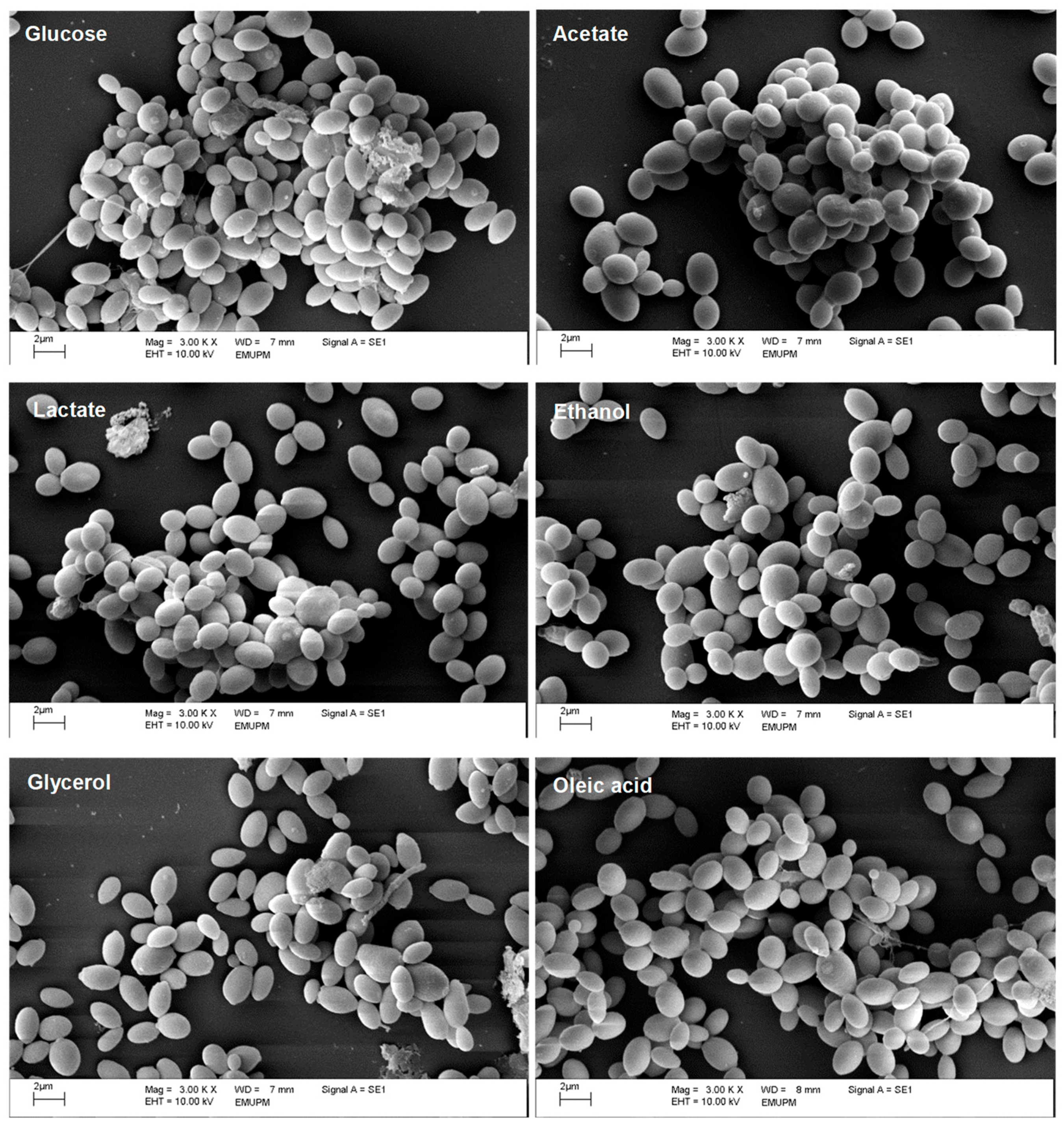
4.4. Visualisation of Biofilm Structures

Biofilm structures of C. glabrata grown in glucose and alternative carbon sources were visualised using SEM. Briefly, overnight culture of C. glabrata was centrifuged, washed twice with PBS, pH 7.4, and resuspended into fresh SC media supplemented with glucose, acetate, lactate, ethanol, glycerol, and oleic acid (OD600nm of 0.1). Cell suspension was dispensed into a pre-sterilised, clear and flat bottom 6-well polystyrene microtiter plate (Becton, Dickinson and Company, USA) with Nunc Thermanox coverslips (Thermo Fisher Scientific, Waltham, MA, USA). The plate was covered with its original lid, sealed with parafilm, and incubated for 48 h at 37 °C. After 48 h of incubation, coverslips were fixed in 4% (v/v) glutaraldehyde (Agar Scientific, Stansted, UK) for 6 h at 4 °C, followed by washing with 0.1 M sodium cacodylate buffer, pH 7.2 (Agar Scientific, UK) for three times of 10 min each. Coverslip was post-fixed in 1% osmium tetroxide (w/v) (Agar Scientific, UK) for 2 h at 4 °C, washed with 0.1 M sodium cacodylate buffer for three times of 10 min each, and dehydrated with increasing concentration of acetone (Friendemann Schmidt, Parkwood, Australia): 35% (v/v) for 10 min, 50% (v/v) for 10 min, 75% (v/v) for 10 min, 95% (v/v) for 10 min and 100% (v/v) for 15 min (three times). Prior to viewing, coverslips were air-dried in EM CPD030 critical point dryer (Leica, Wetzlar, Germany), mounted onto aluminium stubs and sputter coated with gold using EM SCD050 sputter coater (Leica, Germany). Biofilm structures of C. glabrata grown in glucose and alternative carbon sources were observed with LEO 1455 VP SEM (Carl-Zeiss, Oberkochen, Germany).

4.5. Visualisation of Cell Wall Architecture

Cell wall architectures of C. glabrata grown in glucose and alternative carbon sources were investigated by TEM. Briefly, overnight culture of C. glabrata was centrifuged, washed twice with PBS, pH 7.4, and resuspend in SC media supplemented with glucose, acetate, lactate, ethanol, glycerol, and oleic acid (OD600nm of 0.1). After 48 h of incubation at 37 °C, cells were fixed in 4% (v/v) glutaraldehyde for 6 h at 4 °C. Subsequently, fixative was removed, and horse serum was added to coagulate the cell pellets. After overnight incubation, the coagulated cell pellets were diced into 1 mm3 pieces and fixed in 4% (v/v) glutaraldehyde for another 2 h at 4 °C. Following three times of washing with 0.1 M sodium cacodylate buffer, pH 7.2, the samples were post-fixed in 1% osmium tetroxide (w/v) for 2 h at 4 °C, washed three times with 0.1 M sodium cacodylate buffer, and dehydrated with increasing concentration of acetone: 35% (v/v) for 10 min, 50% (v/v) for 10 min, 75% (v/v) for 10 min, 95% (v/v) for 10 min and three times of 100% (v/v) for 15 min. The samples were infiltrated with resin: acetone (1:1) mixture (Agar Scientific, UK) for 1 h, followed by resin: acetone (3:1) mixture for 2 h, 100% resin for overnight and 100% fresh resin for 2 h. Finally, the samples were embedded in BEEM capsules (Agar Scientific, UK) with 100% fresh resin and left to polymerisation for 48 h at 60 °C. Ultrathin sections were cut using EM UC7 ultramicrotome (Leica, Germany) at a thickness of 80 nm. Samples were visualised and imaged with a JEM-2100F field emission electron microscope (JEOL, Tokyo, Japan). Thickness of cell wall layers of C. glabrata (chitin plus β-glucan and mannan) were quantified using Image J by averaging 20 measurements of each cell grown in glucose and alternative carbon sources (n = 10 cells).

4.6. Antifungal Susceptibility Assay

Minimum inhibitory concentration (MIC) of amphotericin B against C. glabrata ATCC 2001 was determined by broth microdilution method according to the Clinical and Laboratory Standards Institute (CLSI) M27-A3 document with slight modification by replacing Roswell Park Memorial Institute (RPMI) 1640 medium with SC medium with 2% glucose. The MIC was confirmed to be 0.5 μg/mL. Antifungal susceptibility assay was performed according to the procedure as previously described [53]. Briefly, overnight culture of C. glabrata was resuspended in SC media supplemented with glucose and alternative carbon sources to OD600nm of 0.1 and regrown to OD600nm of 0.5. Cells growing on acetate, lactate, ethanol, and oleic acid were concentrated by centrifugation to achieve this OD. The cell suspensions were treated with 0.5 μg/mL of amphotericin B for 24 h at 37 °C. Colony-forming-unit (CFU) was determined after incubation and the viability of C. glabrata was defined as (CFU of amphotericin B-treated sample / CFU of untreated sample) × 100%.

4.7. Oxidative Stress Assay

To assess the impact of carbon source on oxidative stress resistance, overnight cultures of C. glabrata grown in YPD were harvested and the cells washed twice with phosphate-buffered saline (PBS), pH 7.4, before resuspension in fresh SC media supplemented with glucose, acetate, lactate, ethanol, glycerol, or oleic acid as sole carbon source (OD600nm of 0.1). Then 200 µL of each C. glabrata cell suspension were transferred into a 96-well microtiter plate and H2O2 (Sigma-Aldrich, USA) added to a final concentration of 0 or 10 mM. Growth was then monitored for 24 h by measuring OD600nm with a microtiter plate reader (Thermo Fisher Scientific, USA).

4.8. Statistical Analyses

Statistical analyses were performed using GraphPad Prism Version 7.0 Software (GraphPad Software Inc., San Diego, CA, USA). All experiments were performed at least in three replicates and all data were expressed as mean values from all replicates with the corresponding standard deviations (SD). Differences between control and samples were assessed by unpaired t-test and a p < 0.05 was considered to be statistically significant. All significant differences were indicated in the figures, with *, **, and *** indicating p < 0.05, p < 0.01 and p < 0.001, respectively.

5. Conclusions

Our data suggest the importance of carbon source adaptation in modulating important fitness and virulence attributes of C. glabrata. These include changes in stress resistance and the cell wall. These observations imply that, as C. glabrata cells adapt to the nutrients available within their local microenvironment within the host, this affects the pathogenicity of this fungus and potentially immune responses against the fungus. Furthermore, our data indicate that carbon source adaptation affects susceptibility of this pathogen to antifungal therapy.

Author Contributions

Conceptualization, S.Y.C., D.S., A.J.P.B. and L.T.L.T.; Methodology, S.Y.C., K.L.H., Y.K.C., D.S., A.J.P.B. and L.T.L.T.; Investigation, S.Y.C.; Resources, K.L.H., Y.K.C. and L.T.L.T.; Formal analysis, S.Y.C. and L.T.L.T.; Funding acquisition, L.T.L.T.; Writing—original draft preparation, S.Y.C.; Writing—review and editing, K.L.H., Y.K.C., D.S., A.J.P.B. and L.T.L.T.

Acknowledgments

This study was funded by Fundamental Research Grant Scheme (FRGS) from Ministry of Education (MOE), Malaysia (Grant number: 01-01-14-1456FR). S.Y.C. is a recipient of the MyBrain 15 Scholarship from MOE, Malaysia. AB was supported by the UK Medical Research Council (www.mrc.ac.uk: MR/M026663/1), the Medical Research Council Centre for Medical Mycology (MR/N006364/1), the Wellcome Trust (www.wellcome.ac.uk: 097377), and the European Commission (FunHoMic: H2020-MSCA-ITN-2018-812969).

Conflicts of Interest

The authors declare no conflict of interest.

References

  1. Abu Kwaik, Y.; Bumann, D. Microbial quest for food in vivo: “Nutritional virulence” as an emerging paradigm. Cell Microbiol. 2013, 15, 882–890. [Google Scholar] [CrossRef]
  2. Ene, I.V.; Adya, A.K.; Wehmeier, S.; Brand, A.C.; MacCallum, D.M.; Gow, N.A.; Brown, A.J. Host carbon sources modulate cell wall architecture, drug resistance and virulence in a fungal pathogen. Cell. Microbiol. 2012, 14, 1319–1335. [Google Scholar] [CrossRef] [PubMed] [Green Version]
  3. Lorenz, M.C.; Bender, J.A.; Fink, G.R. Transcriptional response of Candida albicans upon internalization by macrophages. Eukaryot. Cell 2004, 3, 1076–1087. [Google Scholar] [CrossRef]
  4. Hood, M.I.; Skaar, E.P. Nutritional immunity: Transition metals at the pathogen-host interface. Nat. Rev. Microbiol. 2012, 10, 525–537. [Google Scholar] [CrossRef] [PubMed]
  5. Zhang, Y.J.; Rubin, E.J. Feast or famine: The host-pathogen battle over amino acids. Cell Microbiol. 2013, 15, 1079–1087. [Google Scholar] [CrossRef]
  6. Potrykus, J.; Ballou, E.R.; Childers, D.S.; Brown, A.J. Conflicting interests in the pathogen-host tug of war: Fungal micronutrient scavenging versus mammalian nutritional immunity. PLoS Pathog. 2014, 10, e1003910. [Google Scholar] [CrossRef] [PubMed]
  7. Barelle, C.J.; Priest, C.L.; Maccallum, D.M.; Gow, N.A.; Odds, F.C.; Brown, A.J. Niche-specific regulation of central metabolic pathways in a fungal pathogen. Cell Microbiol. 2006, 8, 961–967. [Google Scholar] [CrossRef]
  8. Almeida, R.S.; Wilson, D.; Hube, B. Candida albicans iron acquisition within the host. FEMS Yeast Res. 2009, 9, 1000–1012. [Google Scholar] [CrossRef]
  9. Ueno, K.; Matsumoto, Y.; Uno, J.; Sasamoto, K.; Sekimizu, K.; Kinjo, Y.; Chibana, H. Intestinal resident yeast Candida glabrata requires Cyb2p-mediated lactate assimilation to adapt in mouse intestine. PLoS ONE 2011, 6, e24759. [Google Scholar] [CrossRef]
  10. Citiulo, F.; Jacobsen, I.D.; Miramón, P.; Schild, L.; Brunke, S.; Zipfel, P.; Brock, M.; Hube, B.; Wilson, D. Candida albicans scavenges host zinc via Pra1 during endothelial invasion. PLoS Pathog. 2012, 8, e1002777. [Google Scholar] [CrossRef]
  11. Sardi, J.C.; Scorzoni, L.; Bernardi, T.; Fusco-Almeida, A.M.; Mendes Giannini, M.J. Candida species: Current epidemiology, pathogenicity, biofilm formation, natural antifungal products and new therapeutic options. J. Med. Microbiol. 2013, 62, 10–24. [Google Scholar] [CrossRef] [PubMed]
  12. Arendrup, M.C. Update on antifungal resistance in Aspergillus and Candida. Clin. Microbiol. Infect. 2014, 20, 42–48. [Google Scholar] [CrossRef] [PubMed]
  13. Beardmore, R.E.; Cook, E.; Nilsson, S.; Smith, A.R.; Tillmann, A.; Esquivel, B.D.; Haynes, K.; Gow, N.A.R.; Brown, A.J.P.; White, T.C.; et al. Drug-mediated metabolic tipping between antibiotic resistant states in a mixed-species community. Nat. Ecol. Evol. 2018, 2, 1312–1320. [Google Scholar] [CrossRef] [PubMed]
  14. Kaur, R.; Ma, B.; Cormack, B.P. A family of glycosylphosphatidylinositol-linked aspartyl proteases is required for virulence of Candida glabrata. Proc. Natl. Acad. Sci. USA 2007, 104, 7628–7633. [Google Scholar] [CrossRef] [PubMed]
  15. Brown, A.J.; Brown, G.D.; Netea, M.G.; Gow, N.A. Metabolism impacts upon Candida immunogenicity and pathogenicity at multiple levels. Trends Microbiol. 2014, 22, 614–622. [Google Scholar] [CrossRef] [PubMed]
  16. Childers, D.S.; Raziunaite, I.; Mol Avelar, G.; Mackie, J.; Budge, S.; Stead, D.; Gow, N.A.; Lenardon, M.D.; Ballou, E.R.; MacCallum, D.M.; et al. The rewiring of ubiquitination targets in a pathogenic yeast promotes metabolic flexibility, host colonization and virulence. PLoS Pathog. 2016, 12, e1005566. [Google Scholar] [CrossRef] [PubMed]
  17. Cunha, D.V.; Salazar, S.B.; Lopes, M.M.; Mira, N.P. Mechanistic insights underlying tolerance to acetic acid stress in vaginal Candida glabrata clinical isolates. Front. Microbiol. 2017, 28, 259. [Google Scholar] [CrossRef]
  18. Ene, I.V.; Cheng, S.C.; Netea, M.G.; Brown, A.J. Growth of Candida albicans cells on the physiologically relevant carbon source lactate affects their recognition and phagocytosis by immune cells. Infect. Immun. 2012, 81, 238–248. [Google Scholar] [CrossRef]
  19. Ballou, E.R.; Avelar, G.M.; Childers, D.S.; Mackie, J.; Bain, J.M.; Wagener, J.; Kastora, S.L.; Panea, M.D.; Hardison, S.E.; Walker, L.A.; et al. Lactate signalling regulates fungal β-glucan masking and immune evasion. Nat. Microbiol. 2016, 12, 16238. [Google Scholar] [CrossRef]
  20. Mota, S.; Alves, R.; Carneiro, C.; Silva, S.; Brown, A.J.; Istel, F.; Kuchler, K.; Sampaio, P.; Casal, M.; Henriques, M.; et al. Candida glabrata susceptibility to antifungals and phagocytosis is modulated by acetate. Front. Microbiol. 2015, 6, 919. [Google Scholar] [CrossRef]
  21. Pfaller, M.A.; Andes, D.R.; Diekema, D.J.; Horn, D.L.; Reboli, A.C.; Rotstein, C.; Franks, B.; Azie, N.E. Epidemiology and outcomes of invasive candidiasis due to non-albicans species of Candida in 2,496 patients: Data from the Prospective Antifungal Therapy (PATH) registry 2004-2008. PLoS ONE 2014, 9, e101510. [Google Scholar] [CrossRef] [PubMed]
  22. Chew, S.Y.; Ho, K.L.; Cheah, Y.K.; Ng, T.S.; Sandai, D.; Brown, A.; Than, L.T.L. Glyoxylate cycle gene ICL1 is essential for the metabolic flexibility and virulence of Candida glabrata. Sci. Rep. 2019, 9, 2843. [Google Scholar] [CrossRef] [PubMed]
  23. Nikolaou, E.; Agrafioti, I.; Stumpf, M.; Quinn, J.; Stansfield, I.; Brown, A.J. Phylogenetic diversity of stress signalling pathways in fungi. BMC Evol. Biol. 2009, 21, 44. [Google Scholar]
  24. Fukuda, Y.; Tsai, H.F.; Myers, T.G.; Bennett, J.E. Transcriptional profiling of Candida glabrata during phagocytosis by neutrophils and in the infected mouse spleen. Infect. Immun. 2013, 81, 1325–1333. [Google Scholar] [CrossRef] [PubMed]
  25. Bertram, G.; Swoboda, R.K.; Gooday, G.W.; Gow, N.A.; Brown, A.J. Structure and regulation of the Candida albicans ADH1 gene encoding an immunogenic alcohol dehydrogenase. Yeast 1996, 12, 115–127. [Google Scholar] [CrossRef]
  26. Askew, C.; Sellam, A.; Epp, E.; Hogues, H.; Mullick, A.; Nantel, A.; Whiteway, M. Transcriptional regulation of carbohydrate metabolism in the human pathogen Candida albicans. PLoS Pathog. 2009, 5, e1000612. [Google Scholar] [CrossRef]
  27. Turcotte, B.; Liang, X.B.; Robert, F.; Soontorngun, N. Transcriptional regulation of nonfermentable carbon utilization in budding yeast. FEMS Yeast Res. 2009, 10, 2–13. [Google Scholar] [CrossRef] [Green Version]
  28. Sandai, D.; Yin, Z.; Selway, L.; Stead, D.; Walker, J.; Leach, M.D.; Bohovych, I.; Ene, I.V.; Kastora, S.; Budge, S.; et al. The evolutionary rewiring of ubiquitination targets has reprogrammed the regulation of carbon assimilation in the pathogenic yeast Candida albicans. MBio 2012, 3, e00495-12. [Google Scholar] [CrossRef]
  29. Pemmaraju, S.C.; Pruthi, P.A.; Prasad, R.; Pruthi, V. Modulation of Candida albicans biofilm by different carbon sources. Mycopathologia 2016, 181, 341–352. [Google Scholar] [CrossRef]
  30. Desai, J.V.; Bruno, V.M.; Ganguly, S.; Stamper, R.J.; Mitchell, K.F.; Solis, N.; Hill, E.M.; Xu, W.; Filler, S.G.; Andes, D.R.; et al. Regulatory role of glycerol in Candida albicans biofilm formation. mBio 2013, 4, e00637-12. [Google Scholar] [CrossRef]
  31. Orlean, P. Architecture and biosynthesis of the Saccharomyces cerevisiae cell wall. Genetics 2012, 192, 775–818. [Google Scholar] [CrossRef] [PubMed]
  32. Sosinska, G.J.; de Groot, P.W.J.; Teixeira de Mattos, M.J.; Dekker, H.L.; de Koster, C.G.; Hellingwerf, K.J.; Klis, F.M. Hypoxic conditions and iron restriction affect the cell-wall proteome of Candida albicans grown under vagina-simulative conditions. Microbiology 2008, 154, 510–520. [Google Scholar] [CrossRef] [PubMed]
  33. Sherrington, S.L.; Sorsby, E.; Mahtey, N.; Kumwenda, P.; Lenardon, M.D.; Brown, I.; Ballou, E.R.; MacCallum, D.M.; Hall, R.A. Adaptation of Candida albicans to environmental pH induces cell wall remodelling and enhances innate immune recognition. PLoS Pathog. 2017, 13, e1006403. [Google Scholar] [CrossRef] [PubMed]
  34. Pradhan, A.; Avelar, G.M.; Bain, J.M.; Childers, D.S.; Larcombe, D.E.; Netea, M.G.; Shekhova, E.; Munro, C.A.; Brown, G.D.; Erwig, L.P.; et al. Hypoxia promotes immune evasion by triggering β-glucan masking on the Candida albicans cell surface via mitochondrial and cAMP-protein kinase A signaling. mBio 2018, 9, e01318. [Google Scholar] [CrossRef] [PubMed]
  35. Kruppa, M.; Greene, R.R.; Noss, I.; Lowman, D.W.; Williams, D.L. Candida albicans increases cell wall mannoprotein, but not mannan, in response to blood, serum and cultivation at physiological temperature. Glycobiology 2011, 21, 1173–1180. [Google Scholar] [CrossRef] [PubMed]
  36. Lowman, D.W.; Ensley, H.E.; Greene, R.R.; Knagge, K.J.; Williams, D.L.; Kruppa, M.D. Mannan structural complexity is decreased when Candida albicans is cultivated in blood or serum at physiological temperature. Carbohydr. Res. 2011, 346, 2752–2759. [Google Scholar] [CrossRef] [PubMed]
  37. Ram, A.F.; Kapteyn, J.C.; Montijn, R.C.; Caro, L.H.; Douwes, J.E.; Baginsky, W.; Mazur, P.; van den Ende, H.; Klis, F.M. Loss of the plasma membrane-bound protein Gas1p in Saccharomyces cerevisiae results in the release of beta 1, 3-glucan into the medium and induces a compensation mechanism to ensure cell wall integrity. J. Bacteriol. 1998, 180, 1418–1424. [Google Scholar] [PubMed]
  38. Okawa, Y.; Goto, K. Antigenicity of cell wall mannans of Candida albicans and Candida stellatoidea cultured at high temperatures in BACTEC medium. Biol. Pharm. Bull. 2006, 29, 1723–1727. [Google Scholar] [CrossRef]
  39. Munro, C.A.; Selvaggini, S.; de Bruijn, I.; Walker, L.; Lenardon, M.D.; Gerssen, B.; Milne, S.; Brown, A.J.; Gow, N.A. The PKC, HOG and Ca2+ signalling pathways co-ordinately regulate chitin synthesis in Candida albicans. Mol. Microbiol. 2007, 63, 1399–1413. [Google Scholar] [CrossRef]
  40. Walker, L.A.; Munro, C.A.; de Bruijn, I.; Lenardon, M.D.; McKinnon, A.; Gow, N.A. Stimulation of chitin synthesis rescues Candida albicans from echinocandins. PLoS Pathog. 2008, 4, e1000040. [Google Scholar] [CrossRef]
  41. Lee, K.K.; Maccallum, D.M.; Jacobsen, M.D.; Walker, L.A.; Odds, F.C.; Gow, N.A.; Munro, C.A. Elevated cell wall chitin in Candida albicans confers echinocandin resistance in vivo. Antimicrob. Agents Chemother. 2012, 56, 208–217. [Google Scholar] [CrossRef] [PubMed]
  42. Ene, I.V.; Walker, L.A.; Schiavone, M.; Lee, K.K.; Martin-Yken, H.; Dague, E.; Gow, N.A.; Munro, C.A.; Brown, A.J. Cell wall remodeling enzymes modulate fungal cell wall elasticity and osmotic stress resistance. mBio 2015, 6, e00986. [Google Scholar] [CrossRef] [PubMed]
  43. Kastora, S.L.; Herrero-de-Dios, C.; Avelar, G.M.; Munro, C.A.; Brown, A.J.P. Sfp1 and Rtg3 reciprocally modulate carbon source-conditional stress adaptation in the pathogenic yeast Candida albicans. Mol. Microbiol. 2017, 105, 620–636. [Google Scholar] [CrossRef] [PubMed]
  44. Kasper, L.; Seider, K.; Hube, B. Intracellular survival of Candida glabrata in macrophages: Immune evasion and persistence. FEMS Yeast Res. 2015, 15, fov042. [Google Scholar] [CrossRef] [PubMed]
  45. Shen, X.X.; Zhou, X.; Kominek, J.; Kurtzman, C.P.; Hittinger, C.T.; Rokas, A. Reconstructing the backbone of the Saccharomycotina yeast phylogeny using genome-scale data. G3 2016, 6, 3927–3939. [Google Scholar] [CrossRef]
  46. Gorner, W.; Durchschlag, E.; Martinez-Pastor, M.T.; Estruch, F.; Ammerer, G.; Hamilton, B.; Ruis, H.; Schüller, C. Nuclear localization of the C2H2 zinc finger protein Msn2p is regulated by stress and protein kinase A activity. Genes Dev. 1998, 12, 586–597. [Google Scholar] [CrossRef]
  47. Garreau, H.; Hasan, R.N.; Renault, G.; Estruch, F.; Boy-Marcotte, E.; Jacquet, M. Hyperphosphorylation of Msn2p and Msn4p in response to heat shock and the diauxic shift is inhibited by cAMP in Saccharomyces cerevisiae. Microbiology 2000, 146, 2113–2120. [Google Scholar] [CrossRef]
  48. Roetzer, A.; Gregori, C.; Jennings, A.M.; Quintin, J.; Ferrandon, D.; Butler, G.; Kuchler, K.; Ammerer, G.; Schüller, C. Candida glabrata environmental stress response involves Saccharomyces cerevisiae Msn2/4 orthologous transcription factors. Mol. Microbiol. 2008, 69, 603–620. [Google Scholar] [CrossRef]
  49. Rodaki, A.; Bohovych, I.M.; Enjalbert, B.; Young, T.; Odds, F.C.; Gow, N.; Brown, A. Glucose promotes stress resistance in the fungal pathogen Candida albicans. Mol. Biol. Cell 2009, 20, 4845–4855. [Google Scholar] [CrossRef]
  50. Brown, A.; Gow, N.; Warris, A.; Brown, G.D. Memory in fungal pathogens promotes immune evasion, colonisation, and infection. Trends Microbiol. 2019, 27, 219–230. [Google Scholar] [CrossRef]
  51. Sherman, F. Getting started with yeast. Methods Enzymol. 1991, 194, 3–21. [Google Scholar] [PubMed]
  52. Pierce, C.G.; Uppuluri, P.; Tristan, A.R.; Wormley, F.L., Jr.; Mowat, E.; Ramage, G.; Lopez-Ribot, J.L. A simple and reproducible 96-well plate-based method for the formation of fungal biofilms and its application to antifungal susceptibility testing. Nat. Protoc. 2008, 3, 1494–1500. [Google Scholar] [CrossRef] [PubMed]
  53. Ng, T.S.; Desa, M.N.M.; Sandai, D.; Chong, P.P.; Than, L.T.L. Growth, biofilm formation, antifungal susceptibility and oxidative stress resistance of Candida glabrata are affected by different glucose concentrations. Infect. Genet. Evol. 2015, 40, 331–338. [Google Scholar] [CrossRef] [PubMed]
Figure 1. Alternative carbon sources modulate the planktonic and biofilm growth of C. glabrata. (a) Planktonic growth of C. glabrata in synthetic complete (SC) media supplemented with 2% glucose, 2% acetate, 2% lactate, 2% ethanol, 2% glycerol, or 0.2% oleic acid and incubated for 72 h at 37 °C; (b) Biofilm formation of C. glabrata in SC media supplemented with 2% glucose, 2% acetate, 2% lactate, 2% ethanol, 2% glycerol, or 0.2% oleic acid. Results were presented as means ± SD. * p < 0.05, ** p < 0.01 and *** p < 0.001 were considered statistically significant relative to the control (2% glucose). All experiments were conducted in triplicate, and each independent experiment was repeated three times.
Ijms 20 03172 g001
Figure 2. Effect of glucose and alternative carbon sources on the biofilm structures of C. glabrat. Representative scanning electron photomicrographs from 48 h biofilms of C. glabrata grown on glucose and alternative carbon sources, viewed under scanning electron microscopy (SEM) at a magnification of 3000×. Scale bar represents 2 µm.
Ijms 20 03172 g002
Figure 3. Alternative carbon sources alter the ultrastructure of the C. glabrata cell wall. (a) Representative transmission electron microscopy (TEM) images of cell wall architectures from C. glabrata grown on glucose and alternative carbon sources, [I] indicates inner cell wall layer (β-glucan and chitin) and [O] indicates outer cell wall layer (mannan). Scale bar represents 100 nm. (b) Quantification of thickness of the inner cell wall. (c) Quantification of the thickness of the outer cell wall. Results are presented as means ± SD from 10 individual cells by averaging 20 measurements obtained from the cell periphery of each C. glabrata cells. * p < 0.05, ** p < 0.01 and *** p < 0.001 were considered statistically significant relative to glucose-grown cells.
Ijms 20 03172 g003
Figure 4. Alternative carbon sources reduce the susceptibility of C. glabrata to amphotericin B. Results are presented as means ± SD. ** p < 0.01 and *** p < 0.001 were considered statistically significant relative to glucose-grown cells.
Ijms 20 03172 g004
Figure 5. Alternative carbon sources affect the oxidative stress resistance of C. glabrata. Growth of C. glabrata in SC media supplemented with glucose or alternative carbon sources in the presence or absence of H2O2 was monitored for 24 h at 37 °C.
Ijms 20 03172 g005

