Transcriptomic Insights into the Response of the Olfactory Bulb to Selenium Treatment in a Mouse Model of Alzheimer’s Disease

Abstract
:1. Introduction
2. Results
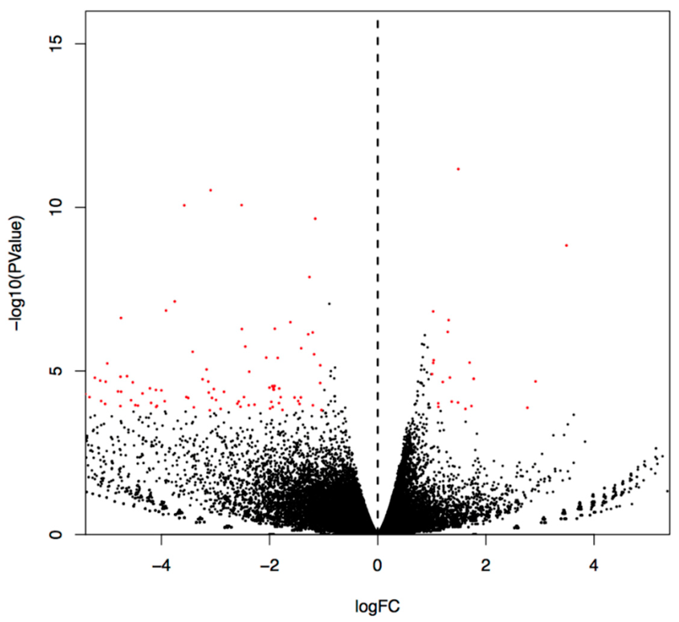
2.1. Differential Gene Expression between 3×Tg-AD Mice and Se-Met-Treated 3×Tg-AD Mice
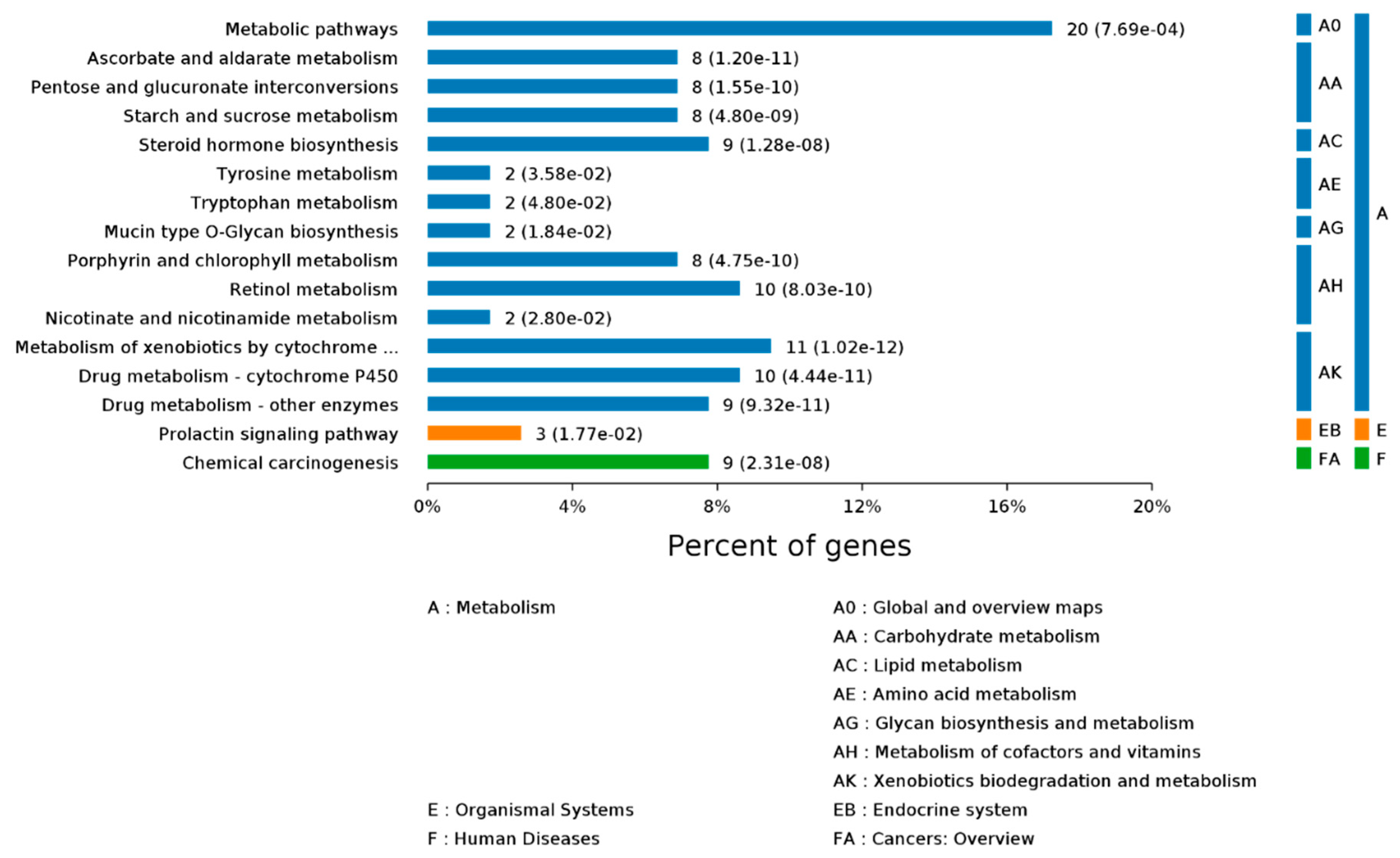
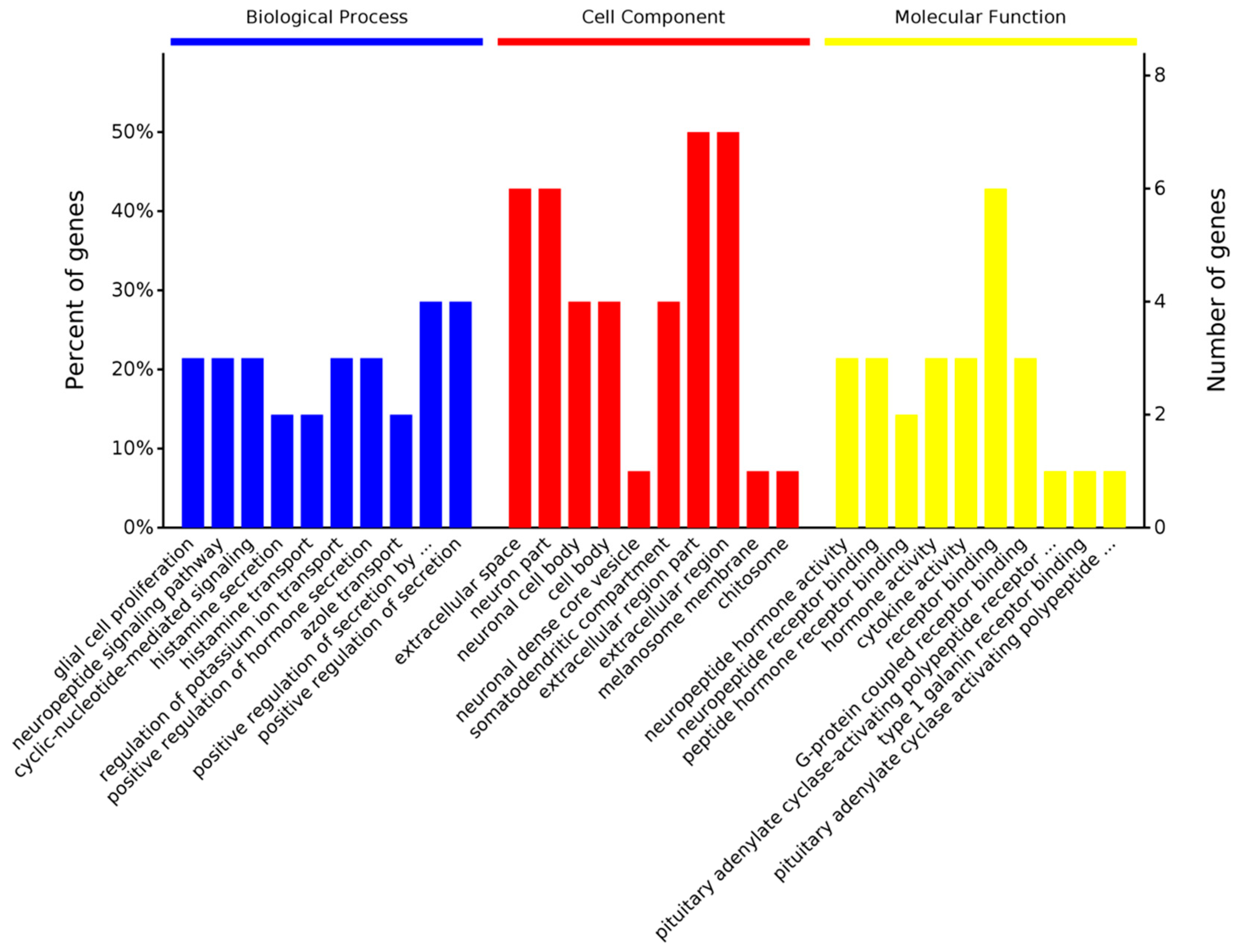
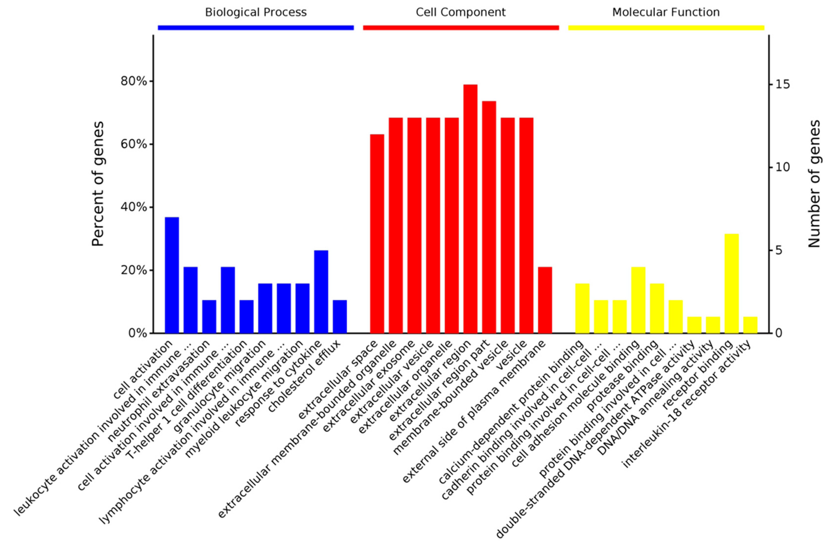
2.2. Overrepresentation Analysis
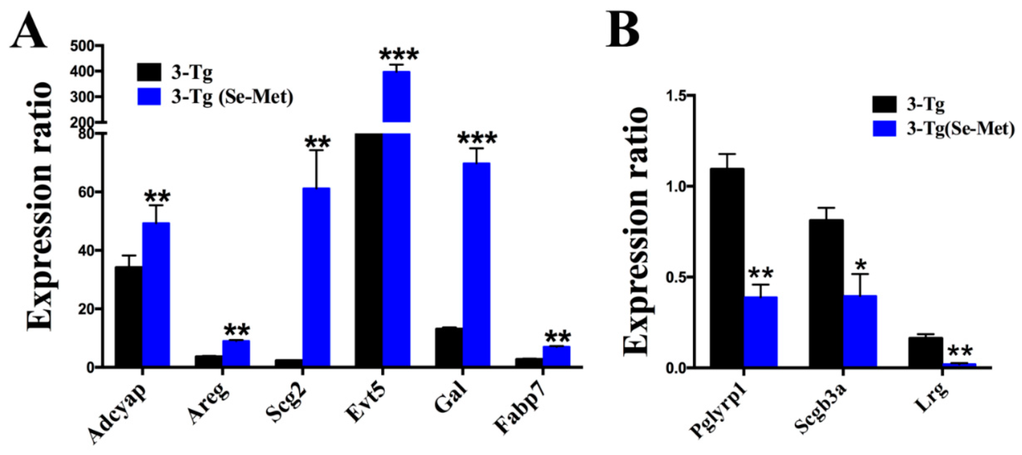
2.3. Real-Time PCR Validation of Differentially Expressed Genes
3. Discussion
3.1. Upregulated Genes in 3×Tg-AD (Se-Met) Mice
3.2. Downregulated Genes in 3×Tg-AD (Se-Met) Mice
4. Material and Methods
4.1. Transgenic Mice
4.2. Tissue Processing
4.3. RNA-Seq Analysis
4.4. Differential Expression Analysis
4.5. Real-Time PCR
4.6. Statistical Analysis
Supplementary Materials
Consent for Publication
Ethics Approval and Consent to Participate
Author Contributions
Funding
Acknowledgments
Conflicts of Interest
References
- Qiu, W.Y.; Yang, Q.; Zhang, W.; Wang, N.; Zhang, D.; Huang, Y.; Ma, C. The correlations between postmortem brain pathologies and cognitive dysfunction in aging and Alzheimer’s disease. Curr. Alzheimer Res. 2017, 15, 462–473. [Google Scholar] [CrossRef] [PubMed]
- Libro, R.; Bramanti, P.; Mazzon, E. Endogenous glucocorticoids: Role in the etiopathogenesis of Alzheimer’s disease. Neuro Endocrinol. Lett. 2017, 38, 1–12. [Google Scholar] [PubMed]
- Jevtic, S.; Sengar, A.S.; Salter, M.W.; McLaurin, J. The role of the immune system in Alzheimer disease: Etiology and treatment. Ageing Res. Rev. 2017, 40, 84–94. [Google Scholar] [CrossRef] [PubMed]
- Rey, N.L.; Wesson, D.W.; Brundin, P. The olfactory bulb as the entry site for prion-like propagation in neurodegenerative diseases. Neurobiol. Dis. 2016, 109, 226–248. [Google Scholar] [CrossRef]
- Arnold, S.E.; Lee, E.B.; Moberg, P.J.; Stutzbach, L.; Kazi, H.; Han, L.Y.; Lee, V.M.; Trojanowski, J.Q. Olfactory epithelium amyloid-beta and paired helical filament-tau pathology in Alzheimer disease. Ann. Neurol. 2010, 67, 462–469. [Google Scholar] [CrossRef]
- Xu, Z.P.; Yang, S.L.; Zhao, S.; Zheng, C.H.; Li, H.H.; Zhang, Y.; Huang, R.X.; Li, M.Z.; Gao, Y.; Zhang, S.J.; et al. Biomarkers for early diagnostic of mild cognitive impairment in type-2 diabetes patients: A multicentre, retrospective, nested case-control study. EBioMedicine 2016, 5, 105–113. [Google Scholar] [CrossRef]
- Dammalli, M.; Dey, G.; Madugundu, A.K.; Kumar, M.; Rodrigues, B.; Gowda, H.; Siddaiah, B.G.; Mahadevan, A.; Shankar, S.K.; Prasad, T.S.K. Proteomic analysis of the human olfactory bulb. OMICS 2017, 21, 440–453. [Google Scholar] [CrossRef] [PubMed]
- Marciniuk, K.; Ryan, T.; Scott, N. Evidence for Prion-Like Mechanisms in Several Neurodegenerative Diseases: Potential Implications for Immunotherapy. Clin. Dev. Immunol. 2013, 2013, 1–20. [Google Scholar] [CrossRef] [Green Version]
- Zhang, Z.H.; Wu, Q.Y.; Zheng, R.; Chen, C.; Chen, Y.; Liu, Q.; Hoffmann, P.R.; Ni, J.Z.; Song, G.L. Selenomethionine mitigates cognitive decline by targeting both tau hyperphosphorylation and autophagic clearance in an Alzheimer’s disease mouse model. J. Neurosci. 2017, 37, 2449–2462. [Google Scholar] [CrossRef]
- Zhang, Z.H.; Chen, C.; Wu, Q.Y.; Zheng, R.; Chen, Y.; Liu, Q.; Ni, J.Z.; Song, G.L. Selenomethionine ameliorates neuropathology in the olfactory bulb of a triple transgenic mouse model of Alzheimer’s disease. Int. J. Mol. Sci. 2016, 17, 1595. [Google Scholar] [CrossRef]
- Zhang, Z.H.; Wu, Q.Y.; Chen, C.; Zheng, R.; Chen, Y.; Ni, J.Z.; Song, G.L. Comparison of the effects of selenomethionine and selenium-enriched yeast in the triple-transgenic mouse model of Alzheimer’s disease. Food Funct. 2018, 9, 3965–3973. [Google Scholar] [CrossRef]
- Zheng, R.; Zhang, Z.H.; Chen, C.; Chen, Y.; Jia, S.Z.; Liu, Q.; Ni, J.Z.; Song, G.L. Selenomethionine promoted hippocampal neurogenesis via the PI3K-Akt-GSK3beta-Wnt pathway in a mouse model of Alzheimer’s disease. Biochem. Biophys. Res. Commun. 2017, 485, 6–15. [Google Scholar] [CrossRef]
- Zhang, Z.H.; Chen, C.; Wu, Q.Y.; Zheng, R.; Liu, Q.; Ni, J.Z.; Hoffmann, P.R.; Song, G.L. Selenomethionine reduces the deposition of beta-amyloid plaques by modulating beta-secretase and enhancing selenoenzymatic activity in a mouse model of Alzheimer’s disease. Metallomics 2016, 8, 782–789. [Google Scholar] [CrossRef] [PubMed]
- Theodore, J.A. A Framework for Comparative Analysis of Gene Expressions and Mutations Linked to Cancer. Ph.D. Thesis, The George Washington University, Washington, DC, USA, 2014. [Google Scholar]
- Van Dijck, A.; Vloeberghs, E.; Van Dam, D.; Staufenbiel, M.; De Deyn, P.P. Evaluation of the APP23-model for Alzheimer’s disease in the odour paired-associate test for hippocampus-dependent memory. Behav. Brain Res. 2008, 190, 147–151. [Google Scholar] [CrossRef] [PubMed]
- Hipp, G.; Diederich, N.J.; Pieria, V.; Vaillant, M. Primary vision and facial emotion recognition in early Parkinson’s disease. J. Neurol. Sci. 2014, 338, 178–182. [Google Scholar] [CrossRef]
- Suto, T.; Meguro, K.; Nakatsuka, M.; Kato, Y.; Tezuka, K.; Yamaguchi, S.; Tashiro, M. Disorders of “taste cognition” are associated with insular involvement in patients with Alzheimer’s disease and vascular dementia: “Memory of food is impaired in dementia and responsible for poor diet”. Int. Psychogeriatr. 2014, 26, 1127–1138. [Google Scholar] [CrossRef]
- Vicente Miranda, H.; El-Agnaf, O.M.A.; Outeiro, T.F. Glycation in Parkinson’s disease and Alzheimer’s disease. Mov. Disord. 2016, 31, 782–790. [Google Scholar] [CrossRef]
- Pintana, H.; Apaijai, N.; Kerdphoo, S.; Pratchayasakul, W.; Sripetchwandee, J.; Suntornsaratoon, P.; Charoenphandhu, N.; Chattipakorn, N.; Chattipakorn, S.C. Hyperglycemia induced the Alzheimer’s proteins and promoted loss of synaptic proteins in advanced-age female Goto-Kakizaki (GK) rats. Neurosci. Lett. 2017, 655, 41. [Google Scholar] [CrossRef] [PubMed]
- Bakir, B.; Sanli, S.; Bakir, V.L.; Ayas, S.; Yildiz, S.O.; Iyibozkurt, A.C.; Kartal, M.G.; Yavuz, E. Role of diffusion weighted MRI in the differential diagnosis of endometrial cancer, polyp, hyperplasia, and physiological thickening. Clin. Imaging 2017, 41, 86–94. [Google Scholar] [CrossRef] [PubMed]
- Arrieta-Cruz, I.; Gutiérrez-Juárez, R. The role of insulin resistance and glucose metabolism dysregulation in the development of Alzheimer´s disease. Revista Investig. Clin. 2016, 68, 53–58. [Google Scholar]
- Tang, Y.; Soroush, F.; Sun, S.; Liverani, E.; Langston, J.C.; Yang, Q.; Kilpatrick, L.E.; Kiani, M.F. Protein kinase C-delta inhibition protects blood-brain barrier from sepsis-induced vascular damage. J. Neuroinflamm. 2018, 15, 309. [Google Scholar] [CrossRef]
- Bennett, C.; Mohammed, F.; Álvarez-Ciara, A.; Nguyen, M.A.; Dietrich, W.D.; Rajguru, S.M.; Streit, W.J.; Prasad, A. Neuroinflammation, oxidative stress, and blood-brain barrier (BBB) disruption in acute Utah electrode array implants and the effect of deferoxamine as an iron chelator on acute foreign body response. Biomaterials 2018, 188, 144–159. [Google Scholar] [CrossRef]
- Martens, E.J.; Hoen, P.W.; Mittelhaeuser, M.; De Jonge, P.; Denollet, J. Symptom dimensions of post-myocardial infarction depression, disease severity and cardiac prognosis. Psychol. Med. 2010, 40, 807. [Google Scholar] [CrossRef]
- Nieweg, K.; Schaller, H.F. Marked differences in cholesterol synthesis between neurons and glial cells from postnatal rats. J. Neurochem. 2010, 109, 125–134. [Google Scholar] [CrossRef]
- Vance, J.E.; Pan, D.; Campenot, R.B.; Bussière, M.; Vance, D.E. Evidence that the major membrane lipids, except cholesterol, are made in axons of cultured rat sympathetic neurons. J. Neurochem. 2010, 62, 329–337. [Google Scholar] [CrossRef]
- Stocker, H.; Mollers, T.; Perna, L.; Brenner, H. The genetic risk of Alzheimer’s disease beyond APOE epsilon4: Systematic review of Alzheimer’s genetic risk scores. Transl. Psychiatr. 2018, 8, 166. [Google Scholar] [CrossRef]
- Kim, Y.; Kim, C.; Jang, H.Y.; Mook-Jung, I. Inhibition of cholesterol biosynthesis reduces γ-secretase activity and amyloid-î² generation. J. Alzheimers Dis. 2016, 51, 1057–1068. [Google Scholar] [CrossRef]
- Wang, C.; Shou, Y.; Pan, J.; Du, Y.; Liu, C.; Wang, H. The relationship between cholesterol level and Alzheimer’s disease-associated APP proteolysis/Aβ metabolism. Nutr. Neurosci. 2018, 22, 1–11. [Google Scholar] [CrossRef]
- Hui, L.; Chen, X.; Geiger, J.D. Endolysosome involvement in LDL cholesterol-induced Alzheimer’s disease-like pathology in primary cultured neurons. Life Sci. 2012, 91, 1159–1168. [Google Scholar] [CrossRef]
- Sawamura, N.; Gong, J.S.; Chang, T.Y.; Yanagisawa, K.; Michikawa, M. Promotion of tau phosphorylation by MAP kinase Erk1/2 is accompanied by reduced cholesterol level in detergent-insoluble membrane fraction in Niemann-Pick C1-deficient cells. J. Neurochem. 2010, 84, 1086–1096. [Google Scholar] [CrossRef]
- Eunjeong, Y.; Sangzin, A.; Junghwa, R.; Moonseok, C.; Shinkyu, C.; Young, H.C.; Jinwon, H.; Moonjeong, C.; Hyesun, K. The reduced protein level of synaptophysin and PSD-95 was restored by phloroglucinol in 5XFAD Tg mouse model. PLoS ONE 2015, 10, 8–22. [Google Scholar]
- Han, P.; Caselli, R.J.; Baxter, L.; Serrano, G.; Yin, J.; Beach, T.G.; Reiman, E.M.; Shi, J. Association of pituitary adenylate cyclase-activating polypeptide with cognitive decline in mild cognitive impairment due to Alzheimer disease. JAMA Neurol. 2015, 72, 333–339. [Google Scholar] [CrossRef]
- Hashimoto, R.; Hashimoto, H.; Shintani, N.; Ohi, K.; Hori, H.; Saitoh, O.; Kosuga, A.; Tatsumi, M.; Iwata, N.; Ozaki, N.; et al. Possible association between the pituitary adenylate cyclase-activating polypeptide (PACAP) gene and major depressive disorder. Neurosci. Lett. 2010, 468, 300–302. [Google Scholar] [CrossRef]
- Rat, D.; Schmitt, U.; Tippmann, F.; Dewachter, I.; Theunis, C.; Wieczerzak, E.; Postina, R.; Van Leuven, F.; Fahrenholz, F.; Kojro, E. Neuropeptide pituitary adenylate cyclase-activating polypeptide (PACAP) slows down Alzheimer’s disease-like pathology in amyloid precursor protein-transgenic mice. FASEB J. 2011, 25, 3208–3218. [Google Scholar] [CrossRef]
- Reglodi, D.; Kiss, P.; Lubics, A.; Tamas, A. Review on the protective effects of PACAP in models of neurodegenerative diseases in vitro and in vivo. Curr. Pharm Des. 2011, 17, 962–972. [Google Scholar] [CrossRef]
- Shieh, P.C.; Tsao, C.W.; Li, J.S.; Wu, H.T.; Wen, Y.J.; Kou, D.H.; Cheng, J.T. Role of pituitary adenylate cyclase-activating polypeptide (PACAP) in the action of ginsenoside Rh2 against beta-amyloid-induced inhibition of rat brain astrocytes. Neurosci. Lett. 2008, 434, 1–5. [Google Scholar] [CrossRef]
- Mao, S.S.; Zhang, Y.M. Research advance of the protective role of PACAP in the nervous system diseases. Sheng Li Ke Xue Jin Zhan Progr. 2011, 42, 276–280. [Google Scholar]
- Koster, K.P.; Thomas, R.; Morris, A.W.; Tai, L.M. Epidermal growth factor prevents oligomeric amyloid-beta induced angiogenesis deficits in vitro. J. Cereb. Blood Flow Metab. 2016, 36, 1865–1871. [Google Scholar] [CrossRef]
- Thomas, R.; Zuchowska, P.; Morris, A.W.; Marottoli, F.M.; Sunny, S.; Deaton, R.; Gann, P.H.; Tai, L.M. Epidermal growth factor prevents APOE4 and amyloid-beta-induced cognitive and cerebrovascular deficits in female mice. Acta Neuropathol. Commun. 2016, 4, 111. [Google Scholar] [CrossRef] [Green Version]
- Li, T.; Wen, H.; Brayton, C.; Das, P.; Smithson, L.A.; Fauq, A.; Fan, X.; Crain, B.J.; Price, D.L.; Golde, T.E.; et al. Epidermal growth factor receptor and notch pathways participate in the tumor suppressor function of gamma-secretase. J. Biol. Chem. 2007, 282, 32264–32273. [Google Scholar] [CrossRef]
- Lachen-Montes, M.; Zelaya, M.V.; Segura, V.; Fernandez-Irigoyen, J.; Santamaria, E. Progressive modulation of the human olfactory bulb transcriptome during Alzheimer’s disease evolution: Novel insights into the olfactory signaling across proteinopathies. Oncotarget 2017, 8, 69663–69679. [Google Scholar] [CrossRef]
- Guillemot, J.; Thouennon, E.; Guerin, M.; Vallet-Erdtmann, V.; Ravni, A.; Montero-Hadjadje, M.; Lefebvre, H.; Klein, M.; Muresan, M.; Seidah, N.; et al. Differential expression and processing of secretogranin II in relation to the status of pheochromocytoma: Implications for the production of the tumoral marker EM66. J. Mol. Endocrinol. 2012, 48, 115–127. [Google Scholar] [CrossRef]
- Willis, M.; Prokesch, M.; Hutter-Paier, B.; Windisch, M.; Stridsberg, M.; Mahata, S.K.; Kirchmair, R.; Wietzorrek, G.; Knaus, H.G.; Jellinger, K.; et al. Chromogranin B and Secretogranin II in transgenic mice overexpressing human APP751 with the London (V717I) and Swedish (K670M/N671L) mutations and in Alzheimer patients. J. Alzheimers Dis. 2008, 13, 123–135. [Google Scholar] [CrossRef]
- Hart, J.E.; Clarke, I.J.; Risbridger, G.P.; Ferneyhough, B.; Vega-Hernandez, M. Mysterious inhibitory cell regulator investigated and found likely to be secretogranin II related. PeerJ 2017, 5, e3833. [Google Scholar] [CrossRef] [PubMed] [Green Version]
- Fischer-Colbrie, R.; Kirchmair, R.; Kahler, C.M.; Wiedermann, C.J.; Saria, A. Secretoneurin: A new player in angiogenesis and chemotaxis linking nerves, blood vessels and the immune system. Curr. Protein Pept. Sci. 2005, 6, 373–385. [Google Scholar] [CrossRef]
- Venditti, P.; Masullo, P.; Di Meo, S.; Agnisola, C. Effects of prolonged aerobic exercise on myocardial responses to ischaemia-reperfusion in the rat. Exp. Physiol. 2001, 86, 341–348. [Google Scholar] [CrossRef]
- Vo, T.; Wang, S.; Poon, G.M.K.; Wilson, W.D. Electrostatic control of DNA intersegmental translocation by the ETS transcription factor ETV6. J. Biol. Chem. 2017, 292, 13187–13196. [Google Scholar] [CrossRef] [Green Version]
- Van Pham, P.; Vu, N.B.; Nguyen, H.T.; Dao, T.T.T.; Le, H.T.N.; Phi, L.T.; Nguyen, O.T.K.; Phan, N.K. ETV-2 activated proliferation of endothelial cells and attenuated acute hindlimb ischemia in mice. Vitro Cell Dev. Biol. Anim. 2017, 53, 616–625. [Google Scholar] [CrossRef]
- Young, J.K.; Heinbockel, T.; Gondre-Lewis, M.C. Astrocyte fatty acid binding protein-7 is a marker for neurogenic niches in the rat hippocampus. Hippocampus 2013, 23, 1476–1483. [Google Scholar] [CrossRef] [Green Version]
- Matsumata, M.; Inada, H.; Osumi, N. Fatty acid binding proteins and the nervous system: Their impact on mental conditions. Neurosci. Res. 2016, 102, 47–55. [Google Scholar] [CrossRef] [Green Version]
- Teunissen, C.E.; Veerhuis, R.; De Vente, J.; Verhey, F.R.J.; Vreeling, F.; van Boxtel, M.P.J.; Glatz, J.F.C.; Pelsers, M.A.L. Brain-specific fatty acid-binding protein is elevated in serum of patients with dementia-related diseases. Eur. J. Neurol. 2011, 18, 865–871. [Google Scholar] [CrossRef] [PubMed]
- Hoeijmakers, L.; Meerhoff, G.F.; de Vries, J.W.; Ruigrok, S.R.; van Dam, A.M.; van Leuven, F.; Korosi, A.; Lucassen, P.J. The age-related slow increase in amyloid pathology in APP.V717I mice activates microglia, but does not alter hippocampal neurogenesis. Neurobiol. Aging 2017, 61, 112–123. [Google Scholar] [CrossRef] [PubMed]
- Horgusluoglu-Moloch, E.; Nho, K.; Risacher, S.L.; Kim, S.; Foroud, T.; Shaw, L.M.; Trojanowski, J.Q.; Aisen, P.S.; Petersen, R.C.; Jack, C.R., Jr.; et al. Targeted neurogenesis pathway-based gene analysis identifies ADORA2A associated with hippocampal volume in mild cognitive impairment and Alzheimer’s disease. Neurobiol. Aging 2017, 60, 92–103. [Google Scholar] [CrossRef] [PubMed]
- Beart, P.M. Synaptic signalling and its interface with neuropathologies: Snapshots from the past, present and future. J. Neurochem. 2016, 139 (Suppl. 2), 76–90. [Google Scholar] [CrossRef]
- Ádori, C.; Glück, L.; Barde, S.; Yoshitake, T.; Kovacs, G.G.; Mulder, J.; Maglóczky, Z.; Havas, L.; Bölcskei, K.; Mitsios, N.; et al. Critical role of somatostatin receptor 2 in the vulnerability of the central noradrenergic system: New aspects on Alzheimer’s disease. Acta Neuropathol. 2015, 129, 541–563. [Google Scholar] [CrossRef] [PubMed]
- Counts, S.E.; Perez, S.E.; Ginsberg, S.D.; Mufson, E.J. Neuroprotective role for galanin in Alzheimer’s disease. EXS 2010, 102, 143–162. [Google Scholar] [PubMed]
- Baraka, A.; ElGhotny, S. Study of the effect of inhibiting galanin in Alzheimer’s disease induced in rats. Eur. J. Pharmacol. 2010, 641, 123–127. [Google Scholar] [CrossRef]
- Manuel, I.; Lombardero, L.; LaFerla, F.M.; Gimenez-Llort, L.; Rodriguez-Puertas, R. Activity of muscarinic, galanin and cannabinoid receptors in the prodromal and advanced stages in the triple transgenic mice model of Alzheimer’s disease. Neuroscience 2016, 329, 284–293. [Google Scholar] [CrossRef]
- Nakajima, M.; Miyajima, M.; Ogino, I.; Watanabe, M.; Miyata, H.; Karagiozov, K.L.; Arai, H.; Hagiwara, Y.; Segawa, T.; Kobayashi, K.; et al. Leucine-rich alpha-2-glycoprotein is a marker for idiopathic normal pressure hydrocephalus. Acta Neurochir Wien 2011, 153, 1339–1346; discussion 46. [Google Scholar] [CrossRef]
- Jingami, N.; Asada-Utsugi, M.; Uemura, K.; Noto, R.; Takahashi, M.; Ozaki, A.; Kihara, T.; Kageyama, T.; Takahashi, R.; Shimohama, S.; et al. Idiopathic normal pressure hydrocephalus has a different cerebrospinal fluid biomarker profile from Alzheimer’s disease. J. Alzheimers Dis. 2015, 45, 109–115. [Google Scholar] [CrossRef]
- Pek, S.L.T.; Cheng, A.K.S.; Lin, M.X.; Wong, M.S.; Chan, E.Z.L.; Moh, A.M.C.; Sum, C.F.; Lim, S.C.; Tavintharan, S. Association of circulating proinflammatory marker, leucine-rich-alpha2-glycoprotein (LRG1), following metabolic/bariatric surgery. Diabetes Metab. Res. Rev. 2018, 34, e3029. [Google Scholar] [CrossRef] [PubMed]
- Read, C.B.; Kuijper, J.L.; Hjorth, S.A.; Heipel, M.D.; Tang, X.; Fleetwood, A.J.; Dantzler, J.L.; Grell, S.N.; Kastrup, J.; Wang, C.; et al. Cutting Edge: Identification of neutrophil PGLYRP1 as a ligand for TREM-1. J. Immunol. 2015, 194, 1417–1421. [Google Scholar] [CrossRef]
- Yin, S.J.; Cho, I.H.; Yang, H.S.; Park, Y.D.; Yang, J.M. Analysis of the peptides detected in atopic dermatitis and various inflammatory diseases patients-derived sera. Int. J. Biol. Macromol. 2017, 106, 1052–1061. [Google Scholar] [CrossRef]
- Kim, S.K.; Seok, H.; Park, H.J.; Han, K.; Kang, S.W.; Ban, J.Y.; Jung, H.J.; Kim, K.I.; Lee, B.J.; Kim, J.; et al. Association between secretoglobin family 3A member 2 (SCGB3A2) gene polymorphisms and asthma in a Korean population. Med. Sci. Monit. 2017, 23, 1880–1885. [Google Scholar] [CrossRef]
- Tokuda, T.; Tanaka, K.; Kametani, F.; Ikeda, S.; Yanagisawa, N. Secretory form of beta-amyloid precursor protein is much abundantly contained in the cerebral white matter in human brain. Neurosci. Lett. 1994, 175, 33–36. [Google Scholar] [CrossRef]
- Haakensen, V.D.; Bjøro, T.; Lüders, T.; Riis, M.; Bukholm, I.K.; Kristensen, V.N.; Troester, M.A.; Homen, M.M.; Ursin, G.; Børresen-Dale, A.L.; et al. Serum estradiol levels associated with specific gene expression patterns in normal breast tissue and in breast carcinomas. BMC Cancer 2011, 11, 332. [Google Scholar] [CrossRef] [PubMed]
- Danielsen, S.; Lind, G.; Berg, E.V.D.; Kolberg, M.; Mertens, F.; Lothe, R. RASSF1A and SCGB3A1 are common target genes for promoter hypermethylation in malignant peripheral nerve sheath tumors. Cancer Res. 2007, 67, 2875. [Google Scholar]
- Oddo, S.; Caccamo, A.; Kitazawa, M.; Tseng, B.P.; LaFerla, F.M. Amyloid deposition precedes tangle formation in a triple transgenic model of Alzheimer’s disease. Neurobiol. Aging 2003, 24, 1063–1070. [Google Scholar] [CrossRef] [PubMed]
- Trapnell, C.; Pachter, L.; Salzberg, S.L. TopHat: Discovering splice junctions with RNA-Seq. Bioinformatics 2009, 25, 1105–1111. [Google Scholar] [CrossRef] [PubMed]
- Anders, S.; Huber, W. Differential expression analysis for sequence count data. Genome Biol. 2010, 11, R106. [Google Scholar] [CrossRef]
- Huang, D.W.; Sherman, B.T.; Tan, Q.; Kir, J.; Liu, D.; Bryant, D.; Guo, Y.; Stephens, R.; Baseler, M.W.; Lane, H.C.; et al. DAVID bioinformatics resources: Expanded annotation database and novel algorithms to better extract biology from large gene lists. Nucleic Acids Res. 2007, 35, W169–W175. [Google Scholar] [CrossRef]
- Huttenrauch, M.; Baches, S.; Gerth, J.; Bayer, T.A.; Weggen, S.; Wirths, O. Neprilysin deficiency alters the neuropathological and behavioral phenotype in the 5XFAD mouse model of Alzheimer’s disease. J. Alzheimers Dis. 2015, 44, 1291–1302. [Google Scholar] [CrossRef] [PubMed]
- Huang da, W.; Sherman, B.T.; Lempicki, R.A. Systematic and integrative analysis of large gene lists using DAVID bioinformatics resources. Nat. Protoc. 2009, 4, 44–57. [Google Scholar] [CrossRef] [PubMed]
- Havugimana, P.C.; Hu, P.; Emili, A. Protein complexes, big data, machine learning and integrative proteomics: Lessons learned over a decade of systematic analysis of protein interaction networks. Expert Rev. Proteom. 2017, 14, 845–855. [Google Scholar] [CrossRef] [PubMed]
- Wirths, O.; Breyhan, H.; Marcello, A.; Cotel, M.C.; Bruck, W.; Bayer, T.A. Inflammatory changes are tightly associated with neurodegeneration in the brain and spinal cord of the APP/PS1KI mouse model of Alzheimer’s disease. Neurobiol. Aging 2010, 31, 747–757. [Google Scholar] [CrossRef]
- Botstein, D.; Cherry, J.M.; Ashburner, M.; Ball, C.A.; Blake, J.A.; Butler, H.; Davis, A.P.; Dolinski, K.; Dwight, S.S.; Eppig, J.T. Gene ontology: Tool for the unification of biology. Nat. Genet. 2000, 25, 25–29. [Google Scholar]
- Faure, A.; Verret, L.; Bozon, B.; El Tayara, N.E.T.; Ly, M.; Kober, F.; Dhenain, M.; Rampon, C.; Delatour, B. Impaired neurogenesis, neuronal loss, and brain functional deficits in the APPxPS1-Ki mouse model of Alzheimer’s disease. Neurobiol. Aging 2011, 32, 407–418. [Google Scholar] [CrossRef]


| Category | GO Class | Function | Sample Number | Background Number | p Value | Gene Names |
|---|---|---|---|---|---|---|
| BP | GO:0052697 | xenobiotic glucuronidation | 4 | 5 | 2.44 × 10⁻⁷ | Ugt1a2; Ugt1a1; Ugt1a10; Ugt1a6a |
| BP | GO:0052696 | flavonoid glucuronidation | 4 | 9 | 1.36 × 10⁻⁶ | Ugt1a2; Ugt1a1; Ugt1a10; Ugt1a6a |
| BP | GO:0009813 | flavonoid biosynthetic process | 4 | 9 | 1.36 × 10⁻⁶ | Ugt1a2; Ugt1a1; Ugt1a10; Ugt1a6a |
| BP | GO:0052695 | cellular glucuronidation | 4 | 10 | 1.89 × 10⁻⁶ | Ugt1a2; Ugt1a1; Ugt1a10; Ugt1a6a |
| BP | GO:0019585 | glucuronate metabolic process | 4 | 11 | 2.56 × 10⁻⁶ | Ugt1a2; Ugt1a1; Ugt1a10; Ugt1a6a |
| BP | GO:0006063 | uronic acid metabolic process | 4 | 11 | 2.56 × 10⁻⁶ | Ugt1a2; Ugt1a1; Ugt1a10; Ugt1a6a |
| BP | GO:0009812 | flavonoid metabolic process | 4 | 11 | 2.56 × 10⁻⁶ | Ugt1a2; Ugt1a1; Ugt1a10; Ugt1a6a |
| BP | GO:0006805 | xenobiotic metabolic process | 5 | 54 | 4.93 × 10⁻⁵ | Ugt1a2; Ugt1a1; Cyp2f2; Ugt1a10; Ugt1a6a |
| BP | GO:0071466 | cellular response to xenobiotic stimulus | 5 | 58 | 6.77 × 10⁻⁵ | Ugt1a2; Ugt1a1; Cyp2f2; Ugt1a10; Ugt1a6a |
| BP | GO:0009410 | response to xenobiotic stimulus | 5 | 64 | 1.04 × 10⁻⁴ | Ugt1a2; Ugt1a1; Cyp2f2; Ugt1a10; Ugt1a6a |
| CC | GO:0005615 | extracellular space | 25 | 1011 | 7.36 × 10⁻⁸ | Pigr; Adcyap1; Tacstd2; Fetub; Pglyrp1; Gpx3; Sftpd; Dmbt1; Pon1; Ces1d; Lbp; Scgb3a1; Mup4; C1s; Bpifb1; Areg; Msln; Anxa1; Tnfsf15; Serpinb11; Bpifa1; Nppa; Il1r1; Scg2; St14 |
| CC | GO:0005576 | extracellular region | 52 | 3593 | 6.55 × 10⁻⁵ | Pigr; Reg3g; Bpifa1; Tspan1; Tacstd2; Krt14; Ccdc3; Vwf; Fetub; Pglyrp1; Gpx3; Scnn1a; Krt5; Sftpd; Dmbt1; Ces1d; Pon1; Agr2; Obp2a; Cd44; Sult1c1; Lbp; Chi3l4; Scgb3a1; Mup4; Wfdc18; C1s; Gsta3; Muc20; Il1r1; Bpifb1; Krt18; Areg; Wfdc2; Msln; Lypd2; Anxa1; Tnfsf15; Epcam; Serpinb11; Steap4; Slc5a9; Cd177; Clic6; Adcyap1; Ugt1a6a; St14; Tmprss2; Slc44a4; Slc39a4; Scg2; Nppa |
| CC | GO:0044421 | extracellular region part | 44 | 3071 | 5.7 × 10⁻⁶ | Pigr; Adcyap1; Tspan1; Tacstd2; Krt14; Vwf; Fetub; Pglyrp1; Gpx3; Scnn1a; Krt5; Sftpd; Dmbt1; Ces1d; Pon1; Cd44; Sult1c1; Lbp; Scgb3a1; Mup4; C1s; Gsta3; Il1r1; Bpifb1; Krt18; Areg; Wfdc2; Msln; Anxa1; Tnfsf15; Epcam; Serpinb11; Steap4; Slc5a9; Cd177; Clic6; Bpifa1; Ugt1a6a; St14; Tmprss2; Slc44a4; Slc39a4; Scg2; Nppa |
| CC | GO:0016323 | basolateral plasma membrane | 8 | 176 | 3.91 × 10⁻⁵ | Tacstd2; Cd44; St14; Cldn8; Epcam; Cldn7; Muc20; Aqp3 |
| CC | GO:0070062 | extracellular vesicular exosome | 32 | 2280 | 1.4 × 10⁻⁴ | Pigr; Pglyrp1; Tspan1; Tacstd2; Krt14; Gsta3; Fetub; Scnn1a; Krt5; Dmbt1; Pon1; Cd44; Sult1c1; Lbp; Scgb3a1; Gpx3; C1s; Vwf; Bpifb1; Krt18; Wfdc2; Anxa1; Epcam; Steap4; Slc5a9; Cd177; Clic6; Ugt1a6a; Tmprss2; Slc44a4; Slc39a4; St14 |
| CC | GO:1903561 | extracellular vesicle | 32 | 2280 | 1.41 × 10⁻⁴ | Pigr; Pglyrp1; Tspan1; Tacstd2; Krt14; Gsta3; Fetub; Scnn1a; Krt5; Dmbt1; Pon1; Cd44; Sult1c1; Lbp; Scgb3a1; Gpx3; C1s; Vwf; Bpifb1; Krt18; Wfdc2; Anxa1; Epcam; Steap4; Slc5a9; Cd177; Clic6; Ugt1a6a; Tmprss2; Slc44a4; Slc39a4; St14 |
| CC | GO:0043230 | extracellular organelle | 32 | 2284 | 1.44 × 10⁻⁴ | Pigr; Pglyrp1; Tspan1; Tacstd2; Krt14; Gsta3; Fetub; Scnn1a; Krt5; Dmbt1; Pon1; Cd44; Sult1c1; Lbp; Scgb3a1; Gpx3; C1s; Vwf; Bpifb1; Krt18; Wfdc2; Anxa1; Epcam; Steap4; Slc5a9; Cd177; Clic6; Ugt1a6a; Tmprss2; Slc44a4; Slc39a4; St14 |
| CC | GO:0065010 | extracellular membrane-bounded organelle | 32 | 2284 | 1.45 × 10⁻⁴ | Pigr; Pglyrp1; Tspan1; Tacstd2; Krt14; Gsta3; Fetub; Scnn1a; Krt5; Dmbt1; Pon1; Cd44; Sult1c1; Lbp; Scgb3a1; Gpx3; C1s; Vwf; Bpifb1; Krt18; Wfdc2; Anxa1; Epcam; Steap4; Slc5a9; Cd177; Clic6; Ugt1a6a; Tmprss2; Slc44a4; Slc39a4; St14 |
| CC | GO:0031988 | membrane-bounded vesicle | 35 | 2756 | 4.34 × 10⁻⁴ | Pigr; Pglyrp1; Tspan1; Tacstd2; Krt14; Th; Gsta3; Fetub; Gpx3; Scnn1a; Krt5; Dmbt1; Pon1; Cd44; Sult1c1; Lbp; Scgb3a1; Scg2; C1s; Vwf; Bpifb1; Krt18; Wfdc2; Galnt15; Anxa1; Epcam; Steap4; Slc5a9; Cd177; Clic6; Ugt1a6a; Tmprss2; Slc44a4; Slc39a4; St14 |
| CC | GO:0098590 | plasma membrane region | 10 | 428 | 8.69 × 10⁻⁴ | Aqp3; Cd44; St14; Cldn8; Slc39a4; Scnn1a; Epcam; Cldn7; Muc20; Tacstd2 |
| MF | GO:0015020 | glucuronosyltransferase activity | 5 | 74 | 1.99 × 10⁻⁴ | Ugt1a2; Ugt1a1; Galnt15; Ugt1a6a; Ugt1a10 |
| MF | GO:0008194 | UDP-glycosyltransferase activity | 6 | 135 | 4.03 × 10⁻⁴ | Ugt1a2; Galnt15; Ugt1a1; Ugt1a6a; Gal; Ugt1a10 |
| MF | GO:0016758 | transferase activity, transferring hexosyl groups | 6 | 165 | 1.11 × 10⁻⁴ | Ugt1a2; Galnt15; Ugt1a1; Ugt1a6a; Gal; Ugt1a10 |
| MF | GO:0005154 | epidermal growth factor receptor binding | 3 | 30 | 1.4 × 10⁻³ | Pigr; Cd44; Areg |
| MF | GO:0016757 | transferase activity, transferring glycosyl groups | 7 | 243 | 1.62 × 10⁻³ | St6galnac1; Ugt1a2; Galnt15; Ugt1a1; Ugt1a6a; Gal; Ugt1a10 |
| MF | GO:0001972 | retinoic acid binding | 2 | 12 | 3.85 × 10⁻³ | Ugt1a2; Ugt1a1 |
| MF | GO:0019865 | immunoglobulin binding | 2 | 14 | 5.03 × 10⁻³ | Vwf; Fcamr |
| MF | GO:0038024 | cargo receptor activity | 3 | 53 | 6.34 × 10⁻³ | Ildr1; Tmprss2; Dmbt1 |
| MF | GO:0051428 | peptide hormone receptor binding | 2 | 16 | 6.36 × 10⁻³ | Adcyap1; Nppa |
| MF | GO:0033293 | monocarboxylic acid binding | 3 | 55 | 6.9 × 10⁻³ | Ugt1a2; Ugt1a1; Fabp7 |
| Gene Name | Gene Description | logFC | p_Value |
|---|---|---|---|
| Adcyap1 | adenylate cyclase activating polypeptide 1 | 1.49 | 6.74 × 10⁻¹² |
| Areg | amphiregulin | 3.49 | 1.46 × 10⁻⁹ |
| Scg2 | secretogranin II | 1.02 | 1.51 × 10⁻⁷ |
| Th | tyrosine hydroxylase | 1.31 | 2.8 × 10⁻⁷ |
| Fabp7 | fatty acid binding protein 7 | 1.29 | 6.41 × 10⁻⁷ |
| Pip5k1b | phosphatidylinositol-4-phosphate 5-kinase, type 1 beta | 1.03 | 4.68 × 10⁻⁶ |
| Trhr2 | thyrotropin releasing hormone receptor 2 | 1.69 | 5.55 × 10⁻⁶ |
| Etv5 | ets variant 5 | 1.02 | 5.68 × 10⁻⁵ |
| fantom3_1110005E01 | /// | 1.01 | 1.25 × 10⁻⁵ |
| Col28a1 | collagen, type XXVIII, alpha 1 | 1.33 | 1.59 × 10⁻⁵ |
| fantom3_D930027K06 | /// | 1.77 | 1.73 × 10⁻⁵ |
| fantom3_D930001D16 | /// | 1.77 | 1.73 × 10⁻⁵ |
| Gm10635 | predicted gene 10635 | 2.91 | 2.09 × 10⁻⁵ |
| Nppa | natriuretic peptide type A | 1.20 | 2.18 × 10⁻⁵ |
| Nt5c1a | 5′-nucleotidase, cytosolic IA | 1.36 | 8.67 × 10⁻⁵ |
| Gal | galanin | 1.48 | 9.23 × 10⁻⁵ |
| fantom3_A430024L20 | /// | 1.12 | 9.76 × 10⁻⁵ |
| Rxfp1 | relaxin/insulin-like family peptide receptor 1 | 1.73 | 1.18 × 10⁻⁴ |
| fantom3_C330006P03 | /// | 1.12 | 1.25 × 10⁻⁴ |
| Tnfsf15 | tumor necrosis factor (ligand) superfamily, member 15 | 2.76 | 1.33 × 10⁻⁴ |
| Gene Name | Gene Description | logFC | p_Value |
|---|---|---|---|
| Lrg1 | leucine-rich alpha-2-glycoprotein 1 | −3.08 | 2.99 × 10⁻¹¹ |
| Anxa1 | annexin A1 | −2.51 | 8.52 × 10⁻¹¹ |
| Cd177 | CD177 antigen | −3.57 | 8.66 × 10⁻¹¹ |
| Vwf | Von Willebrand factor | −1.15 | 2.21 × 10⁻¹⁰ |
| Il1r1 | interleukin 1 receptor, type I | −1.26 | 1.35 × 10⁻⁹ |
| Scgb3a1 | secretoglobin, family 3A, member 1 | −3.75 | 7.5 × 10⁻⁸ |
| fantom3_1110030K16 | /// | −3.91 | 1.42 × 10⁻⁷ |
| Abca13 | ATP-binding cassette, sub-family A (ABC1), member 13 | −4.74 | 2.4 × 10⁻⁷ |
| Pglyrp1 | peptidoglycan recognition protein 1 | −1.61 | 3.23 × 10⁻⁷ |
| Bpifb1 | BPI fold containing family B, member 1 | −6.70 | 4.13 × 10⁻⁷ |
| Gpx3 | glutathione peroxidase 3 | −1.90 | 5.13 × 10⁻⁷ |
| Dlk1 | delta-like 1 homolog (Drosophila) | −2.51 | 5.23 × 10⁻⁷ |
| Gabrp | gamma-aminobutyric acid (GABA) A receptor, pi | −6.28 | 5.51 × 10⁻⁷ |
| Serpina3n | serine (or cysteine) peptidase inhibitor, clade A, member 3N | −1.20 | 6.65 × 10⁻⁷ |
| S100a11 | S100 calcium binding protein A11 | −1.28 | 7.58 × 10⁻⁷ |
| Muc5b | mucin 5, subtype B, tracheobronchial | −6.35 | 7.95 × 10⁻⁷ |
| Lypd2 | Ly6/Plaur domain containing 2 | −5.61 | 8.2 × 10⁻⁷ |
| Pon1 | paraoxonase 1 | −7.07 | 9.8 × 10⁻⁷ |
| Il18r1 | interleukin 18 receptor 1 | −2.44 | 1.79 × 10⁻⁶ |
| Ccdc3 | coiled-coil domain containing 3 | −1.41 | 2.03 × 10⁻⁶ |
| Genes Name | Sequence Information | |
|---|---|---|
| Adcyap1 | Forward | 5′-GGAGAAAAGTGGAGGGAGCA-3′ |
| Reverse | 5′-TGTCTATACCTTTTCCCAAGGACTG-3′ | |
| Areg | Forward | 5′-TGGATTGGACCTCAATGACA-3′ |
| Reverse | 5′-AGCCAGGTATTTGTGGTTCG-3′ | |
| Scg2 | Forward | 5′-CCAATGGTCATGGTATTGACA-3′ |
| Reverse | 5′-TTTGCTCCAGAACTCCACAA-3′ | |
| Fabp7 | Forward | 5′-AGGCTTTCTGTGCTAC-3′ |
| Reverse | 5′-ATTACCGTTGGTTTGG-3′ | |
| Evt5 | Forward | 5′-GAAGTGCCTAACTGCCAGTCACCC-3′ |
| Reverse | 5′-GGCACCACGCAAGTGTCATCGA-3′ | |
| Gal | Forward | 5′-CACTCTGGGACTTGGGATG-3′ |
| Reverse | 5′-CAGGC AAGAGGGAGTTACAA-3′ | |
| GAPDH | Forward | 5′-CAGCCTCGTCTCATAGACAAGATG-3′ |
| Reverse | 5′-CAATGTCCACTTTGTCACAAGAGAA-3′ | |
| Lrg | Forward | 5′-GGAGCAGCTATGGTCTCTTG-3′ |
| Reverse | 5′-AGTATCAGGCATTCCTTGAG-3′ | |
| Scgb3a1 | Forward | 5′-CTCAACCCGCTGAAGCTC-3′ |
| Reverse | 5′-CTTCTGGGAGCCCTCTATGA-3′ | |
| Pglyrp1 | Forward | 5′-GTGGTGATCTCACACACAGC-3′ |
| Reverse | 5′-GTGTGGTCACCCTTGATGTT-3′ | |
© 2019 by the authors. Licensee MDPI, Basel, Switzerland. This article is an open access article distributed under the terms and conditions of the Creative Commons Attribution (CC BY) license (http://creativecommons.org/licenses/by/4.0/).
Share and Cite
Zheng, R.; Zhang, Z.-H.; Zhao, Y.-X.; Chen, C.; Jia, S.-Z.; Cao, X.-C.; Shen, L.-M.; Ni, J.-Z.; Song, G.-L. Transcriptomic Insights into the Response of the Olfactory Bulb to Selenium Treatment in a Mouse Model of Alzheimer’s Disease. Int. J. Mol. Sci. 2019, 20, 2998. https://doi.org/10.3390/ijms20122998
Zheng R, Zhang Z-H, Zhao Y-X, Chen C, Jia S-Z, Cao X-C, Shen L-M, Ni J-Z, Song G-L. Transcriptomic Insights into the Response of the Olfactory Bulb to Selenium Treatment in a Mouse Model of Alzheimer’s Disease. International Journal of Molecular Sciences. 2019; 20(12):2998. https://doi.org/10.3390/ijms20122998
Chicago/Turabian StyleZheng, Rui, Zhong-Hao Zhang, Yu-Xi Zhao, Chen Chen, Shi-Zheng Jia, Xian-Chun Cao, Li-Ming Shen, Jia-Zuan Ni, and Guo-Li Song. 2019. "Transcriptomic Insights into the Response of the Olfactory Bulb to Selenium Treatment in a Mouse Model of Alzheimer’s Disease" International Journal of Molecular Sciences 20, no. 12: 2998. https://doi.org/10.3390/ijms20122998