Article

Prostaglandin D2 Induces Ca2+ Sensitization of Contraction without Affecting Cytosolic Ca2+ Level in Bronchial Smooth Muscle

1
Department of Physiology and Molecular Sciences, School of Pharmacy, Hoshi University, 2-4-41 Ebara, Shinagawa-ku, Tokyo 142-8501, Japan
2
Global Research Center for Innovative Life Science, School of Pharmacy, Hoshi University, 2-4-41 Ebara, Shinagawa-ku, Tokyo 142-8501, Japan
3
Peptide Drug Innovation Global Research Center for Innovative Life Science, School of Pharmacy, Hoshi University, 2-4-41 Ebara, Shinagawa-ku, Tokyo 142-8501, Japan
4
Department of Biomolecular Pharmacology, School of Pharmacy, Hoshi University, 2-4-41 Ebara, Shinagawa-ku, Tokyo 142-8501, Japan
*
Author to whom correspondence should be addressed.
Int. J. Mol. Sci. 2018, 19(10), 3036; https://doi.org/10.3390/ijms19103036
Submission received: 10 September 2018 / Revised: 2 October 2018 / Accepted: 3 October 2018 / Published: 5 October 2018

Abstract

:
Prostaglandin D2 (PGD2) is one of the key lipid mediators of allergic airway inflammation, including bronchial asthma. However, the role of PGD2 in the pathogenesis of asthma is not fully understood. In the present study, the effect of PGD2 on smooth muscle contractility of the airways was determined to elucidate its role in the development of airway hyperresponsiveness (AHR). In isolated bronchial smooth muscles (BSMs) of naive mice, application of PGD2 (10−9–10−5 M) had no effect on the baseline tension. However, when the tissues were precontracted partially with 30 mM K+ (in the presence of 10−6 M atropine), PGD2 markedly augmented the contraction induced by the high K+ depolarization. The PGD2-induced augmentation of contraction was significantly inhibited both by 10−6 M laropiprant (a selective DP1 antagonist) and 10−7 M Y-27632 (a Rho-kinase inhibitor), indicating that a DP1 receptor-mediated activation of Rho-kinase is involved in the PGD2-induced BSM hyperresponsiveness. Indeed, the GTP-RhoA pull-down assay revealed an increase in active form of RhoA in the PGD2-treated mouse BSMs. On the other hand, in the high K+-depolarized cultured human BSM cells, PGD2 caused no further increase in cytosolic Ca2+ concentration. These findings suggest that PGD2 causes RhoA/Rho-kinase-mediated Ca2+ sensitization of BSM contraction to augment its contractility. Increased PGD2 level in the airways might be a cause of the AHR in asthma.

1. Introduction

Augmented airway responsiveness to a wide variety of nonspecific stimuli, called airway hyperresponsiveness (AHR), is a common feature of allergic asthma. A cause of the AHR is hypercontraction of smooth muscle cells of the airways [1,2,3,4,5]. Rapid remission from airway limitation in asthma attack by inhalation of short-acting beta-stimulant also suggests an involvement of increased airway smooth muscle contraction in the airway obstruction. It is thus important for development of asthma therapy to understand the disease-associated alterations of the contractile signaling of airway smooth muscle cells.
Prostaglandin D2 (PGD2), one of the cyclooxygenase (COX) metabolites, is the major lipid mediator released from mast cells in allergic reaction, and has been suggested to be involved in the pathogenesis of bronchial asthma. An increase in PGD2 level in bronchoalveolar lavage (BAL) fluids was demonstrated in experimental asthma models [6,7]. In asthmatic subjects, allergen challenge to the airways caused an increase in PGD2 in BAL fluids [8,9]. It has been suggested that PGD2 mediates allergic inflammation, including the airway inflammation in asthma. In mice lacking receptors for PGD2 (DP1 receptors), both airway eosinophilia and upregulation of proinflammatory cytokines in BAL fluids induced by allergen challenge were diminished as compared to wild-type animals [10]. PGD2 also caused cytokine release via an activation of a PGD2 receptor, CRTH2 (also called as DP2 receptor), in Th2 lymphocytes [11].
On the other hand, the functional role of PGD2 on airway smooth muscle remains unclear. Application of PGD2 to the isolated smooth muscle strips caused contraction in guinea pig trachea [12] and dog bronchus [13]. In contrast, PGD2 elicited a relaxation in the murine tracheal smooth muscle precontracted with carbachol [10]. In the present study, to elucidate its role in the development of AHR in asthma, the effect of PGD2 on smooth muscle contractility was determined using bronchial rings isolated from mice.

2. Results

2.1. Effects of Prostaglandin D2 (PGD2) on Bronchial Smooth Muscle (BSM) Function

The RT-PCR analyses revealed that both DP1 and DP2 receptors were expressed both in hBSMCs and murine BSMs (Figure 1), indicating that PGD2 could directly act on BSM cells. To determine the role of PGD2 on the BSM function, its effect on the isometric tension of smooth muscles was examined in BSM tissues isolated from naive control mice. Application of PGD2 (10−9–10−5 M) had no effect on basal tone of the BSM tissues (Figure 2A). However, when the BSMs were precontracted with 30 mM K+, application of PGD2 caused an enhancement of the contraction induced by high K+ depolarization, in a PGD2 concentration-dependent manner (10−6 and 10−5 M: Figure 2B,C).
PGD2 has been known to act on G protein-coupled receptors (GPCRs), mainly the PGD2 receptor 1 (DP1) and 2 (DP2). To elucidate receptor(s) responsible for the enhanced contraction induced by PGD2, effects of laropiprant (a selective DP1 receptor antagonist [14]) and fevipiprant (a selective DP2 receptor antagonist [15]) on the PGD2-induced augmentation of contraction were tested. As a result, the enhanced contraction induced by PGD2 was inhibited by laropiprant (10−6 M: Figure 3A,B), whereas fevipiprant (10−6 M) had no effect on it (Figure 3C).

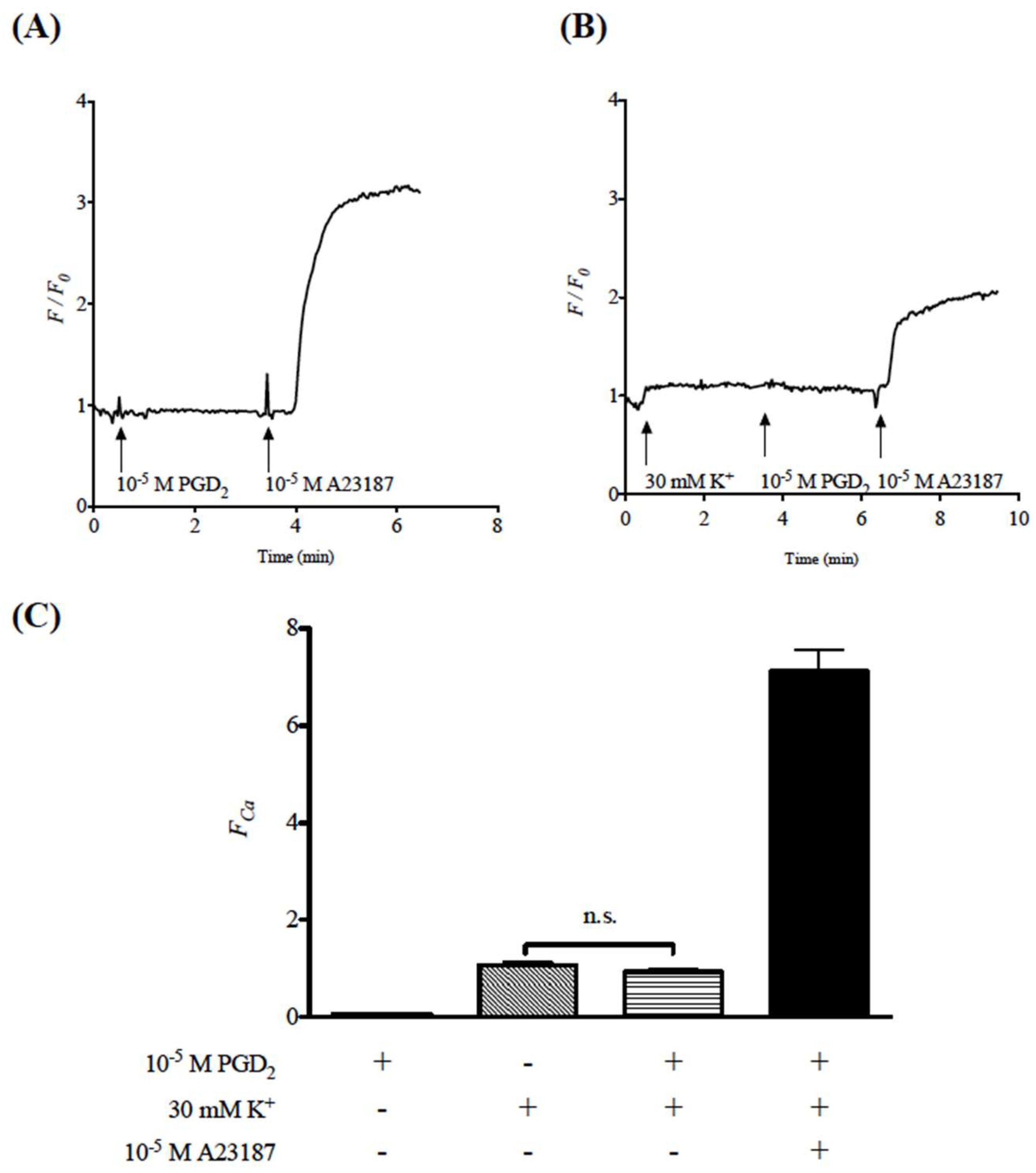
2.2. Effects of Prostaglandin D2 (PGD2) on Cytosolic Ca2+ Level in Human Bronchial Smooth Muscle Cells (hBSMCs)

Due to the difficulty in preparing isolated BSM cells with high purity from the mouse tissues, change in cytosolic Ca2+ level was measured using commercially available human BSM cells (hBSMCs) in the present study. The hBSMCs were loaded with a green fluorescent Ca2+ indicator, Fluo-8 [16]. As shown in Figure 4A,B, in the hBSMCs incubated with Fluo-8/AM, stimulation of the cells with a Ca2+ ionophore A23187 (10−5 M) caused a marked increase in F/F0, that is, an increase in cytosolic Ca2+ concentration, indicating a successful loading of Fluo-8 into the cells. In the Fluo-8-loaded hBSMCs, stimulation of the cells with 30 mM K+ caused a slight but distinct increase in cytosolic Ca2+ concentration (Figure 4B,C). Interestingly, PGD2 had no effect on the K+ depolarization-induced increase in cytosolic Ca2+ level (Figure 4B,C). PGD2 also did not alter the basal cytosolic Ca2+ level in the Fluo-8-loaded cells (Figure 4A).

2.3. Activation of RhoA/Rho-Kinase Signaling by Prostaglandin D2 (PGD2)

The results that PGD2 caused an augmentation of contraction (Figure 2B,C) under the constant cytosolic Ca2+ level (Figure 4) remind us of the Ca2+ sensitization of smooth muscle contraction. In smooth muscle cells including airways, activation of a monomeric G-protein, RhoA, causes Ca2+ sensitization of the contraction by activating its downstream Rho-kinases [17,18]. To determine whether PGD2 activates RhoA protein, the GTP-RhoA pull-down assay was performed in mouse BSMs stimulated by PGD2. As previously reported [19], acetylcholine (ACh: 10−3 M) stimulation caused an increase in GTP-bound, active form of RhoA protein in the BSMs of mice (Figure 5A). Similarly, as shown in Figure 5A, an increase in the active form of RhoA protein was observed when the BSM tissues were stimulated with 10−5 M PGD2, the concentration where no contractile response from baseline tone was observed (see above). The tension study also revealed an activation of RhoA/Rho-kinase signaling by PGD2: the PGD2-induced augmentation of contraction was blocked by Y-27632 (10−7 M), a selective inhibitor of Rho-kinases (Figure 5B).

3. Discussion

The current study was carried out to determine the role of prostaglandin D2 (PGD2) on smooth muscle function of the airways using the bronchial smooth muscles (BSMs) isolated from mice. Although PGD2 had no effect on their baseline tension, PGD2 significantly augmented the BSM contraction induced by high K+ depolarization (Figure 2B,C). The PGD2-induced augmentation of contraction was inhibited both by a DP1 antagonist, laropiprant, and a Rho-kinase inhibitor, Y-27632 (Figure 3 and Figure 5B). Furthermore, PGD2 could cause an activation of RhoA protein (Figure 5A). In the high K+-depolarized cultured human BSM cells, PGD2 caused no further increase in cytosolic Ca2+ concentration (Figure 4). These findings suggest that PGD2 acts on DP1 receptors to cause RhoA/Rho-kinase-mediated Ca2+ sensitization of contraction in BSMs.
PGD2 is an acidic lipid mediator derived from the metabolism of arachidonic acid by the action of cyclooxygenases and downstream PGD2 synthases, and is mainly released from mast cells when activated by antigen stimulation [20]. Allergen challenge to the airways caused an increase in PGD2 level in the airways of asthmatics [8,9]. However, the functional role of PGD2 on airway smooth muscle has not yet been unified. In tracheal smooth muscle strips isolated from the guinea pigs, PGD2 produced a concentration-dependent contraction [12]. Similarly, PGD2 caused a contraction in bronchial rings isolated from the dogs [13]. In contrast, PGD2, at a concentration of 3 µM, elicited a relaxation in the murine tracheal smooth muscle precontracted with carbachol [10]. Currently, PGD2 had no effect on basal tension in BSMs isolated from the mice (see Results section). Differences in the species, region (tracheal versus bronchial smooth muscles), and/or the experimental condition used may be involved in the difference in the PGD2 response in smooth muscles of the airways. Thus, note that the current study also contains a certain limitation: cultured human BSM cells (hBSMCs) were used for cytosolic Ca2+ measurement whereas functional studies were performed using mouse BSM tissues.
The current RT-PCR analyses showed expression of DP1 and DP2 receptors in BSM cells (Figure 1), indicating that PGD2 could directly act on BSM cells. Although PGD2 did not affect the basal tension, it augmented the submaximal contraction induced by 30 mM K+ in BSMs isolated from the mice (Figure 2). The augmented contraction induced by PGD2 was inhibited by laropiprant (Figure 3), a DP1 antagonist [14], but not by fevipiprant (see RESULTS), a DP2 antagonist [15]. An involvement of TP receptor in the PGD2-mediated contraction has also been suggested [21]. However, PGD2 did not increase cytosolic Ca2+ in the present study (Figure 4), whereas an induction of contraction with Ca2+ mobilization by the TP receptor activation has been demonstrated [22]. In addition, our preliminary study revealed that stimulation of TP receptors with a thromboxane A2 (TXA2) mimic, U46619, caused a distinct contraction from baseline tension (without K+ depolarization) in the mouse BSMs. Pretreatment of BSMs with ozagrel, an inhibitor of TXA2 synthase, also did not inhibit the augmented contraction induced by PGD2 (data not shown). It is thus unlikely that the TXA2/TP receptor is involved in the PGD2-mediated response in the mouse BSMs. Thus, an activation of DP1 receptors on the BSM cells might be responsible for the synergistic contraction induced by PGD2.
Currently, PGD2 augmented the contraction induced by high K+ depolarization in mouse BSM tissues (Figure 2B,C). In the high K+-depolarized cultured hBSMCs, PGD2 caused no further increase in cytosolic Ca2+ concentration (Figure 4). Collectively, these findings suggest that PGD2 augmented the BSM contraction induced by K+ depolarization without any increase in cytosolic Ca2+ concentration. The observation that PGD2 caused an augmentation of contraction under the constant cytosolic Ca2+ level reminds us of the Ca2+ sensitization of smooth muscle contraction. Indeed, the augmented contraction induced by PGD2 was inhibited by a Rho-kinase inhibitor, Y-27632 (Figure 5). In addition, stimulation of the BSMs with PGD2 caused an increase in the active form of RhoA, GTP-bound RhoA (Figure 5). The current study for the first time, to our knowledge, demonstrated that PGD2 activates the RhoA/Rho-kinase signaling to induce Ca2+ sensitization of contraction in the BSMs. Previous studies, including ours, demonstrated that muscarinic receptor stimulation of airway smooth muscle caused both an increase in cytosolic Ca2+ concentration and an activation of RhoA/Rho-kinase signaling, resulting in the contraction [17,23,24]. On the other hand, the current study revealed that PGD2 did not have the ability to increase cytosolic Ca2+ level in the BSMs (Figure 4). This may be a reason that PGD2 did not cause any contraction from the baseline tension: the cytosolic Ca2+ level at the baseline tension might not have been enough to induce BSM contraction even if the RhoA/Rho-kinase signaling was activated.
It is a remarkable event that the PGD2-induced augmentation of contraction was inhibited by laropiprant, an antagonist of DP1 receptor that is known as a Gs protein-coupled receptor. In smooth muscle cells including the airways, the Gs protein activation, such as beta-adrenoceptor stimulation by isoprenaline, causes an increase in cAMP level to induce relaxation [25,26,27]. However, the current study indicated that activation of DP1 receptor by PGD2 could cause a response to contractile direction. Although the discrepancy is not explainable now, an activation of extracellular signal-regulated kinase (ERK) signaling by DP1 receptor stimulation has also been reported in nasal epithelial cells [28]. It is thus possible that, in addition to the classical Gs/cAMP pathway, the DP1 receptor stimulation activates multiple intracellular signaling, including the RhoA/Rho-kinase signaling. Further studies are needed to make clear the mechanism of action of PGD2 in the BSMs.
In conclusion, the current study revealed that PGD2 augmented the BSM contraction by activating the RhoA/Rho-kinase-mediated Ca2+ sensitization of contraction via an activation of DP1 receptors on the BSM cells. Increased PGD2 level in the airways might be one of the causes of the enhanced airway responsiveness to nonspecific stimuli, one of the characteristic features of bronchial asthma.

4. Materials and Methods

4.1. Animals

Male BALB/c mice were purchased from the Tokyo Laboratory Animals Science Co., Ltd. (Tokyo, Japan) and housed in a pathogen-free facility. All animal experiments were approved by the Animal Care Committee of the Hoshi University, Tokyo, Japan (permission code: 30-086, permission date: 21 June 2018).

4.2. Pharmacological Reagents

Prostaglandin D2 (PGD2: Cat. No. 12010) and laropiprant (MK-0524: Cat. No. 10009835) were purchased from Cayman Chemical (Ann Arbor, MI, USA). Fevipiprant was purchased from MedChem Express (Monmouth Junction, NJ, USA: Cat. No. HY-16768).

4.3. Determination of Bronchial Smooth Muscle (BSM) Responsiveness

Mice were sacrificed by exsanguination from abdominal aorta under urethane (1.6 g/kg, i.p.) anesthesia and the airway tissues under the larynx to lungs were immediately removed. About 3 mm length of the left main bronchus (about 0.5 mm diameter) was isolated. The resultant tissue ring preparation was then suspended in a 5 mL organ bath by two stainless-steel wires (0.2 mm diameter) passed through the lumen. For all tissues, one end was fixed to the bottom of the organ bath while the other was connected to a force-displacement transducer (TB-612T, Nihon Kohden, Tokyo, Japan) for the measurement of isometric force. A resting tension of 0.5 g was applied. The buffer solution contained modified Krebs–Henseleit solution with the following composition (mM): NaCl 118.0, KCl 4.7, CaCl2 2.5, MgSO4 1.2, NaHCO3 25.0, KH2PO4 1.2, and glucose 10.0. The buffer solution was maintained at 37 °C and oxygenated with 95% O2/5% CO2. After the equilibration period, the tension studies were performed. In case of the high K+ depolarization studies, experiments were conducted in the presence of atropine (10−6 M).

4.4. Determination of Active Form of RhoA in BSM

The active form of RhoA, GTP-bound RhoA, in BSMs was measured by GTP-RhoA pull-down assay as described previously [19]. In brief, the isolated main bronchial tissues were equilibrated in oxygenated Krebs–Henseleit solution at 37 °C for 1 h. After the equilibration period, the tissues were stimulated with PGD2 (10−5 M) or ACh (10−3 M) for 15 min, and were quickly frozen with liquid nitrogen. The tissues were then lysed in lysis buffer with the following composition (mM): HEPES 25.0 (pH 7.5), NaCl 150, IGEPAL CA-630 1%, MgCl2 10.0, EDTA 1.0, glycerol 10%, 1× protease inhibitor cocktail (Nakalai tesque, Kyoto, Japan), and 1× phosphatease inhibitor cocktail (Nakalai tesque). Active RhoA in tissue lysates (200 µg protein) was precipitated with 25 µg GST-tagged Rho binding domain (amino acids residues 7–89 of mouse rhotekin; Upstate, Lake Placid, NY, USA), which was expressed in Escherichia coli and bound to glutathione-agarose beads. The precipitates were washed three times in lysis buffer, and after adding the SDS loading buffer and boiling for 5 min, the bound proteins were resolved in 15% polyacrylamide gels, transferred to nitrocellulose membranes, and immunoblotted with rabbit polyclonal anti-RhoA (Abcam, Cambridge, UK) as primary antibodies.

4.5. Cell Culture and [Ca2+]cyto Measurement

Normal human BSM cells (hBSMCs; a male donor: purchased from Cambrex Bio Science Walkersville, Inc., Walkersville, MD, USA) were maintained in SmBM medium (Cambrex Bio Science Walkersville, Inc., Walkersville, MD, USA) supplemented with 5% fetal bovine serum, 0.5 ng/mL human epidermal growth factor (hEGF), 5 µg/mL insulin, 2 ng/mL human fibroblast growth factor-basic (hFGF-b), 50 µg/mL gentamicin, and 50 ng/mL amphotericin B. Cells were maintained at 37 °C in a humidified atmosphere (5% CO2), fed every 48–72 h, and passaged when cells reached 90–95% confluence. Then the hBSMCs (passages 5–7) were seeded in 24-well plates (Becton Dickinson Labware, Franklin Lakes, NJ, USA) and were cultured without serum. Twenty-four hours after the starvation period, the cells were loaded with Fluo-8/AM (2.5 M: AAT Bioquest, Inc., Sunnyvale, CA, USA) in serum-free SmBM medium for 90 min at 37 °C. The cells were washed with PBS and maintained in Krebs–Henseleit solution described above. The intracellular Fluo-8 fluorescence was monitored using fluorescence microscope (Keyence, Osaka, Japan) with BZ-X filter GFP (470/40, 535/50 nm). Images were pictured using time-lapse imaging (Keyence), and analyzed with BZ-X analyzer (Keyence). Change in the cytosolic Ca2+ level was calculated as ratio to the basal fluorescence intensity.

4.6. RT-PCR Analyses

Total RNAs of hBSMCs and mouse BSM tissues were extracted using NucleoSpin™ miRNA (TaKaRa Bio, Inc., Shiga, Japan) according to the manufacturer’s instruction. cDNAs were prepared from the total RNA by using PrimeScript™ RT reagent Kit (TaKaRa) according to the manufacturer’s instructions. cDNA samples were subjected to PCR with Quick Taq™ HS DyeMix (TOYOBO Co., Ltd., Osaka, Japan) in a final volume of 10 µL. The PCR primer sets used are shown in Table 1 (for human) and Table 2 (for mouse), which was designed from published database, BLAST. The thermal cycle profile used was (1) denaturing for 30 s at 94 °C, (2) annealing primers for 30 s at 60 °C, (3) extending the primers for 1 min at 68 °C, and the reaction was run for 40 cycles. The PCR products were subjected to electrophoresis on 2% agarose gel and visualized by ethidium bromide staining.

4.7. Statistical Analyses

All the data are expressed as means ± SE. Statistical significance of difference was determined by paired t-test (Figure 2B,C) or one-way analysis of variance (ANOVA) with post hoc Bonferroni’s multiple comparison (Figure 3B and Figure 4C) using Prism 5 for Mac OS X (GraphPad Software, La Jolla, CA, USA). A value of p < 0.05 was considered significant.

Author Contributions

Conception and design, W.S. and Y.C.; analysis and interpretation, W.S., Y.A., T.H., F.T., and Y.C.; drafting the manuscript for important intellectual content: W.S., S.S., J.K., H.S., and Y.C. All authors read and approved the final manuscript.

Funding

This work was partly supported by Grant-in-Aid for Scientific Research (C) Grant Number 15K08248 (Y.C.) from the Japan Society for the Promotion of Science (JSPS).

Acknowledgments

We thank Sayaka Endo, Mifuyu Shioda, Misaki Tsuchiyama, Shigeki Fujii, Yamato Yamane, and Yuki Kai for their technical assistance.

Conflicts of Interest

The authors declare no conflict of interest.

Abbreviations

AChacetylcholine
AHRairway hyperresponsiveness
ANOVAanalysis of variance
BALbronchoalveolar lavage
BSMbronchial smooth muscle
COXcyclooxygenase
CRTH2chemoattractant receptor-homologous molecule on Th2 cells
ERKextracellular signal-regulated kinase
FCanormalized ratios of the Ca2+ fluorescence intensities
F/F0ratio of the Ca2+ fluorescence intensity to that at time 0 (baseline)
Fluo-8/AMFluo-8 acetoxymethyl ester
GPCRG protein-coupled receptor
GSTglutathione S-transferase
GTPguanosine triphosphate
PGprostaglandin
SDSsodium dodecyl sulfate
TXA2thromboxane A2

References

  1. Björck, T.; Gustafsson, L.E.; Dahlén, S.E. Isolated bronchi from asthmatics are hyperresponsive to adenosine, which apparently acts indirectly by liberation of leukotrienes and histamine. Am. Rev. Respir. Dis. 1992, 145, 1087–1091. [Google Scholar] [CrossRef] [PubMed]
  2. Seow, C.Y.; Schellenberg, R.R.; Paré, P.D. Structural and functional changes in the airway smooth muscle of asthmatic subjects. Am. J. Respir. Crit. Care Med. 1998, 158, S178–S186. [Google Scholar] [CrossRef] [PubMed]
  3. Martin, J.G.; Duguet, A.; Eidelman, D.H. The contribution of airway smooth muscle to airway narrowing and airway hyperresponsiveness in disease. Eur. Respir. J. 2000, 16, 349–354. [Google Scholar] [CrossRef] [PubMed]
  4. Ma, X.; Cheng, Z.; Kong, H.; Wang, Y.; Unruh, H.; Stephens, N.L.; Laviolette, M. Changes in biophysical and biochemical properties of single bronchial smooth muscle cells from asthmatic subjects. Am. J. Physiol. Lung Cell. Mol. Physiol. 2002, 283, L1181–L1189. [Google Scholar] [CrossRef] [PubMed]
  5. Tang, D.D. Critical role of actin-associated proteins in smooth muscle contraction, cell proliferation, airway hyperresponsiveness and airway remodeling. Respir. Res. 2015, 16, 134. [Google Scholar] [CrossRef] [PubMed]
  6. Dworski, R.; Sheller, J.R.; Wickersham, N.E.; Oates, J.A.; Brigham, K.L.; Roberts, L.J.; Fitzgerald, G.A. Allergen-stimulated release of mediators into sheep bronchoalveolar lavage fluid. Effect of cyclooxygenase inhibition. Am. Rev. Respir. Dis. 1989, 139, 46–51. [Google Scholar] [CrossRef] [PubMed]
  7. Mandal, A.K.; Zhang, Z.; Ray, R.; Choi, M.S.; Chowdhury, B.; Pattabiraman, N.; Mukherjee, A.B. Uteroglobin represses allergen-induced inflammatory response by blocking PGD2 receptor-mediated functions. J. Exp. Med. 2004, 199, 1317–1330. [Google Scholar] [CrossRef] [PubMed]
  8. Chilton, F.H.; Averill, F.J.; Hubbard, W.C.; Fonteh, A.N.; Triggiani, M.; Liu, M.C. Antigen-induced generation of lyso-phospholipids in human airways. J. Exp. Med. 1996, 183, 2235–2245. [Google Scholar] [CrossRef] [PubMed] [Green Version]
  9. Hartert, T.V.; Dworski, R.T.; Mellen, B.G.; Oates, J.A.; Murray, J.J.; Sheller, J.R. Prostaglandin E2 decreases allergen-stimulated release of prostaglandin D2 in airways of subjects with asthma. Am. J. Respir. Crit. Care Med. 2000, 162, 637–640. [Google Scholar] [CrossRef] [PubMed]
  10. Matsuoka, T.; Hirata, M.; Tanaka, H.; Takahashi, Y.; Murata, T.; Kabashima, K.; Sugimoto, Y.; Kobayashi, T.; Ushikubi, F.; Aze, Y.; et al. Prostaglandin D2 as a mediator of allergic asthma. Science 2000, 287, 2013–2017. [Google Scholar] [CrossRef] [PubMed]
  11. Xue, L.; Gyles, S.L.; Wettey, F.R.; Gazi, L.; Townsend, E.; Hunter, M.G.; Pettipher, R. Prostaglandin D2 causes preferential induction of proinflammatory Th2 cytokine production through an action on chemoattractant receptor-like molecule expressed on Th2 cells. J. Immunol. 2005, 175, 6531–6536. [Google Scholar] [CrossRef] [PubMed]
  12. Underwood, D.C.; Muccitelli, R.M.; Luttmann, M.A.; Hay, D.W.; Torphy, T.J.; Wasserman, M.A. Differential antagonism of airway contractile responses to prostaglandin (PG)D2 and 9 α, 11 β-PGF2 by atropine, SK&F 88046 and SQ 29,548 in the guinea pig. J. Pharmacol. Exp. Ther. 1994, 268, 304–310. [Google Scholar] [PubMed]
  13. Tamaoki, J.; Sekizawa, K.; Graf, P.D.; Nadel, J.A. Cholinergic neuromodulation by prostaglandin D2 in canine airway smooth muscle. J. Appl. Physiol. 1987, 63, 1396–1400. [Google Scholar] [CrossRef] [PubMed]
  14. Sturino, C.F.; O’Neill, G.; Lachance, N.; Boyd, M.; Berthelette, C.; Labelle, M.; Li, L.; Roy, B.; Scheigetz, J.; Tsou, N.; et al. Discovery of a potent and selective prostaglandin D2 receptor antagonist, [(3R)-4-(4-chloro-benzyl)-7-fluoro-5-(methylsulfonyl)-1,2,3,4-tetrahydrocyclopenta[b]indol-3-yl]-acetic acid (MK-0524). J. Med. Chem. 2007, 50, 794–806. [Google Scholar] [CrossRef] [PubMed]
  15. Sykes, D.A.; Bradley, M.E.; Riddy, D.M.; Willard, E.; Reilly, J.; Miah, A.; Bauer, C.; Watson, S.J.; Sandham, D.A.; Dubois, G.; et al. Fevipiprant (QAW039), a slowly dissociating CRTh2 antagonist with the potential for improved clinical efficacy. Mol. Pharmacol. 2016, 89, 593–605. [Google Scholar] [CrossRef] [PubMed]
  16. Lacroix, J.J.; Botello-Smith, W.M.; Luo, Y. Probing the gating mechanism of the mechanosensitive channel Piezo1 with the small molecule Yoda1. Nat. Commun. 2018, 9, 2029. [Google Scholar] [CrossRef] [PubMed]
  17. Chiba, Y.; Ueno, A.; Shinozaki, K.; Takeyama, H.; Nakazawa, S.; Sakai, H.; Misawa, M. Involvement of RhoA-mediated Ca2+ sensitization in antigen-induced bronchial smooth muscle hyperresponsiveness in mice. Respir. Res. 2005, 6, 4. [Google Scholar] [CrossRef] [PubMed] [Green Version]
  18. Gerthoffer, W.T.; Solway, J.; Camoretti-Mercado, B. Emerging targets for novel therapy of asthma. Curr. Opin. Pharmacol. 2013, 13, 324–330. [Google Scholar] [CrossRef] [PubMed] [Green Version]
  19. Chiba, Y.; Danno, S.; Suto, R.; Suto, W.; Yamane, Y.; Hanazaki, M.; Katayama, H.; Sakai, H. Intranasal administration of recombinant progranulin inhibits bronchial smooth muscle hyperresponsiveness in mouse allergic asthma. Am. J. Physiol. Lung Cell. Mol. Physiol. 2018, 314, L215–L223. [Google Scholar] [CrossRef] [PubMed]
  20. Modena, B.D.; Dazy, K.; White, A.A. Emerging concepts: Mast cell involvement in allergic diseases. Transl. Res. 2016, 174, 98–121. [Google Scholar] [CrossRef] [PubMed]
  21. Coleman, R.A.; Sheldrick, R.L. Prostanoid-induced contraction of human bronchial smooth muscle is mediated by TP-receptors. Br. J. Pharmacol. 1989, 96, 688–692. [Google Scholar] [CrossRef] [PubMed] [Green Version]
  22. Shiraki, A.; Kume, H.; Oguma, T.; Makino, Y.; Ito, S.; Shimokata, K.; Honjo, H.; Kamiya, K. Role of Ca2+ mobilization and Ca2+ sensitization in 8-iso-PGF-induced contraction in airway smooth muscle. Clin. Exp. Allergy 2009, 39, 236–245. [Google Scholar] [CrossRef] [PubMed]
  23. Chiba, Y.; Sakai, H.; Suenaga, H.; Kamata, K.; Misawa, M. Enhanced Ca2+ sensitization of the bronchial smooth muscle contraction in antigen-induced airway hyperresponsive rats. Res. Commun. Mol. Pathol. Pharmacol. 1999, 106, 77–85. [Google Scholar] [PubMed]
  24. Chiba, Y.; Takada, Y.; Miyamoto, S.; MitsuiSaito, M.; Karaki, H.; Misawa, M. Augmented acetylcholine-induced, Rho-mediated Ca2+ sensitization of bronchial smooth muscle contraction in antigen-induced airway hyperresponsive rats. Br. J. Pharmacol. 1999, 127, 597–600. [Google Scholar] [CrossRef] [PubMed]
  25. Cheah, E.Y.; Mann, T.S.; Burcham, P.C.; Henry, P.J. Influenza A infection attenuates relaxation responses of mouse tracheal smooth muscle evoked by acrolein. Biochem. Pharmacol. 2015, 93, 519–526. [Google Scholar] [CrossRef] [PubMed]
  26. Donovan, C.; Bailey, S.R.; Tran, J.; Haitsma, G.; Ibrahim, Z.A.; Foster, S.R.; Tang, M.L.; Royce, S.G.; Bourke, J.E. Rosiglitazone elicits in vitro relaxation in airways and precision cut lung slices from a mouse model of chronic allergic airways disease. Am. J. Physiol. Lung Cell. Mol. Physiol. 2015, 309, L1219–L1228. [Google Scholar] [CrossRef] [PubMed]
  27. Morgan, S.J.; Deshpande, D.A.; Tiegs, B.C.; Misior, A.M.; Yan, H.; Hershfeld, A.V.; Rich, T.C.; Panettieri, R.A.; An, S.S.; Penn, R.B. Beta-Agonist-mediated relaxation of airway smooth muscle is protein kinase A-dependent. J. Biol. Chem. 2014, 289, 23065–23074. [Google Scholar] [CrossRef] [PubMed]
  28. Choi, Y.H.; Lee, S.N.; Aoyagi, H.; Yamasaki, Y.; Yoo, J.Y.; Park, B.; Shin, D.M.; Yoon, H.G.; Yoon, J.H. The extracellular signal-regulated kinase mitogen-activated protein kinase/ribosomal S6 protein kinase 1 cascade phosphorylates cAMP response element-binding protein to induce MUC5B gene expression via D-prostanoid receptor signaling. J. Biol. Chem. 2011, 286, 34199–34214. [Google Scholar] [CrossRef] [PubMed]
Figure 1. Expression of various prostanoid receptors in cultured human bronchial smooth muscle (BSM) cells (A) and murine BSMs (B) determined by RT-PCR analyses. Marker: M.W. markers (100 bp ladder), GAPDH: glyceraldehyde-3-phosphate dehydrogenase, DP1: prostaglandin D2 (PGD2) receptor 1, DP2: PGD2 receptor 2, EP1: PGE2 receptor 1, EP2: PGE2 receptor 2, EP3: PGE2 receptor 3, EP4: PGE2 receptor 4, FP: PGF2 receptor, IP: PGI2 receptor, and TP: thromboxane A2 receptor. The primer sets used are shown in Materials and Methods section.
Ijms 19 03036 g001
Figure 2. Effects of prostaglandin D2 (PGD2) on the contraction induced by 30 mM K+ depolarization in bronchial smooth muscles (BSMs) isolated from mice. PGD2 (10−9–10−5 M) had no effect on basal tone (A). After the stable contraction induced by K+ depolarization was observed, 10−6 (B) or 10−5 M (C) PGD2 was applied. Representative traces of changes in the active force are shown in respective upper panels, and the data are summarized in the lower panels. Results are presented as mean ± SEM from 5 animals, respectively. * p < 0.05 and *** p < 0.001 versus without PGD2 by paired Student’s t-test.
Ijms 19 03036 g002
Figure 3. Effect of laropiprant (a selective DP1 receptor antagonist) on the augmented contraction induced by prostaglandin D2 (PGD2) in bronchial smooth muscles (BSMs) isolated from mice. After the BSM contraction induced by PGD2 reached to plateau, 10−6 M laropiprant was applied. Representative traces of changes in the active force are shown in (A), and the data are summarized in (B). Results are presented as mean ± SEM from 5 animals. *** p < 0.001 versus 30 mM K+ only group and †† p < 0.01 versus 30 mM K+ + 10−5 M PGD2 group by one-way ANOVA with post hoc Bonferroni’s multiple comparison. Note that fevipiprant (10−6 M, a selective DP2 receptor antagonist) had no effect on the PGD2-induced augmentation of contraction (C).
Ijms 19 03036 g003
Figure 4. Effects of prostaglandin D2 (PGD2) on the cytosolic Ca2+ level in cultured human bronchial smooth muscle cells (hBSMCs) determined by a fluorescent Ca2+ indicator Fluo-8. (A,B) Representative trace of change in cytosolic Ca2+ (F/F0, ratio of the Ca2+ fluorescence intensity to that at time 0 (baseline)). The Fluo-8-loaded hBSMCs were stimulated with 30 mM K+ and, when its stable response was observed, 10−5 M PGD2 was applied. To confirm the maximal response, a Ca2+ ionophore A23187 (10−5 M) was applied at the end of experiments. (C) Summary of normalized ratios of the Ca2+ fluorescence intensities (FCa) data. Results are presented as mean ± SEM from 8 independent experiments. Note that neither the baseline Ca2+ level nor the stable increase in Ca2+ induced by K+ depolarization was affected by PGD2.
Ijms 19 03036 g004
Figure 5. Activation of RhoA/Rho-kinase signaling by prostaglandin D2 (PGD2) in bronchial smooth muscle (BSM) of the mouse. (A) Effect of PGD2 on the level of GTP-bound active form of RhoA. Freshly isolated BSMs of mice were stimulated with acetylcholine (ACh: 10−3 M) or PGD2 (10−5 M) for 10 min, and GTP-RhoA pull-down assay and RhoA immunoblottings were performed as described in Materials and Methods section. The blots for GTP-bound (active: upper) and total RhoA (lower) are shown and representative from 3 independent experiments, respectively. (B) Effect of Y-27632 (a selective Rho-kinase inhibitor) on the augmented contraction induced by prostaglandin D2 (PGD2) in BSMs isolated from mice. After the BSM contraction induced by PGD2 reached to plateau, 10−7 M Y-27632 was applied. A trace of change in the active force is shown and representative from 3 independent experiments.
Ijms 19 03036 g005
Table 1. Primer sequences for RT-PCR used in the present study (human).
Gene NameRefSeq Accession SequenceAmplicon Size
human PTGDRNM_000953Sense5′-TCTGCGCGCTACCTTTCATG-3′85 bp
Antisense5′-TCCTCGTGGACCATCTGGATA-3′
human PTGDR2NM_004778Sense5′-CCTCTGTGCCCAGAGCCCCACGATGTCGGC-3′114 bp
Antisense5′-ATGTAGCGGATGCTGGTGTTG-3′
human PTGER1NM_000955Sense5′-GATGGTGGGCCAGCTTGTC-3′72 bp
Antisense5′-GCCACCAACACCAGCATTG-3′
human PTGER2NM_000956Sense5′-GTGCTGACAAGGCACTTCATGT-3′87 bp
Antisense5′-TGTTCCTCCAAAGGCCAAGTAC-3′
human PTGER3NM_198714Sense5′-AAGGCCACGGCATCTCAGT-3′76 bp
Antisense5′-TGATCCCCATAAGCTGAATGG-3′
human PTGER4NM_000958Sense5′-CTTGGAGGCAGGAATTTGCTT-3′77 bp
Antisense5′-AAAGTCCTCAGTGAGGTGGTGTCT-3′
human PTGFRNM_000959Sense5′-GCACATTGATGGGCAACTAGAA-3′91 bp
Antisense5′-GCACCTATCATTGGCATGTAGCT-3′
human PTGIRNM_000960Sense5′-GCCGATCAGCTGCTGTTTCT-3′75 bp
Antisense5′-TTTCCTCTGTCCCTCACTCTCTTC-3′
human TBXA2RNM_001060Sense5′-ACGGAGAAGGAGCTGCTCATC-3′84 bp
Antisense5′-GCGGCGGAACAGGATATACA-3′
human GAPDHNM_002046Sense5′-GGAGCCAAAAGGGTCATCATCTC-3′282 bp
Antisense5′-AGGGATGATGTTCTGGAGAGCC-3′
Table 2. Primer sequences for RT-PCR used in the present study (mouse).
Gene NameRefSeq Accession SequenceAmplicon Size
mouse Ptgdr1NM_008962Sense5′-CAACCTGGGTGCCATGTAC-3′112 bp
Antisense5′-GGACCCGTGCCTGTAGTCT-3′
mouse Ptgdr2NM_009962Sense5′-CTGCACCTGGCGCTATC-3′174 bp
Antisense5′-GTCCAGGCTAATGGCACT-3′
mouse Ptger1NM_013641Sense5′-TACATGGGATGCTCGAAACA-3′223 bp
Antisense5′-TTTTAGGCCGTGTGGGTAG-3′
mouse Ptger2NM_008964Sense5′-ATGCACCTGCTGCTTATCGT-3′196 bp
Antisense5′-TAATGGCCAGGAGAATGAGG-3′
mouse Ptger3NM_001359745Sense5′-TGCTGGCTCTGGTGGTGAC-3′258 bp
Antisense5′-ACTCCTTCTCCTTTCCCATCTGTG-3′
mouse Ptger4NM_001136079Sense5′-CCATCGCCACATACATGAAG-3′209 bp
Antisense5′-TGCACAGATGGCGAAGAGTG-3′
mouse PtgfrNM_008966Sense5′-CTGCTCCGGACACAACCACTC-3′191 bp
Antisense5′-GGTTCTCCGTCTGGCAGGTTG-3′
mouse PtgirNM_008967Sense5′-GGATGAAGTTTACCACCTGATTCTGC-3′196 bp
Antisense5′-AGCCTTTCGGAAAAGGATGAAGAC-3′
mouse Tbxa2rNM_009325Sense5′-TTTCGCCCGGTGAACATC-3′255 bp
Antisense5′-GGCTCGCCAGTCCAACAA-3′
mouse GapdhNM_001289726Sense5′-CCTCGTCCCGTAGACAAAATG-3′100 bp
Antisense5′-TCTCCACTTTGCCACTGCAA-3′

