Article

CPT-11-Induced Delayed Diarrhea Develops via Reduced Aquaporin-3 Expression in the Colon

1
Global Research Center for Innovative Life Science, Hoshi University, 2-4-41 Ebara, Shinagawa-ku, Tokyo 142-8501, Japan
2
Department of Clinical Pharmacokinetics, Hoshi University, 2-4-41 Ebara, Shinagawa-ku, Tokyo 142-8501, Japan
3
Division of Applied Pharmaceutical Education and Research, Hoshi University, 2-4-41 Ebara, Shinagawa-ku, Tokyo 142-8501, Japan
4
Department of Functional Molecular Kinetics, Hoshi University, 2-4-41 Ebara, Shinagawa-ku, Tokyo 142-8501, Japan
*
Authors to whom correspondence should be addressed.
Int. J. Mol. Sci. 2018, 19(1), 170; https://doi.org/10.3390/ijms19010170
Submission received: 22 November 2017 / Revised: 19 December 2017 / Accepted: 31 December 2017 / Published: 6 January 2018
(This article belongs to the Special Issue Aquaporins: Water Channels Essential for Living Organisms)

Abstract

:
While irinotecan (CPT-11) has a potent anti-cancer effect, it also causes serious diarrhea as an adverse reaction. In this study, we analyzed the pathogenic mechanism of CPT-11-induced delayed diarrhea by focusing on water channel aquaporin-3 (AQP3) in the colon. When rats received CPT-11, the expression level of AQP3 was reduced during severe diarrhea. It was found that the expression levels of inflammatory cytokines and the loss of crypt cells were increased in the colon when CPT-11 was administered. When celecoxib, an anti-inflammatory drug, was concomitantly administered, both the diarrhea and the reduced expression of AQP3 induced by CPT-11 were suppressed. The inflammation in the rat colon during diarrhea was caused via activated macrophage by CPT-11. These results showed that when CPT-11 is administered, the expression level of AQP3 in the colon is reduced, resulting in delayed diarrhea by preventing water transport from the intestinal tract. It was also suggested that the reduced expression of AQP3 might be due to the inflammation that occurs following the loss of colonic crypt cells and to the damage caused by the direct activation of macrophages by CPT-11. Therefore, it was considered that anti-inflammatory drugs that suppress the reduction of AQP3 expression could prevent CPT-11-induced delayed diarrhea.

Graphical Abstract

1. Introduction

Irinotecan (CPT-11) is an anticancer agent that is widely used in the treatment of colon cancer, gastric cancer, pulmonary cancer, and cervical cancer. CPT-11 exerts its anticancer effects through the inhibition of DNA synthesis by inhibiting DNA topoisomerase I [1], but it also causes serious adverse drug reactions. The adverse reactions with high incidence include myelosuppression, nausea, vomiting, and diarrhea [2]. Among these reactions, diarrhea is a serious adverse drug reaction that occurs in approximately 80% of patients and is one of the dose-limiting factors of CPT-11 [3]. CPT-11-induced diarrhea is classified into early-onset diarrhea, occurring within a few hours of drug administration, and late-onset diarrhea, occurring more than 24 h after administration. Early-onset diarrhea is believed to be caused by the inhibition of acetylcholinesterase by CPT-11, leading to acetylcholine accumulation, as well as by the direct binding of CPT-11 to acetylcholine receptors, increasing peristaltic movement [4,5]. This early-onset diarrhea can be treated with anticholinergic agents such as atropine [6]. In contrast, late-onset diarrhea is very serious and is believed to be caused by the following mechanism. CPT-11 is metabolized mainly by carboxylesterases in the liver to form an active metabolite, SN-38, which is subsequently metabolized by uridine diphosphate-glucuronosyltransferase 1A1 (UGT1A1) into SN-38-glucuronide (SN-38-glu) [7,8]. After being excreted into the bile, SN-38-glu is deconjugated by enteric bacteria-derived β-glucuronidase in the intestinal tract and is re-converted into the active SN-38 [9,10]. This SN-38 is believed to induce severe, persistent diarrhea by damaging the intestinal mucosa and by accumulating in the body via the enterohepatic circulation [11]. Various methods to treat CPT-11-induced delayed diarrhea are being discussed based on this mechanism, including the following: (1) suppression of the production of SN-38 by killing enteric bacteria with antibiotics [12,13,14,15,16]; (2) inhibition of β-glucuronidase activity with Hangeshashinto, a traditional Kampo medicine [17,18]; and (3) adsorption of SN-38 in the intestinal tract using activated charcoal [19,20]. Other methods include the high-dose administration of loperamide, an antidiarrheal, as well as magnesium oxide, an antacid, to promote the excretion of SN-38 and CPT-11 by alkalizing the intestine [21,22]. Although these methods can alleviate diarrhea, treatment often becomes difficult in patients with severe diarrhea, and no reliable preventive methods have been established [23]. Severe, persistent diarrhea results in circulatory failure due to dehydration and electrolyte disturbance, which can then lead to death. Accordingly, it is crucial to suppress CPT-11-induced delayed diarrhea, not only because it improves the quality of life (QOL) of patients but also because it allows cancer treatment to be conducted smoothly.
In recent years, it has become clear that the water channel aquaporin (AQP) is expressed at significant levels in the colon, where the final fecal water content is controlled. In a previous study, we discovered that AQP3 is expressed at significant levels in colonic mucosal epithelial cells and that AQP3 plays a major role in the development of diarrhea and constipation [24,25,26,27]. These results led us to consider the possibility that AQP3 in the colon is involved in CPT-11-induced delayed diarrhea and that diarrhea may therefore be prevented from becoming severe by controlling AQP3 expression, thereby preventing dehydration. In this study, we investigated the mechanism of the development of CPT-11-induced delayed diarrhea by focusing on AQP3 to discover new preventive methods and/or treatments for CPT-11-induced delayed diarrhea.

2. Results

2.1. Effect of CPT-11 Dose and Administration Schedule on the Diarrhea Score and Mortality

To create a model for CPT-11-induced delayed diarrhea, the optimal dose and administration schedule of CPT-11 were investigated.
When CPT-11 was administered via the tail vein at various doses and schedules, early-onset diarrhea was observed within 3 h immediately after administration. Although no diarrhea was subsequently observed, it was found that soft stool and diarrhea started on the second day following the final administration of CPT-11, and most rats developed severe diarrhea by the third day. The diarrhea grade was calculated on the third day following the final administration. The results indicated that diarrhea did not develop in rats receiving CPT-11 at 60 or 80 mg/kg/day for four days, although soft stool was observed. In contrast, when CPT-11 was administered at 100 or 120 mg/kg/day for three or four days, most rats developed diarrhea. Although rats receiving CPT-11 at 120 mg/kg/day developed diarrhea at the same level as rats receiving 100 mg/kg/day, the associated weight loss was severe. In addition, rats receiving CPT-11 at 150 mg/kg/day exhibited high mortality (Table 1).
Based on the above results, it was found that the administration of CPT-11 at a dose of 100 mg/kg/day for four days causes a high incidence of diarrhea, while both weight loss and mortality remain low.
Table 2. Diarrhea scale.
ScaleGradeCondition
0NormalNormal stool or absent
1SlightSlightly wet and soft stool
2ModerateWet and unformed stool with moderate perianal staining of the coat
3SevereWatery stool with severe perianal staining of the coat

2.2. Evaluation of Diarrhea and Colitis in the CPT-11-Induced Delayed Diarrhea Rat Model

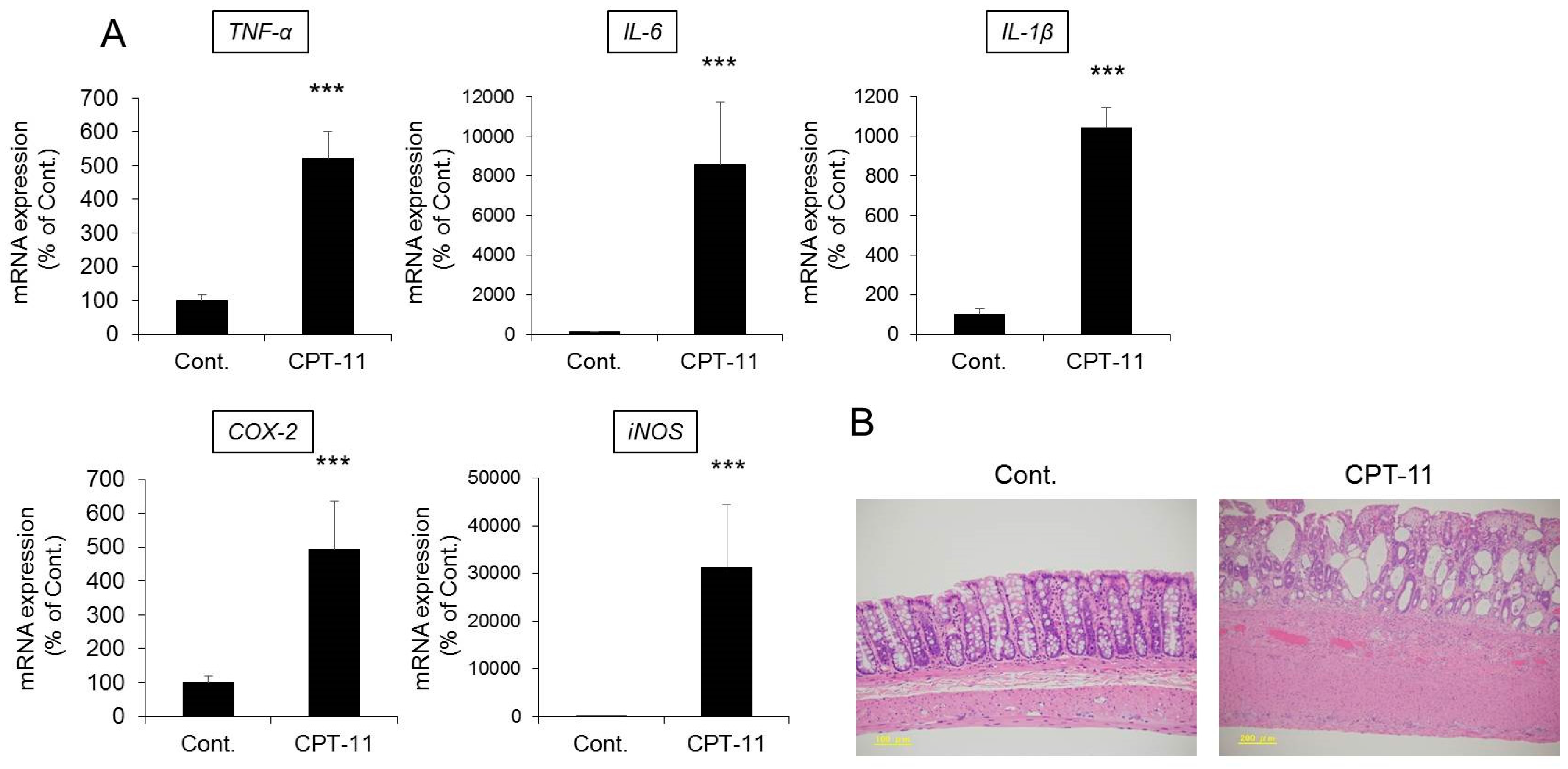
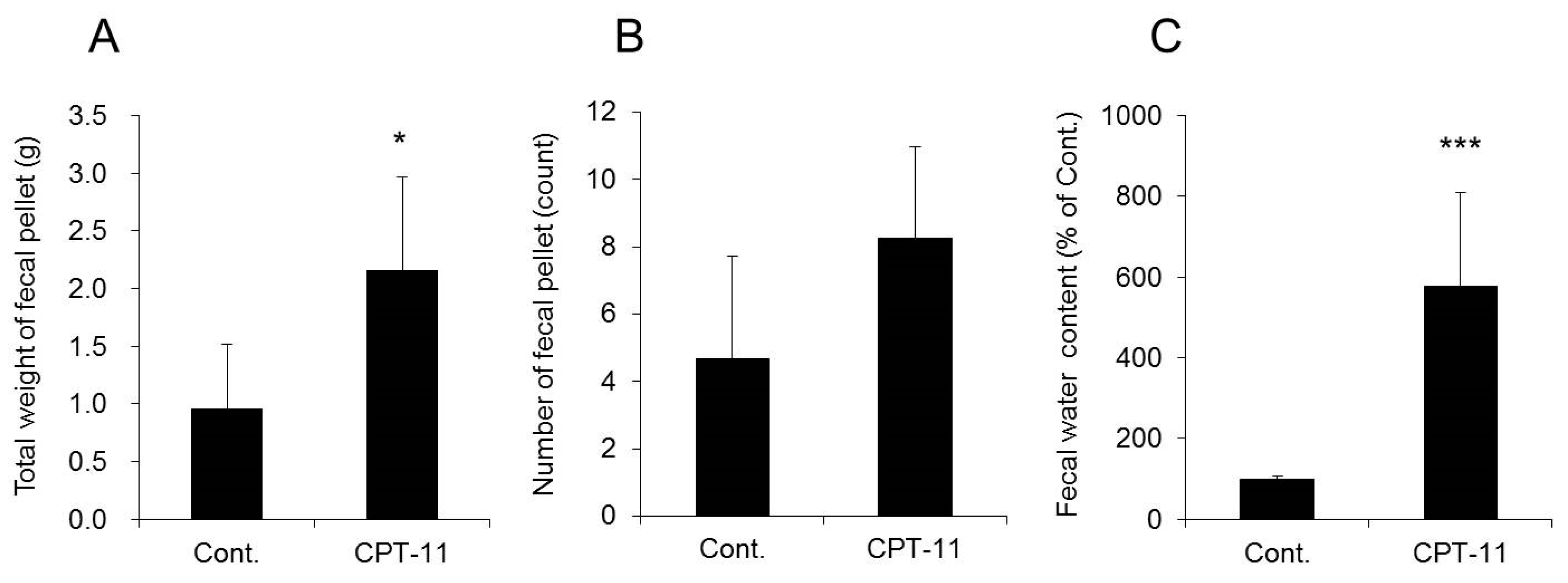
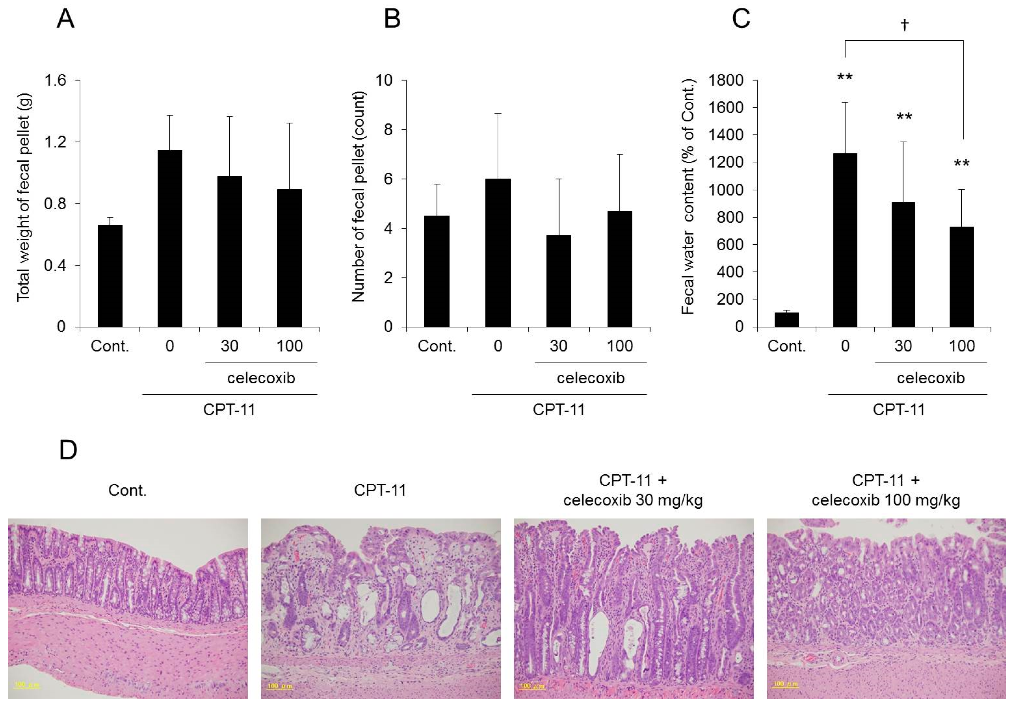
CPT-11 at a dose of 100 mg/kg was administered to rats via the tail vein for four days, and feces were collected on the third day following the last administration to investigate the level of diarrhea by analyzing the total weight of fecal pellets, total number of fecal pellets, and fecal water content. In addition, colitis was assessed by analyzing the condition of the colon tissue and inflammatory cytokines.
Both the total weight and fecal water content were significantly higher in rats in the CPT-11 administration group than in the rats in the control group, indicating the development of severe diarrhea (Figure 1). In addition, it was found that the mRNA expression levels of cyclooxygenase-2 (COX-2), inducible nitric oxide synthase (iNOS), and inflammatory cytokines (e.g., tumor necrosis factor-α (TNF-α), interleukin (IL)-6, and IL-1β) increased significantly in the rat colon in the CPT-11 administration group (Figure 2A). The condition of the colon tissue at that time was investigated by hematoxylin and eosin (HE) staining, and both crypt cell damage and infiltration of inflammatory cells into the lamina propria were observed in the CPT-11 administration group. Fibroplasia and muscle layer thickness in the colon also increased in the CPT-11 administration group (Figure 2B). These changes were consistent with the characteristics of animal models of CPT-11-induced delayed diarrhea that have been previously reported [28,29].
Based on the above results, it was found that severe diarrhea develops in rats when CPT-11 is administered via the tail vein at a dose of 100 mg/kg/day for four days, at which point colitis is present.

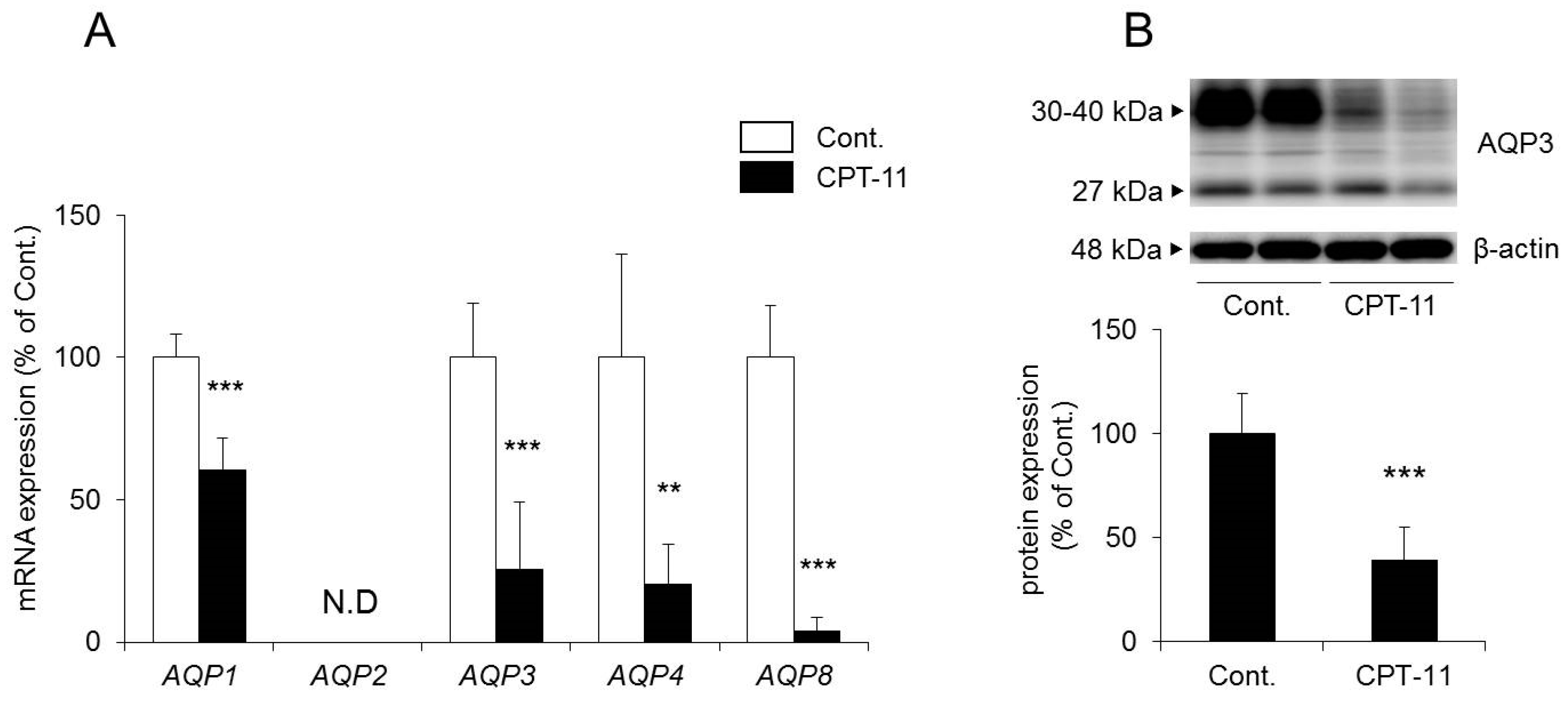
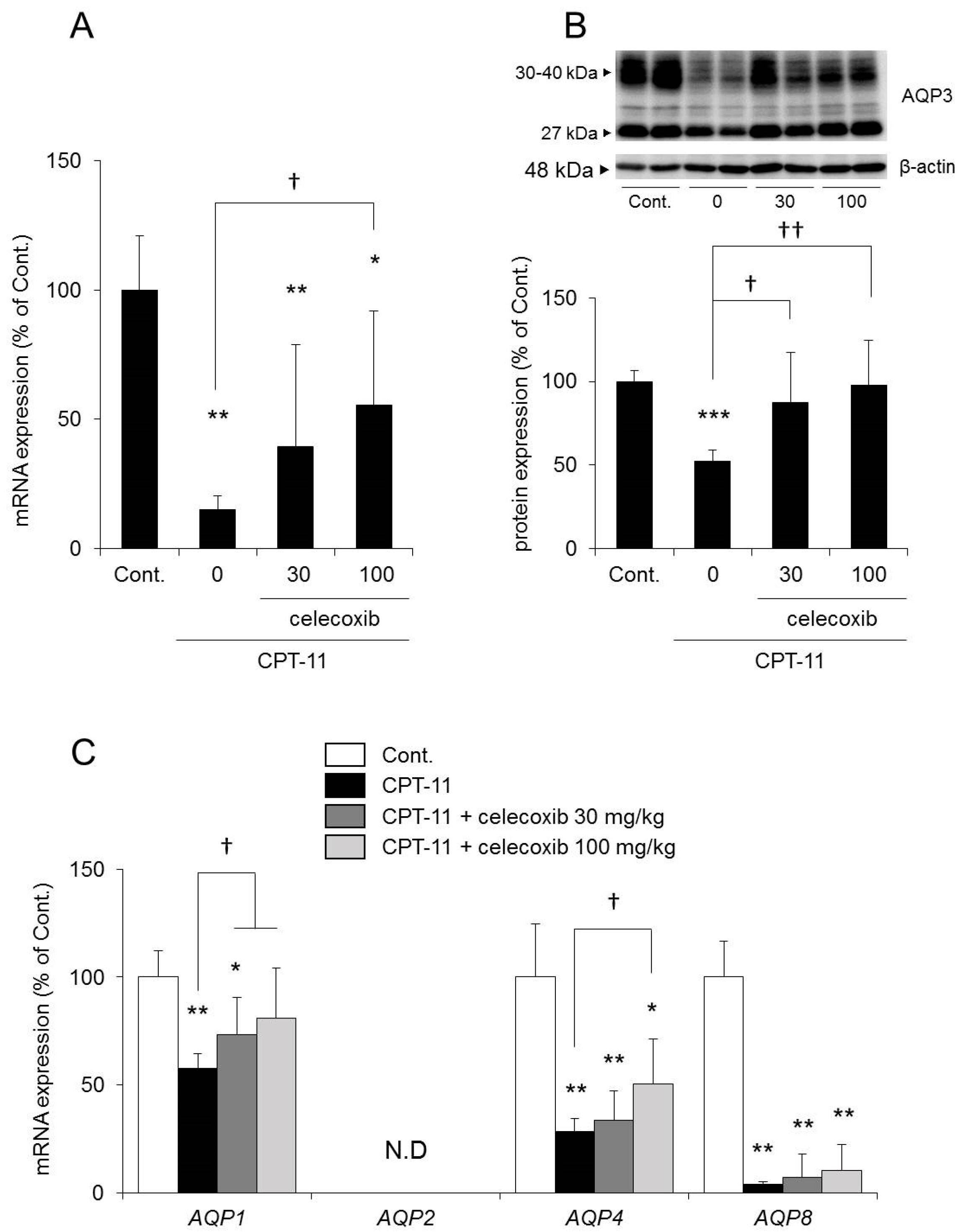
2.3. Changes in AQP in the Colon in the CPT-11-Induced Delayed Diarrhea Rat Model

It has been reported that AQP1, AQP2, AQP3, AQP4, and AQP8 are found in the colon [30,31,32]. Therefore, we investigated the expression levels of these AQP using a rat model of CPT-11-induced delayed diarrhea.
The mRNA expression levels of AQP1, AQP3, AQP4, and AQP8 in the colon decreased significantly in rats in the CPT-11 administration group compared to the levels in the control group. No AQP2 was detected (Figure 3A).
In a previous study, we found that AQP1 was expressed around blood vessels, AQP4 was expressed in muscular layer and AQP8 were low in the rat colon, using immunohistochemistry [24]. In this study, although we were not able to find AQP8, we found AQP1, AQP3, and AQP4 by immunohistochemistry (Figure 4). Among these, AQP3 was expressed especially in colonic mucosal epithelial cells. This distribution of AQP3 in rat colon is similar to this in human colon [32,33]. In addition, AQP3 plays a significant role in the development of diarrhea and constipation [24,25,26,27]. Therefore, we analyzed the protein expression level of AQP3 in colon membrane fractions by Western blotting. The results showed that AQP3 decreased significantly in rats in the CPT-11 administration group to approximately 40% of that in the control group (Figure 3B).
Based on the above results, it was found that the administration of CPT-11 markedly reduced AQPs in the colon. Specifically, AQP3 expression in mucosal epithelial cells was found to have decreased markedly, even at the protein level.

2.4. Effect of Celecoxib on the CPT-11-Induced Delayed Diarrhea Rat Model

Kase et al. reported that prostaglandin E2 (PGE2) production in the colon plays a major role in the development of CPT-11-induced delayed diarrhea and that Hangeshashinto, a traditional Kampo medicine, suppresses CPT-11-induced delayed diarrhea by reducing the production of PGE2 [34,35,36,37]. In addition, Trifan et al. reported that celecoxib, a selective COX-2 inhibitor involved in the production of PGE2, suppresses CPT-11-induced delayed diarrhea [38]. Therefore, we investigated whether CPT-11-induced delayed diarrhea and the reduced expression of AQP3 in the colon improved when celecoxib, an anti-inflammatory drug, and CPT-11 were administered in combination.
The fecal water content increased markedly in the group receiving CPT-11 alone compared with that in the control group, and all rats showed severe diarrhea, with a diarrhea score of three (Table 3). However, it was found that although the fecal water content was increased in the group receiving a combination of CPT-11 and celecoxib compared with that in the control group, it was lower than that in the group receiving CPT-11 alone (Figure 5C). In addition, when celecoxib was administered concomitantly, the diarrhea score decreased, and in particular, the incidence of severe diarrhea (grade 3) decreased (Table 3). This effect of celecoxib in improving diarrhea was found to be dose-dependent. When investigating the condition of the colon tissue, it was found that crypt cell damage, infiltration of inflammatory cells into the lamina propria, and edema caused by CPT-11 were reduced in a celecoxib dose-dependent manner (Figure 5D and Table 4).
The mRNA and protein expression levels of AQP3 in the colon were both significantly reduced in the group receiving CPT-11 alone compared with those in the control group. In contrast, the reduction in AQP3 expression was milder in the group receiving a combination of CPT-11 and celecoxib than in the group receiving CPT-11 alone, and the change was dependent on the dose of celecoxib (Figure 6A,B). In addition, the reduction in AQP1 and AQP4 mRNA expression by CPT-11 recovered after celecoxib administration. AQP8 mRNA expression remained decreasing (Figure 6C).
Based on the above results, it was found that the concomitant administration of celecoxib resulted in a decrease in the level of CPT-11-induced delayed diarrhea and a reduction in AQP3 protein expression in the colon.

2.5. Involvement of Colonic Macrophages in CPT-11-Induced Delayed Diarrhea

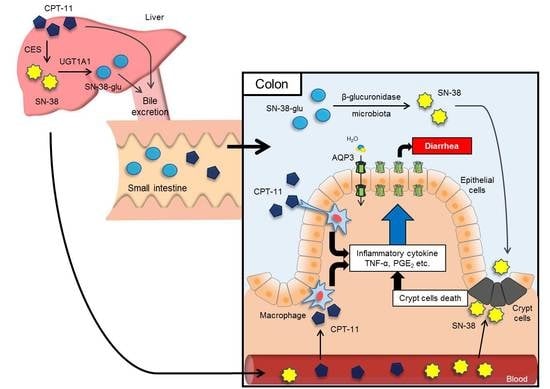
Based on the above results, it was found that the reduced expression of AQP3 in the colon plays a role in the development of CPT-11-induced diarrhea. In a previous study, we found that TNF-α and PGE2, which are secreted when colonic macrophages are activated, reduce the expression of AQP3 in mucosal epithelial cells in the colon [27]. Therefore, we investigated the mechanism of the reduction of AQP3 expression in the colon when CPT-11 is administered by focusing on the activation of macrophages.
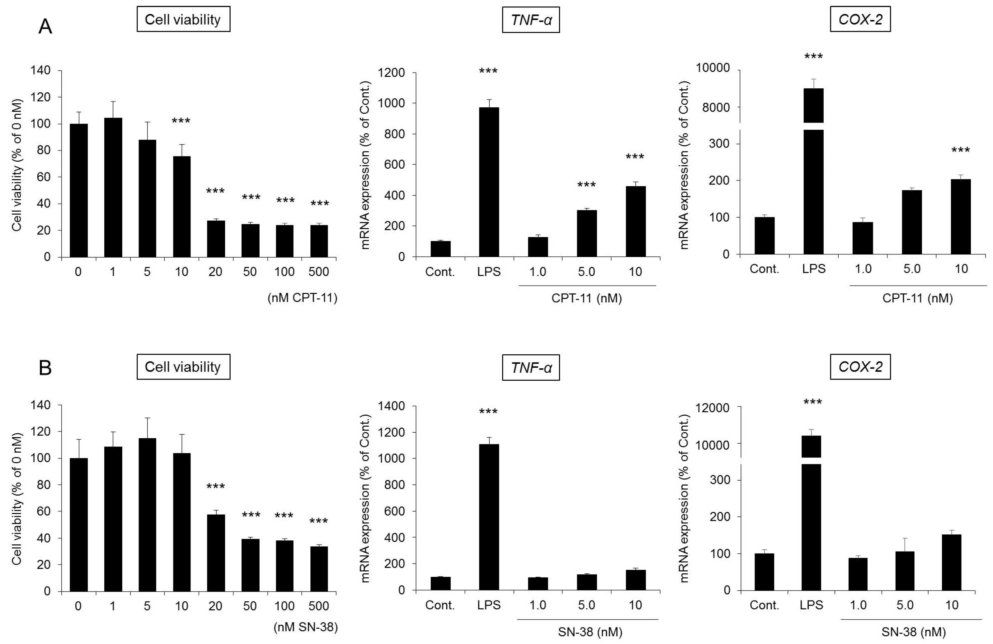
RAW264 cells are monocyte-derived macrophages and are frequently used to evaluate the functions of macrophages [39]. When RAW264 cells were incubated for 48 h after the addition of CPT-11 (0–500 μM) or SN-38 (0–500 nM), cytotoxicity began to be observed at 20 μM in the culture with CPT-11 and at 20 nM in the culture with SN-38, which is 1/1000 of the level in the CPT-11 culture (Figure 7). In addition, when the changes in the mRNA expression of TNF-α and COX-2 were investigated at concentrations at which no toxicity was observed, significant increases in TNF-α and COX-2 expression were observed only in the culture with CPT-11 (Figure 7A).
Based on the above observations, it was found that the cytotoxicity of SN-38 against macrophages was 1000 times greater than that of CPT-11. However, at concentrations at which no cytotoxicity was observed, CPT-11 exerted a more potent effect in activating macrophages. In addition, when CPT-11 or SN-38 was exposed for 12 h, no substantial degree of cytotoxicity or macrophage activation was observed.

3. Discussion

In this study, the mechanism of the development of CPT-11-induced delayed diarrhea was analyzed by focusing on AQPs, which are water channels expressed in the colon, to discover new preventive methods and treatments for the severe delayed diarrhea that occurs at the time of CPT-11 administration.
When CPT-11 was administered at 100 mg/kg/day for four days, severe diarrhea developed by the third day following the final dose, and the model showed the characteristics of CPT-11-induced delayed diarrhea (Figure 1) [28,29]. Next, AQP3 expression in the colonic mucosal epithelial cells in the model was analyzed. AQP3 in the CPT-11 administration group was markedly reduced at both the mRNA and protein levels (Figure 3). Based on this observation, it was hypothesized that when CPT-11 is administered, AQP3 is reduced, which then prevents water transport from the intestinal tract, resulting in retention of water in the colon and leading to the development of delayed diarrhea.
We investigated why AQP3 is reduced when CPT-11 is administered. It was previously believed that the mechanism of the onset of CPT-11-induced delayed diarrhea was damage to mucosal epithelial cells by the active metabolite SN-38 [11]. However, the colonic epithelial cells were present in our model rats (Figure 2B), and CPT-11 and SN-38 had little effect on AQP3 expression in an in vitro study (Figure S1). It was considered that the probability that mucosal damage is involved in the mechanism associated with the reduced AQP3 is low.
It has been reported that the levels of various inflammatory mediators, such as PGE2, are increased in the colon when CPT-11-induced delayed diarrhea develops [29,40,41]. In our model, the degree of CPT-11-induced delayed diarrhea was improved by the administration of celecoxib, an anti-inflammatory, and the reduced expression of AQP3 recovered to a level similar to that of the control (Figure 5 and Figure 6). These findings suggested that AQP3 plays a certain role in CPT-11-induced delayed diarrhea and that the increased production of PGE2 mediated by the increased COX-2 expression is involved in the reduced AQP3 expression. In addition, based on the results of the in vitro study, CPT-11 directly activates macrophages (Figure 7), and this action does not occur within a short period of time. Therefore, it was considered that the inflammation caused by the activation of macrophages and the accompanying reduced expression of AQP3 in the colon are characteristic of CPT-11-induced delayed diarrhea. Although the details of the mechanism of macrophage activation by CPT-11 are not clear, Li et al. reported that CPT-11 caused inflammation by activating NOD-like receptor protein-3 (NLRP3) and nuclear factor-kappa B (NF-κB) via c-Jun N-terminal kinase (JNK) in macrophages [42]. In addition, it is widely known that when cells are damaged, the production of TNF-α increases, causing inflammation. Therefore, it was considered that when CPT-11 was administered, the reduction of AQP3 expression was caused by the activation of macrophages by CPT-11 itself, as well as by inflammation that occurs following the loss of crypt cells.
The expression level of AQP1, AQP4, and AQP8 in the colon was also reduced significantly in the CPT-11 administration group, as was that of AQP3 (Figure 3A). In the colon, AQP1 is expressed in vascular endothelial cells [43,44]. When AQP1 is reduced, it is believed that water in the colonic tissues cannot be transferred to blood vessels efficiently, and this may have caused the swelling of the colon observed in the CPT-11 administration group. Although AQP4 is found in the muscle layers of the colon, when an AQP4 knockout mouse and a transgenic mouse with an overexpression of muscle AQP4 were analyzed, neither showed a change in myofunction [45,46]. It has also been reported that AQP4 was reduced in regenerated muscle cells [47]. AQP4 may be reduced during muscle tissue regeneration caused by the proliferation of active myofibroblasts and by the increase in muscle layers and fibers triggered by chronic inflammation in the colon, which occur at the time of CPT-11 administration. AQP8 is expressed in the mucosal epithelial cells of the colon and Fischer et al. have suggested that AQP8 could be a marker protein for a normal large intestine [48]. Laforenza et al. also reported that AQP8 may play a role in water transport in the proximal colon [49]. However, the role of AQP8 in diarrhea or constipation has not been clarified completely. AQP3 is intensively expressed in the mucosal epithelial cells of the colon, and it was found that the regeneration of epithelial cells was advanced when inflammation was suppressed by celecoxib (Table 4). It has been reported that AQP3 not only functions as a water channel but is also involved in cell proliferation and cell migration [50,51]. Thiagarajah et al. reported that colitis became severe and that the regeneration of epithelial cells was delayed in AQP3 knockout mice [52]. Therefore, it was expected that an increase in AQP3 expression may not only attenuate CPT-11-induced diarrhea but may also play a major role in the regeneration of colonic tissues. In addition, recovery of the expression of other AQPs may also be useful in normalizing the swelling and muscle layers and in attenuating diarrhea.
In this study, the administration of celecoxib improved the reduction of AQP3 expression levels induced by CPT-11; however, it did not completely resolve the diarrhea (Table 3 and Figure 5C). The AQP3 localization in the CPT-11-treated group was similar to that in celecoxib-treated group (Figure S2). Therefore, we considered that the diarrhea was not resolved for the following reasons: (1) AQP8 might play a critical role in CPT-11-induced delayed diarrhea (Figure 6C) [49]; (2) CPT-11 activates cystic fibrosis transmembrane conductance regulator (CFTR), a chloride ion channel, which disrupts the balance of electrolyte transport in the colon [53]; (3) CPT-11 disturbs the composition of mucin, which is the mucus component that protects intestinal epithelial cells [54]; and (4) CPT-11 changes the intestinal microbiota [55]. In addition, it is known that at the time of CPT-11 administration, damage is observed not only in the colon but also in the lower section of the small intestine [28]. Therefore, to improve CPT-11-induced diarrhea, we considered that it is necessary not only to control the expression of AQP3 but also to improve these factors in an integrated manner.
In summary, AQP3 expression in the colonic mucosal epithelial cells is markedly reduced in CPT-11-induced delayed diarrhea. Celecoxib reduces CPT-11-induced delayed diarrhea. As celecoxib has been reported to enhance the anticancer effects of CPT-11 [38], celecoxib is considered useful in combination with CPT-11 as a symptomatic treatment. The results of this study also uncovered the potential of AQP3 in the colon as a new functional molecule in the mechanism of the development of CPT-11-induced delayed diarrhea. We consider the search for a comprehensive treatment to be crucial, including the control of AQP3 for the management of CPT-11-induced delayed diarrhea.

4. Materials and Methods

4.1. Materials

CPT-11 hydrochloride was purchased from Carbosynth Limited (Berkshire, UK). SN-38 was purchased from Tokyo Chemical Industry Co., Ltd. (Tokyo, Japan). Celecoxib was purchased from Combi-Blocks, Inc. (San Diego, CA, USA). d-sorbitol and TRI reagent were purchased from Sigma-Aldrich Corp. (St. Louis, MO, USA). Lactic acid and d-MEM medium were purchased from Wako Pure Chemicals (Osaka, Japan). WST-1 was purchased from Roche Diagnostics (Indianapolis, IN, USA). Rabbit anti-rat AQP3 antibody, rabbit anti-human AQP1 antibody, and rabbit anti-rat AQP4 antibody were purchased from Alomone Labs (Jerusalem, Israel). Rabbit anti-rat AQP8 antibody was purchased from Alpha Diagnostic Inc. (San Antonio, TX, USA). Alexa Fluor 488 donkey anti-rabbit IgG was purchased from Thermo Fisher Scientific Inc. (Waltham, MA, USA). Purified anti-β-actin antibody was purchased from BioLegend Inc. (San Diego, CA, USA). Donkey anti-rabbit IgG-HRP antibody and ECL prime Western blotting detection reagents were purchased from GE Healthcare (Chalfont St. Giles, UK). All primers for real-time PCR were purchased from Hokkaido System Science Co., Ltd. (Hokkaido, Japan). A high-capacity cDNA reverse transcription kit was purchased from Applied Biosystems (Foster City, CA, USA). SsoAdvanced SYBR green supermix was purchased from Bio-Rad Laboratories (Hercules, CA, USA).

4.2. Animals

Male Wistar rats (eight weeks old) were purchased from Japan SLC, Inc. (Shizuoka, Japan). The rats were housed at 24 ± 1 °C and 55 ± 1% humidity with 12 h of light (08:00–20:00). The study was conducted upon approval (approval No. 29–118, 29 June 2017) in accordance with the Hoshi University Guiding Principles for the Care and Use of Laboratory Animals.

4.3. Treatment

Rats were given lactic acid buffer (45 mg/mL d-sorbitol, 0.9 mg/mL lactic acid; pH 3.4) or CPT-11 (60, 80, 100, 120, or 150 mg/kg in lactic acid buffer) via the tail vein, and the level of diarrhea was evaluated on the third day following the final administration.
One day prior to the administration of CPT-11, rats were started on oral celecoxib (30 or 100 mg/kg/day in 0.5% methylcellulose) twice daily for eight days. CPT-11 (100 mg/kg in lactic acid buffer) was given via the tail vein for four days, and the colon was isolated under diethyl ether anesthesia on the third day following the final administration.

4.4. Assessment of Diarrhea

On the third day following the final administration of CPT-11, rat feces were collected to measure the total number of fecal pellets and total fecal weight. The fecal water content was calculated by freeze-drying the collected feces in a lyophilizer for 24 h, and the water content per gram of feces was calculated based on the moist weight and dry weight. The degree of diarrhea was assessed based on past reports (Table 2) [38].

4.5. HE Staining

The colons isolated from the rats were immersed in 10% neutral buffered formalin to fix the tissues. The tissues were embedded in paraffin and sectioned into 3 μm slices on glass slides. The slides were deparaffinized and stained with hematoxylin followed by eosin. The slides were dehydrated in alcohol, cleared in xylene, and covered for microscopic examination. The slides were read blindly by a pathologist and the colonic damage was assessed.

4.6. RAW264 Cell Culture

RAW264 cells (Riken Cell Bank, Tsukuba, Japan) were cultured using d-MEM medium (100 U/mL penicillin G potassium, 100 μg/mL streptomycin, and 10% fetal bovine serum).
RAW264 cells were plated in a 96 well-plate at a cell density of 1 × 10⁴ cells/well and incubated for 24 h. After CPT-11 (0–500 μM) or SN-38 (0–500 nM) was added, cells were incubated for 48 h, and cell viability was measured using the WST-1 assay. RAW264 cells were also plated in a 12 well-plate at a cell density of 2 × 10⁵ cells/well and incubated for 24 h. The expression level of each gene was analyzed using cells that had been incubated for 48 h after the addition of LPS (10 ng/mL), CPT-11 (0–10 μM) or SN-38 (0–10 nM).

4.7. WST-1 Assay

RAW264 cells were plated in a 96 well-plate and incubated for 48 h after the addition of drugs. After washing each well, WST-1 was added at a ratio of 10/100 μL medium, and cells were incubated for 2 h at 37 °C in a CO2 incubator. The absorbance at 450 nm was measured using a microplate reader.

4.8. Total RNA Preparations and Real-Time RT-PCR

TRI reagent was added to colon tissue or RAW264 cells, and total RNA was extracted. A high-capacity cDNA reverse transcription kit was used to synthesize cDNA from 1 μg of RNA.
Real-time PCR was performed using the primers listed in Table 5 and the following mixture: 2 μL of cDNA solution (2.5 ng/μL), 0.6 μL of forward primer (5 pmol/μL) and reverse primer (5 pmol/μL), 5 μL of SsoAdvanced SYBR Green Supermix, and 2.8 μL of RNase-free water. The reaction conditions included denaturation at 95 °C for 15 s, annealing at 56 °C for 30 s, and elongation at 72 °C for 30 s. The fluorescence intensity of the amplification process was monitored using the CFX Connect™ Real-Time PCR Detection System (Bio-Rad Laboratories). β-Actin and 18S rRNA expression levels in reagent-treated group and control group did not differ.

4.9. Extraction of the Plasma Membrane Fraction from the Rat Colons

The mucosa was scraped from each rat colon sample, suspended in dissecting buffer (0.3 M sucrose, 25 mM imidazole, 1 mM ethylenediaminetetraacetic acid, 8.5 μM leupeptin, and 1 μM phenylmethylsulfonyl fluoride; pH 7.2) and homogenized on ice. The homogenate was centrifuged (800× g at 4 °C for 15 min), and the resulting supernatant was further centrifuged (17,000× g at 4 °C for 30 min). The supernatant was removed, and dissecting buffer was added to the precipitate, which was then dispersed using an ultrasonic homogenizer. This solution included the plasma membrane fraction with abundant cell membranes [26,56].

4.10. Western Blotting

Protein concentrations were measured by the bicinchoninic acid methods using bovine serum albumin as standard. Each sample was diluted with loading buffer (84 mM Tris, 20% glycerol, 0.004% bromophenol blue, 4.6% sodium dodecyl sulfate, and 10% 2-mercaptoethanol; pH 6.8), and samples were loaded in each lane. After polyacrylamide gel electrophoresis, the proteins were transferred to a polyvinylidene difluoride membrane. After blocking with skim milk, the resulting membrane was incubated with rabbit anti-rat AQP3 or anti-β-actin antibody for 1 h, followed by washing and incubation with donkey anti-rabbit IgG-HRP antibody for 1 h. The membrane was washed and then reacted with the ECL prime Western blotting detection reagents, and the bands detected by the LAS-3000 mini-imaging system (FUJIFILM, Tokyo, Japan) were analyzed.

4.11. Immunohistochenistry

The colon was post-fixed in 4% paraformaldehyde. The tissues were embedded, and the frozen blocks were sectioned into 10 μm slices on glass slides. The sections were reacted with a rabbit anti-human AQP1 antibody, rabbit anti-rat AQP3 antibody, rabbit anti-rat AQP4 antibody, and rabbit anti-rat AQP8 antibody. The sections were treated with an Alexa Fluor 488 donkey anti-rabbit IgG antibody. The slides were covered and observed under a fluorescent microscope.

4.12. Statistical Analysis

Numerical data are expressed as the mean ± standard deviation (SD). The significance of the differences was examined using Tukey’s test and Student’s t-test.

Supplementary Materials

Supplementary materials can be found at www.mdpi.com/1422-0067/19/1/170/s1.

Acknowledgments

This work was supported by the Takeda Science Foundation. We thank Nanami Inoue, Michiru Yamamoto, Kazuhiro Onuma, Misato Toyonaga, and Mayumi Okuma for their technical assistance.

Author Contributions

Risako Kon conceived and designed the experiments; Risako Kon, Nobutomo Ikarashi, Yuika Tsubota, Moe Minami, Saki Kato, Yukari Matsunaga, Hiroshi Kimura, Yuta Murakami, Tetsuya Fujikawa, Ryoya Sakurai, Rei Tomimoto, and Yoshiaki Machida performed the experiments and analyzed the data; Risako Kon, Nobutomo Ikarashi, and Kiyoshi Sugiyama wrote the paper.

Conflicts of Interest

The authors declare no conflict of interest.

Abbreviations

AQPAquaporin
CFTRCystic fibrosis transmembrane conductance regulator
COXCyclooxygenase
CPT-11Camptothecin-11, irinotecan
DAPI4′,6-diamidino-2-phenylindole
HEHematoxylin and eosin
ILInterleukin
iNOSInducible nitric oxide synthase
JNKc-Jun N-terminal kinase
LPSLipopolysaccharide
NF-κBNuclear factor-kappa B
NLRP3NOD-like receptor protein-3
PCRpolymerase chain reaction
PGEProstaglandin E
QOLQuality of life
SN-387-ethyl-10-hydroxy-camptothecin
SN-38-glu7-ethyl-10-hydroxy-camptothecin glucronide
TNFTumor necrosis factor
UGTUridine diphosphate-glucuronosyltransferase
WST-1Water-soluble tetrazolium salt

References

  1. Kawato, Y.; Aonuma, M.; Hirota, Y.; Kuga, H.; Sato, K. Intracellular roles of SN-38, a metabolite of the camptothecin derivative CPT-11, in the antitumor effect of CPT-11. Cancer Res. 1991, 51, 4187–4191. [Google Scholar] [PubMed]
  2. Shimada, Y.; Yoshino, M.; Wakui, A.; Nakao, I.; Futatsuki, K.; Sakata, Y.; Kambe, M.; Taguchi, T.; Ogawa, N. Phase II study of CPT-11, a new camptothecin derivative, in metastatic colorectal cancer. CPT-11 Gastrointestinal Cancer Study Group. J. Clin. Oncol. 1993, 11, 909–913. [Google Scholar] [CrossRef] [PubMed]
  3. Bleiberg, H.; Cvitkovic, E. Characterisation and clinical management of CPT-11 (irinotecan)-induced adverse events: The European perspective. Eur. J. Cancer 1996, 32A (Suppl. 3), S18–S23. [Google Scholar] [CrossRef]
  4. Gandia, D.; Abigerges, D.; Armand, J.P.; Chabot, G.; Da Costa, L.; De Forni, M.; Mathieu-Boue, A.; Herait, P. CPT-11-induced cholinergic effects in cancer patients. J. Clin. Oncol. 1993, 11, 196–197. [Google Scholar] [CrossRef] [PubMed]
  5. Kawato, Y.; Sekiguchi, M.; Akahane, K.; Tsutomi, Y.; Hirota, Y.; Kuga, H.; Suzuki, W.; Hakusui, H.; Sato, K. Inhibitory activity of camptothecin derivatives against acetylcholinesterase in dogs and their binding activity to acetylcholine receptors in rats. J. Pharm. Pharmacol. 1993, 45, 444–448. [Google Scholar] [CrossRef] [PubMed]
  6. Rothenberg, M.L.; Kuhn, J.G.; Burris, H.A., 3rd; Nelson, J.; Eckardt, J.R.; Tristan-Morales, M.; Hilsenbeck, S.G.; Weiss, G.R.; Smith, L.S.; Rodriguez, G.I.; et al. Phase I and pharmacokinetic trial of weekly CPT-11. J. Clin. Oncol. 1993, 11, 2194–2204. [Google Scholar] [CrossRef] [PubMed]
  7. Iyer, L.; King, C.D.; Whitington, P.F.; Green, M.D.; Roy, S.K.; Tephly, T.R.; Coffman, B.L.; Ratain, M.J. Genetic predisposition to the metabolism of irinotecan (CPT-11). Role of uridine diphosphate glucuronosyltransferase isoform 1A1 in the glucuronidation of its active metabolite (SN-38) in human liver microsomes. J. Clin. Investig. 1998, 101, 847–854. [Google Scholar] [CrossRef] [PubMed]
  8. Rivory, L.P.; Bowles, M.R.; Robert, J.; Pond, S.M. Conversion of irinotecan (CPT-11) to its active metabolite, 7-ethyl-10-hydroxycamptothecin (SN-38), by human liver carboxylesterase. Biochem. Pharmacol. 1996, 52, 1103–2211. [Google Scholar] [CrossRef]
  9. Atsumi, R.; Okazaki, O.; Hakusui, H. Pharmacokinetics of SN-38 [(+)-(4S)-4,11-diethyl-4,9-dihydroxy-1H- pyrano[3′,4′:6,7]-indolizino[1,2-b]quinoline-3,14(4H,12H)-dione], an active metabolite of irinotecan, after a single intravenous dosing of 14C-SN-38 to rats. Biol. Pharm. Bull. 1995, 18, 1114–1119. [Google Scholar] [CrossRef] [PubMed]
  10. Takasuna, K.; Hagiwara, T.; Hirohashi, M.; Kato, M.; Nomura, M.; Nagai, E.; Yokoi, T.; Kamataki, T. Involvement of β-glucuronidase in intestinal microflora in the intestinal toxicity of the antitumor camptothecin derivative irinotecan hydrochloride (CPT-11) in rats. Cancer Res. 1996, 56, 3752–3757. [Google Scholar] [PubMed]
  11. Gupta, E.; Lestingi, T.M.; Mick, R.; Ramirez, J.; Vokes, E.E.; Ratain, M.J. Metabolic fate of irinotecan in humans: Correlation of glucuronidation with diarrhea. Cancer Res. 1994, 54, 3723–3725. [Google Scholar] [PubMed]
  12. De Jong, F.A.; Kehrer, D.F.; Mathijssen, R.H.; Creemers, G.J.; de Bruijn, P.; van Schaik, R.H.; Planting, A.S.; van der Gaast, A.; Eskens, F.A.; Janssen, J.T.; et al. Prophylaxis of irinotecan-induced diarrhea with neomycin and potential role for UGT1A1*28 genotype screening: A double-blind, randomized, placebo-controlled study. Oncologist 2006, 11, 944–954. [Google Scholar] [CrossRef] [PubMed]
  13. Flieger, D.; Klassert, C.; Hainke, S.; Keller, R.; Kleinschmidt, R.; Fischbach, W. Phase II clinical trial for prevention of delayed diarrhea with cholestyramine/levofloxacin in the second-line treatment with irinotecan biweekly in patients with metastatic colorectal carcinoma. Oncology 2007, 72, 10–16. [Google Scholar] [CrossRef] [PubMed]
  14. Furman, W.L.; Crews, K.R.; Billups, C.; Wu, J.; Gajjar, A.J.; Daw, N.C.; Patrick, C.C.; Rodriguez-Galindo, C.; Stewart, C.F.; Dome, J.S.; et al. Cefixime allows greater dose escalation of oral irinotecan: A phase I study in pediatric patients with refractory solid tumors. J. Clin. Oncol. 2006, 24, 563–570. [Google Scholar] [CrossRef] [PubMed]
  15. McGregor, L.M.; Stewart, C.F.; Crews, K.R.; Tagen, M.; Wozniak, A.; Wu, J.; McCarville, M.B.; Navid, F.; Santana, V.M.; Houghton, P.J.; et al. Dose escalation of intravenous irinotecan using oral cefpodoxime: A phase I study in pediatric patients with refractory solid tumors. Pediatr. Blood Cancer 2012, 58, 372–379. [Google Scholar] [CrossRef] [PubMed]
  16. McNall-Knapp, R.Y.; Williams, C.N.; Reeves, E.N.; Heideman, R.L.; Meyer, W.H. Extended phase I evaluation of vincristine, irinotecan, temozolomide, and antibiotic in children with refractory solid tumors. Pediatr. Blood Cancer 2010, 54, 909–915. [Google Scholar] [CrossRef] [PubMed]
  17. Mori, K.; Kondo, T.; Kamiyama, Y.; Kano, Y.; Tominaga, K. Preventive effect of Kampo medicine (Hangeshashin-to) against irinotecan-induced diarrhea in advanced non-small-cell lung cancer. Cancer Chemother. Pharmacol. 2003, 51, 403–406. [Google Scholar] [PubMed]
  18. Sakata, Y.; Suzuki, H.; Kamataki, T. Preventive effect of TJ-14, a kampo (Chinese herb) medicine, on diarrhea induced by irinotecan hydrochloride (CPT-11). Gan Kagaku Ryoho. Cancer Chemother. 1994, 21, 1241–1244. [Google Scholar]
  19. Michael, M.; Brittain, M.; Nagai, J.; Feld, R.; Hedley, D.; Oza, A.; Siu, L.; Moore, M.J. Phase II study of activated charcoal to prevent irinotecan-induced diarrhea. J. Clin. Oncol. 2004, 22, 4410–4417. [Google Scholar] [CrossRef] [PubMed]
  20. Sergio, G.C.; Felix, G.M.; Luis, J.V. Activated charcoal to prevent irinotecan-induced diarrhea in children. Pediatr. Blood Cancer 2008, 51, 49–52. [Google Scholar] [CrossRef] [PubMed]
  21. Abigerges, D.; Chabot, G.G.; Armand, J.P.; Herait, P.; Gouyette, A.; Gandia, D. Phase I and pharmacologic studies of the camptothecin analog irinotecan administered every 3 weeks in cancer patients. J. Clin. Oncol. 1995, 13, 210–221. [Google Scholar] [CrossRef] [PubMed]
  22. Takeda, Y.; Kobayashi, K.; Akiyama, Y.; Soma, T.; Handa, S.; Kudoh, S.; Kudo, K. Prevention of irinotecan (CPT-11)-induced diarrhea by oral alkalization combined with control of defecation in cancer patients. Int. J. Cancer 2001, 92, 269–275. [Google Scholar] [CrossRef]
  23. Hecht, J.R. Gastrointestinal toxicity or irinotecan. Oncology 1998, 12, 72–78. [Google Scholar] [PubMed]
  24. Ikarashi, N.; Baba, K.; Ushiki, T.; Kon, R.; Mimura, A.; Toda, T.; Ishii, M.; Ochiai, W.; Sugiyama, K. The laxative effect of bisacodyl is attributable to decreased aquaporin-3 expression in the colon induced by increased PGE2 secretion from macrophages. Am. J. Physiol.-Gastrointest. Liver Physiol. 2011, 301, G887–G895. [Google Scholar] [CrossRef] [PubMed]
  25. Ikarashi, N.; Kon, R.; Iizasa, T.; Suzuki, N.; Hiruma, R.; Suenaga, K.; Toda, T.; Ishii, M.; Hoshino, M.; Ochiai, W.; et al. Inhibition of aquaporin-3 water channel in the colon induces diarrhea. Biol. Pharm. Bull. 2012, 35, 957–962. [Google Scholar] [CrossRef] [PubMed]
  26. Kon, R.; Ikarashi, N.; Hayakawa, A.; Haga, Y.; Fueki, A.; Kusunoki, Y.; Tajima, M.; Ochiai, W.; Machida, Y.; Sugiyama, K. Morphine-Induced Constipation Develops With Increased Aquaporin-3 Expression in the Colon via Increased Serotonin Secretion. Toxicol. Sci. 2015, 145, 337–347. [Google Scholar] [CrossRef] [PubMed]
  27. Kon, R.; Ikarashi, N.; Nagoya, C.; Takayama, T.; Kusunoki, Y.; Ishii, M.; Ueda, H.; Ochiai, W.; Machida, Y.; Sugita, K.; et al. Rheinanthrone, a metabolite of sennoside A, triggers macrophage activation to decrease aquaporin-3 expression in the colon, causing the laxative effect of rhubarb extract. J. Ethnopharm. 2014, 152, 190–200. [Google Scholar] [CrossRef] [PubMed]
  28. Gibson, R.J.; Bowen, J.M.; Inglis, M.R.; Cummins, A.G.; Keefe, D.M. Irinotecan causes severe small intestinal damage, as well as colonic damage, in the rat with implanted breast cancer. J. Gastroenterol. Hepatol. 2003, 18, 1095–1100. [Google Scholar] [CrossRef] [PubMed]
  29. Logan, R.M.; Gibson, R.J.; Bowen, J.M.; Stringer, A.M.; Sonis, S.T.; Keefe, D.M. Characterisation of mucosal changes in the alimentary tract following administration of irinotecan: Implications for the pathobiology of mucositis. Cancer Chemother. Pharmacol. 2008, 62, 33–41. [Google Scholar] [CrossRef] [PubMed]
  30. Gallardo, P.; Cid, L.P.; Vio, C.P.; Sepulveda, F.V. Aquaporin-2, a regulated water channel, is expressed in apical membranes of rat distal colon epithelium. Am. J. Physiol.-Gastrointest. Liver Physiol. 2001, 281, G856–G863. [Google Scholar] [CrossRef] [PubMed]
  31. Koyama, Y.; Yamamoto, T.; Tani, T.; Nihei, K.; Kondo, D.; Funaki, H.; Yaoita, E.; Kawasaki, K.; Sato, N.; Hatakeyama, K.; et al. Expression and localization of aquaporins in rat gastrointestinal tract. Am. J. Physiol. 1999, 276, C621–C627. [Google Scholar] [CrossRef] [PubMed]
  32. Silberstein, C.; Kierbel, A.; Amodeo, G.; Zotta, E.; Bigi, F.; Berkowski, D.; Ibarra, C. Functional characterization and localization of AQP3 in the human colon. Braz. J. Med. Biol. Res. 1999, 32, 1303–1313. [Google Scholar] [CrossRef] [PubMed]
  33. Yde, J.; Keely, S.; Wu, Q.; Borg, J.F.; Lajczak, N.; O’Dwyer, A.; Dalsgaard, P.; Fenton, R.A.; Moeller, H.B. Characterization of AQPs in Mouse, Rat, and Human Colon and Their Selective Regulation by Bile Acids. Front. Nutr. 2016, 3, 46. [Google Scholar] [CrossRef] [PubMed]
  34. Kase, Y.; Hayakawa, T.; Aburada, M.; Komatsu, Y.; Kamataki, T. Preventive effects of Hange-shashin-to on irinotecan hydrochloride-caused diarrhea and its relevance to the colonic prostaglandin E2 and water absorption in the rat. Jpn. J. Pharmacol. 1997, 75, 407–413. [Google Scholar] [CrossRef] [PubMed]
  35. Kase, Y.; Hayakawa, T.; Ishige, A.; Aburada, M.; Komatsu, Y. The effects of Hange-shashin-to on the content of prostaglandin E2 and water absorption in the large intestine of rats. Biol. Pharm. Bull. 1997, 20, 954–957. [Google Scholar] [CrossRef] [PubMed]
  36. Kase, Y.; Hayakawa, T.; Togashi, Y.; Kamataki, T. Relevance of irinotecan hydrochloride-induced diarrhea to the level of prostaglandin E2 and water absorption of large intestine in rats. Jpn. J. Pharmacol. 1997, 75, 399–405. [Google Scholar] [CrossRef] [PubMed]
  37. Kase, Y.; Saitoh, K.; Ishige, A.; Komatsu, Y. Mechanisms by which Hange-shashin-to reduces prostaglandin E2 levels. Biol. Pharm. Bull. 1998, 21, 1277–1281. [Google Scholar] [CrossRef] [PubMed]
  38. Trifan, O.C.; Durham, W.F.; Salazar, V.S.; Horton, J.; Levine, B.D.; Zweifel, B.S.; Davis, T.W.; Masferrer, J.L. Cyclooxygenase-2 inhibition with celecoxib enhances antitumor efficacy and reduces diarrhea side effect of CPT-11. Cancer Res. 2002, 62, 5778–5784. [Google Scholar] [PubMed]
  39. Zheng, S.; Brown, M.C.; Taffet, S.M. Lipopolysaccharide stimulates both nuclear localization of the nuclear factor kappa B 50-kDa subunit and loss of the 105-kDa precursor in RAW264 macrophage-like cells. J. Biol. Chem. 1993, 268, 17233–17239. [Google Scholar] [PubMed]
  40. Lima-Junior, R.C.; Freitas, H.C.; Wong, D.V.; Wanderley, C.W.; Nunes, L.G.; Leite, L.L.; Miranda, S.P.; Souza, M.H.; Brito, G.A.; Magalhaes, P.J.; et al. Targeted inhibition of IL-18 attenuates irinotecan-induced intestinal mucositis in mice. Br. J. Pharmacol. 2014, 171, 2335–2350. [Google Scholar] [CrossRef] [PubMed]
  41. Sakai, H.; Sato, T.; Hamada, N.; Yasue, M.; Ikari, A.; Kakinoki, B.; Takeguchi, N. Thromboxane A2, released by the anti-tumour drug irinotecan, is a novel stimulator of Cl secretion in isolated rat colon. J. Physiol. 1997, 505 Pt 1, 133–144. [Google Scholar] [CrossRef] [PubMed]
  42. Li, Q.; Zhang, X.; Wang, W.; Li, L.; Xu, Q.; Wu, X.; Gu, Y. CPT-11 activates NLRP3 inflammasome through JNK and NF-kappaB signalings. Toxicol. Appl. Pharmacol. 2015, 289, 133–141. [Google Scholar] [CrossRef] [PubMed]
  43. Bondy, C.; Chin, E.; Smith, B.L.; Preston, G.M.; Agre, P. Developmental gene expression and tissue distribution of the CHIP28 water-channel protein. Proc. Natl. Acad. Sci. USA 1993, 90, 4500–4504. [Google Scholar] [CrossRef] [PubMed]
  44. Mobasheri, A.; Marples, D. Expression of the AQP-1 water channel in normal human tissues: A semiquantitative study using tissue microarray technology. Am. J. Physiol.-Cell Physiol. 2004, 286, C529–C537. [Google Scholar] [CrossRef] [PubMed]
  45. Frigeri, A.; Nicchia, G.P.; Balena, R.; Nico, B.; Svelto, M. Aquaporins in skeletal muscle: Reassessment of the functional role of aquaporin-4. FASEB J. 2004, 18, 905–907. [Google Scholar] [CrossRef] [PubMed]
  46. Wakayama, Y.; Takahashi, J.; Shibuya, S.; Inoue, M.; Kojima, H.; Oniki, H.; Arata, S.; Hara, H.; Jimi, T.; Shioda, S.; et al. Generation of muscle aquaporin 4 overexpressing transgenic mouse: Its characterization at RNA and protein levels including freeze-fracture study. Micron 2007, 38, 257–267. [Google Scholar] [CrossRef] [PubMed]
  47. Jimi, T.; Wakayama, Y.; Murahashi, M.; Shibuya, S.; Inoue, M.; Hara, H.; Matsuzaki, Y.; Uemura, N. Aquaporin 4: Lack of mRNA expression in the rat regenerating muscle fiber under denervation. Neurosci. Lett. 2000, 291, 93–96. [Google Scholar] [CrossRef]
  48. Fischer, H.; Stenling, R.; Rubio, C.; Lindblom, A. Differential expression of aquaporin 8 in human colonic epithelial cells and colorectal tumors. BMC Physiol. 2001, 1, 1. [Google Scholar] [CrossRef]
  49. Laforenza, U.; Cova, E.; Gastaldi, G.; Tritto, S.; Grazioli, M.; LaRusso, N.F.; Splinter, P.L.; D’Adamo, P.; Tosco, M.; Ventura, U. Aquaporin-8 is involved in water transport in isolated superficial colonocytes from rat proximal colon. J. Nutr. 2005, 135, 2329–2336. [Google Scholar] [PubMed]
  50. Hara-Chikuma, M.; Verkman, A.S. Aquaporin-3 facilitates epidermal cell migration and proliferation during wound healing. J. Mol. Med. 2008, 86, 221–231. [Google Scholar] [CrossRef] [PubMed]
  51. Levin, M.H.; Verkman, A.S. Aquaporin-3-dependent cell migration and proliferation during corneal re-epithelialization. Investig. Ophthalmol. Vis. Sci. 2006, 47, 4365–4372. [Google Scholar] [CrossRef] [PubMed]
  52. Thiagarajah, J.R.; Zhao, D.; Verkman, A.S. Impaired enterocyte proliferation in aquaporin-3 deficiency in mouse models of colitis. Gut 2007, 56, 1529–1535. [Google Scholar] [CrossRef] [PubMed]
  53. Moon, C.; Zhang, W.; Ren, A.; Arora, K.; Sinha, C.; Yarlagadda, S.; Woodrooffe, K.; Schuetz, J.D.; Valasani, K.R.; de Jonge, H.R.; et al. Compartmentalized accumulation of cAMP near complexes of multidrug resistance protein 4 (MRP4) and cystic fibrosis transmembrane conductance regulator (CFTR) contributes to drug-induced diarrhea. J. Biol. Chem. 2015, 290, 11246–11257. [Google Scholar] [CrossRef] [PubMed]
  54. Stringer, A.M.; Gibson, R.J.; Bowen, J.M.; Logan, R.M.; Ashton, K.; Yeoh, A.S.; Al-Dasooqi, N.; Keefe, D.M. Irinotecan-induced mucositis manifesting as diarrhoea corresponds with an amended intestinal flora and mucin profile. Int. J. Exp. Pathol. 2009, 90, 489–499. [Google Scholar] [CrossRef] [PubMed]
  55. Stringer, A.M.; Gibson, R.J.; Logan, R.M.; Bowen, J.M.; Yeoh, A.S.; Keefe, D.M. Faecal microflora and β-glucuronidase expression are altered in an irinotecan-induced diarrhea model in rats. Cancer Biol. Ther. 2008, 7, 1919–1925. [Google Scholar] [CrossRef] [PubMed]
  56. Marples, D.; Knepper, M.A.; Christensen, E.I.; Nielsen, S. Redistribution of aquaporin-2 water channels induced by vasopressin in rat kidney inner medullary collecting duct. Am. J. Physiol. 1995, 269, C655–C664. [Google Scholar] [CrossRef] [PubMed]
Figure 1. Assessment of diarrhea parameters after CPT-11 administration. CPT-11 (100 mg/kg/day) was administered in rats via the tail vein for 4 days. The total weight of fecal pellets (A), total number of fecal pellets (B), and fecal water content (C) were measured on the third day following the last administration. The fecal water content is shown with the mean value of the control group set at 100% (mean ± SD, n = 8, Student’s t-test: * p < 0.05, *** p < 0.001 vs. Cont.).
Ijms 19 00170 g001
Figure 2. Changes in the inflammatory response and tissue morphology in the rat colon after CPT-11 administration. CPT-11 (100 mg/kg/day) was administered in rats via the tail vein for four days. The mRNA expressions of TNF-α, IL-6, IL-1β, COX-2, and iNOS in the colon were measured three days later using real-time polymerase chain reaction (PCR). After normalization with β-actin, data are presented with the mean value of the control group set at 100% (A) (mean ± SD, n = 8, Student’s t-test: *** p < 0.001 vs. Cont.). The colonic tissue morphology was observed by HE staining (B).
Ijms 19 00170 g002
Figure 3. Change in AQP expression in the rat colon after CPT-11 administration. CPT-11 (100 mg/kg/day) was administered in rats via the tail vein for four days. The mRNA expressions of AQP1, AQP2, AQP3, AQP4, and AQP8 in the colon were measured three days later using real-time PCR (A). The protein expression of AQP3 in the colon was analyzed by Western blotting (B). After normalization with β-actin, data are shown with the mean value of the control group set at 100% (mean ± SD, n = 8, Student’s t-test: ** p < 0.01, *** p < 0.001 vs. Cont.).
Ijms 19 00170 g003
Figure 4. The distribution of AQP1, AQP3, and AQP4 in the normal rat colon. The AQP1, AQP3, and AQP4 (green) in rat colon were immunostained. The nuclei were counterstained with 4′,6-diamidino-2-phenylindole (DAPI; blue).
Ijms 19 00170 g004
Figure 5. Effect of celecoxib on CPT-11-induced delayed diarrhea and colon tissue. CPT-11 (100 mg/kg/day) was administered to rats either alone or in combination with celecoxib. The total weight of the fecal pellets (A), total number of fecal pellets (B), and fecal water content (C) were measured on the third day following the last administration. The fecal water content is shown with the mean value of the control group set at 100% (mean ± SD, n = 8, Tukey’s test: ** p < 0.01 vs. Cont., † p < 0.05 vs. CPT-11). The colonic tissue was assessed by HE staining (D).
Ijms 19 00170 g005
Figure 6. Effect of celecoxib on AQP expression in the rat colons of rats with CPT-11-induced delayed diarrhea. CPT-11 (100 mg/kg/day) was administered in rats either alone or in combination with celecoxib. The mRNA expression of AQP3 in the colon was measured on the third day following the last administration by real-time PCR and was normalized with β-actin (A). The protein expression of AQP3 in the colon was analyzed by Western blotting and was normalized with β-actin (B). The mRNA expression of AQP1, AQP2, AQP4, and AQP8 in the colon was analyzed by real-time PCR and was normalized with β-actin (C). Data are shown with the mean value of the control group set at 100% (mean ± SD, n = 8, Tukey’s test: * p < 0.05, ** p < 0.01, *** p < 0.001 vs. Cont., † p < 0.05, †† p < 0.01 vs. CPT-11).
Ijms 19 00170 g006
Figure 7. Effect of CPT-11 or SN-38 on RAW264 cell activation. CPT-11 (A), SN-38 (B), or lipopolysaccharide (LPS) as a positive control was added to RAW264 cells. After a 48-h incubation, cell viability was analyzed by the water-soluble tetrazolium salt (WST-1) assay. Data are shown with the mean value of the control group set at 100% (mean ± SD, n = 8, Dunnett’s test: *** p < 0.001 vs. Cont.). Cells were collected after a 48-h incubation, and the mRNA expression of TNF-α and COX-2 was measured by real-time PCR. After normalization with 18S rRNA, data are shown with the mean value for the control group set at 100% (mean ± SD, n = 4, Dunnett’s test: *** p < 0.01 vs. Cont.).
Ijms 19 00170 g007
Table 1. Effect of CPT-11 dose and administration schedule on the diarrhea grade and mortality.
CPT-11 Dose (mg/kg/day)ScheduleTotal CPT-11 (mg/kg rat)Average Diarrhea GradeBody Weight Loss (% of Control)Mortality (%)
604 day2400.25.10
804 day3201.2511.80
1003 day300216.10
1004 day4002.523.30
1203 day3602.526.70
1204 day4802.725.20
1501 day1500050
1502 day30000100
CPT-11 (60, 80, 100, 120, or 150 mg/kg in lactic acid buffer) was administered in rats via the tail vein. The grade of diarrhea was evaluated on the third day following the final administration using the diarrhea scale (Table 2). The rates of weight loss were measured with the control group value set at 100% (n = 4).
Table 3. Effect of celecoxib on CPT-11-induced diarrhea.
Group Incidence of Diarrhea ScaleAverage Diarrhea GradeBody Weight Loss (% of Control)
Grade 2 (%)Grade 3 (%)
Control0000
CPT-11 alone0100328.5
  + celecoxib 30 mg/kg42.957.12.626.7
  + celecoxib 100 mg/kg83.316.72.228.0
CPT-11 (100 mg/kg/day) was administered in rats either alone or in combination with celecoxib. The level of diarrhea was evaluated on the third day following the final administration of CPT-11 using the diarrhea scale (Table 2), and the rate of weight loss was also investigated at that time (n = 8).
Table 4. Effect of celecoxib on colonic damage by CPT-11.
Colon ConditionControlCPT-11 AloneCPT-11 + Celecoxib
30 mg/kg100 mg/kg
Disruption of epithelial cells
Disruption of crypt cells++++
Inflammatory cell infiltration+++++
Swelling+
Regeneration of epithelial cells++++++++
CPT-11 (100 mg/kg/day) was administered in rats alone or in combination with celecoxib. The tissue condition of the colon was assessed by HE staining on the third day following the last administration.
Table 5. Primer sequences for real-time PCR.
GeneForward (5′-3′)Reverse (5′-3′)
rTNF-αGAAACACACGAGACGCTGAAGTCACTGGATCCCGGAATGTCGAT
rIL-1βTCAGGCTTCCTTGTGCAAGTGTACAGGTCATTCTCCTCACTGTC
rIL-6TAGTCCTTCCTACCCCAACTTCGCCGAGTAGACCTCATAGTGAC
rCOX-2GCTGATGACTGCCCAACTCGATCCGGGATGAACTCTCTC
riNOSCAAGCACATTTGGCAATGGAGCCAAATACCGCATACCTGA
rAQP1CTGGTGCTGTGCGTTCTGGTCCAAGAGCCACAGACAAG
rAQP2GGTTCCCAGTGCAGAGTAGGAGGGTAGCTCAAGGCTTC
rAQP3CCCCTTGTGATGCCTCTCCCCTAGCTGGCAGAGTTC
rAQP4AGGAGGACCCAGGCAATGGGCAAGGTCTCATGCCATC
rAQP8TTGGGCTCCGCTCTCTTCCAAGGCCAGCCCATGAG
rβ-actinGCCACTGCCGCATCCTCTTGCGGAACCGCTCATTGCCGAT
mTNF-αATGGACACCAAACATTTCCTGCCCAGTGGAGAGCCGATTCC
mCOX-2CAGGGCCCTTCCTCCCGTAGGCCTTGGGGGTCAGGGATGA
m18S rRNAGTCTGTGATGCCCTTAGATGAGCTTATGACCCGCACTTAC

