Role of Plasminogen Activator Inhibitor Type 1 in Pathologies of Female Reproductive Diseases

 and
and 
Abstract
:1. Introduction
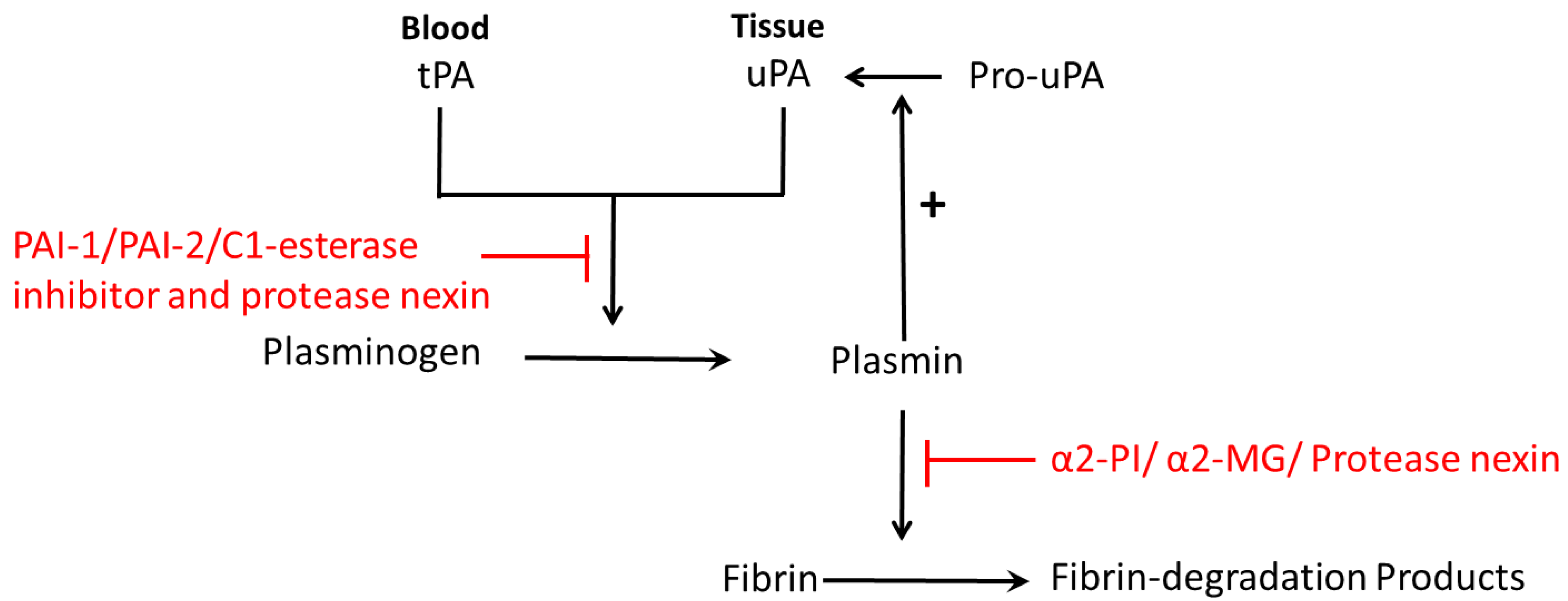
2. Fibrinolytic System and PAI-1 (Plasminogen Activator Inhibitor Type 1)
3. Role of PAI-1 in the Female Reproduction System
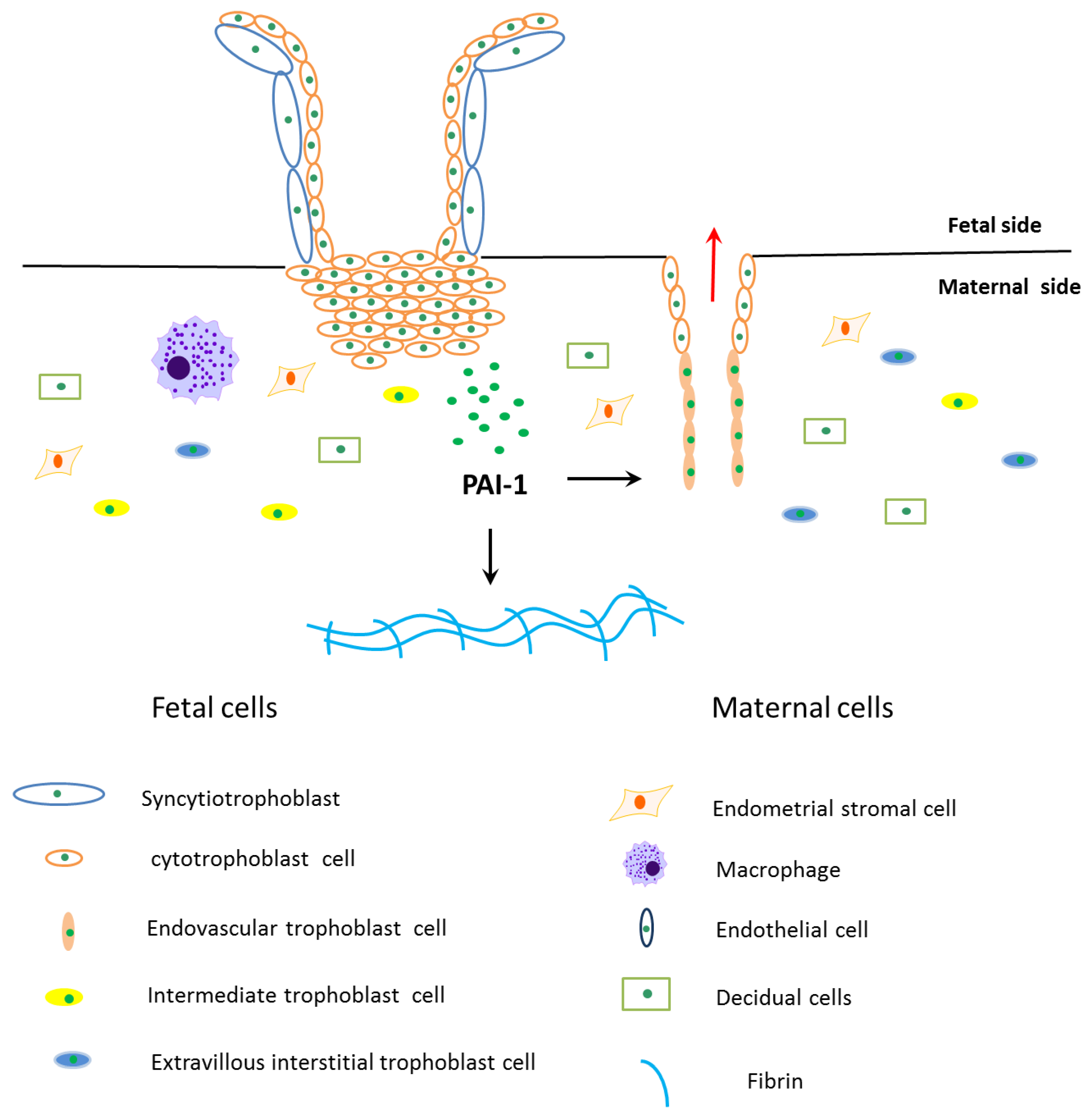
3.1. PAI-1 Inhibits Trophoblast Invasion
3.2. Recurrent Pregnancy Losses
3.3. Preeclampsia
3.4. Intrauterine Growth Restriction
3.5. Gestational Diabetes Mellitus
3.6. Endometriosis
3.7. Polycystic Ovary Syndrome
4. Conclusions
Acknowledgments
Author Contributions
Conflicts of Interest
Abbreviations
| PAI | plasminogen activator inhibitor |
| tPA | tissue-type plasminogen activator |
| uPA | urokinase-type plasminogen activator |
| ECM | extra cellular matrix |
| α2-PI | α2-plasmin inhibitor |
| α2-MG | α2-macroglobulin |
| RCL | reactive center loop |
| LMW | low molecular weight |
| HMW | high molecular weight |
| PCI | protein C inhibitor |
| uNK | uterine natural killer |
| EVT | extravillous cytotrophoblast |
| uPAR | uPA receptor |
| LRP | low density lipoprotein receptor-related protein |
| TIMP | tissue inhibitors of metalloprotease |
| TNF-α | tumor necrosis factor α |
| MMP | metalloproteinase |
| Jak/-Stat | Janus kinase 2/signal transducer and activator of transcription protein |
| NF-κB | nuclear factor κ-light-chain-enhancer of activated B cells |
| ACE | angiotensin I-converting enzyme |
| IL-1β | interleukin 1β |
| VEGF | vascular endothelial growth factor |
| EGF | epidermal growth factor |
| FGF | fibroblast growth factor |
| HIF | hypoxia-inducible transcription factors |
| p38 MAPK | p38 mitogen-activated protein kinases |
| PPAR-γ | peroxisome proliferator-activated receptor γ |
| RKIP | raf kinase inhibitor protein |
| ESR | estrogen receptor |
| PGR | progesterone receptor |
| SI | sensitivity index |
| BMI | body mass index |
| HOMA | homeostasis model assessment |
| CL | corpus luteum |
| RPL | recurrent pregnancy losses |
| IF | implantation failure |
| IUGR | intrauterine growth restriction |
| GDM | gestational diabetes mellitus |
| PCOS | polycystic ovary syndrome |
| MI | myocardial infarction |
References
- Zorio, E.; Gilabert-Estelles, J.; Espana, F.; Ramon, L.A.; Cosin, R.; Estelles, A. Fibrinolysis: The key to new pathogenetic mechanisms. Curr. Med. Chem. 2008, 15, 923–929. [Google Scholar] [CrossRef] [PubMed]
- Hellgren, M. Hemostasis during normal pregnancy and puerperium. Semin. Thromb. Hemost. 2003, 29, 125–130. [Google Scholar] [CrossRef] [PubMed]
- Kluft, C.; Jie, A.F.; Sprengers, E.D.; Verheijen, J.H. Identification of a reversible inhibitor of plasminogen activators in blood plasma. FEBS Lett. 1985, 190, 315–318. [Google Scholar] [CrossRef]
- Jorgensen, M.; Philips, M.; Thorsen, S.; Selmer, J.; Zeuthen, J. Plasminogen activator inhibitor-1 is the primary inhibitor of tissue-type plasminogen activator in pregnancy plasma. Thromb. Haemost. 1987, 58, 872–878. [Google Scholar] [PubMed]
- Labied, S.; Blacher, S.; Carmeliet, P.; Noel, A.; Frankenne, F.; Foidart, J.M.; Munaut, C. Transient reduction of placental angiogenesis in PAI-1-deficient mice. Physiol. Genom. 2011, 43, 188–198. [Google Scholar] [CrossRef] [PubMed]
- Fay, W.P.; Parker, A.C.; Condrey, L.R.; Shapiro, A.D. Human plasminogen activator inhibitor-1 (PAI-1) deficiency: Characterization of a large kindred with a null mutation in the PAI-1 gene. Blood 1997, 90, 204–208. [Google Scholar] [PubMed]
- Heiman, M.; Gupta, S.; Shapiro, A.D. The obstetric, gynaecological and fertility implications of homozygous PAI-1 deficiency: Single-centre experience. Haemophilia 2014, 20, 407–412. [Google Scholar] [CrossRef] [PubMed]
- Cesarman-Maus, G.; Hajjar, K.A. Molecular mechanisms of fibrinolysis. Br. J. Haematol. 2005, 129, 307–321. [Google Scholar] [CrossRef] [PubMed]
- Yasar Yildiz, S.; Kuru, P.; Toksoy Oner, E.; Agirbasli, M. Functional stability of plasminogen activator inhibitor-1. Sci. World J. 2014, 2014, 858293. [Google Scholar] [CrossRef] [PubMed]
- Ghosh, A.K.; Vaughan, D.E. PAI-1 in tissue fibrosis. J. Cell Physiol. 2012, 227, 493–507. [Google Scholar] [CrossRef] [PubMed]
- Duffy, M.J.; McGowan, P.M.; Harbeck, N.; Thomssen, C.; Schmitt, M. uPA and PAI-1 as biomarkers in breast cancer: Validated for clinical use in level-of-evidence-1 studies. Breast Cancer Res. 2014, 16, 428. [Google Scholar] [CrossRef] [PubMed]
- Lyon, C.J.; Hsueh, W.A. Effect of plasminogen activator inhibitor-1 in diabetes mellitus and cardiovascular disease. Am. J. Med. 2003, 115, 62–68. [Google Scholar] [CrossRef]
- Lijnen, H.R.; Collen, D. Mechanisms of physiological fibrinolysis. Baillieres Clin. Haematol. 1995, 8, 277–290. [Google Scholar] [CrossRef]
- Chakraborty, C.; Gleeson, L.M.; McKinnon, T.; Lala, P.K. Regulation of human trophoblast migration and invasiveness. Can. J. Physiol. Pharmacol. 2002, 80, 116–124. [Google Scholar] [CrossRef] [PubMed]
- Blasi, F.; Vassalli, J.D.; Dano, K. Urokinase-type plasminogen activator: Proenzyme, receptor, and inhibitors. J. Cell Biol. 1987, 104, 801–804. [Google Scholar] [CrossRef] [PubMed]
- Petersen, L.C.; Lund, L.R.; Nielsen, L.S.; Dano, K.; Skriver, L. One-chain urokinase-type plasminogen activator from human sarcoma cells is a proenzyme with little or no intrinsic activity. J. Biol. Chem. 1988, 263, 11189–11195. [Google Scholar] [PubMed]
- Brenner, B. Haemostatic changes in pregnancy. Thromb. Res. 2004, 114, 409–414. [Google Scholar] [CrossRef] [PubMed]
- Gils, A.; Declerck, P.J. The structural basis for the pathophysiological relevance of PAI-1 in cardiovascular diseases and the development of potential PAI-1 inhibitors. Thromb. Haemost. 2004, 91, 425–437. [Google Scholar] [CrossRef] [PubMed]
- Andreasen, P.A.; Egelund, R.; Petersen, H.H. The plasminogen activation system in tumor growth, invasion, and metastasis. Cell. Mol. Life Sci. CMLS 2000, 57, 25–40. [Google Scholar] [CrossRef] [PubMed]
- Gils, A. The pathophysiological relevance of PAI-1 in cardiovascular diseases and the development of monoclonal antibodies as PAI-1 inhibitors. Verh K Acad Geneeskd Belg 2006, 68, 179–198. [Google Scholar] [PubMed]
- Eriksson, P.; Nilsson, L.; Karpe, F.; Hamsten, A. Very-low-density lipoprotein response element in the promoter region of the human plasminogen activator inhibitor-1 gene implicated in the impaired fibrinolysis of hypertriglyceridemia. Arterioscler. Thromb. Vasc. Biol. 1998, 18, 20–26. [Google Scholar] [CrossRef] [PubMed]
- Saksela, O.; Rifkin, D.B. Cell-associated plasminogen activation: Regulation and physiological functions. Annu. Rev. Cell Biol. 1988, 4, 93–126. [Google Scholar] [CrossRef] [PubMed]
- Astedt, B.; Lindoff, C.; Lecander, I. Significance of the plasminogen activator inhibitor of placental type (PAI-2) in pregnancy. Semin. Thromb. Hemost. 1998, 24, 431–435. [Google Scholar] [CrossRef] [PubMed]
- Kruithof, E.K.; Baker, M.S.; Bunn, C.L. Biological and clinical aspects of plasminogen activator inhibitor type 2. Blood 1995, 86, 4007–4024. [Google Scholar] [PubMed]
- Suzuki, K. The multi-functional serpin, protein C inhibitor: Beyond thrombosis and hemostasis. J. Thromb. Haemost. 2008, 6, 2017–2026. [Google Scholar] [CrossRef] [PubMed]
- Kruithof, E.K.; Tran-Thang, C.; Gudinchet, A.; Hauert, J.; Nicoloso, G.; Genton, C.; Welti, H.; Bachmann, F. Fibrinolysis in pregnancy: A study of plasminogen activator inhibitors. Blood 1987, 69, 460–466. [Google Scholar] [CrossRef]
- Stirling, Y.; Woolf, L.; North, W.R.; Seghatchian, M.J.; Meade, T.W. Haemostasis in normal pregnancy. Thromb. Haemost. 1984, 52, 176–182. [Google Scholar] [PubMed]
- Hofmann, G.E.; Glatstein, I.; Schatz, F.; Heller, D.; Deligdisch, L. Immunohistochemical localization of urokinase-type plasminogen activator and the plasminogen activator inhibitors 1 and 2 in early human implantation sites. Am. J. Obstet. Gynecol. 1994, 170, 671–676. [Google Scholar] [CrossRef]
- Feinberg, R.F.; Kao, L.C.; Haimowitz, J.E.; Queenan, J.T., Jr.; Wun, T.C.; Strauss, J.F., 3rd; Kliman, H.J. Plasminogen activator inhibitor types 1 and 2 in human trophoblasts. PAI-1 is an immunocytochemical marker of invading trophoblasts. Lab. Investig. J. Tech. Methods Pathol. 1989, 61, 20–26. [Google Scholar]
- Floridon, C.; Nielsen, O.; Holund, B.; Sweep, F.; Sunde, L.; Thomsen, S.G.; Teisner, B. Does plasminogen activator inhibitor-1 (PAI-1) control trophoblast invasion? A study of fetal and maternal tissue in intrauterine, tubal and molar pregnancies. Placenta 2000, 21, 754–762. [Google Scholar] [CrossRef] [PubMed]
- Hu, Z.Y.; Liu, Y.X.; Liu, K.; Byrne, S.; Ny, T.; Feng, Q.; Ockleford, C.D. Expression of tissue type and urokinase type plasminogen activators as well as plasminogen activator inhibitor type-1 and type-2 in human and rhesus monkey placenta. J. Anat. 1999, 194, 183–195. [Google Scholar] [CrossRef] [PubMed]
- Naruse, K.; Lash, G.E.; Bulmer, J.N.; Innes, B.A.; Otun, H.A.; Searle, R.F.; Robson, S.C. The urokinase plasminogen activator (uPA) system in uterine natural killer cells in the placental bed during early pregnancy. Placenta 2009, 30, 398–404. [Google Scholar] [CrossRef] [PubMed]
- Silva, J.F.; Serakides, R. Intrauterine trophoblast migration: A comparative view of humans and rodents. Cell Adh. Migr. 2016, 10, 88–110. [Google Scholar] [CrossRef] [PubMed]
- Lala, P.K.; Lee, B.P.; Xu, G.; Chakraborty, C. Human placental trophoblast as an in vitro model for tumor progression. Can. J. Physiol. Pharmacol. 2002, 80, 142–149. [Google Scholar] [CrossRef] [PubMed]
- Fitzpatrick, T.E.; Graham, C.H. Stimulation of plasminogen activator inhibitor-1 expression in immortalized human trophoblast cells cultured under low levels of oxygen. Exp. Cell Res. 1998, 245, 155–162. [Google Scholar] [CrossRef] [PubMed]
- Crippa, M.P. Urokinase-type plasminogen activator. Int. J. Biochem. Cell Biol. 2007, 39, 690–694. [Google Scholar] [CrossRef] [PubMed]
- Lash, G.E.; Otun, H.A.; Innes, B.A.; Bulmer, J.N.; Searle, R.F.; Robson, S.C. Low oxygen concentrations inhibit trophoblast cell invasion from early gestation placental explants via alterations in levels of the urokinase plasminogen activator system. Biol. Reprod. 2006, 74, 403–409. [Google Scholar] [CrossRef] [PubMed]
- Xia, Y.; Wen, H.Y.; Kellems, R.E. Angiotensin II inhibits human trophoblast invasion through AT1 receptor activation. J. Biol. Chem. 2002, 277, 24601–24608. [Google Scholar] [CrossRef] [PubMed]
- Bauer, S.; Pollheimer, J.; Hartmann, J.; Husslein, P.; Aplin, J.D.; Knofler, M. Tumor necrosis factor-α inhibits trophoblast migration through elevation of plasminogen activator inhibitor-1 in first-trimester villous explant cultures. J. Clin. Endocrinol. Metab. 2004, 89, 812–822. [Google Scholar] [CrossRef] [PubMed]
- Estella, C.; Herrer, I.; Atkinson, S.P.; Quinonero, A.; Martinez, S.; Pellicer, A.; Simon, C. Inhibition of histone deacetylase activity in human endometrial stromal cells promotes extracellular matrix remodelling and limits embryo invasion. PLoS ONE 2012, 7, e30508. [Google Scholar] [CrossRef] [PubMed]
- Degryse, B.; Sier, C.F.; Resnati, M.; Conese, M.; Blasi, F. PAI-1 inhibits urokinase-induced chemotaxis by internalizing the urokinase receptor. FEBS Lett. 2001, 505, 249–254. [Google Scholar] [CrossRef]
- Czekay, R.P.; Kuemmel, T.A.; Orlando, R.A.; Farquhar, M.G. Direct binding of occupied urokinase receptor (uPAR) to LDL receptor-related protein is required for endocytosis of uPAR and regulation of cell surface urokinase activity. Mol. Biol. Cell 2001, 12, 1467–1479. [Google Scholar] [CrossRef] [PubMed]
- Degryse, B.; Neels, J.G.; Czekay, R.P.; Aertgeerts, K.; Kamikubo, Y.; Loskutoff, D.J. The low density lipoprotein receptor-related protein is a motogenic receptor for plasminogen activator inhibitor-1. J. Biol. Chem. 2004, 279, 22595–22604. [Google Scholar] [CrossRef] [PubMed]
- Zhang, Q.; Wu, Y.; Ann, D.K.; Messadi, D.V.; Tuan, T.L.; Kelly, A.P.; Bertolami, C.N.; Le, A.D. Mechanisms of hypoxic regulation of plasminogen activator inhibitor-1 gene expression in keloid fibroblasts. J. Investig. Dermatol. 2003, 121, 1005–1012. [Google Scholar] [CrossRef] [PubMed]
- Renaud, S.J.; Postovit, L.M.; Macdonald-Goodfellow, S.K.; McDonald, G.T.; Caldwell, J.D.; Graham, C.H. Activated macrophages inhibit human cytotrophoblast invasiveness in vitro. Biol. Reprod. 2005, 73, 237–243. [Google Scholar] [CrossRef] [PubMed]
- Huber, A.V.; Saleh, L.; Bauer, S.; Husslein, P.; Knofler, M. TNFα-mediated induction of PAI-1 restricts invasion of HTR-8/SVneo trophoblast cells. Placenta 2006, 27, 127–136. [Google Scholar] [CrossRef] [PubMed]
- Feng, Q.; Liu, Y.; Liu, K.; Byrne, S.; Liu, G.; Wang, X.; Li, Z.; Ockleford, C.D. Expression of urokinase, plasminogen activator inhibitors and urokinase receptor in pregnant rhesus monkey uterus during early placentation. Placenta 2000, 21, 184–193. [Google Scholar] [CrossRef] [PubMed]
- Soares, M.J.; Chakraborty, D.; Kubota, K.; Renaud, S.J.; Rumi, M.A. Adaptive mechanisms controlling uterine spiral artery remodeling during the establishment of pregnancy. Int. J. Dev. Biol. 2014, 58, 247–259. [Google Scholar] [CrossRef] [PubMed]
- Kaufmann, P.; Black, S.; Huppertz, B. Endovascular trophoblast invasion: Implications for the pathogenesis of intrauterine growth retardation and preeclampsia. Biol. Reprod. 2003, 69, 1–7. [Google Scholar] [CrossRef] [PubMed]
- McMahon, G.A.; Petitclerc, E.; Stefansson, S.; Smith, E.; Wong, M.K.; Westrick, R.J.; Ginsburg, D.; Brooks, P.C.; Lawrence, D.A. Plasminogen activator inhibitor-1 regulates tumor growth and angiogenesis. J. Biol. Chem. 2001, 276, 33964–33968. [Google Scholar] [CrossRef] [PubMed]
- Stirrat, G.M. Recurrent miscarriage. Lancet 1990, 336, 673–675. [Google Scholar] [CrossRef]
- Practice Committee of the American Society for Reproductive Medicine. Definitions of infertility and recurrent pregnancy loss: A committee opinion. Fertil. Steril. 2013. [Google Scholar] [CrossRef]
- Rai, R.; Regan, L. Recurrent miscarriage. Lancet 2006, 368, 601–611. [Google Scholar] [CrossRef]
- Jaslow, C.R.; Carney, J.L.; Kutteh, W.H. Diagnostic factors identified in 1020 women with two versus three or more recurrent pregnancy losses. Fertil. Steril. 2010, 93, 1234–1243. [Google Scholar] [CrossRef] [PubMed]
- Gris, J.C.; Neveu, S.; Mares, P.; Biron, C.; Hedon, B.; Schved, J.F. Plasma fibrinolytic activators and their inhibitors in women suffering from early recurrent abortion of unknown etiology. J. Lab. Clin. Med. 1993, 122, 606–615. [Google Scholar] [PubMed]
- Gris, J.C.; Ripart-Neveu, S.; Maugard, C.; Tailland, M.L.; Brun, S.; Courtieu, C.; Biron, C.; Hoffet, M.; Hedon, B.; Mares, P. Respective evaluation of the prevalence of haemostasis abnormalities in unexplained primary early recurrent miscarriages. The Nimes Obstetricians and Haematologists (NOHA) Study. Thromb. Haemost. 1997, 77, 1096–1103. [Google Scholar] [PubMed]
- Li, X.; Liu, Y.; Zhang, R.; Tan, J.; Chen, L.; Liu, Y. Meta-analysis of the association between plasminogen activator inhibitor-1 4G/5G polymorphism and recurrent pregnancy loss. Med. Sci. Monit. Int. Med. J. Exp. Clin. Res. 2015, 21, 1051–1056. [Google Scholar] [CrossRef] [PubMed]
- Khosravi, F.; Zarei, S.; Ahmadvand, N.; Akbarzadeh-Pasha, Z.; Savadi, E.; Zarnani, A.H.; Sadeghi, M.R.; Jeddi-Tehrani, M. Association between plasminogen activator inhibitor 1 gene mutation and different subgroups of recurrent miscarriage and implantation failure. J. Assist. Reprod. Genet. 2014, 31, 121–124. [Google Scholar] [CrossRef] [PubMed]
- Goodman, C.; Hur, J.; Goodman, C.S.; Jeyendran, R.S.; Coulam, C. Are polymorphisms in the ACE and PAI-1 genes associated with recurrent spontaneous miscarriages? Am. J. Reprod. Immunol. 2009, 62, 365–370. [Google Scholar] [CrossRef] [PubMed]
- Su, M.T.; Lin, S.H.; Chen, Y.C.; Kuo, P.L. Genetic association studies of ACE and PAI-1 genes in women with recurrent pregnancy loss: A systematic review and meta-analysis. Thromb. Haemost. 2013, 109, 8–15. [Google Scholar] [CrossRef] [PubMed]
- Coulam, C.B.; Jeyendran, R.S.; Fishel, L.A.; Roussev, R. Multiple thrombophilic gene mutations rather than specific gene mutations are risk factors for recurrent miscarriage. Am. J. Reprod. Immunol. 2006, 55, 360–368. [Google Scholar] [CrossRef] [PubMed]
- Poursadegh Zonouzi, A.; Chaparzadeh, N.; Ghorbian, S.; Sadaghiani, M.M.; Farzadi, L.; Ghasemzadeh, A.; Kafshdooz, T.; Sakhinia, M.; Sakhinia, E. The association between thrombophilic gene mutations and recurrent pregnancy loss. J. Assist. Reprod. Genet. 2013, 30, 1353–1359. [Google Scholar] [CrossRef] [PubMed]
- Buchholz, T.; Lohse, P.; Rogenhofer, N.; Kosian, E.; Pihusch, R.; Thaler, C.J. Polymorphisms in the ACE and PAI-1 genes are associated with recurrent spontaneous miscarriages. Hum. Reprod. 2003, 18, 2473–2477. [Google Scholar] [CrossRef] [PubMed]
- Redman, C.W.; Sargent, I.L. Latest advances in understanding preeclampsia. Science 2005, 308, 1592–1594. [Google Scholar] [CrossRef] [PubMed]
- Sibai, B.M.; Stella, C.L. Diagnosis and management of atypical preeclampsia-eclampsia. Am. J. Obstet. Gynecol. 2009, 200, 481. [Google Scholar] [CrossRef] [PubMed]
- Irani, R.A.; Xia, Y. The functional role of the renin-angiotensin system in pregnancy and preeclampsia. Placenta 2008, 29, 763–771. [Google Scholar] [CrossRef] [PubMed]
- Bodova, K.B.; Biringer, K.; Dokus, K.; Ivankova, J.; Stasko, J.; Danko, J. Fibronectin, plasminogen activator inhibitor type 1 (PAI-1) and uterine artery Doppler velocimetry as markers of preeclampsia. Dis. Mark. 2011, 30, 191–196. [Google Scholar] [CrossRef] [PubMed]
- Purwosunu, Y.; Sekizawa, A.; Koide, K.; Farina, A.; Wibowo, N.; Wiknjosastro, G.H.; Okazaki, S.; Chiba, H.; Okai, T. Cell-free mRNA concentrations of plasminogen activator inhibitor-1 and tissue-type plasminogen activator are increased in the plasma of pregnant women with preeclampsia. Clin. Chem. 2007, 53, 399–404. [Google Scholar] [CrossRef] [PubMed]
- Wikstrom, A.K.; Nash, P.; Eriksson, U.J.; Olovsson, M.H. Evidence of increased oxidative stress and a change in the plasminogen activator inhibitor (PAI)-1 to PAI-2 ratio in early-onset but not late-onset preeclampsia. Am. J. Obstet. Gynecol. 2009, 201, 597. [Google Scholar] [CrossRef] [PubMed]
- Estelles, A.; Gilabert, J.; Aznar, J.; Loskutoff, D.J.; Schleef, R.R. Changes in the plasma levels of type 1 and type 2 plasminogen activator inhibitors in normal pregnancy and in patients with severe preeclampsia. Blood 1989, 74, 1332–1338. [Google Scholar] [PubMed]
- Akolekar, R.; Cruz Jde, J.; Penco, J.M.; Zhou, Y.; Nicolaides, K.H. Maternal plasma plasminogen activator inhibitor-2 at 11 to 13 weeks of gestation in hypertensive disorders of pregnancy. Hypertens. Pregnancy 2011, 30, 194–202. [Google Scholar] [CrossRef] [PubMed]
- Catarino, C.; Rebelo, I.; Belo, L.; Rocha, S.; Castro, E.B.; Patricio, B.; Quintanilha, A.; Santos-Silva, A. Relationship between maternal and cord blood hemostatic disturbances in preeclamptic pregnancies. Thromb. Res. 2008, 123, 219–224. [Google Scholar] [CrossRef] [PubMed]
- Wei, J.; Liu, C.X.; Gong, T.T.; Wu, Q.J.; Wu, L. Cigarette smoking during pregnancy and preeclampsia risk: A systematic review and meta-analysis of prospective studies. Oncotarget 2015, 6, 43667–43678. [Google Scholar] [CrossRef] [PubMed]
- Simpson, A.J.; Gray, R.S.; Moore, N.R.; Booth, N.A. The effects of chronic smoking on the fibrinolytic potential of plasma and platelets. Br. J. Haematol. 1997, 97, 208–213. [Google Scholar] [CrossRef] [PubMed]
- Said, J.M.; Tsui, R.; Borg, A.J.; Higgins, J.R.; Moses, E.K.; Walker, S.P.; Monagle, P.T.; Brennecke, S.P. The PAI-1 4G/5G polymorphism is not associated with an increased risk of adverse pregnancy outcome in asymptomatic nulliparous women. J. Thromb. Haemost. 2012, 10, 881–886. [Google Scholar] [CrossRef] [PubMed]
- Ciarmela, P.; Marzioni, D.; Islam, M.S.; Gray, P.C.; Terracciano, L.; Lorenzi, T.; Todros, T.; Petraglia, F.; Castellucci, M. Possible role of RKIP in cytotrophoblast migration: Immunohistochemical and in vitro studies. J. Cell. Physiol. 2012, 227, 1821–1828. [Google Scholar] [CrossRef] [PubMed]
- Prutsch, N.; Fock, V.; Haslinger, P.; Haider, S.; Fiala, C.; Pollheimer, J.; Knofler, M. The role of interleukin-1β in human trophoblast motility. Placenta 2012, 33, 696–703. [Google Scholar] [CrossRef] [PubMed]
- Anteby, E.Y.; Greenfield, C.; Natanson-Yaron, S.; Goldman-Wohl, D.; Hamani, Y.; Khudyak, V.; Ariel, I.; Yagel, S. Vascular endothelial growth factor, epidermal growth factor and fibroblast growth factor-4 and -10 stimulate trophoblast plasminogen activator system and metalloproteinase-9. Mol. Hum. Reprod. 2004, 10, 229–235. [Google Scholar] [CrossRef] [PubMed]
- Meade, E.S.; Ma, Y.Y.; Guller, S. Role of hypoxia-inducible transcription factors 1α and 2α in the regulation of plasminogen activator inhibitor-1 expression in a human trophoblast cell line. Placenta 2007, 28, 1012–1019. [Google Scholar] [CrossRef] [PubMed]
- Guller, S. Role of the syncytium in placenta-mediated complications of preeclampsia. Thromb. Res. 2009, 124, 389–392. [Google Scholar] [CrossRef] [PubMed]
- Gerhardt, A.; Goecke, T.W.; Beckmann, M.W.; Wagner, K.J.; Tutschek, B.; Willers, R.; Bender, H.G.; Scharf, R.E.; Zotz, R.B. The G20210A prothrombin-gene mutation and the plasminogen activator inhibitor (PAI-1) 5G/5G genotype are associated with early onset of severe preeclampsia. J. Thromb. Haemost. 2005, 3, 686–691. [Google Scholar] [CrossRef] [PubMed]
- Morgan, J.A.; Bombell, S.; McGuire, W. Association of plasminogen activator inhibitor-type 1 (-675 4G/5G) polymorphism with pre-eclampsia: Systematic review. PLoS ONE 2013, 8, e56907. [Google Scholar] [CrossRef] [PubMed]
- Dall’Asta, A.; Brunelli, V.; Prefumo, F.; Frusca, T.; Lees, C.C. Early onset fetal growth restriction. Matern. Health Neonatol. Perinat. 2017, 3, 2. [Google Scholar] [CrossRef] [PubMed]
- Seferovic, M.D.; Gupta, M.B. Increased Umbilical Cord PAI-1 Levels in Placental Insufficiency Are Associated with Fetal Hypoxia and Angiogenesis. Dis. Markers 2016, 2016, 7124186. [Google Scholar] [CrossRef] [PubMed]
- Bernstein, I.M.; Horbar, J.D.; Badger, G.J.; Ohlsson, A.; Golan, A. Morbidity and mortality among very-low-birth-weight neonates with intrauterine growth restriction. The Vermont Oxford Network. Am. J. Obstet. Gynecol. 2000, 182, 198–206. [Google Scholar] [CrossRef]
- Murphy, V.E.; Smith, R.; Giles, W.B.; Clifton, V.L. Endocrine regulation of human fetal growth: The role of the mother, placenta, and fetus. Endocr. Rev. 2006, 27, 141–169. [Google Scholar] [CrossRef] [PubMed]
- Scifres, C.M.; Nelson, D.M. Intrauterine growth restriction, human placental development and trophoblast cell death. J. Physiol. 2009, 587, 3453–3458. [Google Scholar] [CrossRef] [PubMed]
- Sheppard, B.L.; Bonnar, J. Uteroplacental hemostasis in intrauterine fetal growth retardation. Semin. Thromb. Hemost. 1999, 25, 443–446. [Google Scholar] [CrossRef] [PubMed]
- Estelles, A.; Gilabert, J.; Keeton, M.; Eguchi, Y.; Aznar, J.; Grancha, S.; Espna, F.; Loskutoff, D.J.; Schleef, R.R. Altered expression of plasminogen activator inhibitor type 1 in placentas from pregnant women with preeclampsia and/or intrauterine fetal growth retardation. Blood 1994, 84, 143–150. [Google Scholar] [PubMed]
- Coolman, M.; Timmermans, S.; de Groot, C.J.; Russcher, H.; Lindemans, J.; Hofman, A.; Geurts-Moespot, A.J.; Sweep, F.C.; Jaddoe, V.V.; Steegers, E.A. Angiogenic and fibrinolytic factors in blood during the first half of pregnancy and adverse pregnancy outcomes. Obstet. Gynecol. 2012, 119, 1190–1200. [Google Scholar] [CrossRef] [PubMed]
- Gilabert, J.; Estelles, A.; Ayuso, M.J.; Espana, F.; Chirivella, M.; Grancha, S.; Mico, J.M.; Aznar, J. Evaluation of plasminogen activators and plasminogen activator inhibitors in plasma and amniotic fluid in pregnancies complicated with intrauterine fetal growth retardation. Gynecol. Obstet. Investig. 1994, 38, 157–162. [Google Scholar] [CrossRef]
- De Boer, K.; ten Cate, J.W.; Sturk, A.; Borm, J.J.; Treffers, P.E. Enhanced thrombin generation in normal and hypertensive pregnancy. Am. J. Obstet. Gynecol. 1989, 160, 95–100. [Google Scholar] [CrossRef]
- Estelles, A.; Gilabert, J.; Espana, F.; Aznar, J.; Galbis, M. Fibrinolytic parameters in normotensive pregnancy with intrauterine fetal growth retardation and in severe preeclampsia. Am. J. Obstet. Gynecol. 1991, 165, 138–142. [Google Scholar] [CrossRef]
- Infante-Rivard, C.; Rivard, G.E.; Guiguet, M.; Gauthier, R. Thrombophilic polymorphisms and intrauterine growth restriction. Epidemiology 2005, 16, 281–287. [Google Scholar] [CrossRef] [PubMed]
- Larciprete, G.; Rossi, F.; Deaibess, T.; Brienza, L.; Barbati, G.; Romanini, E.; Gioia, S.; Cirese, E. Double inherited thrombophilias and adverse pregnancy outcomes: Fashion or science? J. Obstet. Gynaecol. Res. 2010, 36, 996–1002. [Google Scholar] [CrossRef] [PubMed]
- Miehle, K.; Stepan, H.; Fasshauer, M. Leptin, adiponectin and other adipokines in gestational diabetes mellitus and pre-eclampsia. Clin. Endocrinol. 2012, 76, 2–11. [Google Scholar] [CrossRef] [PubMed]
- Moon, J.H.; Kwak, S.H.; Jang, H.C. Prevention of type 2 diabetes mellitus in women with previous gestational diabetes mellitus. Korean J. Intern. Med. 2017, 32, 26–41. [Google Scholar] [CrossRef] [PubMed]
- Vrachnis, N.; Belitsos, P.; Sifakis, S.; Dafopoulos, K.; Siristatidis, C.; Pappa, K.I.; Iliodromiti, Z. Role of adipokines and other inflammatory mediators in gestational diabetes mellitus and previous gestational diabetes mellitus. Int. J. Endocrinol. 2012, 2012, 549748. [Google Scholar] [CrossRef] [PubMed]
- Seely, E.W.; Solomon, C.G. Insulin resistance and its potential role in pregnancy-induced hypertension. J. Clin. Endocrinol. Metab. 2003, 88, 2393–2398. [Google Scholar] [CrossRef] [PubMed]
- Cesari, M.; Pahor, M.; Incalzi, R.A. Plasminogen activator inhibitor-1 (PAI-1): A key factor linking fibrinolysis and age-related subclinical and clinical conditions. Cardiovasc. Ther. 2010, 28, 72–91. [Google Scholar] [CrossRef] [PubMed]
- Salmi, A.A.; Zaki, N.M.; Zakaria, R.; Nor Aliza, A.G.; Rasool, A.H. Arterial stiffness, inflammatory and pro-atherogenic markers in gestational diabetes mellitus. Vasa 2012, 41, 96–104. [Google Scholar] [CrossRef] [PubMed]
- Bugatto, F.; Quintero-Prado, R.; Visiedo, F.M.; Vilar-Sanchez, J.M.; Figueroa-Quinones, A.; Lopez-Tinoco, C.; Torrejon, R.; Bartha, J.L. The influence of lipid and proinflammatory status on maternal uterine blood flow in women with late onset gestational diabetes. Reprod. Sci. 2017. [Google Scholar] [CrossRef] [PubMed]
- McManus, R.; Summers, K.; de Vrijer, B.; Cohen, N.; Thompson, A.; Giroux, I. Maternal, umbilical arterial and umbilical venous 25-hydroxyvitamin D and adipocytokine concentrations in pregnancies with and without gestational diabetes. Clin. Endocrinol. 2014, 80, 635–641. [Google Scholar] [CrossRef] [PubMed]
- Farhan, S.; Winzer, C.; Tura, A.; Quehenberger, P.; Bieglmaier, C.; Wagner, O.F.; Huber, K.; Waldhausl, W.; Pacini, G.; Kautzky-Willer, A. Fibrinolytic dysfunction in insulin-resistant women with previous gestational diabetes. Eur. J. Clin. Investig. 2006, 36, 345–352. [Google Scholar] [CrossRef] [PubMed]
- Morimitsu, L.K.; Fusaro, A.S.; Sanchez, V.H.; Hagemann, C.C.; Bertini, A.M.; Dib, S.A. Fibrinolytic dysfunction after gestation is associated to components of insulin resistance and early type 2 diabetes in latino women with previous gestational diabetes. Diabetes Res. Clin. Pract. 2007, 78, 340–348. [Google Scholar] [CrossRef] [PubMed]
- Lowe, L.P.; Metzger, B.E.; Lowe, W.L., Jr.; Dyer, A.R.; McDade, T.W.; McIntyre, H.D.; Group, H.S.C.R. Inflammatory mediators and glucose in pregnancy: Results from a subset of the Hyperglycemia and Adverse Pregnancy Outcome (HAPO) Study. J. Clin. Endocrinol. Metab. 2010, 95, 5427–5434. [Google Scholar] [CrossRef] [PubMed]
- Cawyer, C.R.; Horvat, D.; Leonard, D.; Allen, S.R.; Jones, R.O.; Zawieja, D.C.; Kuehl, T.J.; Uddin, M.N. Hyperglycemia impairs cytotrophoblast function via stress signaling. Am. J. Obstet. Gynecol. 2014, 211, 541. [Google Scholar] [CrossRef] [PubMed]
- Leipold, H.; Knoefler, M.; Gruber, C.; Klein, K.; Haslinger, P.; Worda, C. Plasminogen activator inhibitor 1 gene polymorphism and gestational diabetes mellitus. Obstet. Gynecol. 2006, 107, 651–656. [Google Scholar] [CrossRef] [PubMed]
- Gingras, V.; Vigneault, J.; Weisnagel, S.J.; Tchernof, A.; Robitaille, J. Accelerometry-measured physical activity and inflammation after gestational diabetes. Med. Sci. Sports Exerc. 2013, 45, 1307–1312. [Google Scholar] [CrossRef] [PubMed]
- Glueck, C.J.; Phillips, H.; Cameron, D.; Wang, P.; Fontaine, R.N.; Moore, S.K.; Sieve-Smith, L.; Tracy, T. The 4G/4G polymorphism of the hypofibrinolytic plasminogen activator inhibitor type 1 gene: An independent risk factor for serious pregnancy complications. Metabolism 2000, 49, 845–852. [Google Scholar] [CrossRef] [PubMed]
- Greene, A.D.; Lang, S.A.; Kendziorski, J.A.; Sroga-Rios, J.M.; Herzog, T.J.; Burns, K.A. Endometriosis: Where are we and where are we going? Reproduction 2016, 152, R63–R78. [Google Scholar] [CrossRef] [PubMed]
- Bruse, C.; Guan, Y.; Carlberg, M.; Carlstrom, K.; Bergqvist, A. Basal release of urokinase plasminogen activator, plasminogen activator inhibitor-1, and soluble plasminogen activator receptor from separated and cultured endometriotic and endometrial stromal and epithelial cells. Fertil. Steril. 2005, 83, 1155–1160. [Google Scholar] [CrossRef] [PubMed]
- Gilabert-Estelles, J.; Estelles, A.; Gilabert, J.; Castello, R.; Espana, F.; Falco, C.; Romeu, A.; Chirivella, M.; Zorio, E.; Aznar, J. Expression of several components of the plasminogen activator and matrix metalloproteinase systems in endometriosis. Hum. Reprod. 2003, 18, 1516–1522. [Google Scholar] [CrossRef] [PubMed]
- Ramon, L.A.; Gilabert-Estelles, J.; Cosin, R.; Gilabert, J.; Espana, F.; Castello, R.; Chirivella, M.; Romeu, A.; Estelles, A. Plasminogen activator inhibitor-1 (PAI-1) 4G/5G polymorphism and endometriosis. Influence of PAI-1 polymorphism on PAI-1 antigen and mRNA expression. Thromb. Res. 2008, 122, 854–860. [Google Scholar] [CrossRef] [PubMed]
- Gentilini, D.; Vigano, P.; Castaldi, D.; Mari, D.; Busacca, M.; Vercellini, P.; Somigliana, E.; di Blasio, A.M. Plasminogen activator inhibitor-1 4G/5G polymorphism and susceptibility to endometriosis in the Italian population. Eur. J. Obstet. Gynecol. Reprod. Biol. 2009, 146, 219–221. [Google Scholar] [CrossRef] [PubMed]
- Goncalves-Filho, R.P.; Brandes, A.; Christofolini, D.M.; Lerner, T.G.; Bianco, B.; Barbosa, C.P. Plasminogen activator inhibitor-1 4G/5G polymorphism in infertile women with and without endometriosis. Acta Obstet. Gynecol. Scand. 2011, 90, 473–477. [Google Scholar] [CrossRef] [PubMed]
- Goodarzi, M.O.; Dumesic, D.A.; Chazenbalk, G.; Azziz, R. Polycystic ovary syndrome: Etiology, pathogenesis and diagnosis. Nat. Rev. Endocrinol. 2011, 7, 219–231. [Google Scholar] [CrossRef] [PubMed]
- Koiou, E.; Tziomalos, K.; Katsikis, I.; Delkos, D.; Tsourdi, E.A.; Panidis, D. Disparate effects of pharmacotherapy on plasma plasminogen activator inhibitor-1 levels in women with the polycystic ovary syndrome. Hormones 2013, 12, 559–566. [Google Scholar] [CrossRef] [PubMed]
- Koiou, E.; Tziomalos, K.; Dinas, K.; Katsikis, I.; Kandaraki, E.A.; Tsourdi, E.; Mavridis, S.; Panidis, D. Plasma plasminogen activator inhibitor-1 levels in the different phenotypes of the polycystic ovary syndrome. Endocr. J. 2012, 59, 21–29. [Google Scholar] [CrossRef] [PubMed]
- Orio, F., Jr.; Palomba, S.; Cascella, T.; Tauchmanova, L.; Nardo, L.G.; di Biase, S.; Labella, D.; Russo, T.; Savastano, S.; Tolino, A.; et al. Is plasminogen activator inhibitor-1 a cardiovascular risk factor in young women with polycystic ovary syndrome? Reprod. Biomed. Online 2004, 9, 505–510. [Google Scholar] [CrossRef]
- Tarkun, I.; Canturk, Z.; Arslan, B.C.; Turemen, E.; Tarkun, P. The plasminogen activator system in young and lean women with polycystic ovary syndrome. Endocr. J. 2004, 51, 467–472. [Google Scholar] [CrossRef] [PubMed]
- Palomba, S.; Orio, F., Jr.; Falbo, A.; Russo, T.; Tolino, A.; Zullo, F. Plasminogen activator inhibitor 1 and miscarriage after metformin treatment and laparoscopic ovarian drilling in patients with polycystic ovary syndrome. Fertil. Steril. 2005, 84, 761–765. [Google Scholar] [CrossRef] [PubMed]
- Fauser, B.C.; Tarlatzis, B.C.; Rebar, R.W.; Legro, R.S.; Balen, A.H.; Lobo, R.; Carmina, E.; Chang, J.; Yildiz, B.O.; Laven, J.S.; et al. Consensus on women’s health aspects of polycystic ovary syndrome (PCOS): The Amsterdam ESHRE/ASRM-Sponsored 3rd PCOS Consensus Workshop Group. Fertil. Steril. 2012, 97, 28–38. [Google Scholar] [CrossRef] [PubMed]
- Liu, Y.X. Plasminogen activator/plasminogen activator inhibitors in ovarian physiology. Front. Biosci. 2004, 9, 3356–3373. [Google Scholar] [CrossRef] [PubMed]
- Atiomo, W.U.; Hilton, D.; Fox, R.; Lee, D.; Shaw, S.; Friend, J.; Wilkin, T.J.; Prentice, A.G. Immunohistochemical detection of plasminogen activator inhibitor-1 in polycystic ovaries. Gynecol. Endocrinol. 2000, 14, 162–168. [Google Scholar] [CrossRef] [PubMed]
- Glueck, C.J.; Wang, P.; Fontaine, R.N.; Sieve-Smith, L.; Tracy, T.; Moore, S.K. Plasminogen activator inhibitor activity: An independent risk factor for the high miscarriage rate during pregnancy in women with polycystic ovary syndrome. Metabolism 1999, 48, 1589–1595. [Google Scholar] [CrossRef]
- Meigs, J.B.; O’Donnell, C.J.; Tofler, G.H.; Benjamin, E.J.; Fox, C.S.; Lipinska, I.; Nathan, D.M.; Sullivan, L.M.; D’Agostino, R.B.; Wilson, P.W. Hemostatic markers of endothelial dysfunction and risk of incident type 2 diabetes: The Framingham Offspring Study. Diabetes 2006, 55, 530–537. [Google Scholar] [CrossRef] [PubMed]
- Smith, A.; Patterson, C.; Yarnell, J.; Rumley, A.; Ben-Shlomo, Y.; Lowe, G. Which hemostatic markers add to the predictive value of conventional risk factors for coronary heart disease and ischemic stroke? The Caerphilly Study. Circulation 2005, 112, 3080–3087. [Google Scholar] [CrossRef] [PubMed]
- Yarmolinsky, J.; Bordin Barbieri, N.; Weinmann, T.; Ziegelmann, P.K.; Duncan, B.B.; Ines Schmidt, M. Plasminogen activator inhibitor-1 and type 2 diabetes: A systematic review and meta-analysis of observational studies. Sci. Rep. 2016, 6, 17714. [Google Scholar] [CrossRef] [PubMed]
- Sales, M.F.; Soter, M.O.; Candido, A.L.; Fernandes, A.P.; Oliveira, F.R.; Ferreira, A.C.; Sousa, M.O.; Ferreira, C.N.; Gomes, K.B. Correlation between plasminogen activator inhibitor-1 (PAI-1) promoter 4G/5G polymorphism and metabolic/proinflammatory factors in polycystic ovary syndrome. Gynecol. Endocrinol. 2013, 29, 936–939. [Google Scholar] [CrossRef] [PubMed]
- Lee, Y.H.; Song, G.G. Plasminogen activator inhibitor-1 4G/5G and the MTHFR 677C/T polymorphisms and susceptibility to polycystic ovary syndrome: A meta-analysis. Eur. J. Obstet. Gynecol. Reprod. Biol. 2014, 175, 8–14. [Google Scholar] [CrossRef] [PubMed]

© 2017 by the authors. Licensee MDPI, Basel, Switzerland. This article is an open access article distributed under the terms and conditions of the Creative Commons Attribution (CC BY) license (http://creativecommons.org/licenses/by/4.0/).
Share and Cite
Ye, Y.; Vattai, A.; Zhang, X.; Zhu, J.; Thaler, C.J.; Mahner, S.; Jeschke, U.; Von Schönfeldt, V. Role of Plasminogen Activator Inhibitor Type 1 in Pathologies of Female Reproductive Diseases. Int. J. Mol. Sci. 2017, 18, 1651. https://doi.org/10.3390/ijms18081651
Ye Y, Vattai A, Zhang X, Zhu J, Thaler CJ, Mahner S, Jeschke U, Von Schönfeldt V. Role of Plasminogen Activator Inhibitor Type 1 in Pathologies of Female Reproductive Diseases. International Journal of Molecular Sciences. 2017; 18(8):1651. https://doi.org/10.3390/ijms18081651
Chicago/Turabian StyleYe, Yao, Aurelia Vattai, Xi Zhang, Junyan Zhu, Christian J. Thaler, Sven Mahner, Udo Jeschke, and Viktoria Von Schönfeldt. 2017. "Role of Plasminogen Activator Inhibitor Type 1 in Pathologies of Female Reproductive Diseases" International Journal of Molecular Sciences 18, no. 8: 1651. https://doi.org/10.3390/ijms18081651