Article

Comprehensive Identification of Immunodominant Proteins of Brucella abortus and Brucella melitensis Using Antibodies in the Sera from Naturally Infected Hosts

1
Institute of Animal Hygiene and Environmental Health, Centre for Infectious Medicine, Freie Universität Berlin, Robert-von-Ostertag-Str. 7-13, Berlin 14163, Germany
2
Friedrich-Loeffler-Institut, Federal Research Institute for Animal Health, Institute of Bacterial Infections and Zoonoses, Naumburger Str. 96a, Jena 07743, Germany
3
Faculty of Veterinary Medicine, Benha University, Moshtohor, Toukh 13736, Egypt
4
Institute of Chemistry and Biochemistry, Freie Universität Berlin, Thielallee 63, Berlin 14195, Germany
*
Author to whom correspondence should be addressed.
Int. J. Mol. Sci. 2016, 17(5), 659; https://doi.org/10.3390/ijms17050659
Submission received: 11 March 2016 / Revised: 15 April 2016 / Accepted: 22 April 2016 / Published: 30 April 2016
(This article belongs to the Special Issue Host-Microbe Interaction)

Abstract

:
Brucellosis is a debilitating zoonotic disease that affects humans and animals. The diagnosis of brucellosis is challenging, as accurate species level identification is not possible with any of the currently available serology-based diagnostic methods. The present study aimed at identifying Brucella (B.) species-specific proteins from the closely related species B. abortus and B. melitensis using sera collected from naturally infected host species. Unlike earlier reported investigations with either laboratory-grown species or vaccine strains, in the present study, field strains were utilized for analysis. The label-free quantitative proteomic analysis of the naturally isolated strains of these two closely related species revealed 402 differentially expressed proteins, among which 63 and 103 proteins were found exclusively in the whole cell extracts of B. abortus and B. melitensis field strains, respectively. The sera from four different naturally infected host species, i.e., cattle, buffalo, sheep, and goat were applied to identify the immune-binding protein spots present in the whole protein extracts from the isolated B. abortus and B. melitensis field strains and resolved on two-dimensional gel electrophoresis. Comprehensive analysis revealed that 25 proteins of B. abortus and 20 proteins of B. melitensis were distinctly immunoreactive. Dihydrodipicolinate synthase, glyceraldehyde-3-phosphate dehydrogenase and lactate/malate dehydrogenase from B. abortus, amino acid ABC transporter substrate-binding protein from B. melitensis and fumarylacetoacetate hydrolase from both species were reactive with the sera of all the tested naturally infected host species. The identified proteins could be used for the design of serological assays capable of detecting pan-Brucella, B. abortus- and B. melitensis-specific antibodies.

Graphical Abstract

1. Introduction

Brucellosis is a zoonosis affecting a wide range of mammals including humans [1]. The genus Brucella currently includes 12 accepted nomo-species, with Brucella (B.) abortus and B. melitensis representing the species in the majority of notified human cases. These two species possess strikingly similar genomes [2] but display differences in host specificity and their proteomes [3]. B. melitensis is the most virulent species of all brucellae, one of the major causes of abortions in small ruminants and the causative agent of severe infections in humans [4]. B. abortus infections occur in cattle while infections in small ruminants and camels are rare [5]. In humans the course of B. abortus infections is milder [4].
The conventional methods for species identification include cultivation, as well as genome-based assays [6]. All these methods are hazardous, time-consuming and not suitable for ‘high-throughput analysis’; moreover, the routinely utilized bacterial lipopolysaccharide (LPS)-based serological methods are hampered by cross-reactivity with the LPS of other Gram-negative bacteria such as Yersinia enterocolitica, Salmonella spp, and Escherichia coli O:157 [7]. Furthermore, serological tests cannot distinguish between B. abortus and B. melitensis infection or between naturally infected and vaccinated animals [8,9].
The aim of this study was to identify B. abortus- and B. melitensis-specific proteins reacting with circulating antibodies in naturally infected animal host species. This immunoblot-based approach identified several immunodominant proteins from B. abortus and B. melitensis, which could be used to design a new diagnostic brucellosis assay.

2. Results

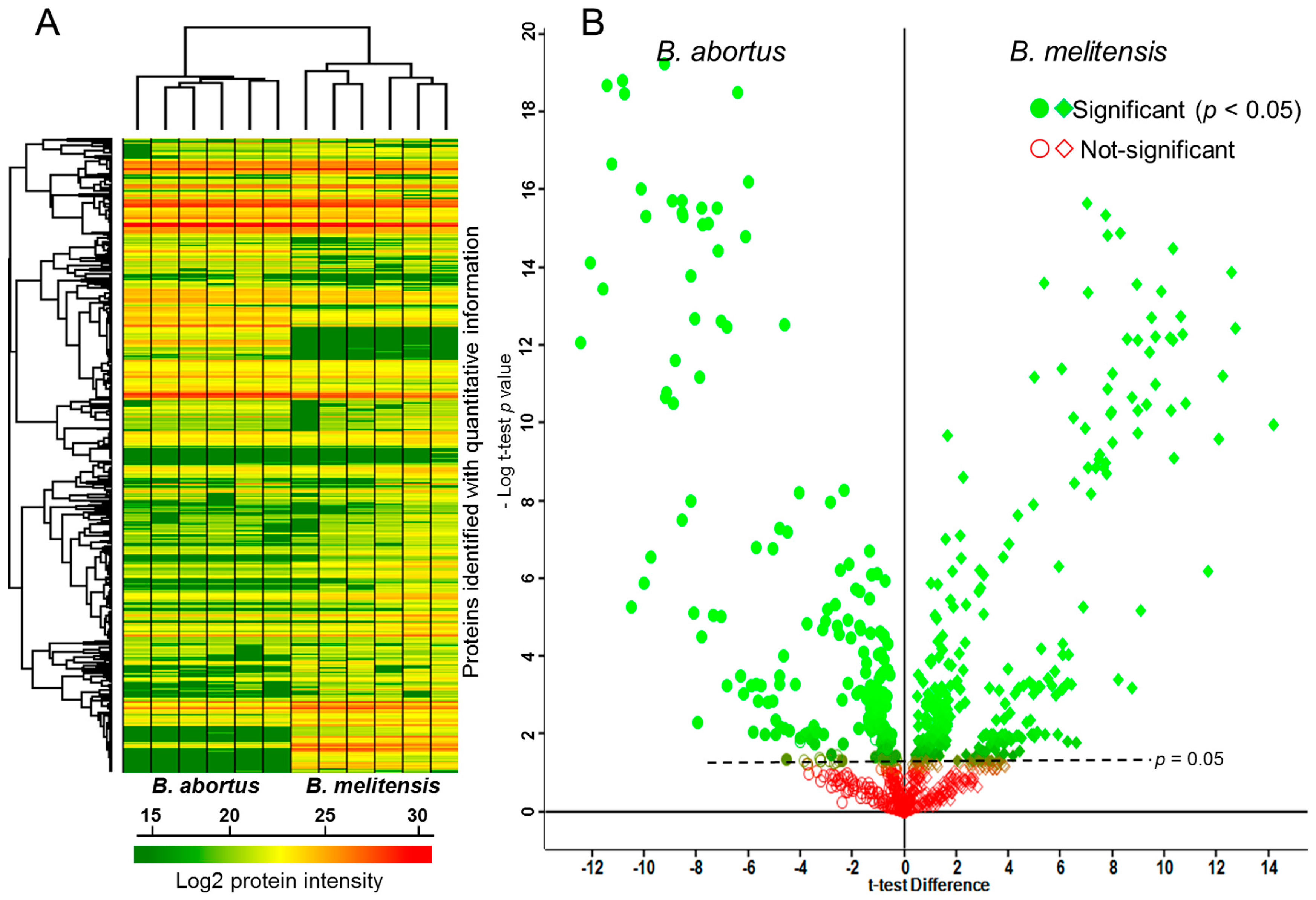
2.1. Comparative Proteomics Analysis of Brucella (B.) abortus and B. melitensis Field Strains

Label-free proteomic analyses involving trypsin digestion, separation of peptides by liquid chromatography (LC) coupled to electrospray ionization, and peptide analysis applying mass spectrometry revealed proteome level differences between B. abortus and B. melitensis field strains. Figure 1 illustrates the heat map and the hierarchical clustering of the 828 proteins identified in at least three of the six replicates for B. abortus and for B. melitensis. The volcano plot displays the negative log10 t-test p value over the log2 fold change. Proteins with p values above the dotted line (p < 0.05) were considered to be differentially expressed between the two Brucella species. Initially the two species differed in the expression of 568 proteins (t-test significance); upon application of a 1% false discovery rate (FDR) filter [10] the intensities of 402 proteins were still found to be significantly different between B. abortus and B. melitensis. Of note, 63 of these were found exclusively in the B. abortus and 103 exclusively in the B. melitensis field strain. The complete list of identified proteins is given in the supplementary table (Supplementary Table S1, sheets 1–4).

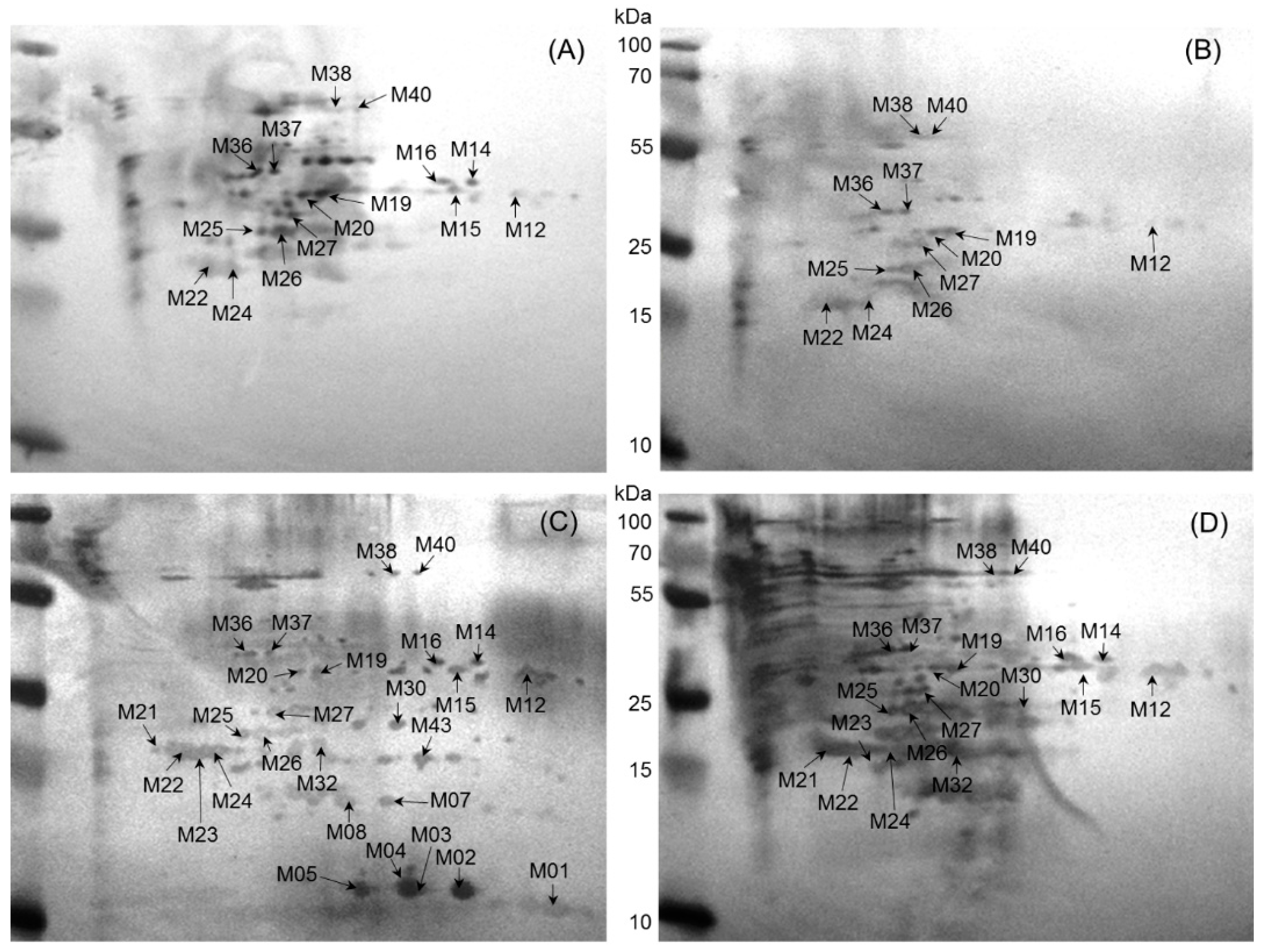
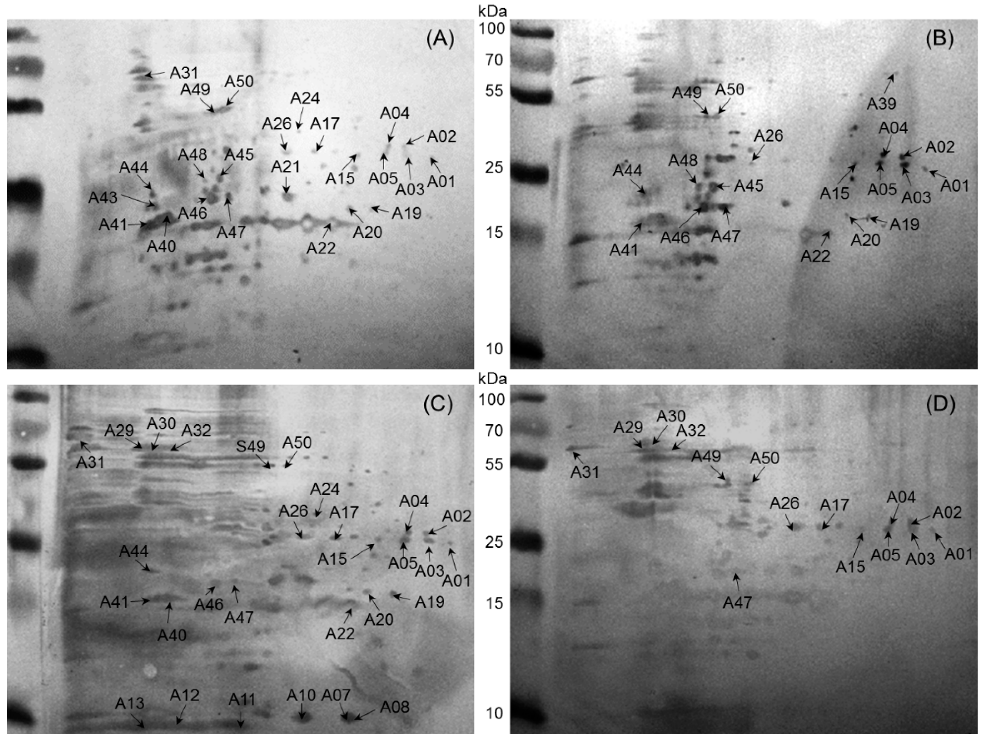
2.2. Immunoreactive Proteins of B. abortus

A total of 50 immunoreactive protein spots, corresponding to different 25 proteins, were detected by two dimensional electrophoresis -immunoblotting with a cell lysate from a B. abortus field strain and sera from naturally infected cows, buffaloes, sheep, and goats (Figure 2). Total numbers of proteins identified were 24, 19, 29, and 15 for cow, buffalo, sheep, and goat, respectively. Subsequent Western blot matching revealed 10 spots (A01–05, A15, A26, A47, A49, A50) (bold highlighted in Table 1), which corresponded to five proteins detected in the tested sera from all four naturally infected animal species. There was no unique host-specific immunodominant protein for buffalo and goat, whereas two (A43; A21) and four proteins (A08; A10, A11, A12) were specific for cow and sheep, respectively (Table 1).

2.3. Immunoreactive Proteins of B. melitensis

A total of 43 immunoreactive protein spots corresponding to 20 different proteins were identified in the cell lysate of a B. melitensis field strain. Total numbers of proteins identified were 27, 19, 15, and 12 using sera from sheep, goat, cow, and buffalo, respectively (Figure 3). Subsequent Western blot matching revealed 12 spots (M12; M19; M20; M24; M25; M26; M27; M36; M37; M38; M40; M22) (bold highlighted in Table 2) common to all four tested animal species, corresponding to 10 proteins. There was no unique host-specific immunodominant protein for buffalo and cow, whereas three (M32; M21; M23) and six proteins (M01; M02; M05; M07; M08; M43) were specific for sheep and goat, and sheep only (Table 2).

2.4. Identification of Cross-Reactive Proteins between B. abortus and B. melitensis

The cell lysates of the B. abortus and B. melitensis field strains generated a total of 61 immunoreactive spots which could be assigned to 36 proteins. Nine proteins (A47/M25; A22/M43; A41/M36; A45/M27; A40/M24; A07/M01; A10/M05; A13/M12; A21/M21) were detected in cell lysates of B. abortus and B. melitensis (Table 3), while 16 and 11 proteins were only detected in cell lysates of B. abortus or B. melitensis, respectively (Table 1 and Table 2). Spot ID A47/M25 (fumarylacetoacetate hydrolase domain-containing protein 2) was found in cell lysates of B. abortus and B. melitensis and reacted with the sera of all four tested animal species (Table 3 and Table 4). All immunogenic spots reacted only with sera of Brucella-positive animals and no reactions were detected with sera from Brucella-negative animals.

2.5. Comparative Basic Local Alignment Search Tool (BLAST) Analysis

In order to identify similar or identical epitope structures between Brucella spp., Ochrobactrum spp. and putative cross-reacting bacterial species, five B. abortus proteins (spot ID A47; A01; A02; A26; A49) and five B. melitensis proteins (spot ID M20; M37; M40; M22; M38) reacting with the sera of all four naturally infected animal host species (i.e., cattle, buffalo, sheep, goat) were selected and submitted to a comparative protein BLAST search (Table 4).
With the exception of the proteins (spot ID) A01, M22, M38, and M40, all proteins displayed identity values ≥95% for Brucella spp., B. suis, B. ovis and Ochrobactrum spp. Identity values of all ten proteins with the possibly cross-reacting bacterial species Y. enterocolitica, Y. pseudotuberculosis, S. enterica and E. coli O:157 were between 26% and 62%.

3. Discussion

Diagnosis of brucellosis in veterinary medicine is still a challenging process as it is based on serology and isolation of the agent [6]. The serological assays have their limitations with regard to sensitivity and specificity, moreover, they are not able to distinguish between infected and vaccinated animals [19,20,21]. Hence, the aim of this study was to identify immunodominant proteins in a B. abortus and a B. melitensis field strain by immunoproteomic screening to detect specific proteins for a future diagnostic assay. Among the 61 proteins found to be immunoreactive, four proteins expressed in B. abortus, five proteins expressed in B. melitensis cell lysates, and one protein present in both B. abortus and B. melitensis cell lysates were identified as promising candidates for further analysis.
In contrast to previous studies on the Brucella proteome which focussed mainly on vaccine or museum strains with altered immunogenic properties, i.e., diminished or loss of virulence [3,7,12,14,16,19,22,23,24], the present study used a fully virulent B. abortus field strain and a fully virulent B. melitensis field strain from a naturally infected cow and sheep, respectively. Sera obtained from naturally infected ruminants which had recently aborted and shown strong positive reactions in the RBT, CFT, and ELISA, were subsequently tested against both field strains. The strains and sera used in the present study can therefore be expected to represent the acute phase of the disease. Since naturally infected hosts generally show a stronger immunoreaction than hosts challenged with inactivated antigen [7], it can be assumed that the sera used in the present study contained antibodies against all immunoreactive proteins involved in infection.
In the present study LC/MS-based quantitative proteomic analysis revealed considerable differences in protein expression in the two Brucella field strains. Based on these findings a 2D-PAGE immunoblotting approach was used to determine immunodominant proteins in both field strains. Several studies using two-dimensional gel electrophoresis and mass spectrometry were also able to determine proteome level differences among the laboratory-grown strains B. abortus 2308 and B. melitensis 16M and revealed a species-specific protein expression pattern [3,14].
The present study identified a total of 61 immunoreactive protein spots from the proteomic profiles of B. abortus and B. melitensis using MALDI-TOF MS and the NCBI database search corresponding to 36 proteins. When performing a data base search against the sequence information of all entries in the NCBI database, the likelihood of identifying suitable proteins was significantly increased by applying MS/MS matched to at least one unique peptide. This approach contrasts that of Al-Dahouk et al., Yang et al. and Connolly et al., [7,12,16], who searched only against data sets of the Brucella species used in their experiments, and Zhao et al. [22], who selected proteins containing more than five peptide matches.
Each Brucella species can be associated with a specific host, i.e., B. abortus usually infects bovines, whereas B. melitensis is the most predominant species in sheep and goat [5]. Despite the close genetic relationship among Brucella spp. one could speculate that certain proteins induce a host species-specific immunoreaction. This hypothesis is corroborated by the findings of Zhao et al. [22], who demonstrated that some proteins are themselves immunogenic and induce high immunogenicity in the host species but not in others. The sera obtained from sheep were the most reactive, with 56 identified immunogenic protein spots, whereas 39, 31, and 34 spots were found in the sera of cow, buffalo and goat, respectively. Previous studies using the same immunoproteomic techniques as in the present study identified a differing range of immunoreactive proteins in various Brucella spp. and animal species [7,12,16,19,22,23]. These observed differences can be attributed to the technical procedures during protein preparation and the source/type of sera samples used, i.e., field or experimental, early or late stage of infection [23]. Moreover, these findings are indicative of a host species-specific immunoreaction.
Ten immunogenic proteins specific either from B. abortus (n = 4), B. melitensis (n = 5) or both (n = 1) were reactive with the sera of all four tested host species, i.e., cattle, buffalo, sheep, and goat. The mitochondrial catalytic enzyme, fumarylacetoacetate hydrolase domain-containing protein (FAHD2) was found in both B. abortus and B. melitensis cell lysates. To date, the role of this mitochondrial protein in the pathogenesis of Brucella infections is not known. Four proteins were identified in B. abortus only, i.e., dihydrodipicolinate synthase (DHDPS), glyceraldehyde-3-phosphate dehydrogenase (GAPDH), lactate/malate dehydrogenase, and phosphopyruvate hydratase. DHDPS is essential for bacterial growth and involved in the lysine biosynthesis pathway. This protein has been isolated from various Gram positive and Gram negative bacteria and is considered to be an attractive antibiotic target [25]. GAPDH is a protein of the Brucella-containing vacuole (BCV) and essential for B. abortus virulence [26]. Studies using recombinant B. abortus derived GAPDH induced both humoral and cellular immune responses during experimental infection with B. abortus in natural hosts (cattle and sheep) and mice [11]. However, when used as DNA vaccine it provided only partial protection against experimental B. abortus infection in mice [11]. lactate/malate dehydrogenase from B. abortus is considered to be a promising candidate for serodiagnosis and vaccine development due to its immunogenic characteristics [27], but further studies are required. Phosphopyruvate hydratase proteins participate in glycolysis, but their importance as possible diagnostic candidates is not known [28].
Five proteins found only in B. melitensis cell lysates were immunoreactive in all four host species: thiosulfate-binding protein precursor, which specifically binds thiosulfate and is involved in its transmembrane transport; amidohydrolase 3, a member of the amidohydrolase superfamily. These proteins catalyse the hydrolysis of amide or amine bonds in a large number of different substrates [29]. Amino-acid ABC transporter substrate-binding protein, a transmembrane protein previously found via proteome analysis in B. melitensis and B. ovis [30], and two hypothetical proteins closely related to the ABC transporter substrate-binding proteins. The function of these differentially expressed proteins in natural B. melitensis infection is not known to date.
LPS is the major cell surface antigen of Brucella and one of the main reasons for serological cross-reactions with other Gram-negative bacteria such as E. coli, Salmonella spp., and Y. enterocolitica [7,21,31]. LPS has been shown to bind to sodium dodecyl sulfate (SDS), and may mimic protein spots in SDS polyacrylamide gels [32,33]. LPS closely associates with the protein and traces of LPS are expected to be present in the whole cell protein extract irrespective of the extraction method used. In order to exclude the likelihood of LPS interference during immunostaining, the LPS concentration was measured in the extract. The whole cell protein extracts of B. abortus and B. melitensis contained 0.9 and 100 ng/mL of LPS, respectively. This LPS concentration is below the limit of detection of monoclonal antibodies [34] and therefore any interference of LPS can be excluded.
BLAST search to assess the similarity of these immunoreactive proteins identified among several Brucella species and possibly cross-reacting bacteria revealed that by combining various proteins it is possible to design a pan-Brucella test as well as a species-differentiating assay. Glyceraldehyde-3-phosphate dehydrogenase, lactate/malate dehydrogenase, thiosulfate-binding protein precursor, the amino acid ABC transporter substrate-binding proteins, and FAHD2 are suitable candidates for designing a pan-Brucella test. Aminohydrolase 3 on the other hand, might be useful for the differentiation of B. ovis and Ochrobactrum spp. from B. abortus, B. melitensis, and B. suis.

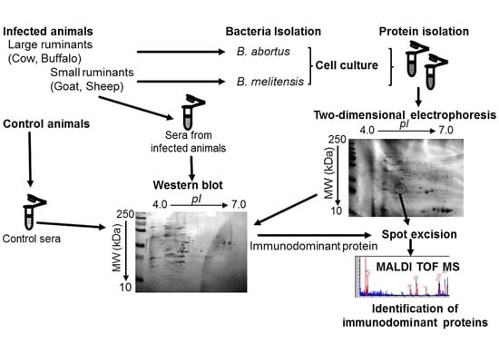
4. Materials and Methods

4.1. Bacterial Strains and Sera Selection

The strains and sera used in the present study were obtained from the Friedrich-Loeffler-Institut (FLI), Federal Research Institute for Animal Health, Institute of Bacterial Infections and Zoonoses, Jena, Germany. The utilised Brucella field strains were isolated from an outbreak in Turkey (B. abortus; cow) and an outbreak in China (B. melitensis; ewe). Identification and biotyping of these Brucella isolates was carried out as previously described [6,35]. The serum samples were collected in Egypt from naturally infected ruminants which had recently aborted (i.e., acute stage of infection); 35 sera were sent to the FLI for further analysis. Sera were considered positive if the Rose Bengal test (RBT) showed strong agglutination; the complement fixation test (CFT) showed more than 1000 sensE/mL; and the ELISA an OD ≥ 3. For each animal species (i.e., cattle, buffalo, sheep, goat) three positive serum samples were selected and used separately in the present study [6]. Negative serum samples were collected from non-infected animals in a non-endemic region. Experiments were run in three replicates. No sera could be obtained from the outbreaks in Turkey and China, and no isolates could be obtained from the outbreak in Egypt.

4.2. Ethics Statement

The sera samples collected during routine diagnosis from Egypt were approved (no 11/2012) by the Ethics committee at the Dean’s office, Faculty of Veterinary Medicine, Benha University, Ministry of Higher Education, Qalyobia, Egypt. All further sera used in this study were samples originally submitted for diagnostic purposes to and subsequently stored at FLI. According to German law, ethical approval or special permission concerning animal welfare is not required for sera meant for diagnostic purposes.

4.3. Cell Culture and Protein Extraction

Whole cell protein of the B. abortus and B. melitensis field strains was extracted as described previously [36]. The strains were cultured in Tryptic Soy media for 48 h at 37 °C; bacteria were harvested by centrifugation, washed twice with phosphate buffer saline, spun down, and the resulting pellets resuspended in 80% ethanol (v/v). Following centrifugation, the ethanol was discarded and the cell pellet air dried to ensure the removal of traces of ethanol. The cell pellets were then resuspended in 250 µL of HEPES buffer (20 mM, pH 7.4) and sonicated (duty cycle: 1.0, amplitude: 100%, UP100H; Hielscher Ultrasound Technology, Teltow, Germany) for 45 s on ice. After centrifugation at 11,290× g for 10 min at 4 °C, the clear supernatant was collected. The protein content was determined using the modified Bradford method (Bio-Rad, München, Germany) [37]. The concentration of LPS was determined as concentration of endotoxin using the recombinant factor C fluorescence assay (Haemotox rFC Haemochrom Diagnostica GmbH, Essen, Germany). The sample was spiked with an endotoxin standard (PPC-Spike) and LPS determination was carried out according to the manufacturer’s instructions.

4.4. In-Solution Trypsin Digestion

Ten µg of the protein extract (B. abortus and B. melitensis) were acetone precipitated and reconstituted in 20 µL of denaturation buffer (6 M urea/2 M thiourea in 10 mM HEPES, pH 8.0). The following steps were carried out with incubation steps at room temperature and gentle shaking. Reduction of disulfide bridges was performed by adding 0.2 µL of 10 mM dithiothreitol in 50 mM of ammonium bicarbonate (ABC) and incubation for 30 min, followed by an alkylation step with 0.4 µL of 55 mM iodacemtamide in ABC and incubation in the dark for 20 min. Then 0.4 µL of LysC (Sigma, Taufkirchen Germany) solution (0.5 µg/µL in ABC) was added and subjected to overnight incubation at RT. After pre-digestion with LysC, the sample was diluted by adding 75 µL of ABC to bring down the urea concentration to <2 M and digestion was started by adding 0.4 µL of 0.5 µg/µL trypsin in 50 mM ABC. Following overnight incubation, trypsin activity was stopped by adding 100 µL of 5% acetonitrile in 3% trifluoroacetic acid.

4.5. Liquid Chromatography-Electrospray Ionization-Tandem Mass Spectrometry (LC-ESI-MS/MS)

After digestion peptide samples were desalted by solid phase extraction (SPE), using C18 stage tips [38]. Desalted peptide mixtures were separated by reverse-phase chromatography using a Dionex Ultimate 3000 nanoLC on in-house manufactured 25 cm fritless silica microcolumns with an inner diameter of 100 µm. Columns were packed with ReproSil-Pur C18-AQ 3 µm resin (Dr. Maisch GmbH, Ammerbuch-Entringen, Germany). Peptides were separated on a 5%–60% acetonitrile gradient with 0.1% formic acid at a flow rate of 350 nL/min for 90 min. Eluting peptides were ionized on-line by electrospray ionization and transferred into an LTQ Orbitrap Velos mass spectrometer (Thermo Fisher Scientific, Bremen, Germany). The LTQ-Orbitrap was operated in the positive mode to simultaneously measure full scan MS spectra (from m/z 300–1700) in the Orbitrap analyzer at resolution R = 60,000 following isolation and fragmentation of the twenty most intense ions in the LTQ part by collision-induced dissociation.

4.6. Protein Identification and Data Analysis

A freely available software suit, MaxQuant (version. 1.3.0.5) (Max-Planck-Institute of Biochemistry, Martinsried, Germany) was used to process the raw MS files and the search engine, Andromeda [39] was utilized to search the peak list files against forward and backward protein sequences of Brucella (Reference proteome Brucella abortus (strain 2308) with protein count of 3022 and Brucella melitensis (strain M28) with a protein count of 3351) downloaded from Uniprot database and 248 frequently observed laboratory contaminants (accession date: August 2015). Initial maximum precursor and fragment mass deviations were set to 7 ppm and 0.5 Da, respectively. Methionine oxidation/acetylation of peptide N-termini and cysteine carbamidomethylation were set as variable and fixed modification, respectively, for the search. Furthermore, enzyme specificity was set to trypsin and a maximum of two missed cleavages was allowed for searching. The target-decoy-based false discovery rate (FDR) for peptide and protein identification was set to 1% for peptides and proteins and the minimum peptide length was set to 7 amino acids. Precursor mass tolerance was set to 20 ppm. The mass tolerance for fragment ions was set to 0.5 Da. MS-quantification of proteins was performed using the label-free quantification algorithm of the MaxQuant software package (Max-Planck-Institute of Biochemistry, Martinsried, Germany) [40].
The statistical analysis was performed by unpaired Student´s t Test using the Perseus software [41] version 1.4.1.3 package (Max-Planck-Institute of Biochemistry, Martinsried, Germany). For FDR ccorrections of the significant p-values (p <0.05) the Benjamini-Hochberg procedure [10] was applied. Heat map and volcano plots were calculated for further assessment and visualization.

4.7. Two-Dimensional Electrophoresis

Two-dimensional electrophoresis (2D-PAGE) was performed as described [42]. Briefly, the first dimension of 2D-PAGE was performed by applying 100 µg of acetone-precipitated protein per sample to pI, 4–7, 7.0 cm immobilized pH gradient (IPG) strips (Immobiline™ Dry Strip, GE Healthcare Bio-sciences AB; Uppsala, Sweden). The strips were rehydrated overnight at room temperature with 135 µL DeStreak Rehydration Solution. Isoelectric focusing (IEF) was performed by using the Ettan™ IPGphor3™ Unit (GE Healthcare Europe; Freiburg, Germany) and carried out at 20 °C for 6.5 h at 5000 V and 50 µA/strip.
Then the strips were sequentially equilibrated for 20 min in 2 mL equilibration buffer 1 (0.05 M trichloroethylene HCl (pH 8.8), 6 M urea, 30% glycerol, 4% SDS, 2% DTE, 0.002% bromophenol blue) and equilibration buffer 2 (0.05 M trichloroethylene HCl (pH 8.8), 6 M urea, 30% glycerol, 4% SDS, 2.5% iodoacetamid, 0.002% bromophenol blue). Standard molecular weight prestained protein ladder marker (10–250 kDa; Page Ruler™ Plus, ThermoScientific; Germany) and IPG strips were loaded onto homogeneous 12% polyacrylamide gels and sealed with 1% agarose solution. Electrophoresis was carried out at room temperature and 10 mA/gel until the tracking dye reached the bottom of the gels (1.5 h). 2D-PAGE protein profiles were visualized using Coomassie blue stain as previously described [43].

4.8. 2-D-PAGE Western Blotting

2D-PAGE Immunoblotting was carried out as previously described [19,36] with minor modifications. Briefly, proteins were separated on 2D-PAGE gels and transferred at 80 mA/gel for 90 min to nitrocellulose membranes (0.2 µM Bio-Rad laboratories; München, Germany) using Towbin transfer buffer (0.025 M Tris, 0.192 M glycine, 2.33% SDS, 20% (v/v) methanol, pH 8.3). The nitrocellulose membrane was blocked overnight at room temperature with gentle shaking in 1% skimmed milk in Tris buffered saline (TBS). The membrane was washed twice using TBS with Tween (TBST; 20 mM Tris, pH 7.5; 500 mM NaCl; 0.05% Tween-20; 10 min). Next, the nitrocellulose membrane was placed for 90 min at room temperature in a diluted solution of the respective sera in TBST. Bovine sera (1:200 dilution) and small ruminants sera (1:5000 dilution) were used as primary antibody source while 1:1000 diluted anti-bovine IgG (H and L) (Chicken) peroxidase-conjugated, anti-sheep IgG (H and L) (Donkey) peroxidase-conjugated and anti-goat IgG (H and L) (Chicken) peroxidase-conjugated antibody served as secondary antibody source. All the secondary antibodies were obtained from Biomol-Rockland, Hamburg, Germany. After washing the nitrocellulose membrane twice with TBST for 10 min, the detection of signals was carried out using the TMB kit™ (3,3′,5,5′-tetramethylbenzidine liquid substrate; Sigma-Aldrich; Steinheim, Germany) according to the manufacturer’s description.

4.9. In-Gel Trypsin Digestion and Matrix-Assisted Laser Desorption/ionization-Tine of Flight-Mass Spectrometry (MALDI-TOF MS/MS)

Following the selection of the spots of interest, i.e., spots detected in all three replicates, the protein spots corresponding to the Western blots were excised from the gel, destained, and subjected to overnight trypsin digestion (0.01 µg/µL) (Promega; Mannheim, Germany) as previously described [44]. The digested precipitates were reconstituted in 3.5 µL 5% acetonitrile in 0.1% TFA (trifluoroacetic acid; Merck; Darmstadt, Germany). The reconstituted precipitates were then spotted on to target plates for matrix-assisted laser desorption/ionization-time-of-flight mass spectrometry (MALDI-TOF MS) on a Bruker Ultraflex II instrument (Bruker Daltonik; Bremen, Germany) using HCCA (α-cyano-4-hydroxycinnamic acid; Sigma-Aldrich; Steinheim, Germany) as matrix. A database search was conducted against all entries using the MS/MS ion search mode (MASCOT, http://www.matrixscience.com) as previously described [36]. Protein identification was considered valid if more than two peptides matched and the MOWSE score was significant (p < 0.05).

4.10. Comparison of the Identified Proteins and Other Cross-Reactive Bacteria

BLAST search was done as previously described [3] to compare the identified proteins against Brucella spp., B. suis, B. ovis, Ochrobactrum spp., Y. enterocolitica, Y. pseudotuberculosis, S. enterica, and E. coli O:157, the latter five species being the most cross-reactive bacteria with Brucella. Query cover and identity values were evaluated and cut-off values set between 31%–54%.

5. Conclusions

The sera from naturally infected host species appear to possess different antibodies against B. abortus and B. melitensis. The presence of specific antibodies against four proteins of B. abortus, fumarylacetoacetate hydrolase 2, dihydrodipicolinate synthase, glyceraldehyde-3-phosphate dehydrogenase, and lactate/malate dehydrogenase as well as one protein from B. melitensis, ABC transporter substrate-binding protein, indicates that these proteins might be useful for designing a serology-based assay for the rapid species determination of Brucella. As suggested earlier, these results should be further verified using bacterial strains and sera collected from various geographical regions and sera from a variety of host species.
Cross-reactivity with other Gram negative bacteria and within the species of the genus is the major hindrance for the serological diagnosis of brucellosis. The results presented here open up new possibilities for the serodiagnosis of brucellosis by providing Brucella species-specific immunodominant protein candidates reacting only with sera collected from naturally infected cattle, buffaloes, sheep, and goats. The study provides information on new protein candidates and could help to improve the serological diagnosis of brucellosis.

Supplementary Materials

Supplementary materials can be found at https://www.mdpi.com/1422-0067/17/5/659/s1.

Acknowledgments

The authors thank Michael Kühl for technical support. We would like to thank the Deutscher Akademischer Austausch Dienst (DAAD) for financial support of Gamal Wareth, grant no. A/11/92495 and the Egyptian Ministry of Higher Education for partially funding. The author, Murat Eravci is supported by the Deutsche Forschungsgemeinschaft (DFG, SFB-958). For mass spectrometry (Christoph Weise and Murat Eravci), we would like to acknowledge the assistance of the Core Facility BioSupraMol supported by the Deutsche Forshungsgemeinschaft (DFG).

Author Contributions

Gamal Wareth: sample collection, study design, bacterial culturing, 2D-PAGE Western blotting, BLAST analysis and manuscript drafting; Murat Eravci: LC-MS analysis and data interpretation; Christoph Weise: MALDI-TOF MS analysis and manuscript drafting; Falk Melzer: selection of bacteria, serum samples and bacteria cultivation; Uwe Roesler, Lisa D. Sprague and Heinrich Neubauer: study design, manuscript drafting and manuscript revision; Jayaseelan Murugaiyan: study design, data collection, data analysis, manuscript drafting and coordination of the study. All authors read and approved the final version of the manuscript.

Conflicts of Interest

The authors declare no conflict of interest.

References

  1. Cutler, S.J.; Whatmore, A.M.; Commander, N.J. Brucellosis—New aspects of an old disease. J. Appl. Microbiol. 2005, 98, 1270–1281. [Google Scholar]
  2. Halling, S.M.; Peterson-Burch, B.D.; Bricker, B.J.; Zuerner, R.L.; Qing, Z.; Li, L.L.; Kapur, V.; Alt, D.P.; Olsen, S.C. Completion of the genome sequence of Brucella abortus and comparison to the highly similar genomes of Brucella melitensis and Brucella suis. J. Bacteriol. 2005, 187, 2715–2726. [Google Scholar] [CrossRef] [PubMed]
  3. Eschenbrenner, M.; Horn, T.A.; Wagner, M.A.; Mujer, C.V.; Miller-Scandle, T.L.; DelVecchio, V.G. Comparative proteome analysis of laboratory grown Brucella abortus 2308 and Brucella melitensis 16 m. J. Proteome Res. 2006, 5, 1731–1740. [Google Scholar] [CrossRef] [PubMed]
  4. Galińska, E.; Zagórski, J. Brucellosis in humans—Etiology, diagnostics, clinical forms. Ann. Agric. Environ. Med. 2013, 20, 233–238. [Google Scholar] [PubMed]
  5. Diaz, A.E. Epidemiology of brucellosis in domestic animals caused by Brucella melitensis, Brucella suis and Brucella abortus. Rev. Sci. Tech. 2013, 32, 43–51, 53–60. [Google Scholar]
  6. Alton, G.G.; Jones, L.M.; Angus, R.D.; Verger, J.M. Techniques for the Brucellosis Laboratory; Instituttional de la Recherche Agronomique: Paris, France, 1988; pp. 17–62. [Google Scholar]
  7. Al Dahouk, S.; Nöckler, K.; Scholz, H.C.; Tomaso, H.; Bogumil, R.; Neubauer, H. Immunoproteomic characterization of Brucella abortus 1119-3 preparations used for the serodiagnosis of Brucella infections. J. Immunol. Methods 2006, 309, 34–47. [Google Scholar] [CrossRef] [PubMed]
  8. Samartino, L.; Gall, D.; Gregoret, R.; Nielsen, K. Validation of enzyme-linked immunosorbent assays for the diagnosis of bovine brucellosis. Vet. Microbiol. 1999, 70, 193–200. [Google Scholar] [CrossRef]
  9. Von Bargen, K.; Gorvel, J.P.; Salcedo, S.P. Internal affairs: Investigating the Brucella intracellular lifestyle. FEMS Microbiol. Rev. 2012, 36, 533–562. [Google Scholar] [CrossRef] [PubMed]
  10. Benjamini, Y.; Hochberg, Y. Controlling the false discovery rate—A practical and powerful approach to multiple testing. J. R. Stat. Soc. Ser. B Methodol. 1995, 57, 289–300. [Google Scholar]
  11. Rosinha, G.; Myioshi, A.; Azevedo, V.; Splitter, G.; Oliveira, S. Molecular and immunological characterisation of recombinant Brucella abortus glyceraldehyde-3-phosphate-dehydrogenase, a T- and B-cell reactive protein that induces partial protection when co-administered with an interleukin-12-expressing plasmid in a DNA vaccine formulation. J. Med. Microbiol. 2002, 51, 661–671. [Google Scholar] [PubMed]
  12. Yang, Y.; Wang, L.; Yin, J.; Wang, X.; Cheng, S.; Lang, X.; Wang, X.; Qu, H.; Sun, C.; Wang, J.; et al. Immunoproteomic analysis of Brucella melitensis and identification of a new immunogenic candidate protein for the development of brucellosis subunit vaccine. Mol. Immunol. 2011, 49, 175–184. [Google Scholar] [CrossRef] [PubMed]
  13. Al-Dahouk, S.; Loisel-Meyer, S.V.; Scholz, H.C.; Tomaso, H.; Kersten, M.; Harder, A.; Neubauer, H.; Köhler, S.; Jubier-Maurin, V.R. Proteomic analysis of Brucella suis under oxygen deficiency reveals flexibility in adaptive expression of various pathways. Proteomics 2009, 9, 3011–3021. [Google Scholar] [CrossRef] [PubMed]
  14. Eschenbrenner, M.; Wagner, M.A.; Horn, T.A.; Kraycer, J.A.; Mujer, C.V.; Hagius, S.; Elzer, P.; DelVecchio, V.G. Comparative proteome analysis of Brucella melitensis vaccine strain rev 1 and a virulent strain, 16 M. J. Bacteriol. 2002, 184, 4962–4970. [Google Scholar] [CrossRef] [PubMed]
  15. Del Vecchio, V.G.; Wagner, M.A.; Eschenbrenner, M.; Horn, T.; Kraycer, J.A.; Estock, F.; Elzer, P.; Mujer, C.V. Brucella proteomes—A review. Vet. Microbiol. 2002, 90, 593–603. [Google Scholar] [CrossRef]
  16. Connolly, J.P.; Comerci, D.; Alefantis, T.G.; Walz, A.; Quan, M.; Chafin, R.; Grewal, P.; Mujer, C.V.; Ugalde, R.A.; DelVecchio, V.G. Proteomic analysis of Brucella abortus cell envelope and identification of immunogenic candidate proteins for vaccine development. Proteomics 2006, 6, 3767–3780. [Google Scholar] [CrossRef] [PubMed]
  17. Teixeira-Gomes, A.; Cloeckaert, A.; Bézard, G.; Bowden, R.A.; Dubray, G.; Zygmunt, M.S. Identification and characterization of Brucella ovis immunogenic proteins using two-dimensional electrophoresis and immunoblotting. Electrophoresis 1997, 18, 1491–1497. [Google Scholar] [CrossRef] [PubMed]
  18. Teixeira-Gomes, A.; Cloeckaert, A.; Bézard, G.; Dubray, G.; Zygmunt, M.S. Mapping and identification of Brucella melitensis proteins by two-dimensional electrophoresis and microsequencing. Electrophoresis 1997, 18, 156–162. [Google Scholar] [CrossRef] [PubMed]
  19. Ko, K.Y.; Kim, J.W.; Her, M.; Kang, S.I.; Jung, S.C.; Cho, D.H.; Kim, J.Y. Immunogenic proteins of Brucella abortus to minimize cross-reactions in brucellosis diagnosis. Vet. Microbiol. 2012, 156, 374–380. [Google Scholar] [CrossRef] [PubMed]
  20. Olsen, S.C.; Stoffregen, W.S. Essential role of vaccines in brucellosis control and eradication programs for livestock. Expert Rev. Vaccines 2005, 4, 915–928. [Google Scholar] [CrossRef] [PubMed]
  21. Nielsen, K.; Smith, P.; Widdison, J.; Gall, D.; Kelly, L.; Kelly, W.; Nicoletti, P. Serological relationship between cattle exposed to Brucella abortus, Yersinia enterocolitica O:9 and Escherichia coli O157:H7. Vet. Microbiol. 2004, 100, 25–30. [Google Scholar] [CrossRef] [PubMed]
  22. Zhao, Z.; Yan, F.; Ji, W.; Luo, D.; Liu, X.; Xing, L.; Duan, Y.; Yang, P.; Shi, X.; Li, Z.; et al. Identification of immunoreactive proteins of Brucella melitensis by immunoproteomics. Sci. China Life Sci. 2011, 54, 880–887. [Google Scholar] [CrossRef] [PubMed]
  23. Lee, J.J.; Simborio, H.L.; Reyes, A.W.; Kim, D.G.; Hop, H.T.; Min, W.; Her, M.; Jung, S.C.; Yoo, H.S.; Kim, S. Immunoproteomic identification of immunodominant antigens independent of the time of infection in Brucella abortus 2308-challenged cattle. Vet. Res. 2015, 46, 17. [Google Scholar] [CrossRef] [PubMed]
  24. Kim, J.Y.; Sung, S.R.; Lee, K.; Lee, H.K.; Kang, S.I.; Lee, J.J.; Jung, S.C.; Park, Y.H.; Her, M. Immunoproteomics of brucella abortus RB51 as candidate antigens in serological diagnosis of brucellosis. Vet. Immunol. Immunopathol. 2014, 160, 218–224. [Google Scholar] [CrossRef] [PubMed]
  25. Skovpen, Y.; Palmer, D. Dihydrodipicolinate synthase from Campylobacter jejuni: Kinetic mechanism of cooperative allosteric inhibition and inhibitor-induced substrate cooperativity. Biochemistry 2013, 52, 5454–5462. [Google Scholar] [CrossRef] [PubMed]
  26. Fugier, E.; Salcedo, S.; de Chastellier, C.; Pophillat, M.; Muller, A.; Arce-Gorvel, V.; Fourquet, P.; Gorvel, J. The glyceraldehyde-3-phosphate dehydrogenase and the small GTPase Rab 2 are crucial for Brucella replication. PLoS Pathog. 2009, 5, e1000487. [Google Scholar] [CrossRef] [PubMed]
  27. Han, X.; Tong, Y.; Tian, M.; Sun, X.; Wang, S.; Ding, C.; Yu, S. Characterization of the immunogenicity and pathogenicity of malate dehydrogenase in Brucella abortus. World J. Microbiol. Biotechnol. 2014, 30, 2063–2070. [Google Scholar] [CrossRef] [PubMed]
  28. Tomm, J.; Krause, C.; Simon, J.; Treudler, R.; von Bergen, M.; Averbeck, M. Pyruvate kinase and phosphopyruvate hydratase as novel IGE reactive proteins in prawn. J. Investig. Allergol. Clin. Immunol. 2013, 23, 443–445. [Google Scholar] [PubMed]
  29. Kim, G.J.; Kim, H.S. Identification of the structural similarity in the functionally related amidohydrolases acting on the cyclic amide ring. Biochem. J. 1998, 330, 295–302. [Google Scholar] [CrossRef] [PubMed]
  30. Schneider, E.; Hunke, S. ATP-binding-cassette (ABC) transport systems: Functional and structural aspects of the ATP-hydrolyzing subunits/domains. FEMS Microbiol. Rev. 1998, 22, 1–20. [Google Scholar] [CrossRef] [PubMed]
  31. Kittelberger, R.; Reichel, M.; Joyce, M.; Staak, C. Serological crossreactivity between Brucella abortus and Yersinia enterocolitica 0:9. III. Specificity of the in vitro antigen-specific γ interferon test for bovine brucellosis diagnosis in experimentally Yersinia enterocolitica 0:9-infected cattle. Vet. Microbiol. 1997, 57, 361–371. [Google Scholar] [CrossRef]
  32. Wu, A.M.; MacKenzie, N.E.; Adams, G.; Pugh, R. Structural and immunochemical aspects of Brucella abortus endotoxins. Adv. Exp. Med. Biol. 1988, 228, 551–576. [Google Scholar] [PubMed]
  33. Sowa, B.A.; Crawforda, R.P.; Heck, F.C.; Williams, J.D.; Wu, A.M.; Kelly, K.A.; Adams, L.G. Size, charge and structural heterogeneity of Brucella abortus lipopolysaccharides demonstrated by twodimensional gel electrophoresis. Electrophoresi 1986, 7, 283–288. [Google Scholar] [CrossRef]
  34. Palmer, D.A.; Douglas, J.T. Analysis of Brucella lipopolysaccharide with specific and cross-reacting monoclonal antibodies. J. Clin. Microbiol. 1989, 27, 2331–2337. [Google Scholar] [PubMed]
  35. Probert, W.S.; Schrader, K.N.; Khuong, N.Y.; Bystrom, S.L.; Graves, M.H. Real-time multiplex PCR assay for detection of Brucella spp., B. abortus, and B. melitensis. J. Clin. Microbiol. 2004, 42, 1290–1293. [Google Scholar] [CrossRef] [PubMed]
  36. Murugaiyan, J.; Rockstroh, M.; Wagner, J.; Baumann, S.; Schorsch, K.; Trump, S.; Lehmann, I.; Bergen, M.V.; Tomm, J.M. Benzo[a]pyrene affects jurkat T cells in the activated state via the antioxidant response element dependent Nrf2 pathway leading to decreased IL-2 secretion and redirecting glutamine metabolism. Toxicol. Appl. Pharmacol. 2013, 269, 307–316. [Google Scholar] [CrossRef] [PubMed]
  37. Markwell, M.A.; Haas, S.M.; Bieber, L.L.; Tolbert, N.E. A modification of the lowry procedure to simplify protein determination in membrane and lipoprotein samples. Anal. Biochem. 1978, 87, 206–210. [Google Scholar] [CrossRef]
  38. Rappsilber, J.; Ishihama, Y.; Mann, M. Stop and go extraction tips for matrix-assisted laser desorption/ionization, nanoelectrospray, and LC/MS sample pretreatment in proteomics. Anal. Chem. 2003, 75, 663–670. [Google Scholar] [CrossRef] [PubMed]
  39. Cox, J.; Neuhauser, N.; Michalski, A.; Scheltema, R.A.; Olsen, J.V.; Mann, M. Andromeda: A peptide search engine integrated into the maxquant environment. J Proteome Res. 2011, 10, 1794–1805. [Google Scholar] [CrossRef] [PubMed]
  40. Cox, J.; Hein, M.Y.; Luber, C.A.; Paron, I.; Nagaraj, N.; Mann, M. Accurate proteome-wide label-free quantification by delayed normalization and maximal peptide ratio extraction, termed maxlfq. Mol. Cell. Proteom. 2014, 13, 2513–2526. [Google Scholar] [CrossRef] [PubMed]
  41. Cox, J.; Matic, I.; Hilger, M.; Nagaraj, N.; Selbach, M.; Olsen, J.V.; Mann, M. A practical guide to the maxquant computational platform for silac-based quantitative proteomics. Nat. Protoc. 2009, 4, 698–705. [Google Scholar] [CrossRef] [PubMed]
  42. Murugaiyan, J.; Weise, C.; von Bergen, M.; Roesler, U. Two-dimensional proteome reference map of Prototheca zopfii revealed reduced metabolism and enhanced signal transduction as adaptation to an infectious life style. Proteomics 2013, 13, 2664–2669. [Google Scholar] [CrossRef] [PubMed]
  43. Candiano, G.; Ruschi, M.; Musante, L.; Santucci, L.; Ghiggeri, G.; Carnemolla, B.; Orecchia, P.; Zardi, L.; Righetti, P. Blue silver: A very sensitive colloidal coomassie G-250 staining for proteome analysis. Electrophoresis 2004, 25, 1327–1333. [Google Scholar] [CrossRef] [PubMed]
  44. Wareth, G.; Melzer, F.; Weise, C.; Neubauer, H.; Roesler, U.; Murugaiyan, J. Proteomics-based identification of immunodominant proteins of brucellae using sera from infected hosts points towards enhanced pathogen survival during the infection. Biochem. Biophys. Res. Commun. 2015, 456, 202–206. [Google Scholar] [CrossRef] [PubMed]
Figure 1. Brucella field strain proteome. (A) Heat map and hierarchical clustering of 828 quantified proteins from 6 replicates of each field strain; (B) Volcano plot comparing the field strains of B. abortus and B. melitensis. The p-values were calculated by unpaired t-test analysis using log2 transformed peptide intensities. Multiple testing correction was applied [10].
Ijms 17 00659 g001
Figure 2. Representative two dimensional electrophoresis immunoblotting images of whole cell proteins from B. abortus field strain extracts separated on a 12% polyacrylamide gel. The blot was developed using the TMB (3,3′,5,5′-tetramethylbenzidine liquid substrate) kit after immuno-blotting with serum from (A) cattle; (B) buffalo; (C) sheep; and (D) goat and the respective peroxidase-conjugated secondary antibodies.
Ijms 17 00659 g002
Figure 3. Representative 2D-PAGE immunoblotting images of whole cell proteins from B. melitensis field strain extracts separated on a 12% polyacrylamide gel. The blot was developed using the TMB kit after immuno-blotting with serum from (A) cattle; (B) buffalo; (C) sheep; and (D) goat and the respective peroxidase-conjugated secondary antibodies.
Ijms 17 00659 g003
Table 1. Immunoreactive proteins from Brucella (B.) abortus using two dimensional electrophoresis Western blot and Matrix-assisted laser desorption/ionization-Tine of Flight-Mass Spectrometry (MALDI-TOF-MS). Spot ID: Spot identification; A: B. abortus; M: B. melitensis; Acc.ID: Accession number at the National Center for Biotechnology Information (NCBI) databank; Proteins: Description as in the NCBI database and the bold highlighted proteins were those considered as a promising candidate for future sero-diagnostics assays; Mw: Molecular weight; MOlecular Weight Search (MOWSE) score: −10*Log (p), where p is the probability that the observed match is a random event. This list includes only bands with a MOWSE score greater than (p <0.05); pI: Isoelectric point; Sequence coverage (%): Is the percentage of peptide that covers the complete sequence of the protein; No. of peptides matching: Is the number of peptides that matched with the protein sequence.
No.Spot IDAcc.IDProteinMwMOWSE Score apISequence Coverage (%)No. of Peptides MatchingHostReference/Cross-Matching Spot ID
1A01gi|256369084Dihydrodipicolinate synthase31,8922446.266713Cow, Buffalo, Sheep, Goat
A03gi|256369084Dihydrodipicolinate synthase31,8924266.266713Cow, Buffalo, Sheep, Goat
A05gi|493692811Dihydrodipicolinate synthase33,5391327.086814Cow, Buffalo, Sheep, Goat
A15gi|495149454Dihydrodipicolinate synthase31,753755.94285Cow, Buffalo, Sheep, Goat
2A02gi|496823699Glyceraldehyde-3-phosphate dehydrogenase36,3853566.264813Cow, Buffalo, Sheep, Goat[10]
A04gi|4165122Glyceraldehyde-3-phosphate dehydrogenase36,3441075.89388Cow, Buffalo, Sheep, Goat[10]
3A26gi|226887955Lactate malate dehydrogenase34,1522435.24315Cow, Buffalo, Sheep, Goat
4A47gi|493015116Hypothetical protein (fumarylacetoacetate hydrolase family protein)29,3833435.09367Cow, Buffalo, Sheep, GoatM25
5A49gi|17987134Phosphopyruvate hydratase45,4624214.995318Cow, Buffalo, Sheep, Goat
A50gi|148560469Phosphopyruvate hydratase45,4314945.034716Cow, Buffalo, Sheep, Goat
6A20gi|148558534Metal-dependent hydrolase25,2571035.58447Cow, Buffalo, Sheep
7A17gi|82700282Choloylglycine hydrolase36,8681085.62299Cow, Sheep, Goat
8A22gi|490830157Hydrolase27,7311346.07508Cow, Buffalo, Sheep[11] M43
A19gi|490830157Hydrolase27,7313836.07488Cow, Buffalo, Sheep[11]
9A44gi|320161003Putative DNA processing protein40,919565.85427Cow, Buffalo, Sheep
10A46gi|4890553322-Hydroxyhepta-2,4-diene-1,7-dioate isomerase30,0923805.08488Cow, Buffalo, Sheep
11A41gi|493691811Sugar ABC transporter substrate-binding protein33,2584405.115211Cow, Buffalo, SheepM36
12A45gi|384211119Lysine-arginine-ornithine-binding periplasmic protein36,6844405.095715Cow, BuffaloM27
13A48gi|152013695ADP/ATP translocase20,876639.63314Cow, Buffalo
14A39gi|62317242Urocanate hydratase61,5891736.041911Buffalo, Sheep
15A24gi|493147262Sulfate transporter subunit37,7271325.923811Cow, Sheep
16A31gi|493053174Catalase55,5562236.623618Cow, Goat
17A40gi|17988780d-ribose-binding periplasmic protein precursor31,0301935.60579Cow, Sheep[12,13,14] M24
18A29gi|148558491Chaperonin GroEL57,505995.08187Sheep, Goat[7,15,16,17]
A30gi|14855849Chaperonin GroEL57,505925.08209Sheep, Goat[7,15,16,17]
19A32gi|144108Heat shock protein57,534945.332712Sheep, Goat
20A43gi|492987884Protein grpE24,8831284.704012Cow
21A07gi|384446825Superoxide dismutase, copper/zinc binding protein17,2553706.10647Sheep[7,16,17,18] M01
A8gi|489058379Superoxide dismutase copper/zinc binding protein18,2052426.24545Sheep[7,16,17,18]
22A10gi|1798923019 kDa periplasmic protein20,238686.0681SheepM05
23A11gi|222447132Ferritin (bacterioferritin)20,895686.05335Sheep
A13gi|222447132Ferritin (bacterioferritin)20,8951835.05364SheepM12
24A12gi|493690773Bacterioferritin, partial16,1182204.81333Sheep[16,17]
25A21gi|8925817531 kDa cell surface protein31,0842935.50389Cow[16] M21
Table 2. Immunoreactive proteins from B. melitensis using 2D-PAGE Western blot and MALDI-TOF-MS. Spot ID: Spot identification; A: B. abortus; M: B. melitensis; Acc.ID: Accession number at NCBI; sequence in NCBI databank; Proteins: Description as in the NCBI database and the bold highlighted proteins were those considered as a promising candidate for future sero-diagnostics assays; Mw: Molecular weight; MOWSE score: −10*Log (p), where p is the probability that the observed match is a random event. This list includes only bands with a MOWSE score greater than (p <0.05); pI: Isoelectric point; Sequence coverage (%): Is the percentage of peptide that covers the complete sequence of the protein; No. of peptides matching: Is the number of peptides that matched with the protein sequence.
No.Spot IDAcc.IDProteinMwMOWSE Score apISequence Coverage (%)No. of Peptides MatchingHostReference/Cross-Matching Spot ID
1M12gi|222447132Ferritin (Bacterioferritin)20,8951835.05364Sheep, Goat, Cow, BuffaloA13
2M19gi|225852817Sulfate ABC transporter substrate-binding protein37,1513245.514412Sheep, Goat, Cow, Buffalo
3M20gi|17986956Thiosulfate-binding protein precursor37,152345.3151Sheep, Goat, Cow, Buffalo
4M24gi|17988780d-ribose-binding periplasmic protein precursor31,0302805.60295Sheep, Goat, Cow, Buffalo[12,13,14] A40
5M25gi|225851771Fumarylacetoacetate hydrolase domain-containing protein 230,1184715.006111Sheep, Goat, Cow, BuffaloA47
M26gi|225851771Fumarylacetoacetate hydrolase domain-containing protein 230,1184925.005611Sheep, Goat, Cow, Buffalo
6M27gi|384211119Lysine-arginine-ornithine-binding periplasmic protein36,6842405.093110Sheep, Goat, Cow, BuffaloA45
7M36gi|516360216Sugar ABC transporter substrate-binding protein, partial44,9631215.155015Sheep, Goat, Cow, BuffaloA41
8M37gi|493172683Amino acid ABC transporter substrate-binding protein31,3311785.24487Sheep, Goat, Cow, Buffalo[16]
9M40gi|384410242Amidohydrolase 363,5672655.474220Sheep, Goat, Cow, Buffalo
10M22gi|493003797Hypothetical protein similar to amino acid ABC transporter substrate-binding protein21,946905.06334Sheep, Goat, Cow, Buffalo
M38gi|493155701Hypothetical protein similar to ABC transporter substrate-binding protein58,9474374.974324Sheep, Goat, Cow, Buffalo
M32gi|492818336Hypothetical protein similar to ABC transporter substrate-binding protein31,9051135.57337Sheep, Goat
11M14gi|490823297Alcohol dehydrogenase36,5371166.07217Sheep, Goat, Cow
M16gi|489059662Alcohol dehydrogenase43,149997.66259Sheep, Goat, Cow
12M15gi|493009422Thiamine-binding periplasmic protein36,8291645.71439Sheep, Goat, Cow
13M21gi|8925817531 kDa cell surface protein31,084965.505210Sheep, Goat[16] A21
M30gi|8925817531 kDa cell surface protein31,0841665.50165Sheep, Goat[16]
14M23gi|225686619rhizopine-binding protein33,2942575.115511Sheep, Goat
15M01gi|384446825Superoxide dismutase, copper/zinc binding protein17,2552226.10516Sheep[7,16,17,18] A07
M03gi|384446825Superoxide dismutase, copper/zinc binding protein17,255866.1576Sheep[7,16,17,18]
16M02gi|118137288Copper/zinc superoxide dismutase16,1762976.11637Sheep
M04gi|551701922Copper/zinc superoxide dismutase16,176836.11616Sheep
17M05gi|38444651619 kDa periplasmic protein18,7352195.65204SheepA10
18M07gi|495782928Transaldolase23,5542645.47418Sheep[18]
19M08gi|493009465Fructose-6-phosphate aldolase23,5542445.47225Sheep
20M43gi|490830157Hydrolase27,7313716.07488SheepA22
Table 3. Cross-reactive proteins identified by immunoblotting in the B. abortus and B. melitensis field strains using sera from different naturally infected host species (A: B. abortus; M: B. melitensis); Acc.ID: accession number at NCBI.
No.Acc.IDProteinB. abortusB. melitensis
Spot IDHostSpot IDHost
1gi|493015116Fumarylacetoacetate hydrolase family proteinA47Cow, Buffalo, Sheep, GoatM25Cow, Buffalo, Sheep, Goat
2gi|490830157HydrolaseA22Cow, Buffalo, SheepM43Sheep
3gi|493691811Sugar ABC transporter substrate-binding proteinA41Cow, Buffalo, SheepM36Cow, Buffalo, Sheep, Goat
4gi|384211119Lysine-arginine-ornithine-binding periplasmic proteinA45Cow, BuffaloM27Cow, Buffalo, Sheep, Goat
5gi|17988780d-ribose-binding periplasmic protein precursorA40Cow, SheepM24Cow, Buffalo, Sheep, Goat
6gi|384446825Superoxide dismutase, copper/zinc binding proteinA07SheepM01Sheep
7gi|38444651619 kDa periplasmic proteinA10SheepM05Sheep
8gi|222447132Ferritin (Bacterioferritin)A13SheepM12Cow, Buffalo, Sheep, Goat
9gi|8925817531 kDa cell surface proteinA21CowM21Sheep, Goat
Table 4. Comparative Blast search between the identified proteins obtained from B. abortus and B. melitensis field strains and from proteins of putatively cross-reacting bacteria (A: B. abortus; M: B. melitensis); low cross-reactivity % values are in bold text. Acc.ID: Accession number at NCBI; Y: Yersinia; S: Salmonella.
No.Spot IDAcc.IDProteinLocus, Query Cover (QC) and Identity (I)Host
100% IdentityBrucella spp.B. suisB. ovisOchrobactrum spp.Y. enterocoliticaY. pseudotuberculosisS. entericaE. coli O:157
1A47/M25gi|493015116
MW 29383
FAHD2WP_006093223
B. Abortus
QC 100%
I 100%
WP_006162877
QC 80%
I 96%
WP_006200925
QC 100%
I 96%
YP_001258270
FAHD
QC 100%
I 96%
WP_006470802
QC 100%
I 92%
Not found
16.04.2014
YP_001401380
FAHD
QC 98%
I 62%
YP_001588666
QC 78%
I 41%
Not found
16.04.2014
Cow, Buffalo, Sheep, Goat
2A01gi|256369084
MW 31892
Dihydrodi-picolinate synthaseYP_003106592
B. microti
CCM 4915
WP_006165259
QC 100%
I 99%
NP_697660
QC 100%
I 99%
YP_001257393
QC 98%
I 29%
WP_021587874
QC 100%
I 95%
YP_006003506
QC 99%
I 46%
YP_071290
QC 99%
I 46%
WP_023259918
QC 99%
I 45%
NP_311367
QC 99%
I 45%
Cow, Buffalo, Sheep, Goat
3A02gi|496823699
MW 36385
Glyceraldehyde-3-phosphate dehydrogenaseWP_009374365
Brucella spp.
QC 100%
I 100%
WP_009374365
QC 100%
I 100%
NP_698712
QC 100%
I 99%
NP_698712.1
QC 100%
I 99%
WP_021588015
QC 100%
I 96%
WP_019083593Q
C 98%
I 54%
YP_071698
QC 99%
I 46%
WP_000218344
QC 99%
I 46%
ELW37260
QC 97%
I 52%
Cow, Buffalo, Sheep, Goat
4A26gi|226887955
MW 34152
Lactate malate dehydrogenaseWP_002970355
B. abortus
QC 98%
I 100%
3GVH_A
B. melitensis
QC 100%
I 100%
YP_001628354
QC 98%
I 99%
YP_001259751
QC 98%
I 99%
WP_007872232
L/M dehydrogenase QC 98%
I 98%
WP_019080697
QC 67%
I 33%
YP_069003
QC 67%
I 33%
YP_218284.1
QC 86%
I 31%
ELV66131
QC 86%
I 31%
Cow, Buffalo, Sheep, Goat
5A49gi|17987134
MW 45462
Phosphopyruvate hydrataseNP_539768
B. melitensis
QC 100%
I 100%
YP_008839865
QC 100%
I 99%
enolase
NP_698137
QC 100%
I 99%
YP_001259054
QC 100%
I 99%
YP_001370601
QC 100%
I 97%
YP_001005091.
QC 99%
I 60%
YP_069296.1
QC 99%
I 60%
WP_016735109
QC 99%
I 61%
ELV67289
QC 99%
I 61%
Cow, Buffalo, Sheep, Goat
6M20gi|17986956
MW 37152
Thiosulfate-binding protein precursorNP_539590.1
B. melitensis
QC 100%
I 100%
WP_008934207
QC 100%
I 99%
WP_020628554
QC 100%
I 100%
YP_001259236
QC 100%
I 99%
WP_021586689
QC 100%
I 91%
AHM75213.1
QC 98%
I 55%
YP_071244.1
QC 99%
I 55%
WP_000290287
QC 93%
I 57%
NP_288986
QC 92%
I 57%
Sheep, Goat, Cow, Buffalo
7M37gi|493172683
MW 31331
Amino acid ABC transporter substrate-binding proteinWP_004685846
B. melitensis
QC 100%
I 94%
WP_006161567
Brucella spp
QC 100%
I 95%
NP_698767
putative branch QC 100%
I 95%
NP_698767
putative branch QC 100%
I 95%
WP_006467797
QC 100%
I 90%
WP_019080170
QC 95%
I 40%
Not found
16.04.2014
WP_000822979
leucine branch QC 95%
I 40%
ELV65532
leucine specific QC 95%
I 42%
Sheep, Goat, Cow, Buffalo
8M40gi|384410242
MW 63567
Amidohydrolase 3YP_005602224
B. melitensis M5
QC 100%
I 100%
YP_005114197
B. abortus
QC 100%
I 99%
WP_004689025
QC 100%
I 99%
YP_001257534
amidohydrolase QC 88%
I 31%
YP_001371888
QC 100%
I 53%
Not found
16.04.2014
Not found
16.04.2014
WP_023220860
amidohydrolase QC 90%
I 26%
Not found
16.04.2014
Sheep, Goat, Cow, Buffalo
9M22gi|493003797
MW 21946
Hypothetical protein (amino acid ABC transporter substrate-binding protein)WP_023080384
B. melitensis
QC 100%
I 100%
WP_006085596
B. abortus
QC 100%
I 100%
WP_023080435
QC 100%
I 84%
YP_001258837
ABC transporter
QC 100%
I 100%
WP_006466755
ABC transporter
QC 99%
I 94%
YP_001006291.
ABC transporter
QC 98%
I 39%
Not found
16.04.2014
Not found
16.04.2014
Not found
16.04.2014
sheep, goat, cow, buffalo
10M38gi|493155701
MW 58947
Hypothetical protein (ABC transporter substrate-binding protein)WP_006256535
B. melitensis
QC 100%
I 99%
WP_006164780
QC 100%
I 100%
WP_006197818
QC 100%
I 99%
WP_006157758
QC 99%
I 70%
WP_010658797
ABC transporter QC 100%
I 89%
WP_019083182
ABC transporter QC 97%
I 40%
Not found
16.04.2014
WP_023210061
ABC transporter QC 93%
I 40%
Not found 16.04.2014sheep, goat, cow, buffalo

