Review

The Role of Docosahexaenoic Acid (DHA) in the Control of Obesity and Metabolic Derangements in Breast Cancer

1
Department of Clinical Medicine, Sapienza University of Rome, viale dell’Università 37, 00185 Rome, Italy
2
Department of Surgical Sciences, Sapienza University of Rome, viale Regina Margherita 324, 00161 Rome, Italy
*
Author to whom correspondence should be addressed.
Int. J. Mol. Sci. 2016, 17(4), 505; https://doi.org/10.3390/ijms17040505
Submission received: 29 February 2016 / Revised: 24 March 2016 / Accepted: 31 March 2016 / Published: 5 April 2016
(This article belongs to the Special Issue The Mechanism of Action of Food Components in Disease Prevention)

Abstract

:
Obesity represents a major under-recognized preventable risk factor for cancer development and recurrence, including breast cancer (BC). Healthy diet and correct lifestyle play crucial role for the treatment of obesity and for the prevention of BC. Obesity is significantly prevalent in western countries and it contributes to almost 50% of BC in older women. Mechanisms underlying obesity, such as inflammation and insulin resistance, are also involved in BC development. Fatty acids are among the most extensively studied dietary factors, whose changes appear to be closely related with BC risk. Alterations of specific ω-3 polyunsaturated fatty acids (PUFAs), particularly low basal docosahexaenoic acid (DHA) levels, appear to be important in increasing cancer risk and its relapse, influencing its progression and prognosis and affecting the response to treatments. On the other hand, DHA supplementation increases the response to anticancer therapies and reduces the undesired side effects of anticancer therapies. Experimental and clinical evidence shows that higher fish consumption or intake of DHA reduces BC cell growth and its relapse risk. Controversy exists on the potential anticancer effects of marine ω-3 PUFAs and especially DHA, and larger clinical trials appear mandatory to clarify these aspects. The present review article is aimed at exploring the capacity of DHA in controlling obesity-related inflammation and in reducing insulin resistance in BC development, progression, and response to therapies.

Graphical Abstract

1. Introduction

Relation between Obesity, Insulin-Resistance, Inflammation, and Breast Cancer Development

Research on modifiable risk factors for mortality in breast cancer (BC) [1], including the role of diet, has received increasing intensive attention by researchers worldwide [2], as they linearly affect the prognosis of the disease.
Obesity represents a major under-recognized preventable risk factor for cancer development and mortality and it increases cancer risk, including BC development [3]. Risk factors related to diet, particularly obesity, and low physical activity are often underestimated in assessing BC risk. The impact of obesity on the incidence and evolution of BC is not completely developed, due to multiple confounding factors, including biological differences of cancer subtypes [4]. During pre-menopause period, the subtype aggressive and estrogen receptor (ER) negative BC is more prevalent, whereas ER positive tumor subtypes are more frequent among post-menopausal women. Effects of obesity in postmenopausal women may be mediated by endogenous sex hormones (high levels of circulating estrogens and decreased levels of sex hormone binding globulin) [5,6]. This condition suggests that obesity is considered a relevant risk factor in post-menopausal BC. Plasma estrogen levels are increased in obese women and they are associated with almost a two-fold increased risk for BC development in postmenopausal phase. Moreover, aromatase expression is four to five times higher in BC tissue with respect to non-neoplastic areas of the same mammary gland [5]. In this light, estrogen concentrations of the mammary tissue resulted to be up to 50 times greater compared to plasma levels in healthy women in postmenopause. This condition has been demonstrated to be a crucial determinant in BC proliferation [7,8].
Instead, obesity in premenopause enhances the risk for developing the subtype of BC hormone receptor-negative [9,10].
This evidence supports that, independently from the hormonal derangements, obesity negatively influences BC risk, which is in part related to inflammation, insulin resistance and hyperinsulinemia related to obesity, and in part to hormonal interactions between cell types of the mammary gland [4].
Dietary polyunsaturated fatty acids (PUFAs) have been considered to play a key role in the pathophysiology of BC malignancy. Recent evidences [4,11] indicate that omega (ω)-3 PUFAs, including eicosapentaenoic acid (EPA) and docosahexaenoic acid (DHA), potentially reduce the circulating concentrations of eicosanoids produced by ω-6 PUFAs pathway [12] (Table 1). Moreover, the cytotoxic environment generated by ω-3 PUFAs has been shown to induce apoptosis and reduce cell proliferation in BC cells [12,13]. Long-chain ω-3 PUFAs showed high capability to sensitize BC cells to chemo- and radio-therapy, and thus potentially improve treatment efficacy, suggesting that ω-3 PUFAs oral intake, for which fish is the most important dietary source, might provide improvement of survival among women with BC [13].
Moreover, ω-3 PUFAs may be useful in a pan-anti-inflammatory approach to mitigate the inflammatory interactions of obesity on mammary tissue and subsequently to reduce tumorigenesis. It has been documented that increased intake of ω-3 PUFA, and especially of DHA, might reduce the incidence of BC associated to obesity. An American study found that higher intake of EPA and DHA from fish was associated with lower BC relapse and mortality [14].
High intake of ω-3 PUFAs is related to lower concentrations of proinflammatory biomarkers, i.e., interleukin (IL)-1, IL-6, tumor necrosis factor (TNF)-α, C reactive protein (CRP), and to higher concentrations of anti-inflammatory cytokines, i.e., IL-10 (Table 1) [11,15,16].
In addition, experimental data suggest a strong tumor-enhancing effect of ω-6 PUFAs and protective effects of ω-3 PUFAs on BC development [17]. Altering the balance between dietary ω-3 and ω-6 PUFAs has received attention as an approach for disease prevention, and tailored dietary intervention may be indicated as a promising tool to reduce BC risk [17].
The aim of the present article is to explore the role of DHA in BC development, progression and response to therapies. In particular, we focus on DHA capacity in controlling obesity-related inflammation, as well as insulin resistance in BC.

2. Relation between Overweight and Obesity during and after Treatment for BC and the Relative Risk of Recurrence

Obesity is a risk factor not only in influencing BC incidence, but also affecting poor clinical outcomes during and after therapies [18], and it is predictive of increased all-cause mortality [3]. Despite the higher survival rates in the last decades, BC survivors remain at considerable high risk of recurrence, as well as new primary BC, and the risk of mortality is higher compared to women without a prior diagnosis of BC [19]. Based on the available observational studies, patients who are overweight or obese at the diagnosis of BC, or who gain weight during therapies and follow-up, have a higher risk of relapse or mortality with respect to patients who do not increase their body weight after the diagnosis of BC [20,21,22]. Hormonal derangements, which are directly linked to the presence of obesity, and specifically the increased conversion of androgens to estrogens in peripheral adipose tissue, may play a crucial role in enhancing BC growth [1,23].
Studies documented that weight gain during BC treatments decreases the survival rate and increases the risk of recurrence [18,22,24].
Heideman et al. in a retrospective study conducted in Netherlands on patients treated for BC showed a significant increase in body weight occurred since the first year after the diagnosis. The highest gain of body weight in the five years after diagnosis was observed in patients who underwent combined systemic chemotherapy and endocrine therapy [24]. Rooney et al. confirmed that weight gain and changes in body composition are common during the first year after the diagnosis of BC [22], and that these conditions are associated with a poorer prognosis and poorer overall survival of BC patients [22]. Studies confirmed the association between BC and body weight gain for obese patients and, in particular, a risk of recurrence almost two times greater and a risk of death at 10-year follow-up greater with respect to patients maintaining usual body weight [18]. These data link obesity to BC mortality. In addition, weight gain contributes to the risk of comorbidities such as diabetes, cardiovascular disease, and depression [22].
The suggested mechanisms of weight gain after BC diagnosis and during cancer treatments include changes of the hormonal status, decreased physical activity, increased intakes, and low basal metabolic rate [21,23]. Increased circulating levels of leptin, insulin, and insulin-like growth factors (IGF-1) promote tumor cell proliferation, negatively affecting the prognosis of BC. In overweight and obese women the higher circulating levels of IGF-1 activate the phosphatidylinositol-3-kinases (PI3K)/Protein kinase B (Akt) pathway [24]. On the contrary, several studies indicate an increased overall survival in patients who are physically active after BC diagnosis [25,26], showing that physical activity and weight maintenance are associated with reduced BC recurrence and mortality [27]. An improvement in quality of life has been documented as a result of practicing aerobic exercise during and after therapies for BC. Physical activity improves mood and functional capacity, and reduces fatigue and nausea. Moreover, lifestyle interventions aimed at avoiding weight gain and increasing or maintaining a quite good level of physical activity, reduce BC incidence and ameliorate prognosis. These effects were associated with the modulation of inflammatory mediators, insulin, IGF-1 and estrogens [21].

3. Effect of ω-3 PUFAs and DHA on Inflammation in BC

Although hormonal modifications seem to play a driving role in breast carcinogenesis, the cytokines production and the consequent increased inflammatory status are recognized as clinically relevant features during BC growth and progression [28,29].
Chronic inflammation contributes to cancer growth and progression, playing a major role in the neoplastic process. Almost 20% of tumors are associated with inflammation [30]. Considering the elevated inflammatory state, which is present in obesity [31,32], the production of adipose tissue inflammatory mediators related to obesity could exacerbate tumorigenic effects associated with inflammation [4]. High body fat content determines insulin-resistance and increased circulating concentrations of insulin-like growth factor (IGF) and adipokines, including leptin and adiponectin, and abnormal values of TNF-α and IL-6 [33]. Insulin-like growth factor, IL-6, and leptin promote neoangiogenesis and trascriptional factors and increase aromatase activity determining production of estrogens [23]. It is known that leptin concentrations are directly proportional to total body fat [16] and high leptin levels are related with greater risk of BC growth and progression [34]. Most of these adipokines are upregulated in overweight and obese BC patients and modulate intracellular pathways influencing cell malignant transformation associated with inflammation [35].
Observational studies linked inflammation to imbalance of dietary intake of ω-3 and ω-6 PUFAs [2] (Figure 1), and specifically a higher intake of ω-3 fatty acids is associated with lower levels of proinflammatory cytokines (IL-2, IL-6, TNF-α) and with higher anti-inflammatory patterns (IL-10, TGF-β) [16]. Imbalance of dietary intake of ω-3 and ω-6 PUFAs (also expressed as ω-6/ω-3 ratio) reached with a high ω-6 PUFAs diets is associated with increased adipokines levels, higher proinflammatory cytokines secretion, and hyperinsulinemia, which represent factors all contributing to chronic inflammation [1,23]. Dietary ω-6 PUFAs increase the eicosanoids synthesis, increasing inflammatory status and the immune response [2]. These events condition cell angiogenic and proliferation properties [2]. The ω-6/ω-3 ratio may be a stronger predictor of inflammation with respect to the fatty acid alone [16]. Alfano et al. [16] documented in a prospective study on more than 600 BC survivor women, a significant linear association between inflammatory markers, particularly CRP, and ω-6 and ω-3 intake. Women with the highest ω-6/ω-3 ratio who were not supplemented with ω-3, had the highest serum CRP levels, whereas women with the lowest ω-6/ω-3 ratio and who were assuming ω-3 supplements showed the lowest serum CRP levels [16].
In the recent years, western diets are significantly enriched in ω-6 PUFAs content, determining an imbalance in the ω-6/ω-3 PUFA ratio. Increased ω-6 PUFA contents and a high ω-6/ω-3 ratio up to a 16:1 ratio, have been indicated to favor the development of several diseases, including cardiovascular disease and different types of tumor, whereas higher ω-3 PUFAs concentrations have been shown to exert protective effects [36].
Experimental studies showed that dietary supplementation of ω-3 PUFAs was related to reduced adipose tissue inflammation and increased insulin sensitivity in obese mice [37], highlighting that ω-3 PUFAs might be useful in reducing obesity-associated inflammation, metabolic derangements during cancer [38], and tumorigenic risk [28,39]. Similarly, clinical studies conducted in overweight patients confirmed the anti-tumorigenic effects mediated by ω-3 PUFAs, showing that ω-3 PUFA supplementation up-regulated the expression of genes involved in cell cycle control [40].
In human clinical trials, dietary ω-3 PUFAs increased adiponectin levels in obese and overweight subjects, demonstrating the potential utility of ω-3 PUFA to restore adiponectin function and to stimulate its anti-inflammatory properties [4], and consequently the anti-tumorigenic effects of this adipokine [41].
Nevertheless, a randomized trial in healthy young adults, providing increasing doses of EPA + DHA up to 1.8 g/day for five months, found a marginal decrease in TNF-α levels and no change in IL-6 levels, documenting no effect of marine PUFAs on circulating inflammatory molecules [42]. However, we acknowledge that healthy adults are unlikely to have high levels of inflammatory markers, hence less likelihood of showing a response. In this light, we found several positive results, when considering the effects of omega-3 fatty acids administration on inflammation associated with several diseases [43], including obesity [44].
Also, it is known that the effects of ω-3 PUFAs, which are able to inhibit cancer development and progression, are related to the number of their skipped dienes [45]. Studies documented that the negative control of cancer cells proliferation was more evident during a treatment with DHA, than with alpha-linolenic acid or with EPA [46]. This indicates that PUFAs need a peroxidation process to become active [47]. Lipids peroxidation, which is followed by secondary metabolites transformation, represents an important pathway able to reduce cancer cell proliferation rate [48]. A synthesis of non-enzymatic metabolites, as neuroprostanes or isoprostanes [49], might explain the anti-proliferative action of DHA in BC (Figure 1). Neuroprostanes and isoprostanes are able to decrease cell growth through damaging cell membranes, by altering membrane composition and structure. These changes in cell membranes can inactivate its transport systems and/or enzymes activity [48,49]. In addition, metabolites of lipid peroxidation reduce cancer cell proliferation through the inactivation of DNA polymerase reactions, inducing intramolecular linkages between nucleic acids and proteins [49].
Further investigations will need to address the role of additional markers of inflammation, including cellular adhesion molecules, and of lipid peroxidation to better clarify the clinical effects of DHA supplementation on inflammatory status and cancer cell growth.

4. Effect of ω-3 PUFAs and DHA on Insulin-Resistance and Obesity in BC

Omega-3 PUFAs are considered to play a protective role on BC risk in obese women which might be related to increased uptake of fatty acids by the cells and to decreased inflammation and enhanced insulin sensitivity in adipose tissue [4] (Figure 2). Increased estrogen synthesis, as a consequence of higher aromatase expression in adipose tissue, is associated with increased obesity related BC risk [4]. High intake of ω-3 PUFAs might decrease endogenous estrogen production via inhibition of aromatase activity/expression [4]. However, this aspect needs to be better clarified in further studies.
Inverse associations between ω-3 PUFA dietetic intake and cancer risk have been also reported in Asian women having a dietary intake of marine PUFAs 40 times greater than women from Western countries, using validated questionnaires of dietary intakes [50,51,52]. In particular, Gago-Dominguez et al. administered a validated questionnaire on food frequency to Singapore Chinese women, consisting of more than 160 food and beverage items, to assess patient’s fatty acids intake and BC risk [50].
Chajès et al. conducted a case–control study in Mexico on 1000 patients affected by BC and over 1000 control healthy subjects to specifically investigate the relation between obesity, ω-3 PUFA intake and BC risk [53]. It was found that an increased risk of BC associated with greater ω-6 PUFA dietary intake in premenopause (p = 0.04) and, on the other hand, a decreased risk of BC associated with higher ω-3 PUFA intake in obese women (p = 0.008), but not in overweight nor in normal weight women. In particular, DHA intake in Mexican women was about 10 times lower than the one reported in Western populations [53].
Patterson et al. showed that in BC survivors the intake of marine PUFAs related to improved BC prognosis [14]. More than 3000 women diagnosed and treated for non-metastatic BC were considered, documenting as higher dietary intakes of EPA and DHA were correlated with lower BC recurrence and with a dose-dependent reduction of mortality risk in overweight women (average body mass index of 27.3 kg/m2). From the other side, DHA and EPA intakes from fish oil supplements were not associated with BC relapse or mortality [14].
MacLean et al. have reviewed the literature to assess the role of ω-3 PUFAs in preventing cancer [54] and found no overall trend across different cohorts and categories of ω-3 PUFA consumption, suggesting that low ω-3 PUFA levels might not be associated with cancer risk.
In contrast, a recent meta-analysis by Zheng et al. supports a protective role of marine derived ω-3 PUFA on the incidence of BC [55], confirming that ω-3 PUFA intake was significantly inversely associated with BC in postmenopausal but not in premenopausal women. This observation highlights also that specific benefit of marine ω-3 PUFA may be obtained usually after long-term exposure. This finding might be determined by a possible different role of premenopausal and postmenopausal adiposity in conditioning risk of BC. One recent meta-analysis suggested that high body mass index appears protective against premenopausal BC but it still represents a risk factor for postmenopausal BC [56]. A mechanistic explanation of these findings is still lacking and further studies are needed.
In a French study, including approximately 56,000 women, Thiebaut et al. investigated the relationship between individual PUFA intakes, evaluated by the administration of dietary history questionnaires, and BC risk and found no association [57].
The Women’s Healthy Eating and Living (WHEL) was a randomized trial conducted among more than 3000 BC survivor women, who were assigned to receive a phone call counseling program, supplemented with cooking classes participation and promotional newsletters about diet (intervention group), or to receive only print materials describing dietary guidelines (control group) [58]. Although the first group significantly increased fruit and vegetables intakes and reduced fat intakes, no differences in BC recurrence nor mortality were documented after a mean follow-up period of seven years [58].
Interpretation of the reported data is limited by differences regarding the characteristics of the populations studied and by differences in the methods used to assess exposure to ω-3 fatty acids and the risk of BC.
Monk et al. [4] provided findings that dietary ω-3 PUFAs, particularly marine-derived EPA and DHA, which present anti-inflammatory properties in obesity [41] and anti-neoplastic role in BC [59,60], may be an additional strategy in the prevention and treatment of obesity-related BC, by reducing inflammatory status within the mammary gland.
Omega-3 PUFAs have been demonstrated to target multiple aspects of the obese BC phenotype [4]. Nutritional interventions appear able to modulate treatment toxicity in different type of cancers [11,61]. In particular, DHA supplementation during chemotherapy enhanced the toxic effects of anti-mitotic therapies [59]. This effect was determined by the incorporation of DHA into membrane lipids, altering membrane permeability, and improving cellular drugs uptake [59]. In this view, a clinical study showed that DHA supplementation during chemotherapy increased survival in metastatic BC patients. The anti-cancer property of DHA may be determined by its ability to counteract cell growth and facilitate apoptosis [13]. In addition, Patterson et al. indicated that BC patients with greater intakes of EPA and DHA from food, but not from other sources such as supplements, showed a significant dose-dependent reduction in mortality from any cause [14].
Therefore, other studies, evaluating the role of dietary PUFAs on BC risk, found inconclusive epidemiological evidence [62]. Although several findings indicate that DHA does not present modulating effects on insulin sensitivity, it does not exclude the possibility that DHA provides a positive effect on insulin metabolism [63]. More importantly, obesity is a cause and consequence of inflammation which is a key determinant in the pathogenesis of insulin resistance. Understanding the influence of these modifiable risk factors in BC appears of high importance as they directly affect patients’ prognosis.

5. Conclusions

The prevention and treatment of BC represent highly relevant public health issues. These aspects are important in the research field especially regarding the relations between diet, lifestyle, and BC.
A possible preventative and therapeutic role of DHA in BC development and progression has been highlighted. Robust data indicate several anti-inflammatory effects of DHA suggesting that this ω-3 PUFA may represent a valid candidate for primary and secondary BC prevention in subjects at increased risk as well as BC survivors [64]. It remains uncertain whether such biological effects of DHA, consisting in altering inflammatory pathways, are clinically relevant at typical dietary doses.
Controversy exists regarding the potential anticancer effects of marine ω-3 PUFAs and especially DHA, and larger clinical trials appear necessary to clarify these aspects.

Acknowledgments

Alessio Molfino’s work was supported in part by Umberto Veronesi Foundation, Milan, Italy (Young Investigator Programme 2014–2015).

Author Contributions

All authors contributed to the writing and editing of this review. The final manuscript was approved by all authors.

Conflicts of interest

The authors declare no conflict of interest.

References

  1. Ghose, A.; Kundu, R.; Toumeh, A.; Hornbeck, C.; Mohamed, I. A review of obesity, insulin resistance and the role of exercise in breast cancer patients. Nutr. Cancer 2015, 67, 197–202. [Google Scholar] [CrossRef] [PubMed]
  2. Makarem, N.; Chandran, U.; Bandera, E.V.; Parekh, N. Dietary fat in breast cancer survival. Annu. Rev. Nutr. 2013, 33, 319–348. [Google Scholar] [CrossRef] [PubMed]
  3. Benedetto, C.; Salvagno, F.; Canuto, E.M.; Gennarelli, G. Obesity and female malignancies. Best Pract. Res. Clin. Obst. Gyn. 2015, 29, 528–540. [Google Scholar] [CrossRef] [PubMed]
  4. Monk, J.M.; Turk, H.F.; Liddle, D.M.; de Boer, A.A.; Power, K.A.; Ma, D.W.; Robinson, L.E. n-3 polyunsaturated fatty acids and mechanisms to mitigate inflammatory paracrine signaling in obesity-associated breast cancer. Nutrients 2014, 6, 4760–4793. [Google Scholar] [PubMed]
  5. Ferguson, R.D.; Gallagher, E.J.; Scheinman, E.J.; Damouni, R.; LeRoith, D. The epidemiology and molecular mechanisms linking obesity, diabetes, and cancer. Vitam. Horm. 2013, 93, 51–98. [Google Scholar] [PubMed]
  6. Rose, D.P.; Vona-Davis, L. Biochemical and molecular mechanisms for the association between obesity, chronic inflammation, and breast cancer. BioFactors 2014, 40, 1–12. [Google Scholar] [CrossRef] [PubMed]
  7. Macaulay, V.M.; Nicholls, J.E.; Gledhill, J.; Rowlands, M.G.; Dowsett, M.; Ashworth, A. Biological effects of stable overexpression of aromatase in human hormone-dependent breast cancer cells. Br. J. Cancer 1994, 69, 77–83. [Google Scholar] [CrossRef] [PubMed]
  8. Simpson, E.R.; Mahendroo, M.S.; Nichols, J.E.; Bulun, S.E. Aromatase gene expression in adipose tissue: Relationship to breast cancer. Int. J. Fertil. Menopausal Stud. 1994, 39, S75–S83. [Google Scholar]
  9. Daling, J.R.; Malone, K.E.; Doody, D.R.; Johnson, L.G.; Gralow, J.R.; Porter, P.L. Relation of body mass index to tumor markers and survival among young women with invasive ductal breast carcinoma. Cancer 2001, 92, 720–729. [Google Scholar] [CrossRef]
  10. Fagherazzi, G.; Chabbert-Buffet, N.; Fabre, A.; Guillas, G.; Boutron-Ruault, M.C.; Mesrine, S.; Clavel-Chapelon, F. Hip circumference is associated with the risk of premenopausal ER-/PR-breast cancer. Int. J. Obes. 2012, 36, 431–439. [Google Scholar] [CrossRef] [PubMed]
  11. Laviano, A.; Rianda, S.; Molfino, A.; Rossi Fanelli, F. Omega-3 fatty acid in cancer. Curr. Opin. Clin. Nutr. Metab. Care 2013, 16, 156–161. [Google Scholar] [CrossRef] [PubMed]
  12. Bougnoux, P.; Hajjaji, N.; Maheo, K.; Couet, C.; Chevalier, S. Fatty acids and breast cancer: Sensitization to treatments and prevention of metastatic re-growth. Prog. Lipid Res. 2010, 49, 76–86. [Google Scholar] [CrossRef] [PubMed]
  13. Bougnoux, P.; Hajjaji, N.; Ferrasson, M.N.; Giraudeau, B.; Couet, C.; Le, F.O. Improving outcome of chemotherapy of metastatic breast cancer by docosahexaenoic acid: A phase II trial. Br. J. Cancer 2009, 101, 1978–1985. [Google Scholar] [CrossRef] [PubMed]
  14. Patterson, R.E.; Flatt, S.W.; Newman, V.A.; Natarajan, L.; Rock, C.L.; Thomson, C.A.; Caan, B.J.; Parker, B.A.; Pierce, J.P. Marine fatty acid intake is associated with breast cancer prognosis. J. Nutr. 2011, 141, 201–206. [Google Scholar] [CrossRef] [PubMed]
  15. Makhoul, Z.; Kristal, A.R.; Gulati, R.; Luick, B.; Bersamin, A.; Boyer, B.; Mohatt, G.V. Associations of very high intakes of eicosapentaenoic and docosahexaenoic acids with biomarkers of chronic disease risk among Yup’ik Eskimos. Am. J. Clin. Nutr. 2011, 91, 777–785. [Google Scholar] [CrossRef] [PubMed]
  16. Alfano, C.M.; Imayama, I.; Neuhouser, M.L.; Kiecolt-Glaser, J.K.; Smith, A.W.; Meeske, K.; McTiernan, A.; Bernstein, L.; Baumgartner, K.B.; Ulrich, C.M.; et al. Fatigue, inflammation, and ω-3 and ω-6 fatty acid intake among breast cancer survivors. J. Clin. Oncol. 2012, 30, 1280–1287. [Google Scholar] [CrossRef] [PubMed]
  17. Fay, M.; Freedman, L.; Clifford, C.; Midthune, D. Effect of different types and amounts of fat on the development of mammary tumors in rodents: A review. Cancer Res. 1997, 57, 3979–3988. [Google Scholar] [PubMed]
  18. Carmichael, A.R. Obesity and prognosis of breast cancer. Obes. Rev. 2006, 7, 333–340. [Google Scholar] [CrossRef] [PubMed]
  19. DeSantis, C.; Siegel, R.; Bandi, P.; Jemal, A. Breast cancer statistics, 2011. CA Cancer J. Clin. 2011, 61, 409–418. [Google Scholar] [CrossRef] [PubMed]
  20. Chlebowski, R.T.; Aiello, E.; McTiernan, A. Weight loss in breast cancer patient management. J. Clin. Oncol. 2002, 20, 1128–1143. [Google Scholar] [CrossRef] [PubMed]
  21. Chlebowski, R.T. Nutrition and physical activity influence on breast cancer incidence and outcome. Breast 2013, 22, 30–37. [Google Scholar] [CrossRef] [PubMed]
  22. Rooney, M.; Wald, A. Interventions for the management of weight and body composition changes in women with breast cancer. Clin. J. Oncol. Nurs. 2007, 11, 41–52. [Google Scholar] [CrossRef] [PubMed]
  23. Chan, D.S.; Norat, T. Obesity and breast cancer: not only a risk factor of the disease. Curr. Treat. Opt. Oncol. 2015, 16. [Google Scholar] [CrossRef] [PubMed]
  24. Heideman, W.H.; Russell, N.S.; Gundy, C.; Rookus, M.A.; Voskuil, D.W. The frequency, magnitude and timing of post-diagnosis body weight gain in Dutch breast cancer survivors. Eur. J. Cancer 2009, 45, 119–126. [Google Scholar] [CrossRef] [PubMed]
  25. Ibrahim, E.M.; Al-Homaidh, A. Physical activity and survival after breast cancer diagnosis: Meta-analysis of published studies. Med. Oncol. 2011, 28, 753–765. [Google Scholar] [CrossRef] [PubMed]
  26. Patterson, R.E.; Cadmus, L.A.; Emond, J.A.; Pierce, J.P. Physical activity, diet, adiposity and female breast cancer prognosis: A review of the epidemiologic literature. Maturitas 2010, 66, 5–15. [Google Scholar] [CrossRef] [PubMed]
  27. Bradshaw, P.T.; Ibrahim, J.G.; Stevens, J.; Cleveland, R.; Abrahamson, P.E.; Satia, J.A.; Teitelbaum, S.L.; Neugut, A.I.; Gammon, M.D. Postdiagnosis change in bodyweight and survival after breast cancer diagnosis. Epidemiology 2012, 23, 320–327. [Google Scholar] [CrossRef] [PubMed]
  28. Howe, L.R.; Subbaramaiah, K.; Hudis, C.A.; Dannenberg, A.J. Molecular pathways: Adipose inflammation as a mediator of obesity-associated cancer. Clin. Cancer Res. 2013, 19, 6074–6083. [Google Scholar] [CrossRef] [PubMed]
  29. Baumgarten, S.C.; Frasor, J. Inflammation: An instigator of more aggressive estrogen receptor (ER) positive breast cancers. Mol. Endocrinol. 2012, 26, 360–371. [Google Scholar] [CrossRef] [PubMed]
  30. Grivennikov, S.I.; Karin, M. Inflammatory cytokines in cancer: Tumour necrosis factor and interleukin 6 take the stage. Ann. Rheum. Dis. 2011, 70, i104–i108. [Google Scholar] [CrossRef] [PubMed]
  31. Horng, T.; Hotamisligil, G.S. Linking the inflammasome to obesity-related disease. Nat. Med. 2011, 17, 164–165. [Google Scholar] [CrossRef] [PubMed]
  32. Hotamisligil, G.S. Inflammation and metabolic disorders. Nature 2006, 444, 860–867. [Google Scholar] [CrossRef] [PubMed]
  33. Pasanisi, P.; Berrino, F.; de Petris, M.; Venturelli, E.; Mastroianni, A.; Panico, S. Metabolic syndrome as a prognostic factor for breast cancer recurrences. Int. J. Cancer 2006, 119, 236–238. [Google Scholar] [CrossRef] [PubMed]
  34. Widjaja, A.; Stratton, I.M.; Horn, R.; Holman, R.R.; Turner, R.; Brabant, G. UKPDS 20: Plasma leptin, obesity, and plasma insulin in type 2 diabetic subjects. J. Clin. Endocrinol. Metab. 1997, 82, 654–657. [Google Scholar] [CrossRef] [PubMed]
  35. Jarde, T.; Caldefie-Chezet, F.; Damez, M.; Mishellany, F.; Penault-Llorca, F.; Guillot, J.; Vasson, M.P. Leptin and leptin receptor involvement in cancer development: A study on human primary breast carcinoma. Oncol. Rep. 2008, 19, 905–911. [Google Scholar] [CrossRef] [PubMed]
  36. Simopoulos, A.P. The importance of the ratio of omega-6/omega-3 essential fatty acids. Biomed. Pharmacother. 2002, 56, 365–379. [Google Scholar] [CrossRef]
  37. Kalupahana, N.S.; Claycombe, K.; Newman, S.J.; Stewart, T.; Siriwardhana, N.; Matthan, N.; Lichtenstein, A.H.; Moustaid-Moussa, N. Eicosapentaenoic acid prevents and reverses insulin resistance in high-fat diet-induced obese mice via modulation of adipose tissue inflammation. J. Nutr. 2010, 140, 1915–1922. [Google Scholar] [CrossRef] [PubMed]
  38. Laviano, A.; Seelaender, M.; Sanchez-Lara, K.; Gioulbasanis, I.; Molfino, A.; Rossi Fanelli, F. Beyond anorexia-cachexia. Nutrition and modulation of cancer patients’ metabolism: Supplementary, complementary or alternative anti-neoplastic therapy? Eur. J. Pharmacol. 2011, 668, s87–s90. [Google Scholar] [CrossRef] [PubMed]
  39. Park, J.; Morley, T.S.; Kim, M.; Clegg, D.J.; Scherer, P.E. Obesity and cancer—Mechanisms underlying tumour progression and recurrence. Nat. Rev. Endocrinol. 2014, 10, 455–465. [Google Scholar] [CrossRef] [PubMed]
  40. Bouwens, M.; van de Rest, O.; Dellschaft, N.; Bromhaar, M.G.; de Groot, L.C.; Geleijnse, J.M.; Muller, M.; Afman, L.A. Fish-oil supplementation induces antiinflammatory gene expression profiles in human blood mononuclear cells. Am. J. Clin. Nutr. 2009, 90, 415–424. [Google Scholar] [CrossRef] [PubMed]
  41. Puglisi, M.J.; Hasty, A.H.; Saraswathi, V. The role of adipose tissue in mediating the beneficial effects of dietary fish oil. J. Nutr. Biochem. 2011, 22, 101–108. [Google Scholar] [CrossRef] [PubMed]
  42. Flock, M.R.; Skulas-Ray, A.C.; Harris, W.S.; Gaugler, T.L.; Fleming, J.A.; Kris-Etherton, P.M. Effects of supplemental long-chain omega-3 fatty acids and erythrocyte membrane fatty acid content on circulating inflammatory markers in a randomized controlled trial of healthy adults. Prostaglandins Leukot. Essent. Fatty Acids 2014, 91, 161–168. [Google Scholar] [CrossRef] [PubMed]
  43. Wall, R.; Ross, R.P.; Fitzgerald, G.F.; Stanton, C. Fatty acids from fish: The anti-inflammatory potential of long-chain omega-3 fatty acids. Nutr. Rev. 2010, 68, 280–289. [Google Scholar] [CrossRef] [PubMed]
  44. Simopoulos, A.P. An increase in the omega-6/omega-3 fatty acid ratio increases the risk for obesity. Nutrients 2016, 2. [Google Scholar] [CrossRef] [PubMed]
  45. Hawkins, R.A.; Sangster, K.; Arends, M.J. Apoptotic death of pancreatic cancer cells induced by polyunsaturated fatty acids varies with double bond number and involves an oxidative mechanism. J. Pathol. 1998, 185, 61–70. [Google Scholar] [CrossRef]
  46. Grammatikos, S.I.; Subbaiah, P.V.; Victor, T.A.; Miller, W.M. n-3 and n-6 fatty acid processing and growth effects in neoplastic and non-cancerous human mammary epithelial cell lines. Br. J. Cancer 1994, 70, 219–227. [Google Scholar] [CrossRef] [PubMed]
  47. Gonzalez, M.J.; Schemmel, R.A.; Dugan, L., Jr.; Gray, J.I.; Welsch, C.W. Dietary fish oil inhibits human breast carcinoma growth: A function of increased lipid peroxidation. Lipids 1993, 28, 827–832. [Google Scholar] [CrossRef] [PubMed]
  48. Roy, J.; le Guennec, J.Y.; Galano, J.M.; Thireau, J.; Bultel-Poncé, V.; Demion, M.; Oger, C.; Lee, J.C.; Durand, T. Non-enzymatic cyclic oxygenated metabolites of omega-3 polyunsaturated fatty acid: Bioactive drugs? Biochimie 2016, 120, 56–61. [Google Scholar] [CrossRef] [PubMed]
  49. Roy, J.; Oliveira, L.T.; Oger, C.; Galano, J.M.; Bultel-Poncé, V.; Richard, S.; Guimaraes, A.G.; Vilela, J.M.; Andrade, M.S.; Durand, T.; et al. Polymeric nanocapsules prevent oxidation of core-loaded molecules: Evidence based on the effects of docosahexaenoic acid and neuroprostane on breast cancer cells proliferation. J. Exp. Clin. Cancer Res. 2015, 34. [Google Scholar] [CrossRef] [PubMed] [Green Version]
  50. Gago-Dominguez, M.; Yuan, J.M.; Sun, C.L.; Lee, H.P.; Yu, M.C. Opposing effects of dietary n-3 and n-6 fatty acids on mammary carcinogenesis: The Singapore Chinese Health Study. Br. J. Cancer 2003, 89, 1686–1692. [Google Scholar] [CrossRef] [PubMed]
  51. Hirose, K.; Takezaki, T.; Hamajima, N.; Miura, S.; Tajima, K. Dietary factors protective against breast cancer in Japanese premenopausal and postmenopausal women. Int. J. Cancer 2003, 107, 276–282. [Google Scholar] [CrossRef] [PubMed]
  52. Wakai, K.; Tamakoshi, K.; Date, C.; Fukui, M.; Suzuki, S.; Lin, Y.; Niwa, Y.; Nishio, K.; Yatsuya, H.; Kondo, T.; et al. Dietary intakes of fat and fatty acids and risk of breast cancer: A prospective study in Japan. Cancer Sci. 2005, 96, 590–599. [Google Scholar] [CrossRef] [PubMed]
  53. Chajes, V.; Torres-Mejía, G.; Biessy, C.; Ortega-Olvera, C.; Angeles-Llerenas, A.; Ferrari, P.; Lazcano-Ponce, E.; Romieu, I. ω-3 and ω-6 polyunsaturated fatty acid intakes and the risk of breast cancer in Mexican women: Impact of obesity status. Cancer Epidemiol. Biomark. Prev. 2011, 21, 319–326. [Google Scholar] [CrossRef] [PubMed]
  54. MacLean, C.H.; Newberry, S.J.; Mojica, W.A.; Khanna, P.; Issa, A.M.; Suttorp, M.J.; Lim, Y.W.; Traina, S.B.; Hilton, L.; Garland, R.; et al. Effects of omega-3 fatty acids on cancer risk: A systematic review. JAMA 2006, 295, 403–415. [Google Scholar] [CrossRef] [PubMed]
  55. Zheng, J.S.; Hu, X.J.; Zhao, Y.M.; Yang, J.; Li, D. Intake of fish and marine n-3 polyunsaturated fatty acids and risk of breast cancer: Meta-analysis of data from 21 independent prospective cohort studies. BMJ 2013, 346. [Google Scholar] [CrossRef] [PubMed]
  56. Cheraghi, Z.; Poorolajal, J.; Hashem, T.; Esmailnasab, N.; Doosti Irani, A. Effect of body mass index on breast cancer during premenopausal and postmenopausal periods: A meta-analysis. PLoS ONE 2012, 7, e51446. [Google Scholar] [CrossRef] [PubMed]
  57. Thiebaut, A.C.; Chajes, V.; Gerber, M.; Boutron-Ruault, M.C.; Joulin, V.; Lenoir, G.; Berrino, F.; Riboli, E.; Benichou, J.; Clavel-Chapelon, F. Dietary intakes of omega-6 and omega-3 polyunsaturated fatty acids and the risk of breast cancer. Int. J. Cancer 2009, 124, 924–931. [Google Scholar] [CrossRef] [PubMed]
  58. Pierce, J.P.; Natarajan, L.; Caan, B.J.; Parker, B.A.; Greenberg, E.R.; Flatt, S.W.; Rock, C.L.; Kealey, S.; Al-Delaimy, W.K.; Bardwell, W.A.; et al. Influence of a diet very high in vegetables, fruit, and fiber and low in fat on prognosis following treatment for breast cancer: The Women’s Healthy Eating and Living (WHEL) randomized trial. JAMA 2007, 298, 289–298. [Google Scholar] [CrossRef] [PubMed]
  59. Menendez, J.A.; Lupu, R.; Colomer, R. Exogenous supplementation with omega-3 polyunsaturated fatty acid docosahexaenoic acid (DHA; 22:6n-3) synergistically enhances taxane cytotoxicity and downregulates Her-2/neu (c-erbB-2) oncogene expression in human breast cancer cells. Eur. J. Cancer Prev. 2005, 14, 263–270. [Google Scholar] [CrossRef] [PubMed]
  60. MacLennan, M.B.; Clarke, S.E.; Perez, K.; Wood, G.A.; Muller, W.J.; Kang, J.X.; Ma, D.W. Mammary tumor development is directly inhibited by lifelong n-3 polyunsatured fatty acids. J. Nutr. Biochem. 2013, 24, 388–395. [Google Scholar] [CrossRef] [PubMed]
  61. Laviano, A.; Molfino, A.; Rossi Fanelli, F. Cancer treatment toxicity: Can nutrition help? Nat. Rev. Clin. Oncol. 2012, 9. [Google Scholar] [CrossRef] [PubMed]
  62. McCullough, M.L.; Giovannucci, E.L. Diet and cancer prevention. Oncogene 2004, 23, 6349–6364. [Google Scholar] [CrossRef] [PubMed]
  63. Flock, M.R.; Rogers, C.J.; Prabhu, K.S.; Kris-Etherton, P.M. Immunometabolic role of long-chain omega-3 fatty acids in obesity-induced inflammation. Diabetes Metab. Res. Rev. 2013, 29, 431–445. [Google Scholar] [CrossRef] [PubMed]
  64. Fabian, C.J.; Kimler, B.F.; Hursting, S.D. Omega-3 fatty acids for breast cancer prevention and survivorship. Breast Cancer Res. 2015, 17. [Google Scholar] [CrossRef] [PubMed]
Figure 1. Enzymatic way and non-enzymatic way for ω-3 PUFAs. DHA, docosahexaenoic acid; EPA, eicosapentaenoic acid; IL-6, interleukin-6; IL-1, interleukin-1; TNF-alfa, tumour necrosis factor-α.
Ijms 17 00505 g001
Figure 2. The role of DHA in insulin and glucose metabolism. DHA, docosahexaenoic acid.
Ijms 17 00505 g002
Table 1. Docosahexaenoic acid (DHA) in breast cancer—Key points.
Research on modifiable risk factors in breast cancer is receiving clinical relevance because they affect the prognosis of the disease [1,2].
Data indicate that obesity increases breast cancer risk in part due to hormonal interactions and in part to inflammatory and insulin-resistance mechanisms [3,4,5,6].
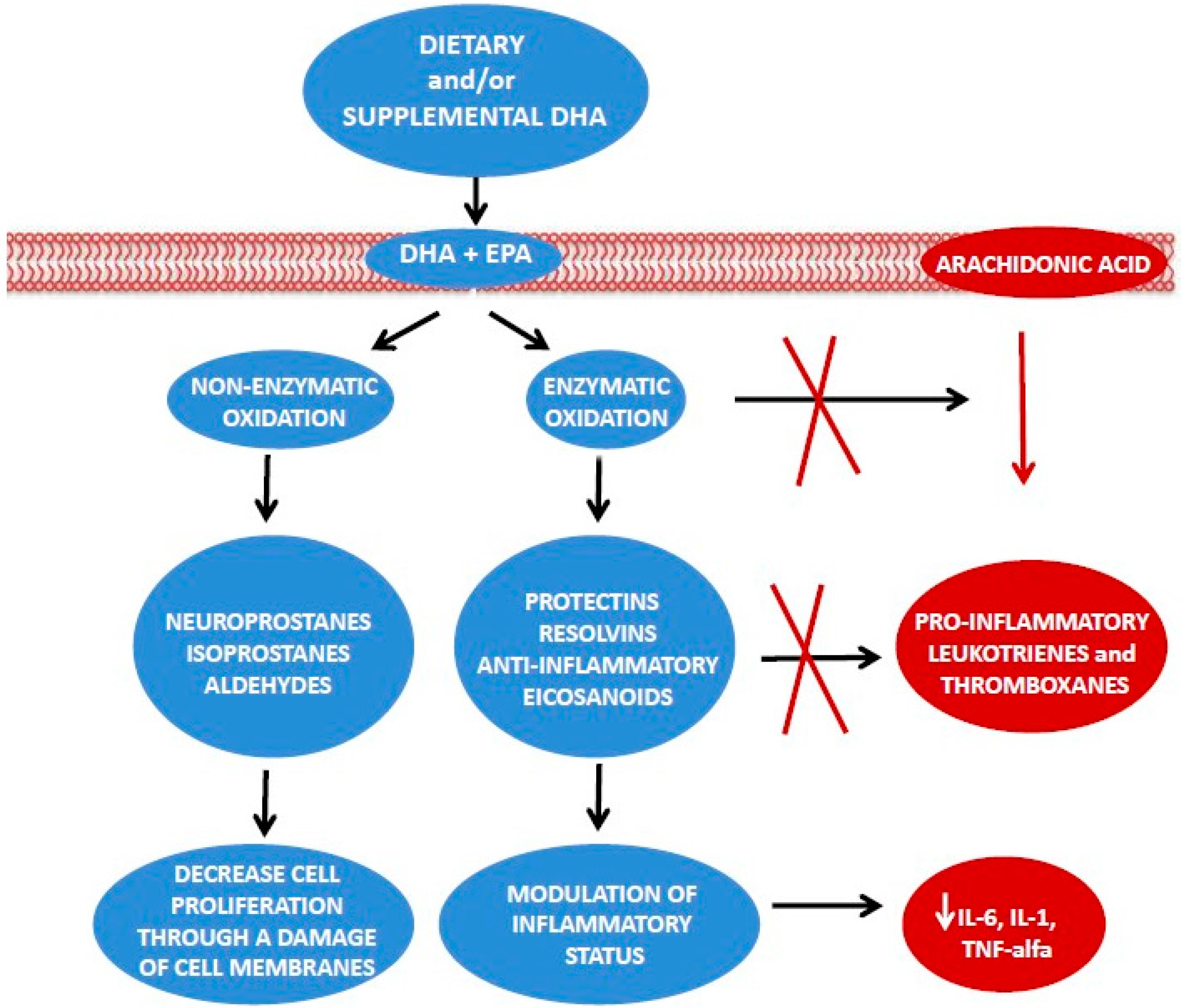
Omega-3 fatty acids are metabolically active lipids with anti-inflammatory properties [13,14,15,16].
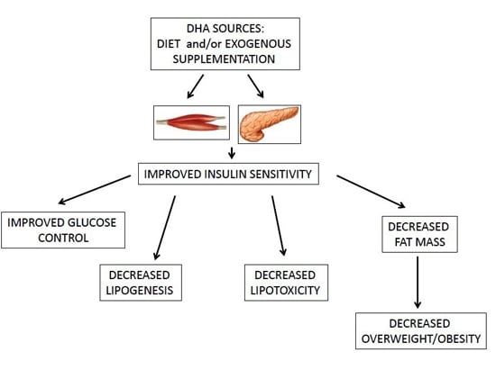
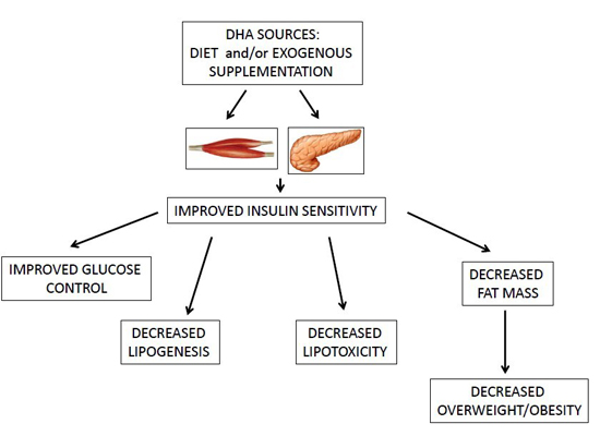
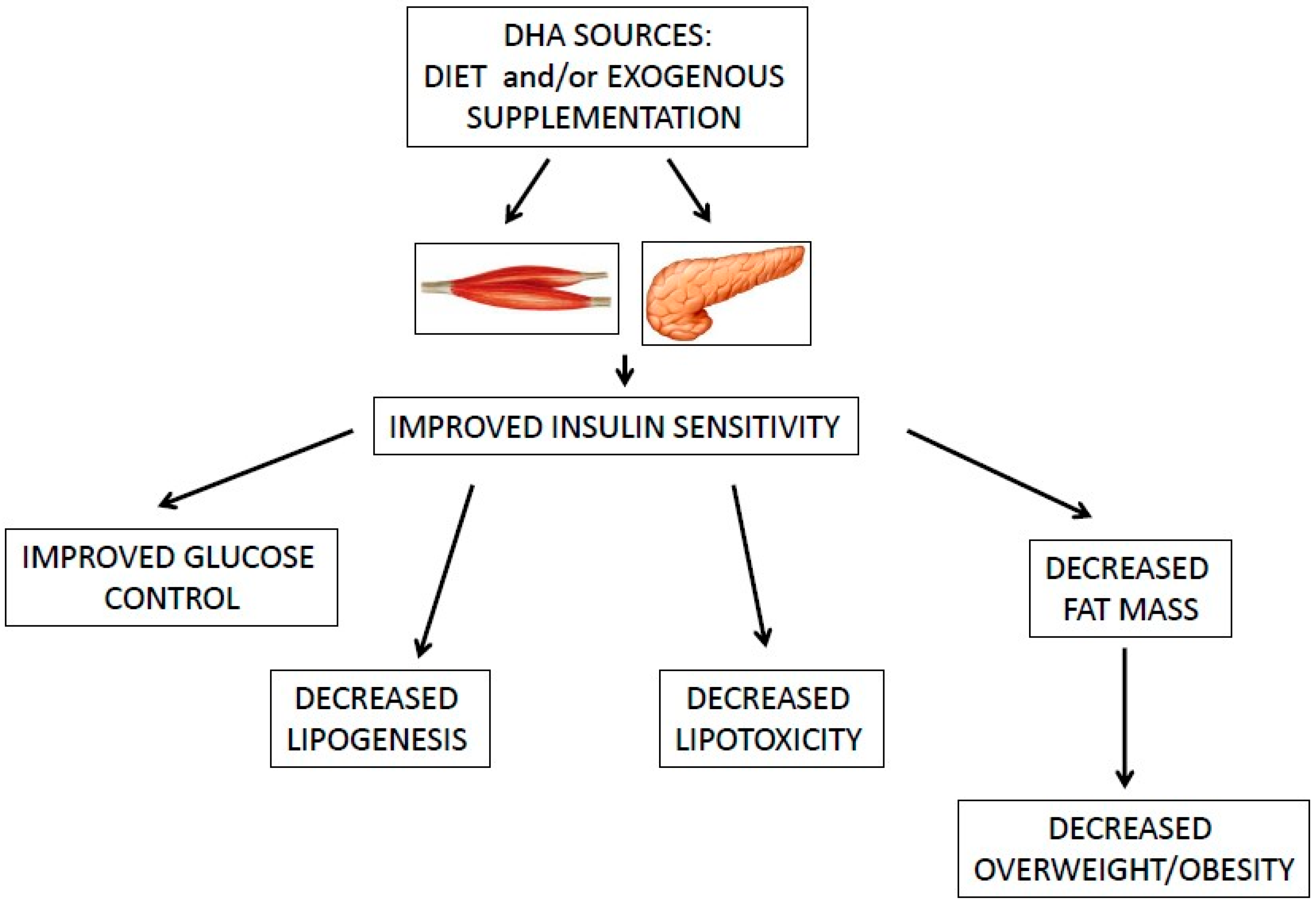
Omega-3 fatty acids and, in particular, DHA ameliorate obesity-induced inflammation and insulin-resistance [4,36,37].
Considering the role of inflammation in breast cancer, there is an increasing rationale for the use of DHA in combination with anticancer therapies [14,15,16,64].
Additional evidence is needed to assess the protective role of DHA breast cancer.

