Article

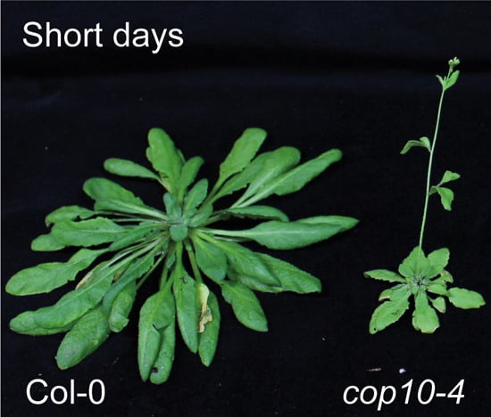
CONSTITUTIVE PHOTOMORPHOGENIC 10 (COP10) Contributes to Floral Repression under Non-Inductive Short Days in Arabidopsis

Department of Plant Science, Plant Genomics and Breeding Institute, Research Institute of Agriculture and Life Sciences, Seoul National University, Seoul 08826, Korea
*
Authors to whom correspondence should be addressed.
Int. J. Mol. Sci. 2015, 16(11), 26493-26505; https://doi.org/10.3390/ijms161125969
Submission received: 9 September 2015 / Revised: 26 October 2015 / Accepted: 27 October 2015 / Published: 5 November 2015
(This article belongs to the Special Issue Plant Molecular Biology)

Abstract

:
In Arabidopsis, CONSTITUTIVE PHOTOMORPHOGENIC/DE-ETIOLATED/FUSCA (COP/DET/FUS) genes act in repression of photomorphogenesis in darkness, and recent reports revealed that some of these genes, such as COP1 and DET1, also have important roles in controlling flowering time and circadian rhythm. The COP/DET/FUS protein COP10 interacts with DET1 and DNA DAMAGE-BINDING PROTEIN 1 (DDB1) to form a CDD complex and represses photomorphogenesis in darkness. The cop10-4 mutants flower normally in inductive long days (LD) but early in non-inductive short days (SD) compared with wild type (WT); however, the role of COP10 remains unknown. Here, we investigate the role of COP10 in SD-dependent floral repression. Reverse transcription-quantitative PCR revealed that in SD, expression of the LD-dependent floral inducers GI, FKF1, and FT significantly increased in cop10-4 mutants, compared with WT. This suggests that COP10 mainly regulates FT expression in a CO-independent manner. We also show that COP10 interacts with GI in vitro and in vivo, suggesting that COP10 could also affect GI function at the posttranslational level. Moreover, FLC expression was repressed drastically in cop10-4 mutants and COP10 interacts with MULTICOPY SUPPRESSOR OF IRA1 4 (MSI4)/FVE (MSI4/FVE), which epigenetically inhibits FLC expression. These data suggest that COP10 contributes to delaying flowering in the photoperiod and autonomous pathways by downregulating FT expression under SD.

Graphical Abstract

1. Introduction

Most flowering plants have evolved to synchronize their growth and development with seasonal environmental changes, especially changes in the intensity and period of daylight and temperature. The precise control of flowering time strongly affects regional adaptation and several signaling pathways, including photoperiod, vernalization, gibberellin, ambient temperature, and autonomous pathways, regulate floral induction [1]. Work to date has identified many genes that control flowering time and the major floral inducers GIGANTEA (GI), CONSTANS (CO), and FLOWERING LOCUS T (FT) have been studied widely and intensively [2,3,4]. The florigen FT acts in multiple flowering pathways [2,5]. CO protein directly binds to the FT promoter and upregulates FT mRNA, but this role differs somewhat under LD and SD conditions. Under LD conditions, CO expression coincides with light at the end of the day, and expressed CO protein promotes FT expression to induce flowering. Under SD, by contrast, the peak of CO transcription occurs after dusk and CO protein is unstable in the dark; therefore, CO cannot induce FT expression [6,7,8,9].
GI and FLAVIN-BINDING, KELCH REPEAT, and F-BOX PROTEIN1 (FKF1) have essential functions in the timing of daily CO expression. GI and FKF1 form a complex to destabilize CYCLING DOF FACTOR1 (CDF1), a key CO repressor [10,11]. Under LD conditions, expression of GI and FKF1 peaks in the afternoon and the GI-FKF1 complex is recruited to the CO chromatin, where it degrades CDF1 to activate CO expression. Conversely, under SD, the peaks of GI and FKF1 expression overlap less than they do in LD, leading to minimal formation of the GI-FKF1 complex [10,12]. This indicates that GI acts as a floral inducer with FKF1 in the CO-FT photoperiod pathway under LD. However, Sawa et al. (2011) reported that under SD conditions, the overexpression of GI increased FT expression without increasing CO expression; GI directly regulates FT expression by binding to the FT promoter region near the binding sites of FT repressors such as SHORT VEGETATIVE PHASE (SVP), TEMPRANILLO1 (TEM1), and TEM2 [13,14,15]. Thus, that GI regulates FT expression through both CO-dependent and CO-independent pathways. In addition, genes involved in the autonomous and vernalization pathways also regulate FT expression. FLOWERING LOCUS C (FLC) acts at a central place in the autonomous and vernalization pathways and FLC directly regulates FT and SOC1 expression by binding to their promoters [13,16,17]. FLC expression is mainly regulated by histone modification factors [18]. For example, MULTICOPY SUPPRESSOR OF IRA1 4/FVE (MSI4/FVE) represses FLC expression by histone modification of FLC locus with DDB1 [19,20].
CONSTITUTIVE PHOTOMORPHOGENIC/DE-ETIOLATED/FUSCA (COP/DET/FUS) genes have important roles in the repression of seedling photomorphogenesis in darkness [21]. We recently reported that DE-ETIOLATED1 (DET1), a COP/DET/FUS family protein, negatively regulates flowering, because det1-1 weak mutants (note that the det1 null mutant is lethal) flower early, especially much earlier in SD, compared with the wild type [22]. In det1-1 mutants, FT expression is significantly up-regulated in SD, but the expression levels of GI and FKF1 do not change. DET1 physically interacts with GI to inhibit its binding to the FT promoter [22]. Furthermore, the expression level of FLC, a repressor of FT, is significantly down-regulated in det1-1 mutants, probably due to the lack of interaction between DET1 and MSI4/FVE. Collectively, these observations indicate that DET1 acts in both photoperiod (post-translational regulation of GI) and autonomous (MSI4/FVE-FLC) pathways to repress the expression of FT. Similar to DET1, the COP/DET/FUS protein COP1 also regulates SD flowering in Arabidopsis; cop1 mutants flowered much earlier than WT only under SD conditions [23]. COP1 is an E3 ubiquitin ligase and forms a complex with SUPPRESSOR OF PHYA1 (SPA1) for its E3 function [24]. The COP1-SPA1 complex is required for the ubiquitination and degradation of CO and GI in the night [7,23]. These results indicate that some COP/DET/FUS proteins also have important roles in repressing flowering, as well as the repression of photomorphogenesis in darkness.
DET1 forms a multi-protein complex with COP10 and DAMAGED DNA BINDING PROTEIN1 (DDB1). The COP/DET/FUS family protein COP10 is an E2-like protein that lacks E2 activity [25]. DDB1 is required for the interaction of the COP10-DET1-DDB1 complex (termed the CDD complex) with CULLIN4 (CUL4), and acts as E3 ubiquitin ligase (CUL4-CDD E3 ligase) [26]. In addition, the CDD complex maintains the circadian rhythm with LHY and CCA1 in the photoperiod pathway [27,28]. These previous reports indicate that, like DET1, COP10 also affects the regulation of flowering time. However, the molecular mechanism of COP10 in flowering remains unclear.
In this study, we show that COP10 delays flowering time in SD by modulating GI at both transcriptional and post-translational levels in the photoperiod pathway. In addition, COP10 indirectly up-regulates FLC expression by interacting with MSI4/FVE, which functions in histone modification of the FLC locus. Our results show that COP10 functions in both the photoperiod and autonomous pathways to repress FT expression in SD.

2. Results and Discussion

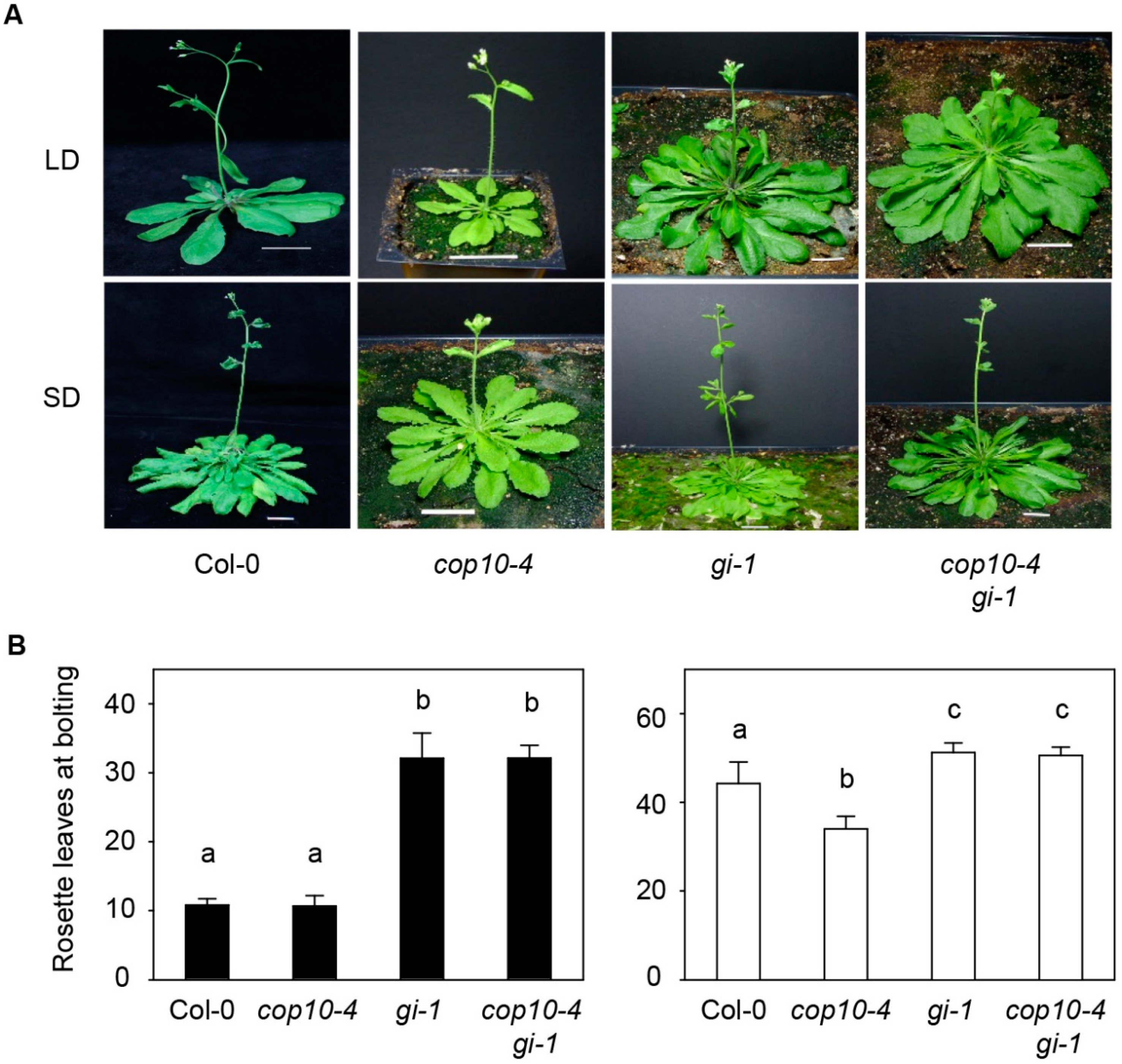
2.1. GIGANTEA (gi-1) Is Epistatic to cop10-4 in the Photoperiodic Pathway of Flowering

cop10-4 has been isolated as one of the cop10 mutant alleles, which has a mutation in the second exon of COP10, resulting in a weak allele although transcriptional and protein levels of COP10 do not alter in cop10-4 mutant [25]. cop10-4 mutants show photomorphogenic development in darkness, with phenotypes such as short hypocotyls and opened cotyledons [29,30,31,32]. However, the function of COP10 in the regulation of flowering time remains unexamined. Null mutants of COP10 are lethal [25]; therefore, to examine the function of COP10 in flowering, we studied flowering time using the weak, viable allele cop10-4. We counted the number of rosette leaves (RLs) at bolting under both LD (16-h light:8-h dark) and SD (10-h light:14-h dark) conditions (Figure 1; Table S1). We found that under SD, cop10-4 mutants flowered earlier, with 34.0 ± 2.8 RLs, about 10 fewer than wild type (WT, 44.3 ± 4.9 RLs). By contrast, WT and cop10-4 flowered with almost the same number of RLs under LD, with 10.8 ± 0.9 RLs for WT and 10.7 ± 0.5 RLs for cop10-4, indicating that COP10 acts as a floral repressor mainly under non-inductive SD conditions, and plays an important role in photoperiod sensitivity in Arabidopsis.
Figure 1. The early flowering phenotype of cop10-4 and gi-1 is epistatic to cop10-4. Phenotypes (A) and the number of rosette leaves (B) of wild type (WT, Col-0 ecotype), cop10-4, gi-1, and cop10-4 gi-1 mutants in long days (LD) (black bars) and short days (SD) (white bars) Plants were grown at 22 °C under cool-white fluorescent light (90–100 μmol·m2·s1) in LD (16-h light:8-h dark) or SD (10-h light:14-h dark), and photographed at two to four days after bolting. Scale bars = 2 cm; and (B) Flowering time was measured as the number of rosette leaves at bolting. Means and standard deviations were obtained from more than 20 plants. These experiments were repeated three times with the same results. Bars with different letters are significantly different according to Duncan’s multiple range test (p < 0.05).
Ijms 16 25969 g001
DET1 acts as a floral repressor upstream of GI and attenuates binding of GI to the FT promoter [22]. COP10 interacts with DET1 and DDB1 to form a CDD complex, which represses photomorphogenesis in darkness [33]; COP10 could also act with GI in flowering. To test whether COP10 interacts genetically with GI, we generated cop10-4 gi-1 double mutants to compare with WT (and each single mutant) in flowering time under LD and SD conditions (Figure 1). As previously reported, gi-1 mutants, which has 5-bp deletion resulting in premature stop codon, showed a strong late-flowering phenotype [3,31]; gi-1 had 32.1 ± 3.7 RLs at bolting in LD and 51.2 ± 2.2 RLs in SD (Figure 1A,B). We found that cop10-4 gi-1 mutants showed a late-flowering phenotype, with 32.1 ± 1.8 RLs in LD and 50.6 ± 1.9 RLs in SD, almost the same as the gi-1 mutant, but many more than the cop10-4 mutant. These results indicate that gi-1 is epistatic to cop10-4 in the photoperiodic pathway of floral induction.

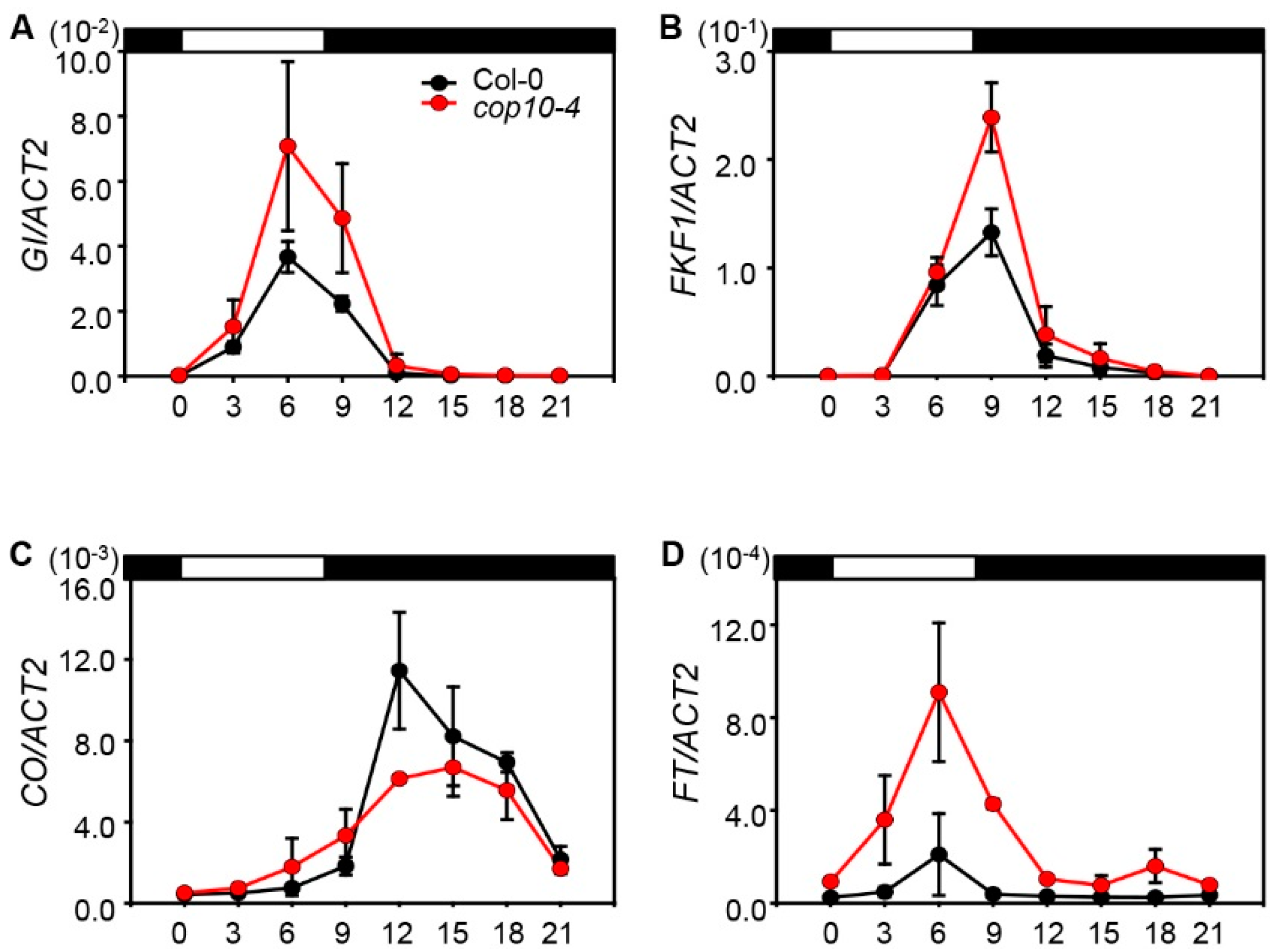
2.2. The cop10 Mutation Alters the Expression of Flowering Time Genes

To investigate the effect of cop10-4 on the expression of floral inducers in the photoperiod pathway, we analyzed the phases and amplitudes of GI, FKF1, CO, and FT mRNA levels in WT and cop10-4 mutants grown under SD conditions (Figure 2). In SD conditions, the transcript levels of GI and FKF1 were the highest at ZT6 (zeitgeber time; 6 h after dawn) and ZT9, respectively [10]. We found that the transcript levels of GI at ZT6 and FKF1 at ZT9 were almost the same in WT and cop10-4 mutants. However, the transcript levels of GI and FKF1 significantly increased in the cop10-4 mutant compared with WT (Figure 2A,B). The timing of the peaks in CO and FT transcript levels did not change in cop10-4 mutants. However, the cop10-4 mutants showed higher expression of FT, although CO expression at ZT6 did not differ in cop10-4 mutants compared with WT (Figure 2C,D).
Figure 2. Effect of cop10-4 on GIGANTEA (GI), F-BOX PROTEIN1 (FKF1), CONSTANS (CO), and FLOWERING LOCUS T (FT) expression under short days (SD). The expression of GI (A); FKF1 (B); CO (C); and FT (D) was analyzed in Col-0 and cop10-4 mutants by real-time PCR using three-week-old plants. Plants were grown at 22 °C under SD (8-h light:16-h dark) conditions, and plant tissues were harvested every 3 h. ACT2 expression was used for normalization. Means and standard deviations were obtained from three biological replicates. These experiments were repeated twice with the same results.
Ijms 16 25969 g002
Our results show that in cop10-4 mutants, the expression levels of GI and FT, as well as FKF1, were significantly up-regulated during daytime in SD conditions (Figure 2A,D). By contrast, CO expression did not differ in cop10-4 mutants compared with WT (Figure 2C), indicating that COP10 delays flowering by down-regulating the GI-FT regulatory module in non-inductive SD conditions. Interestingly, it appears that the expression patterns of GI, FKF1, and CO in cop10-4 mutants differ considerably from those in det1-1 mutants [22]. Under SD conditions, the expression levels of GI and FKF1 did not significantly change in det1-1 mutants compared with WT. By contrast, the CO transcript levels significantly increased in det1-1 mutants, compared with WT [22]. As COP10 and DET1 form a complex together with DDB1 (CDD complex) [33], we expected that the expression levels of key flowering genes, such as GI, FKF1, and CO, were similarly regulated in cop10-4 and det1-1 mutants. However, our results indicate that the COP10- and DET1-dependent mechanisms regulating flowering in SD differ somewhat, and that COP10 probably regulates flowering in SD without forming the CDD complex.
In this experiment, we focused on the transcriptional regulation of key flowering genes in cop10-4 mutants. However, COP10 is a subunit of the E3 CUL4-CDD E3 ligase [26]. Thus, it is also important to examine whether the protein levels of key flowering factors, such as GI, FKF1, CO, and FT, were altered in cop10-4 mutants, which will be investigated in future work.

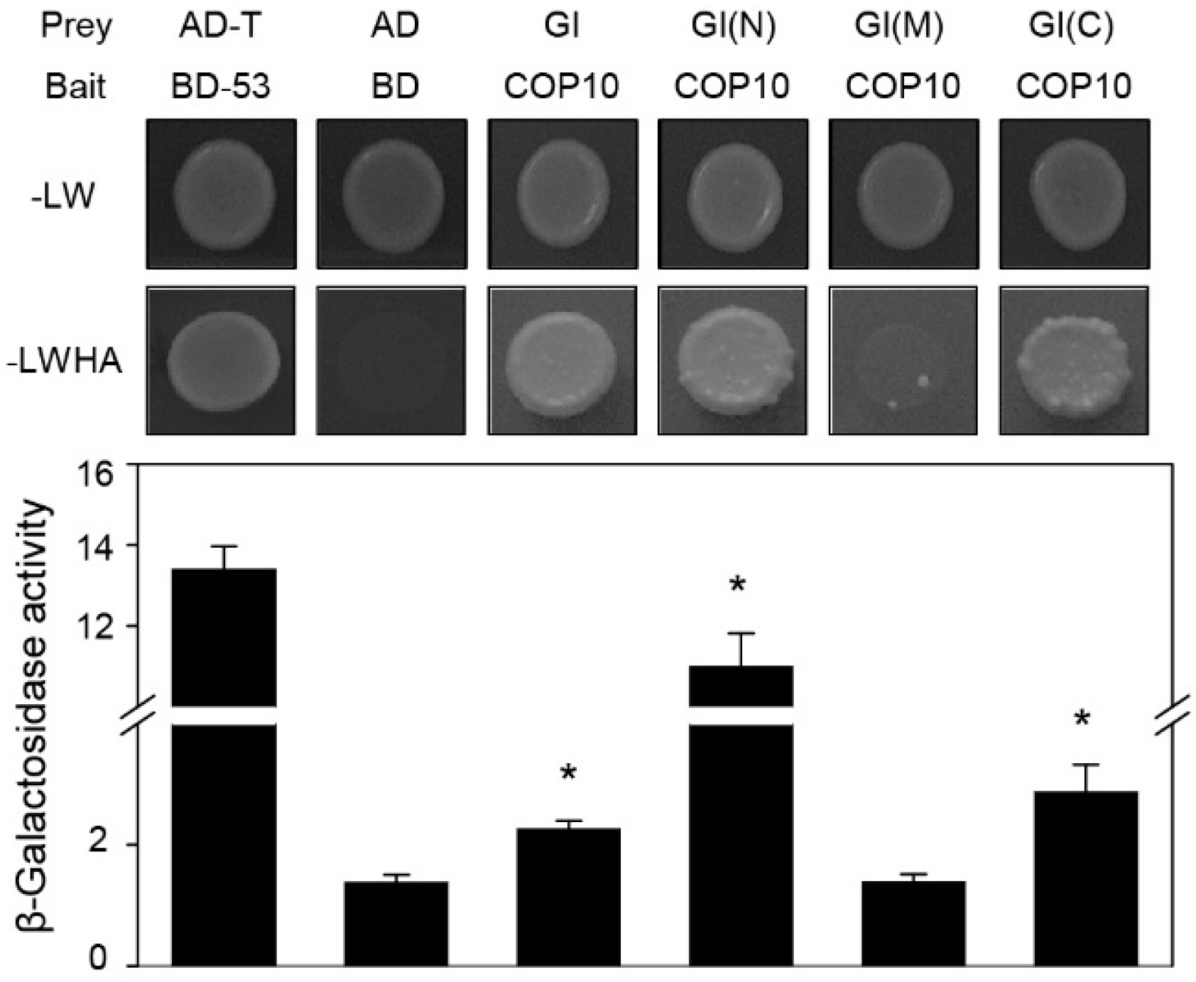
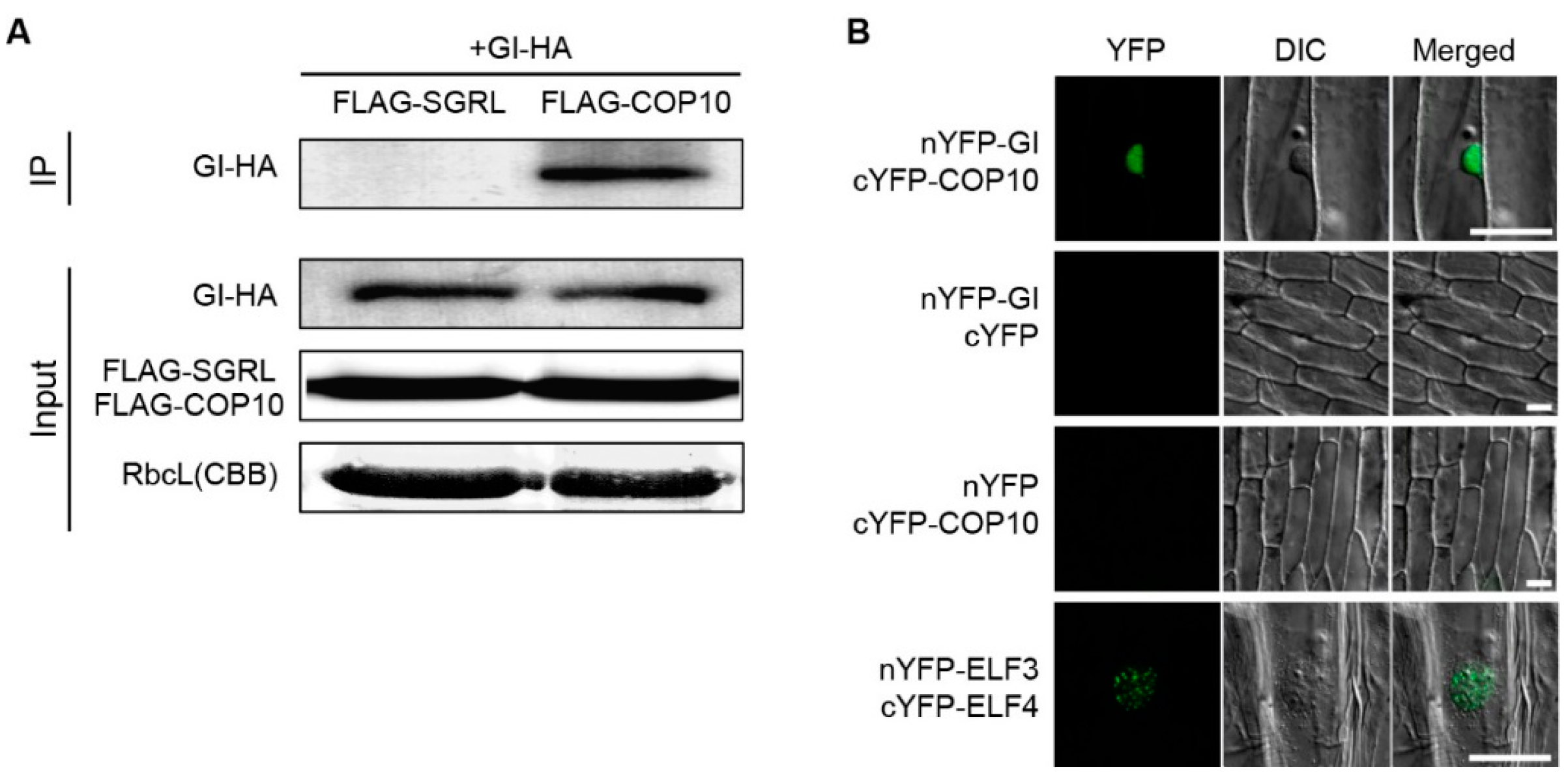
2.3. COP10 Interacts with GI

DET1 physically interacts with GI, leading to decreased GI binding to the promoter of FT [22]. Although we found that mutation of COP10 causes up-regulation of GI expression in SD conditions (Figure 2), it is also possible that COP10 physically associates with GI to down-regulate its function in flowering, similar to DET1. To test this possibility, we first examined the physical interaction between COP10 and GI by yeast two-hybrid assays. We used GI for the prey and COP10 for the bait. Yeast cells transformed with COP10 and GI grew on SD-4 selection media lacking adenine, leucine, histidine, and tryptophan. Especially, yeast cells, in which COP10 was co-transformed with constructs expressing the full-length, N-terminal (aa 1–507), and C-terminal (aa 801–1173) regions of GI, but not the middle region (aa 401–907), grew on the interaction media. We additionally performed chlorophenol red-β-d-galactopyranoside (CPRG) assays to qualitatively measure these interactions, which revealed that COP10 interacts most strongly with the N-terminal (aa 1–507) region of GI (Figure 3).
Figure 3. COP10 directly interacts with GI in a yeast two-hybrid assay. The bait was full-length COP10. For prey, GI was divided into three pieces: N-terminal (N; aa 1–507), middle (M; aa 401–907), and C-terminal (C; aa 801–1173). pGBKT7-53 (BD-53) and pGADT7-T were used as a positive control. Empty pGBKT7 (BD) and pGADT7 (AD) vectors were the negative control. SD medium (-LWHA; lacking tryptophan, leucine, histidine, and adenine) was used to select for the interaction between baits and preys. β-Galactosidase assays were performed according to the manufacturer’s protocol. Means and standard deviations were obtained from three individual colonies. Asterisks indicate statistically significant differences compared to negative control as determined by Student’s t-test (* p < 0.05). These experiments were repeated twice with the same results.
Ijms 16 25969 g003
To test the in vivo interaction of COP10 and GI, we subsequently performed co-immunoprecipitation (co-IP) assays using transgenic plants overexpressing HA-GI or FLAG-COP10. FLAG-tagged STAY-GREEN LIKE (SGRL-FLAG), a chloroplast-localized protein [34], was used for a negative control. 35S:FLAG-COP10, 35S:HA-GI, and 35S:SGRL-FLAG (a negative control) transgenic plants were grown for 2 weeks in SD, and then sampled at ZT8. We found that HA-GI co-immunoprecipitated with FLAG-COP10, but not with SGRL-FLAG (Figure 4A). To further confirm this in vivo interaction, we performed bimolecular fluorescence complementation (BiFC) assays using onion epidermal cells. COP10 and GI localize in the cytosol and nucleus, respectively [25,34]; thus we also investigated where they interact. Using a transient expression assay in onion epidermal cells, we detected strong reconstituted YFP fluorescence in the nucleus when nYFP-GI and cYFP-COP10 plasmids were co-transformed, but we detected no YFP fluorescence in the onion cells when we co-transformed nYFP-GI/cYFP or nYFP/cYFP-COP10 (Figure 4B). Taken together, these results indicate that COP10 physically interacts with GI in vitro and in vivo, and this interaction occurs in the nucleus.
To date, the significance of this COP10-GI interaction remains unclear, but we can consider a few possibilities. We previously found that DET1 physically interacts with GI in the nucleus to decrease GI binding to the promoter of FT for repression of the induction of flowering [22]. DET1 and COP10, together with DDB1, also form a CDD complex in the nucleus to repress photomorphogenesis [33]. Thus, COP10 could act with DET1 to modulate GI binding to the FT promoter. COP10 also acts as an enhancer of a ubiquitin-conjugating E2 enzyme and interacts with COP1, a known a RING E3 ubiquitin ligase, in the ubiquitination pathway [25,33]. Furthermore, we previously suggested that COP1 may destabilize GI protein by interacting with EARLY FLOWERING 3 (ELF3) [23]. Thus, COP10 (in the CDD complex or another form) could also function with COP1 to control GI in the photoperiod pathway. Further biochemical analyses of the interaction between GI and COP10 will be necessary to reveal the significance of the GI-COP10 interaction.
Figure 4. COP10 interacts with GI in plants. (A) Co-immunoprecipitation of COP10 and GI using 35S:FLAG-COP10, 35S:HA-GI, and 35S:SGRL-FLAG. Total protein was extracted from two-week-old seedlings. FLAG-beads were used for pull-down. Anti-HA antibody was used for GI-HA protein band. 35S:SGRL-FLAG plants served as a negative control. The upper panel is co-immunoprecipitated GI-HA protein using an anti-HA antibody after an anti-FLAG-bead pull-down assay. As a loading control, RbcL protein levels were visualized by staining the immunoblot with Coomassie Brilliant Blue; (B) BiFC analysis of the interaction between COP10 and GI in the nucleus of an onion epidermal cell. nYFP-ELF3 and cYFP-ELF4 plasmids served as a positive control [21]. For the negative controls, empty nYFP-GI/cYFP and nYFP/cYFP-COP10 were used. Each pair of recombinant plasmids encoding nYFP and cYFP fusions was mixed 1:1 (w/w) and co-bombarded into onion epidermal cell layers. The transformed onion epidermal layers were incubated at 22 °C for 16–24 h under dark condition. YFP fluorescence was indicated by green color. Scale bar = 50 μm. These experiments were repeated three times with the same results.
Ijms 16 25969 g004

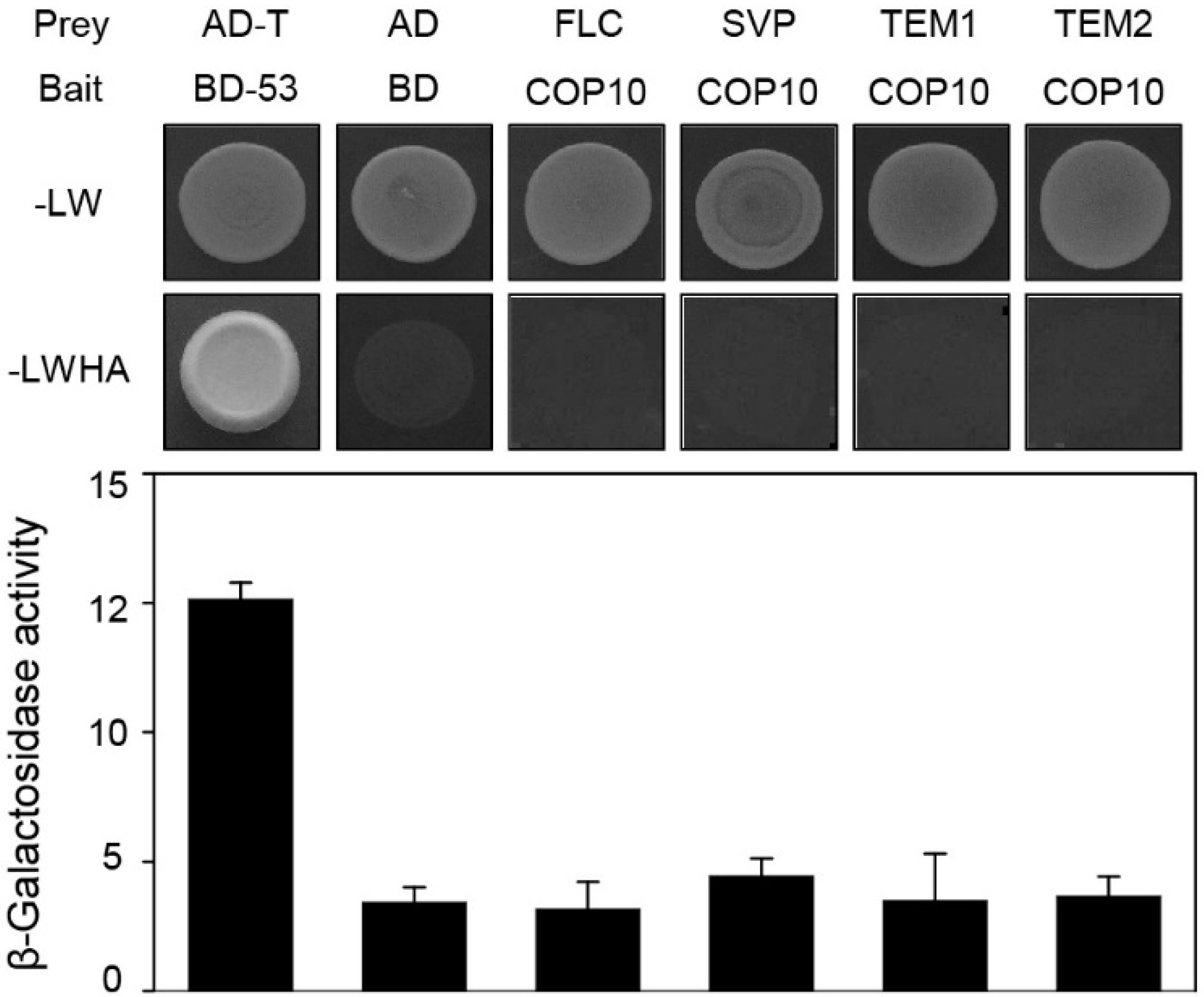
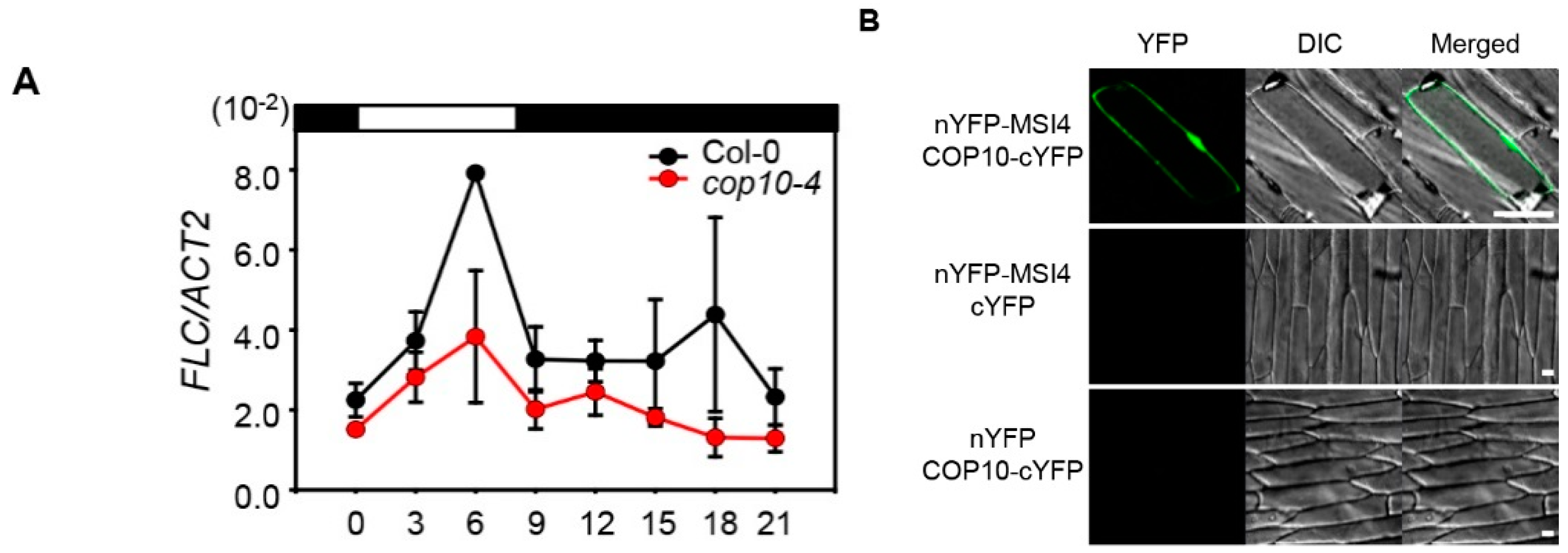
2.4. COP10 Regulates FLOWERING LOCUS C (FLC) Expression through Interaction with MULTICOPY SUPPRESSOR OF IRA1 4 (MSI4) in the Autonomous Pathway

The expression of FT is intricately regulated; GI and CO activate FT transcription and FLC, SVP, TEM, and TEM2 repress FT transcription [13,14,15,35]. GI directly interacts with SVP, TEM1, and TEM2, probably to deactivate or destabilize these FT repressors [35]. Therefore, COP10 could act with these FT repressors to down-regulate FT expression. Thus, we examined the possible interaction of COP10 and repressors of FT transcription in yeast two-hybrid assays. These revealed that COP10 does not interact with FLC, SVP, TEM1, or TEM2 (Figure 5), indicating that COP10 likely does not directly affect the activity of these FT repressors. We next used RT-qPCR to examine whether mutation of COP10 alters the expression levels of these FT repressors in WT and cop10-4 mutants grown in SD conditions. We found that FLC mRNA levels significantly decreased in cop10-4 mutants compared with WT (Figure 6A). By contrast, levels of SVP, TEM1, and TEM2 mRNAs did not significantly differ in cop1-4 mutants and WT (Figure S1).
Figure 5. COP10 does not interact with FT repressors in yeast. The bait was full-length COP10. For prey, FLC, SVP, TEM1, and TEM2 were used. pGBKT7-53 (BD-53) and pGADT7-T were used as a positive control. Empty pGBKT7 (BD) and pGADT7 (AD) vectors were the negative control. SD medium (-LWHA; lacking tryptophan, leucine, histidine, and adenine) was used to select for the interaction between bait and prey proteins. β-Galactosidase activity assays were performed according to the manufacturer’s protocol. Means and standard deviations were obtained from three individual colonies. These experiments were repeated twice with the same results.
Ijms 16 25969 g005
FLC expression is regulated in the autonomous pathway, which consists of several factors involved in RNA processing and epigenetic regulation [36]. MSI4/FVE acts as a key regulator of the autonomous pathway to reduce FLC expression [37]. Furthermore, both DET1 and DDB1, members of CDD complex, interact with MSI4/FVE to reduce its activity and consequently up-regulate FLC expression [20,22]. Thus, COP10 could also interact with MSI4, as well as DET1 and DDB1, possibly in a CDD complex. To examine this possibility, we used BiFC assays to examine the physical interaction between COP10 and MSI4/FVE. Using transient expression in onion epidermal cells, we detected strong reconstituted YFP fluorescence in both cytosol and nucleus when nYFP-MSI4 and COP10-cYFP plasmids were co-transformed (Figure 6B).
Figure 6. COP10 regulates FLOWERING LOCUS C (FLC) expression through interaction with MULTICOPY SUPPRESSOR OF IRA1 4 (MSI4)/FVE (MSI4/FVE). (A) The expression of FLC analyzed in Col-0 and cop10-4 mutants by real-time PCR using three-week-old plants. Plants were grown at 22 °C under SD (8-h light:16-h dark) conditions, and plant tissues were harvested every 3 h. ACT2 expression was used for normalization. Means and standard deviations were obtained from three biological replicates. ZT, zeitgeber time (h after dawn); (B) COP10 interacts with MSI4 in plants. BiFC analysis of the interaction between MSI4 and COP10 in onion epidermal cells. nYFP-ELF3 and cYFP-ELF4 plasmids served as a positive control [21]. For the negative controls, empty nYFP-GI/cYFP and nYFP/cYFP-COP10 were used. Each pair of recombinant plasmids encoding nYFP and cYFP fusions was mixed 1:1 (w/w) and co-bombarded into onion epidermal cell layers. The transformed onion epidermal layers were incubated at 22 °C for 16–24 h under light conditions. YFP fluorescence was indicated by green color. Each bar indicates 50 μm. DIC, differential interference contrast. These experiments were repeated three times with the same results.
Ijms 16 25969 g006
DET1 participates in chromatin remodeling by interacting with unmodified histone H2B [38]. Furthermore, the CDD complex acts in chromatin remodeling with CUL4 through the interaction with non-acetylated H2B, to repress photomorphogenesis under light conditions [39,40]. A recent study showed that MSI4/FVE interacts with HDA6 and directly binds to the FLC locus to repress FLC transcription via chromatin remodeling [19,20]. In this scenario, the CDD complex and MSI4/FVE act in the autonomous pathway by histone modification of the FLC promoter with HDA6. Our findings provide evidence that COP10 also has an important role in regulating FLC expression through interaction with MSI4/FVE in the autonomous pathway, possibly as a part of the CDD complex. In this study, we also found by BiFC analysis that COP10 physically interacts with MSI4/FVE in both the cytosol and nucleus (Figure 6A). COP10 originally localizes in the cytosol and MSI4/FVE l localizes to the cytosol and nucleus [25,37]. Thus, MSI4/FVE likely acts in the translocation of COP10 from the cytosol to nucleus. Many circadian-clock and flowering components localize in the nucleus and the cytosol, and this subcellular compartmentalization has important roles in the regulation of photoperiodic flowering. For example, the Arabidopsis flowering regulator EARLY-FLOWERING4 (ELF4) acts as a regulator of the nucleus/cytosol distribution of GI through a physical interaction [41]. Analogous to ELF4, it is possible that the subcellular compartmentalization of COP10 is also regulated by the interaction with MSI4/FVE or other components in flowering pathway, which will be investigated in more detail in future work.

3. Experimental Section

3.1. Plant Materials and Growth Conditions

Arabidopsis thaliana WT and mutant lines used in this study are in the Columbia (Col-0) genetic background. The gi-1 seeds (CS3123) were obtained from the Arabidopsis Biological Resource Center (Columbus, OH, USA). The cop10-4 mutants were obtained from Xing Wang Deng. cop10-4 has been analyzed as one of the cop10 mutant alleles, which has a mutation in the second exon of COP10, resulting in a weak allele [25]. To create cop10-4 gi-1 double mutants, F1 heterozygotes were obtained by crossing the cop10-4 mutant as the female plant with gi-1 mutants as pollen donors. Plants were grown on soil at a constant 22 °C under white fluorescent light (90–100 μmol·m−2·s−1) in LD (16-h light:8-h dark) and SD (10-h light:14-h dark) conditions.

3.2. Analysis of Flowering Time

Flowering time was measured by counting the total number of rosette leaves (RLs) at bolting. Data were obtained from three experimental replications (20 plants per replication).

3.3. RNA Preparation, Reverse Transcription, and Quantitative Real-Time PCR (qPCR) Analysis

Green tissues of 3-week-old WT and cop10-4 plants grown on agar plates (containing 4.3 g/L Murashige Skoog, Duchefa Biochemie, Haarlem, The Nederland) under LD or SD conditions were collected every 3 h. Total RNA was extracted with the MG Total RNA Extraction Kit (Macrogen, Seoul, Korea) according to the manufacturer’s instructions. For each sample, 2 μL of total mRNA was reverse-transcribed using oligo (dT)15 primers and M-MLV reverse transcriptase (Promega, Madison, WI, USA). The levels of the transcripts were measured by the relative quantification method, using GoTaq qPCR Master Mix (Promega) and a Light Cycler 480 (Roche Applied Science, Penzberg, Upper Bavaria, Germany). The qPCR conditions were: 95 °C for 2 min, and then 45 cycles of 95 °C for 10 s and 60 °C for 1 min. Each qPCR amplification was repeated at least three times with biologically independent samples for statistical analysis. ACTIN2 (ACT2) was used for an internal control. The amount of each mRNA was determined using specific primers (Table S2).

3.4. Yeast Two-Hybrid Assays

Yeast two-hybrid assays were performed according to the Yeast Protocols Handbook (Clontech, Mountain View, CA, USA). The full-length cDNA of COP10 was amplified from WT total RNA using RT-PCR, and cloned into the pGBKT7 (bait) vector. The full-length cDNAs of FLC, SVP, TEM1, and TEM2, and three partial fragments of GI (encoding the N-terminal, aa 1–507; middle aa 401–907; and C-terminal, aa 801–1173) were cloned into the pGADT7 (prey) vector (MATCHMAKER GAL4 two-hybrid system 3, Clontech) as described previously [21,35]. For the interaction study, plasmids expressing fusion proteins were transformed into the yeast (Saccharomyces cerevisiae) strain AH109 [42] by the LiAc-mediated method [43] and grown on media lacking adenine, leucine, histidine, and tryptophan. Chlorophenol red-β-d-galactopyranoside (CPRG; Roche Biochemicals, Penzberg, Upper Bavaria, Germany) was used to measure the β-galactosidase activity according to the manufacturer’s protocol.

3.5. In Vivo Pull-Down Assays

Arabidopsis (Col-0 ecotype) plants were used in this study. The transgenic plants containing the 35S:FLAG-COP10 or 35S:HA-GI constructs were previously described [33,44]. In addition, 35S:SGRL-FLAG, which we previously generated [45], was used as a negative control. For the COP10-GI interaction assays, 35S:FLAG-COP10, 35S:HA-GI, and 35S:SGRL-FLAG plants were grown on 0.5× Murashige-Skoog (MS) phytoagar medium in SD (8-h light:16-h dark) for 10 days and then vacuum infiltrated for 10 min in MS liquid medium (Duchefa Biochemie, Duchefa Biochemie, Haarlem, The Nederland) supplemented with 50 mM MG132 (Sigma-Aldrich, St. Louis, MO, USA) for proteasome inhibitor treatment. After treatment, plants were incubated for 10 h under light conditions, and then homogenized; total proteins were extracted in total protein extract buffer (50 mM Tris-HCl (pH 7.5), 100 mM NaCl, 10 mM MgCl2, 1 mM EDTA (pH 8.0), 10% glycerol, 1 mM PMSF, 1 mM DTT). These experiments were performed with FLAG-M2 magnetic beads (Sigma-Aldrich) for FLAG-IP. After washing, the immunoprecipitated fractions were determined by immunoblot analysis. The HA-GI fusion proteins were immunodetected by an anti-HA antibody (Cell Signaling Technology, Danvers, MA, USA).

3.6. Bimolecular Fluorescence Complementation Assays in Onion Epidermal Cells

The full-length cDNA of COP10 was cloned into the BiFC gateway vectors [46] to examine COP10 in vivo interactions. Cloned vectors containing GI, ELF3, ELF4, and MSI4 were previously prepared [22,23]. For partial YFP-tagged COP10 and MSI4 constructs, the cDNA of each gene was obtained by RT-PCR from WT (Col-0) plants and fused into four BiFC plasmid sets, pSAT5-DEST-cEYFP(175-end)-C1(B) (pE3130), pSAT5(A)-DEST-cEYFP(175-end)-N1 (pE3132), pSAT4(A)-DEST-nEYFP(1-174)-N1(pE3134), and pSAT4-DEST-nEYFP(1-174)-C1 (pE3136) to generate constructs fused with the N- or C-terminal fragments of YFP. Partial YFP-tagged ELF3 and GI constructs were previously described [23]. Purified gold particles were coated with each pair of recombinant plasmids encoding nEYFP and cEYFP fusions, which were mixed 1:1 (w/w), and co-bombarded into onion epidermal layers using a DNA Particle Delivery System (Biolistic PDS-1000/He, BioRad, Hercules, CA, USA). The transformed onion epidermal cells were incubated on MS solid media with 50 mM MG132 (Sigma-Aldrich) for 16–24 h at 22 °C. Onion cells used in Figure 4 (nYFP-GI/cYFP-COP10 and positive/negative control) and in Figure 6 (nYFP-MSI4/COP10-cYFP and positive/negative controls) were incubated under dark and light conditions, respectively. These experimental conditions were selected because nYFP-GI/cYFP-COP10 was predominantly expressed in dark-incubated cells while nYFP-MSI4/COP10-cYFP was predominantly expressed in the light. Then, the fluorescence was detected using a confocal laser scanning microscope (Carl Zeiss LSM710, Germany). nYFP-ELF3/cYFP-ELF4 served as a positive control [22], and empty nYFP-GI/cYFP and empty nYFP/cYFP-COP10 were used for the negative control.

3.7. Accession Numbers
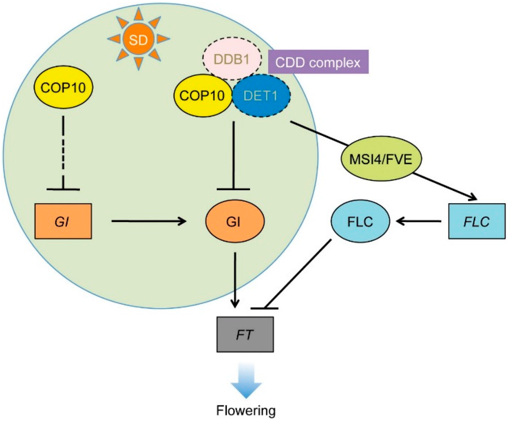
Sequence data from this article can be found in the Arabidopsis Genome Initiative or GenBank/EMBL databases under the following accession numbers: CO (At5g15840), COP10 (At3g13550), ELF3 (At2g25930), ELF4 (At2g40080), FKF1 (At1g68050), FLC (AT5G10140), FT (At1g65480), GI (At3g13550), MSI4 (At2g1952), SVP (At2g22540), TEM1 (At1g25560), and TEM2 (At1g68840).

4. Conclusions

Here we showed that COP10 negatively regulates the SD-dependent flowering pathways. Because COP10 and DET1 act together in the CDD complex [33], we expected COP10 and DET1 to act similarly in the regulation of SD-dependent repression of flowering. However, we found that in SD conditions, GI and FKF1 expression levels were significantly upregulated in cop10-4 mutants, but we observed no change in CO expression level (Figure 2). These expression patterns are quite different from those in the det1-1 mutant [22], suggesting that COP10 and DET1 may regulate the main cascades of photoperiodic flowering differently, and probably not as part of the CDD complex (Figure 7). On the other hand, we found that mutation of COP10 decreases the expression of FLC (Figure 6A), probably due to the lack of interaction of COP10 with MSI4/FVE, a key regulator of the autonomous pathway (Figure 6B). Furthermore, COP10 directly interacts with GI in vitro and in vivo (Figure 3 and Figure 4). Such a mode of action of COP10 is considerably similar to DET1 [22]. Thus, it is possible that COP10 and DET1 act together, in the CDD complex, for posttranslational regulation of the function of GI and the MSI4-FLC cascade in the autonomous pathway (Figure 7). However, further experiments, such as expression analysis of flowering time genes in det1-1 cop10-4 double mutants and time course co-IP of COP10, DET1, and GI, will be needed to understand the role of the CDD complex in repression of flowering pathways.
Figure 7. Working model of COP10 function in flowering repression. COP10 suppresses FT expression via multiple routes in the photoperiod and autonomous pathways. In the photoperiod flowering pathway, COP10 functions in floral repression by regulation of GI at the transcriptional and post-translational levels during daytime under SD. COP10, probably without forming the CDD complex, blocks formation of the FKF1-GI complex by repressing expression of FKF1 and GI during the daytime. Second, COP10 physically interacts with GI, and might repress its binding to the FT promoter. In the autonomous pathway, COP10 modulates FLC chromatin through interaction with MSI4 to induce FLC expression. Genes and proteins are represented as rectangles and ovals, respectively.
Ijms 16 25969 g007

Supplementary Materials

Supplementary materials can be found at https://www.mdpi.com/1422-0067/16/11/25969/s1.

Acknowledgments

We thank Xing Wang Deng for 35S:FLAG-COP10 seeds and Woe-Yeon Kim for 35S:HA-GI seeds. This work was supported by the National Research Foundation of Korea (NRF) grant funded by the Ministry of Science, ICT and Future Planning (NRF-2011-0017308).

Author Contributions

Min-Young Kang and Nam-Chon Paek conceived the study and designed the research. Min-Young Kang, Hye-Young Kwon, and Na-Yun Kim performed experiments. Min-Young Kang and Yasuhito Sakuraba analyzed data with suggestions by Nam-Chon Paek. All authors read and approved the final manuscript.

Conflicts of Interest

The authors declare no conflict of interest.

References

  1. Golembeski, G.S.; Imaizumi, T. Photoperiodic regulation of florigen function in Arabidopsis thaliana. Arabidopsis Book 2015, 13, e0178. [Google Scholar] [CrossRef] [PubMed]
  2. Kardailsky, I.; Shukla, V.K.; Ahn, J.H.; Dagenais, N.; Christensen, S.K.; Nguyen, J.T.; Chory, J.; Harrison, M.J.; Weigel, D. Activation tagging of the floral inducer FT. Science 1999, 286, 1962–1965. [Google Scholar] [CrossRef] [PubMed]
  3. Park, D.H.; Somers, D.E.; Kim, Y.S.; Choy, Y.H.; Lim, H.K.; Soh, M.S.; Kim, H.J.; Kay, S.A.; Nam, H.G. Control of circadian rhythms and photoperiodic flowering by the Arabidopsis GIGANTEA gene. Science 1999, 285, 1579–1582. [Google Scholar] [CrossRef] [PubMed]
  4. Suárez-López, P.; Wheatley, K.; Robson, F.; Onouchi, H.; Valverde, F.; Coupland, G. CONSTANS mediates between the circadian clock and the control of flowering in Arabidopsis. Nature 2001, 410, 1116–1120. [Google Scholar] [CrossRef] [PubMed]
  5. Corbesier, L.; Vincent, C.; Jang, S.; Fornara, F.; Fan, Q.; Searle, I.; Giakountis, A.; Farrona, S.; Gissot, L.; Turnbull, C.; et al. FT protein movement contributes to long-distance signaling in floral induction of Arabidopsis. Science 2007, 316, 1030–1033. [Google Scholar] [CrossRef] [PubMed]
  6. Valverde, F.; Mouradov, A.; Soppe, W.; Ravenscroft, D.; Samach, A.; Coupland, G. Photoreceptor regulation of CONSTANS protein in photoperiodic flowering. Science 2004, 303, 1003–1006. [Google Scholar] [CrossRef] [PubMed]
  7. Jang, S.; Marchal, V.; Panigrahi, K.C.; Wenkel, S.; Soppe, W.; Deng, X.W.; Valverde, F.; Coupland, G. Arabidopsis COP1 shapes the temporal pattern of CO accumulation conferring a photoperiodic flowering response. EMBO J. 2008, 27, 1277–1288. [Google Scholar] [CrossRef] [PubMed]
  8. Yanovsky, M.J.; Kay, S.A. Molecular basis of seasonal time measurement in Arabidopsis. Nature 2002, 419, 308–312. [Google Scholar] [CrossRef] [PubMed]
  9. Liu, L.J.; Zhang, Y.C.; Li, Q.H.; Sang, Y.; Mao, J.; Lian, H.L.; Wang, L.; Yang, H.Q. COP1-mediated ubiquitination of CONSTANS is implicated in cryptochrome regulation of flowering in Arabidopsis. Plant Cell 2008, 20, 292–306. [Google Scholar] [CrossRef] [PubMed]
  10. Sawa, M.; Nusinow, D.A.; Kay, S.A.; Imaizumi, T. FKF1 and GIGANTEA complex formation is required for day-length measurement in Arabidopsis. Science 2007, 318, 261–265. [Google Scholar] [CrossRef] [PubMed]
  11. Imaizumi, T.; Schultz, T.F.; Harmon, F.G.; Ho, L.A.; Kay, S.A. FKF1 F-box protein mediates cyclic degradation of a repressor of CONSTANS in Arabidopsis. Science 2005, 309, 293–297. [Google Scholar] [CrossRef] [PubMed]
  12. Sawa, M.; Kay, S.A.; Imaizumi, T. Photoperiodic flowering occurs under internal and external coincidence. Plant Signal. Behav. 2008, 3, 269–271. [Google Scholar] [CrossRef] [PubMed]
  13. Searle, I.; He, Y.; Turck, F.; Vincent, C.; Fornara, F.; Kröber, S.; Amasino, R.A.; Coupland, G. The transcription factor FLC confers a flowering response to vernalization by repressing meristem competence and systemic signaling in Arabidopsis. Genes Dev. 2006, 20, 898–912. [Google Scholar] [CrossRef] [PubMed]
  14. Lee, J.H.; Yoo, S.J.; Park, S.H.; Hwang, I.; Lee, J.S.; Ahn, J.H. Role of SVP in the control of flowering time by ambient temperature in Arabidopsis. Genes Dev. 2007, 21, 397–402. [Google Scholar] [CrossRef] [PubMed]
  15. Castillejo, C.; Pelaz, S. The balance between CONSTANS and TEMPRANILLO activities determines FT expression to trigger flowering. Curr. Biol. 2008, 18, 1338–1343. [Google Scholar] [CrossRef] [PubMed]
  16. Samach, A.; Onouchi, H.; Gold, S.E.; Ditta, G.S.; Schwarz-Sommer, Z.; Yanofsky, M.F.; Coupland, G. Distinct roles of CONSTANS target genes in reproductive development of Arabidopsis. Science 2000, 288, 1613–1616. [Google Scholar] [CrossRef] [PubMed]
  17. Hepworth, S.R.; Valverde, F.; Ravenscroft, D.; Mouradov, A.; Coupland, G. Antagonistic regulation of flowering-time gene SOC1 by CONSTANS and FLC via separate promoter motifs. EMBO J. 2002, 21, 4327–4337. [Google Scholar] [CrossRef] [PubMed]
  18. He, Y. Chromatin regulation of flowering. Trends Plant Sci. 2012, 17, 556–562. [Google Scholar] [CrossRef] [PubMed]
  19. Gu, X.; Jiang, D.; Yang, W.; Jacob, Y.; Michaels, S.D.; He, Y. Arabidopsis homologs of retinoblastoma-associated protein 46/48 associate with a histone deacetylase to act redundantly in chromatin silencing. PLoS Genet. 2011, 7, e1002366. [Google Scholar] [CrossRef] [PubMed]
  20. Pazhouhandeh, M.; Molinier, J.; Berr, A.; Genschik, P. MSI4/FVE interacts with CUL4-DDB1 and a PRC2-like complex to control epigenetic regulation of flowering time in Arabidopsis. Proc. Natl. Acad. Sci. USA 2011, 108, 3430–3435. [Google Scholar] [CrossRef] [PubMed]
  21. Chory, J.; Peto, C.; Feinbaum, R.; Pratt, L.; Ausubel, F. Arabidopsis thaliana mutant that develops as a light-grown plant in the absence of light. Cell 1989, 58, 991–999. [Google Scholar] [CrossRef]
  22. Kang, M.Y.; Yoo, S.C.; Kwon, H.Y.; Lee, B.D.; Cho, J.N.; Noh, Y.S.; Paek, N.C. Negative regulatory roles of DE-ETIOLATED1 in flowering time in Arabidopsis. Sci. Rep. 2015, 5, 9728. [Google Scholar] [CrossRef] [PubMed]
  23. Yu, J.W.; Rubio, V.; Lee, N.Y.; Bai, S.; Lee, S.Y.; Kim, S.S.; Liu, L.; Zhang, Y.; Irigoyen, M.L.; Sullivan, J.A.; et al. COP1 and ELF3 control circadian function and photoperiodic flowering by regulating GI stability. Mol. Cell 2008, 32, 617–630. [Google Scholar] [CrossRef] [PubMed]
  24. Saijo, Y.; Sullivan, J.A.; Wang, H.; Yang, J.; Shen, Y.; Rubio, V.; Ma, L.; Hoecker, U.; Deng, X.W. The COP1-SPA1 interaction defines a critical step in phytochrome A-mediated regulation of HY5 activity. Genes Dev. 2003, 17, 2642–2647. [Google Scholar] [CrossRef] [PubMed]
  25. Suzuki, G.; Yanagawa, Y.; Kwok, S.F.; Matsui, M.; Deng, X.W. Arabidopsis COP10 is a ubiquitin-conjugating enzyme variant that acts together with COP1 and the COP9 signalosome in repressing photomorphogenesis. Genes Dev. 2002, 16, 554–559. [Google Scholar] [CrossRef] [PubMed]
  26. Chen, H.; Shen, Y.; Tang, X.; Yu, L.; Wang, J.; Guo, L.; Zhang, Y.; Zhang, H.; Feng, S.; Strickland, E.; et al. Arabidopsis CULLIN4 forms an E3 ubiquitin ligase with RBX1 and the CDD complex in mediating light control of development. Plant Cell 2006, 18, 1991–2004. [Google Scholar] [CrossRef] [PubMed]
  27. Lau, O.S.; Huang, X.; Charron, J.B.; Lee, J.H.; Li, G.; Deng, X.W. Interaction of Arabidopsis DET1 with CCA1 and LHY in mediating transcriptional repression in the plant circadian clock. Mol. Cell 2011, 43, 703–712. [Google Scholar] [CrossRef] [PubMed]
  28. McClung, C.R. The photomorphogenic protein, DE-ETIOLATED 1, is a critical transcriptional corepressor in the central loop of the Arabidopsis circadian clock. Mol. Cell 2011, 43, 693–694. [Google Scholar] [CrossRef] [PubMed]
  29. Castle, L.A.; Meinke, D.W. A FUSCA gene of Arabidopsis encodes a novel protein essential for plant development. Plant Cell 1994, 6, 25–41. [Google Scholar] [CrossRef] [PubMed]
  30. Wei, N.; Kwok, S.F.; von Arnim, A.G.; Lee, A.; McNellis, T.W.; Piekos, B.; Deng, X.W. Arabidopsis COP8, COP10, and COP11 genes are involved in repression of photomorphogenic development in darkness. Plant Cell 1994, 6, 629–643. [Google Scholar] [CrossRef] [PubMed]
  31. Kwok, S.F.; Piekos, B.; Misera, S.; Deng, X.W. A complement of ten essential and pleiotropic Arabidopsis COP/DET/FUS genes is necessary for repression of photomorphogenesis in darkness. Plant Physiol. 1996, 110, 731–742. [Google Scholar] [CrossRef] [PubMed]
  32. Vogel, J.P.; Schuerman, P.; Woeste, K.; Brandstatter, I.; Kieber, J.J. Isolation and characterization of Arabidopsis mutants defective in the induction of ethylene biosynthesis by cytokinin. Genetics 1998, 149, 417–427. [Google Scholar] [PubMed]
  33. Yanagawa, Y.; Sullivan, J.A.; Komatsu, S.; Gusmaroli, G.; Suzuki, G.; Yin, J.; Ishibashi, T.; Saijo, Y.; Rubio, V.; Kimura, S.; et al. Arabidopsis COP10 forms a complex with DDB1 and DET1 in vivo and enhances the activity of ubiquitin conjugating enzymes. Genes Dev. 2004, 18, 2172–2181. [Google Scholar] [CrossRef] [PubMed]
  34. Mizoguchi, T.; Wright, L.; Fujiwara, S.; Cremer, F.; Lee, K.; Onouchi, H.; Mouradov, A.; Fowler, S.; Kamada, H.; Putterill, J.; et al. Distinct roles of GIGANTEA in promoting flowering and regulating circadian rhythms in Arabidopsis. Plant Cell 2005, 17, 2255–2270. [Google Scholar] [CrossRef] [PubMed]
  35. Sawa, M.; Kay, S.A. GIGANTEA directly activates Flowering Locus T in Arabidopsis thaliana. Proc. Natl. Acad. Sci. USA 2011, 108, 11698–11703. [Google Scholar] [CrossRef] [PubMed]
  36. Simpson, G.G. The autonomous pathway: Epigenetic and post-transcriptional gene regulation in the control of Arabidopsis flowering time. Curr. Opin. Plant Boil. 2004, 7, 570–574. [Google Scholar] [CrossRef] [PubMed]
  37. Ausin, I.; Alonso-Blanco, C.; Jarillo, J.A.; Ruiz-Garcia, L.; Martinez-Zapater, J.M. Regulation of flowering time by FVE, a retinoblastoma-associated protein. Nat. Genet. 2004, 36, 162–166. [Google Scholar] [CrossRef] [PubMed] [Green Version]
  38. Benvenuto, G.; Formiggini, F.; Laflamme, P.; Malakhov, M.; Bowler, C. The photomorphogenesis regulator DET1 binds the amino-terminal tail of histone H2B in a nucleosome context. Curr. Biol. 2002, 12, 1529–1534. [Google Scholar] [CrossRef]
  39. Schroeder, D.F.; Gahrtz, M.; Maxwell, B.B.; Cook, R.K.; Kan, J.M.; Alonso, J.M.; Ecker, J.R.; Chory, J. De-etiolated 1 and damaged DNA binding protein 1 interact to regulate Arabidopsis photomorphogenesis. Curr. Biol. 2002, 12, 1462–1472. [Google Scholar] [CrossRef]
  40. Jarillo, J.A.; Piñeiro, M.; Cubas, P.; Martinez-Zapater, J.M. Chromatin remodeling in plant development. Int. J. Dev. Biol. 2009, 53, 1581–1596. [Google Scholar] [CrossRef] [PubMed] [Green Version]
  41. Kim, Y.; Lim, J.; Yeom, M.; Kim, H.; Kim, J.; Wang, L.; Kim, W.Y.; Somers, D.E.; Nam, H.G. ELF4 regulates GIGANTEA chromatin access through subnuclear sequestration. Cell Rep. 2013, 3, 671–677. [Google Scholar] [CrossRef] [PubMed]
  42. James, P.; Halladay, J.; Craig, E.A. Genomic libraries and a host strain designed for highly efficient two-hybrid selection in yeast. Genetics 1996, 144, 1425–1436. [Google Scholar] [PubMed]
  43. Gietz, D.; St Jean, A.; Woods, R.A.; Schiestl, R.H. Improved method for high efficiency transformation of intact yeast cells. Nucleic Acids Res. 1992, 20, 1425. [Google Scholar] [CrossRef]
  44. Kim, W.Y.; Ali, Z.; Park, H.J.; Park, S.J.; Cha, J.Y.; Perez-Hormaeche, J.; Quintero, F.J.; Shin, G.; Kim, M.R.; Qiang, Z.; et al. Release of SOS2 kinase from sequestration with GIGANTEA determines salt tolerance in Arabidopsis. Nat. Commun. 2013, 4, 1352. [Google Scholar] [CrossRef] [PubMed] [Green Version]
  45. Sakuraba, Y.; Kim, D.; Kim, Y.S.; Hörtensteiner, S.; Paek, N.C. Arabidopsis STAYGREEN-LIKE (SGRL) promotes abiotic stress-induced leaf yellowing during vegetative growth. FEBS Lett. 2014, 588, 3830–3837. [Google Scholar] [CrossRef] [PubMed]
  46. Citovsky, V.; Lee, L.Y.; Vyas, S.; Glick, E.; Chen, M.H.; Vainstein, A.; Gafni, Y.; Gelvin, S.B.; Tzfira, T. Subcellular localization of interacting proteins by bimolecular fluorescence complementation in planta. J. Mol. Biol. 2006, 362, 1120–1131. [Google Scholar] [CrossRef] [PubMed]

