Review

The Anticancer Drug Ellipticine Activated with Cytochrome P450 Mediates DNA Damage Determining Its Pharmacological Efficiencies: Studies with Rats, Hepatic Cytochrome P450 Reductase Null (HRN™) Mice and Pure Enzymes

1
Department of Biochemistry, Faculty of Science, Charles University, Hlavova 2030, CZ-12843 Prague 2, Czech Republic
2
Analytical and Environmental Sciences Division, MRC-PHE Centre for Environmental & Health, King’s College London, 150 Stamford Street, London SE1 9NH, UK
3
Division of Preventive Oncology, National Center for Tumor Diseases, German Cancer Research Center (DKFZ), Im Neuenheimer Feld 280, 69120 Heidelberg, Germany
*
Author to whom correspondence should be addressed.
Int. J. Mol. Sci. 2015, 16(1), 284-306; https://doi.org/10.3390/ijms16010284
Submission received: 19 October 2014 / Accepted: 17 December 2014 / Published: 25 December 2014
(This article belongs to the Special Issue DNA Damage and Repair in Degenerative Diseases 2014)

Abstract

:
Ellipticine is a DNA-damaging agent acting as a prodrug whose pharmacological efficiencies and genotoxic side effects are dictated by activation with cytochrome P450 (CYP). Over the last decade we have gained extensive experience in using pure enzymes and various animal models that helped to identify CYPs metabolizing ellipticine. In this review we focus on comparison between the in vitro and in vivo studies and show a necessity of both approaches to obtain valid information on CYP enzymes contributing to ellipticine metabolism. Discrepancies were found between the CYP enzymes activating ellipticine to 13-hydroxy- and 12-hydroxyellipticine generating covalent DNA adducts and those detoxifying this drug to 9-hydroxy- and 7-hydroellipticine in vitro and in vivo. In vivo, formation of ellipticine-DNA adducts is dependent not only on expression levels of CYP3A, catalyzing ellipticine activation in vitro, but also on those of CYP1A that oxidize ellipticine in vitro mainly to the detoxification products. The finding showing that cytochrome b5 alters the ratio of ellipticine metabolites generated by CYP1A1/2 and 3A4 explained this paradox. Whereas the detoxification of ellipticine by CYP1A and 3A is either decreased or not changed by cytochrome b5, activation leading to ellipticine-DNA adducts increased considerably. We show that (I) the pharmacological effects of ellipticine mediated by covalent ellipticine-derived DNA adducts are dictated by expression levels of CYP1A, 3A and cytochrome b5, and its own potency to induce these enzymes in tumor tissues, (II) animal models, where levels of CYPs are either knocked out or induced are appropriate to identify CYPs metabolizing ellipticine in vivo, and (III) extrapolation from in vitro data to the situation in vivo is not always possible, confirming the need for these animal models.

1. Introduction

A plant alkaloid ellipticine (5,11-dimethyl-6H-pyrido[4,3-b]carbazole, Figure 1) found in several Apocynaceae plants and its derivatives are efficient anticancer compounds that function through multiple mechanisms participating in cell cycle arrest and the initiation of apoptosis (for a summary see: [1,2,3,4,5,6]). Ellipticine was found (I) to arrest cell cycle progression due to modulation of levels of cyclinB1 and Cdc2, and phosphorylation of Cdc2 in human mammary adenocarcinoma MCF-7 cells [7]; (II) to initiate apoptosis by several mechanisms such as formation of reactive oxygen species (ROS) inducing DNA damage, the activation of mitogen-activated protein kinases (MAPKs), release of cytochrome c and apoptosis-inducing factor (AIF) from the mitochondrial membrane, caspase activation as well as a caspase-independent pathway [8,9], triggering of Fas/Fas ligand pathway and modulation of proteins of the Bcl-2 family in several tumor cell lines [10]; (III) to disrupt mitochondrial function and (IV) to cause the apoptotic signaling that is amplified by cross-talk between the Fas death receptor and the mitochondrial apoptotic pathway (for a summary see: [3,4,7,8,9,10]).
Figure 1. Ellipticine. Numbers 1–11 indicate locations of carbon and nitrogen atoms in the ellipticine molecule.
Ijms 16 00284 g001
Several studies also demonstrated that the p53 tumor suppressor protein is involved in ellipticine-mediated induction of cell cycle arrest and apoptosis [9,10,11,12,13,14,15,16,17,18,19,20]. Ellipticine inhibits p53 protein phosphorylation by a selective inhibition of CDK2 kinase in Lewis lung carcinoma and the human colon cancer cell line SW480 [11], and this effect on p53 correlated with cytotoxic activity of ellipticine [11]. Treatment of Saos 2 cells transfected with mutant p53 with ellipticine restored the transactivation function of p53, resulting in the induction of p53-responsive p21Waf1 and MDM2 genes at protein levels and activation of a p53-responsive luciferase reporter [15]. The results found in the study of Sugikawa and coworkers [15] indicate that ellipticine induces a shift of mutant p53 conformation towards wild-type and this activity is not caused by its function as an inhibitor of topoisomerase II, which is one of the DNA-damaging effects of ellipticine (for a summary see [1,2,3,4,5,6]). More importantly, ellipticine can even activate mutant p53 and induces p21Waf1 and MDM2 gene expression in vivo, in nude mouse tumor xenografts [16]. Moreover, Wang et al. [19] demonstrate that in mutant-p53 lymphoma cells, ellipticine-mediated reactivation of mutant p53 sensitizes these cells to treatment with further DNA-damaging drugs (i.e., doxorubicin).
Ellipticine elevated the nuclear localization of endogenous p53 and exogenous mutant p53 in HCT116 colon cancer cells leading to transactivation of the p21 promoter. Nuclear localization of p53 is frequently the consequence of a genotoxic stress by compounds inducing DNA damage (i.e., inhibitors of topoisomerase II) [21]. The ellipticine-mediated abundance of nuclear p53 was not associated with an increase in DNA double strand breaks. Therefore, this effect of ellipticine seems not to be dependent on the mechanism mediated by topoisomerase II inhibition, but on another genotoxic stress [22]. Further, ellipticine induced nuclear translocalization of p53 and of the serine/threonine kinase Akt (an enzyme providing a survival signal protecting cells from stress induced apoptosis) and recruitment of autophagosomes in human non-small cell lung cancer (NSCLC) epithelial cells A549 [18]. Akt-related cell death also occurred in p53-deficient cells with stable expression of exogenous p53. Hence, as a DNA-damaging agent, ellipticine is a regulator in autophagy-related cell death by cooperation of p53 and Akt [18]. Ellipticine also activates the p53 pathway in glioblastoma cells; its impact on these cancer cells depends on the p53 status [14]. In a U87MG glioblastoma cell line expressing wild-type p53, ellipticine provoked an early G0/G1 cell cycle arrest, whereas in a U373 cell line expressing a p53 mutant it induced arrest in S and G2/M phases of the cell cycle [14].
All studies investigating the mechanism of ellipticine antitumor action indicate complex pathways leading to cancer cell death by this drug. Chemotherapy-induced cell cycle arrest and induction of apoptosis were shown to frequently result from DNA damage caused by exposure to a variety of chemotherapeutics including ellipticine. In addition, genotoxic stress as a result of multiple DNA-damage increases levels of nuclear p53 [21,22], the tumor suppressor protein shown to be involved in ellipticine-mediated induction of cell cycle arrest and apoptosis [10,11,12,13,14,15,16]. These findings suggest that DNA damage by ellipticine is crucial for its cytotoxic effects.

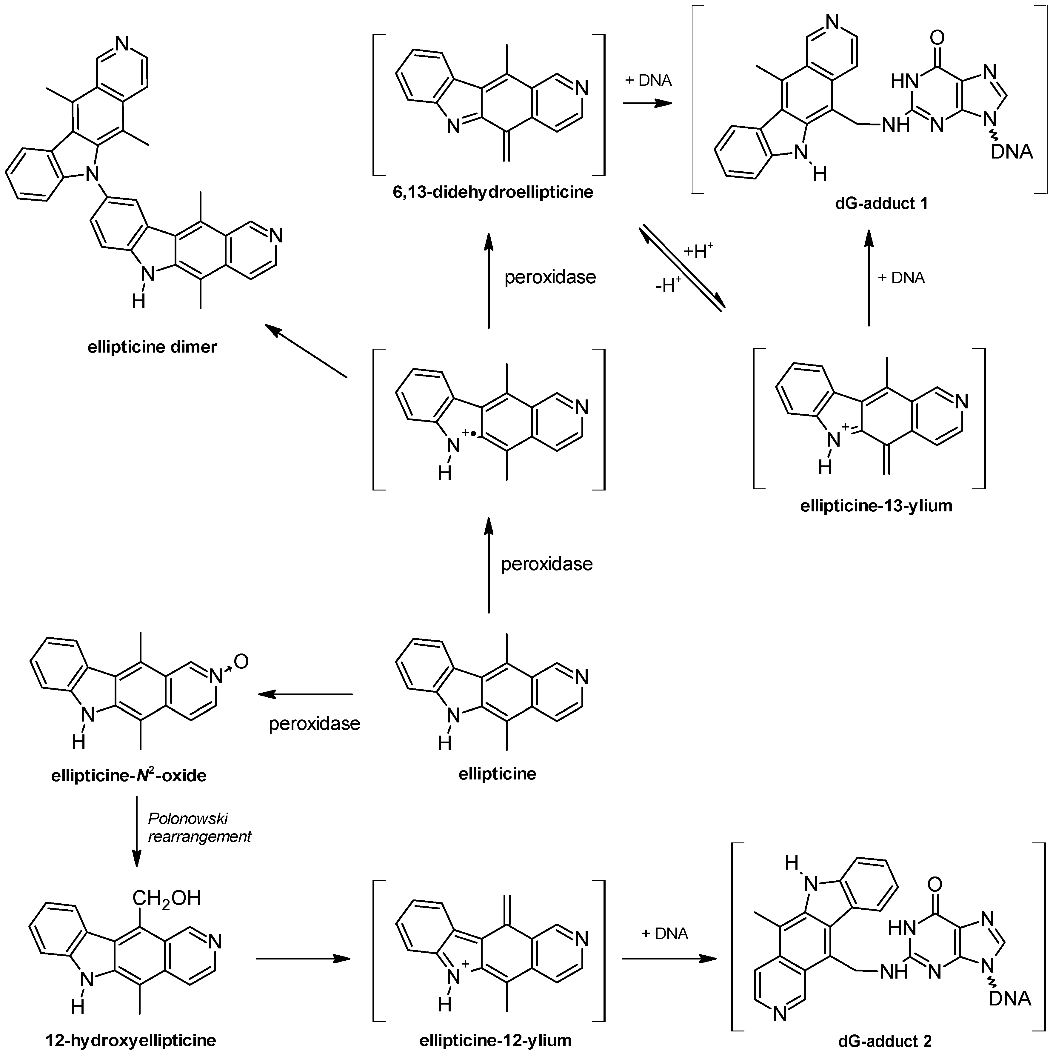
2. DNA-Damaging Mechanisms of Ellipticine Cytotoxicity to Cancer Cells

The most important DNA-damaging mechanisms of ellipticine were considered to be intercalation into DNA [5,23,24,25,26] and inhibition of DNA topoisomerase II activity [5,27,28]. Recently, Andrews and co-workers [29] demonstrated, however, that ellipticine and some of its derivatives are potent and specific inhibitors of RNA polymerase I (Pol-I) transcription and that this Pol-I inhibition occurs by a p53- and topoisomerase II-independent mechanism. They found that the drug influences the assembly and stability of preinitiation complexes by targeting the interaction between promoter recognition essential transcription factor SL1 and the rRNA promoter. In addition, Ghosh et al. [30] showed that along with DNA intercalation and/or topoisomerase II inhibition, interaction with the telomeric DNA region and the resultant inhibition of telomerase activity might be an additional mode of action of ellipticine. Moreover, we showed that this antitumor agent also causes damage to the structural integrity of DNA through covalent binding, by forming covalent DNA adducts after its enzymatic activation with cytochrome P450 (CYP) or peroxidases [1,2,3,4,31,32,33,34,35,36,37,38,39,40]. Cytotoxicity of ellipticine in cells of several cancer lines sensitive to this drug such as HL-60 promyelocytic leukemia, T-cell leukemia CCRF-CEM, glioblastoma U87MG, neuroblastoma UKF-NB-3 and UKF-NB-4, thyroid cancer BHT-101, B-CPAP and 8505-C and breast adenocarcinoma MCF-7 corresponded to levels of ellipticine-derived DNA adducts generated after its enzymatic activation in most of these cell lines [41]. This indicates that covalent binding to DNA of reactive species generated by enzymatic bioactivation of ellipticine is one of the most important mechanisms responsible for ellipticine cytotoxicity in these cancer cells. The formation of ellipticine-DNA adducts ultimately forces cancer cells to initiate cell death signaling [9]. Based on these results, we suggest that ellipticine acts as a prodrug, which is metabolically activated to reactive species forming covalent DNA adducts causing genotoxic stress. Therefore, information on which enzymes are involved in the metabolism of ellipticine is critical to identify the pharmacological effects of ellipticine. Several in vitro and in vivo approaches have been developed to study the role of specific CYP and peroxidase enzymes in ellipticine metabolism.
Over the past 10 years we have gained extensive experience in using the pure enzymes and the various animal models to study the ellipticine metabolism. During these studies, ellipticine was found to be oxidized by CYP and peroxidase enzymes to both electrophilic species forming covalent DNA adducts detected by 32P-postlabeling (Figure 2) and to detoxification metabolites [1,2,3,4,32,33,34,35,36,37,38,39,40,42,43,44,45,46,47,48,49,50,51]. Moreover, we characterized the reactions leading to their formation.
Figure 2. Autoradiographs of thin layer chromatography (TLC) maps of 32P-labeled digests of calf thymus DNA reacted with ellipticine activated by hepatic microsomes from wild-type (WT) mice (A), with those from Hepatic Cytochrome P450 Reductase Null (HRN) mice pre-treated with benzo[a]pyrene (BaP) (B), from calf thymus DNA reacted with 13-hydroxyellipticine (C) [31] or 12-hydroxyellipticine (D) [32] of DNA from livers of WT (E) and HRN (F) mice treated intraperitoneally (i.p.) with 10 mg ellipticine/kg body weight [48] and of DNA from liver of Wistar rats treated i.p. with 40 mg ellipticine per kilogram body weight (G) [33,37]. Analyses were performed by the nuclease P1 version of the 32P-postlabeling assay. Adduct spots 1–7 and A correspond to the ellipticine-derived DNA adducts. Besides adduct 2, another strong adduct (spot X in panel D), which was not found in any other activation systems or in vivo was generated by 12-hydroxyellipticine.
Ijms 16 00284 g002
In this review we focus on comparison between the data found in the in vitro and in vivo studies investigating ellipticine metabolism and show a necessity of both approaches to obtain valid information on CYP enzymes participating in this process.

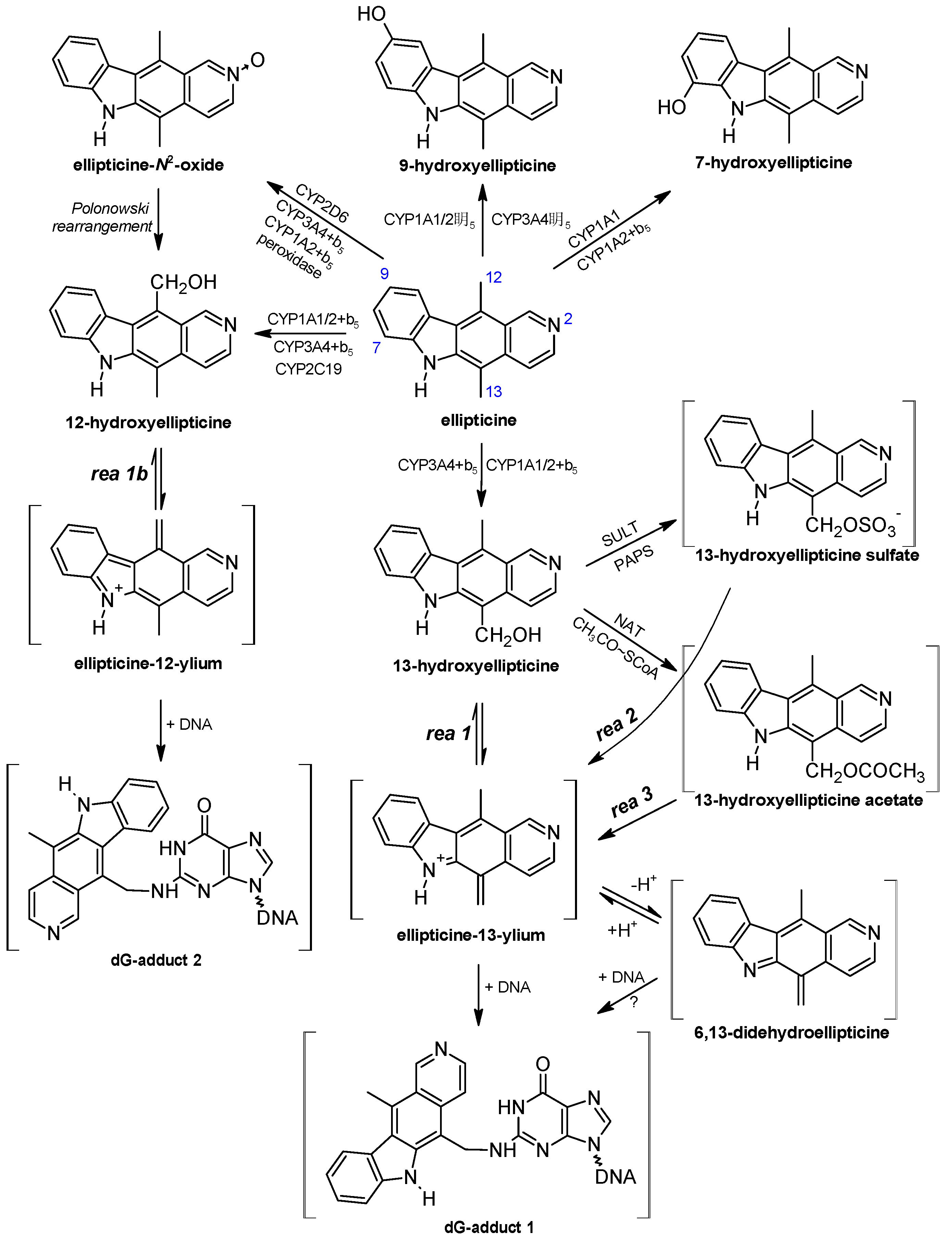
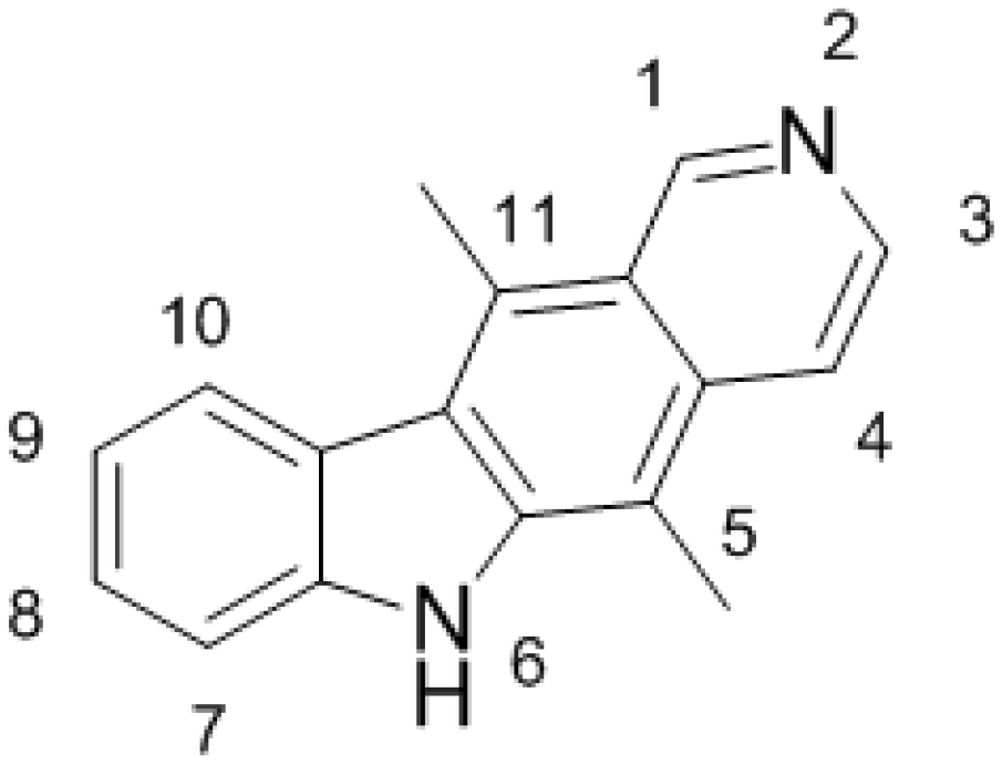
3. Metabolism of Ellipticine by Cytochromes P450 (CYPs), Peroxidases and Conjugation Enzymes in Vitro

Utilizing numerous in vitro systems such as subcellular microsomal fractions and cells in culture expressing CYPs, isolated CYPs reconstituted with other components of the mixed-function-oxidase system [NADPH:CYP reductase (POR), cytochrome b5], and recombinant CYPs, human, rat, rabbit, and mouse CYP enzymes were found to oxidize ellipticine. Ellipticine is oxidized to five metabolites, 7-hydroxy-, 9-hydroxy-, 12-hydroxy-, 13-hydroxyellipticine, and ellipticine N2-oxide (Figure 3), and at least two major ellipticine DNA-adducts were generated by these enzymatic systems [3,31,33,34,35,36,38,39,40,48,49]. 7-Hydroxy- and 9-hydroxyellipticine are efficiently excreted by experimental animals and considered to be the detoxification products of ellipticine [52,53]. But 9-hydroxyellipticine is also an efficient inhibitor of Pol-I transcription in vitro with IC50 values in cells in the nanomolar range [29], intercalates into DNA, and inhibits topoisomerase II activity [54,55,56]. It is therefore a pharmacologically important metabolite. 13-Hydroxy- and 12-hydroxyellipticine are the active metabolites, which spontaneously form ellipticine-13-ylium and ellipticine-12-ylium, which reacts with DNA to produce two major deoxyguanosine adducts (see adduct spots 1 and 2 in Figure 2 and the proposed structures of these DNA adducts in Figure 3) [2,3,4,31,32,34,35,36,39,40,49]. In addition, ellipticine N2-oxide is also considered a potent active ellipticine metabolite, since it converts to 12-hydroxyellipticine [31], by the Polonowski rearrangement [57] (Figure 3). All these results suggest that the enzymes activating or detoxifying ellipticine are crucial for its pharmacological effects. Thus, the identification of enzymes necessary for ellipticine metabolism is of great importance.
Using a variety of human recombinant CYPs, inhibitors of these enzymes in human hepatic microsomes and correlation analyses, the roles of individual CYPs in the formation of ellipticine metabolites was identified [31,34,35,40]. Human recombinant CYP1A1 and 1A2, followed by CYP1B1, are most effective in formation of 7-hydroxy- and 9-hydroxyellipticine detoxifying ellipticine (Figure 3) [31]. The active metabolite, 13-hydroxyellipticine, forming the ellipticine-DNA adduct 1 (Figure 2C), is generated predominantly by CYP3A4. Oxidation of ellipticine to another activation metabolite, 12-hydroxyellipticine, generating DNA adduct 2 (Figure 2D) is also catalyzed by CYP3A4, but more efficiently by CYP2C19. The N2-oxide of ellipticine is generated mainly by CYP2D6 beside CYP3A4 [31,34,35,39]. These results demonstrate that CYP3A4 is the most effective enzyme leading to ellipticine-DNA adducts 1 and 2, while adduct 2 is also generated by CYP2C19 and 2D6. Moreover, orthologous CYP enzymes of rats and mice catalyze formation of these metabolites and DNA adducts [1,2,3,4,36,38]. This indicates that these animals might be suitable models mimicking the fate of ellipticine in human.
Recently, we could show that levels of the DNA adduct formed by 13-hydroxyellipticine increased if this ellipticine metabolite was conjugated with sulfate or acetate by human sulfotransferases 1A1, 1A2, 1A3 and 2A1, or N,O-acetyltransferases 1 and 2 (Figure 3) [34,58].
Besides CYP enzymes, peroxidases are able to oxidize ellipticine to metabolites generating covalent DNA adducts. Human myeloperoxidase, bovine lactoperoxidase, ovine cyclooxygenase (COX)-1, human COX-2 and plant horseradish peroxidase oxidize ellipticine in vitro to form up to four DNA adducts [32]. Even though mechanisms of oxidation of ellipticine by peroxidases and CYPs are different, two of the DNA adducts formed during oxidation of ellipticine by peroxidases are identical to those produced by 13-hydroxy- and 12-hydroxyellipticine generated by CYPs [32]. Ellipticine oxidation to 6,13-didehydroellipticine (the ellipticine methylene-imine) and ellipticine N2-oxide [32,59] explain the mechanisms of peroxidase-mediated formation of DNA adducts identical to those formed by CYPs (Figure 4). The two minor DNA adducts that are formed by peroxidases (spots 6 and 7 in Figure 2) [32], are also generated after ellipticine activation with hepatic microsomes from humans [31,60], rats [1,38,60], rabbits [1,39], and mice [48,61], and in several organs of mice (Figure 2E,F) and rats (Figure 2G) treated with ellipticine [33,37,48].
Figure 3. Ellipticine metabolism by CYPs showing the identified metabolites and those proposed to form DNA adducts. The compounds shown in brackets were not detected under the experimental conditions and/or are not yet structurally characterized. The CYP enzymes predominantly oxidizing ellipticine shown were identified in our previous studies [31,34,35,38,39]. Reactions 1, 2 and 3 lead to ellipticine-13-ylium from 13-hydroxyellipticine, 13-hydroxyellipticine sulfate and 13-hydroxyellipticine acetate, respectively, and rea 1b to ellipticine 12-ylium. ? indicates that the mechanisms of this reaction are not known.
Ijms 16 00284 g003
Figure 4. Oxidation of ellipticine by peroxidases showing the characterized metabolites and those proposed to form DNA adducts. The compounds shown in brackets were not detected under the experimental conditions and are the electrophilic metabolites postulated as ultimate arylating species or the postulated N²-deoxyguanosine adducts (adapted from reference [32]).
Ijms 16 00284 g004

4. Oxidation of Ellipticine by CYP Enzymes in Vivo

In order to extrapolate from the in vitro data to the in vivo situation, additional factors have to be considered such as route of administration, absorption, renal clearance, and tissue-specific expression of enzymes metabolizing ellipticine. To identify CYP enzymes responsible for activation of ellipticine in vivo, several animal models were used: (I) wild-type (WT) and Hepatic P450 Reductase Null (HRN) mice; (II) the same mouse models, in which expression of enzymes of the mixed-function oxidase system was induced by benzo[a]pyrene (BaP); and (III) Wistar rats. The data obtained with these animal models revealed a paradox: namely, that CYP1A enzymes appear to be more important for activation of ellipticine in vivo, despite being involved in its metabolic detoxification in vitro.

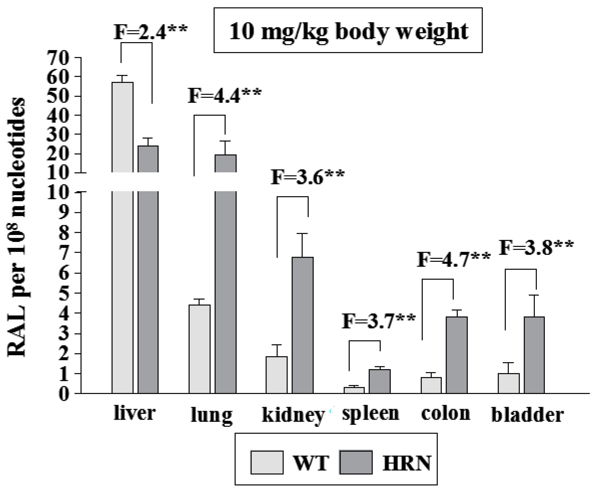
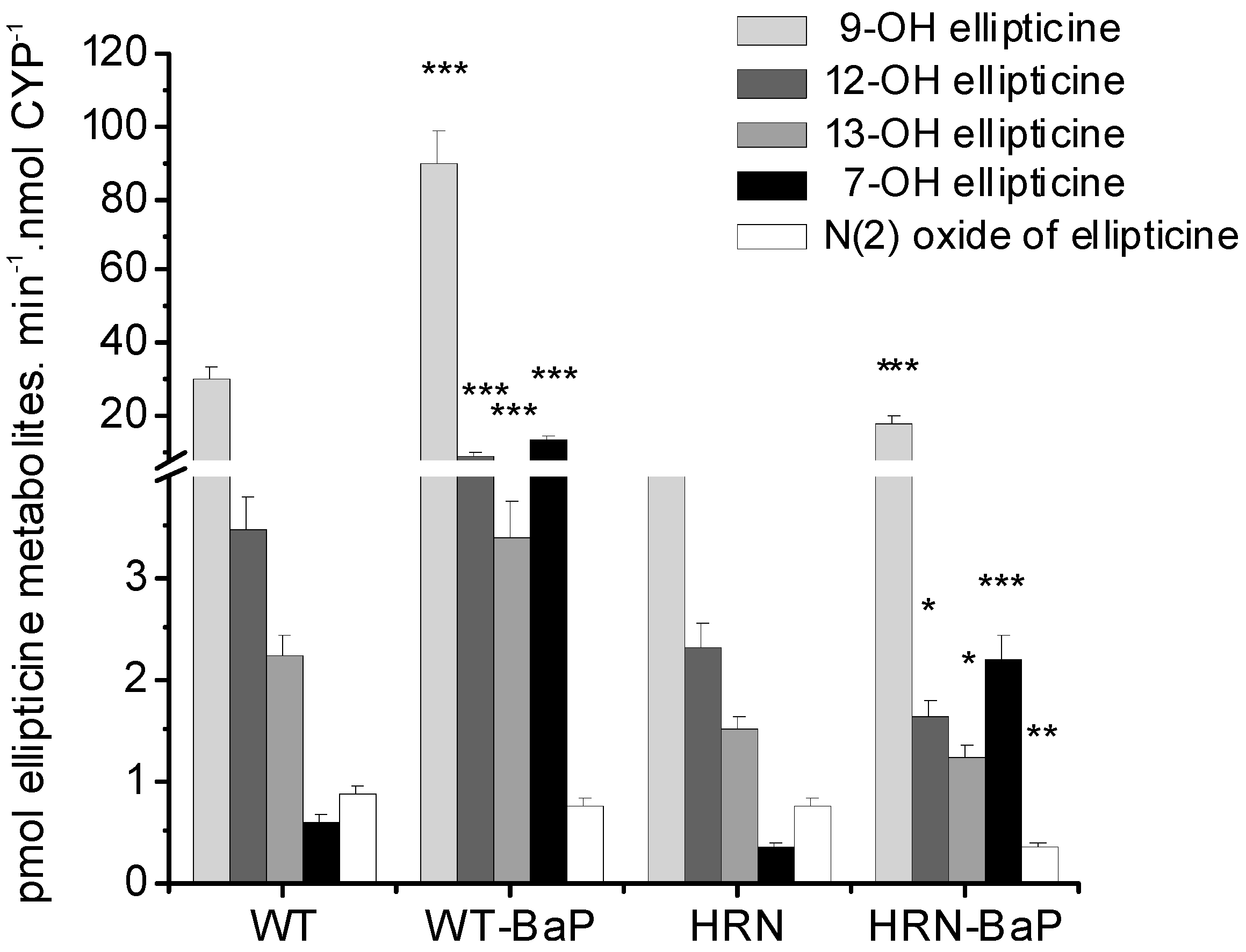
4.1. Utilization of Wild-Type (WT) and Hepatic P450 Reductase Null (HRN) Mice to Identify Enzymes Metabolizing Ellipticine in Vivo

In HRN mice, POR, the most important electron donor to mouse CYPs, is deleted specifically in hepatocytes. This model was developed by Henderson et al. [62] to evaluate the role of both hepatic POR and CYPs in xenobiotic metabolism. Deletion of this enzyme results not only in the loss of essentially all hepatic CYP function, but also in the lack of direct reduction of xenobiotics by POR, an additional property of this enzyme. This mouse model has been used successfully to investigate the role of hepatic versus extra-hepatic CYP-catalyzed metabolism and the disposition of several carcinogens and drugs including ellipticine [48,62,63,64,65,66,67,68,69].
Using WT and HRN mouse lines, hepatic CYPs were demonstrated to be important in ellipticine-derived DNA adduct formation also in vivo, because up to seven ellipticine-specific DNA adducts were observed in liver, lung, kidney, spleen, colon and bladder (see Figure 2E,F for liver of WT and HRN mice). Deoxyguanosine adduct spots 1 and 2 derived from 13-hydroxy- and 12-hydroxyellipticine, respectively (Figure 2 and Figure 3), were the predominant adducts in all mouse tissues examined.
The finding that ellipticine-DNA adducts are formed in all organs tested in these animals suggest that ellipticine or its metabolites are distributed via the blood stream to different organs and that these tissues may have the metabolic capacity to oxidatively activate ellipticine. As found by Chadwick and co-workers [52,53] ellipticine is very rapidly distributed from the blood, and its excretion is essentially complete by 24 h in several species including mice, rats, dogs, and monkeys. The rate of ellipticine elimination from blood was found to reflect the rate of metabolism of this drug [52]. The main organ responsible for its biotransformation was found to be the liver, forming predominantly 9-hydroxyellipticine, which is excreted mainly in bile as its glucuronide or sulfate conjugate [52,53]. Other in vivo pathways involving hydroxylation at as yet unknown positions in the molecule have also been found [52,53]. As mentioned above, in in vitro experiments, ellipticine is oxidized by CYPs in hepatic microsomes from a variety of species, including humans, rats, rabbits and mice [31,39,48,49,60,61] to several hydroxylated derivatives, with 9-hydroxy-, 12-hydroxy- and 13-hydroxyellipticine as the major metabolites in most species. However, because 13-hydroxy- and 12-hydroxyellipticine are reactive and have been found to form the two major ellipticine-DNA adducts [31,32,39,47], they will not be easily detectable in vivo. In addition, in these early studies radioactively labelled ellipticine was found to be deposited in a number of organs with the highest levels in the liver, followed by kidney, lung, intestine and spleen, and was located primarily in the nuclear fraction [52]. Our more recent data would suggest that covalent binding of ellipticine to DNA can explain this localization [32,33,48].
These tissues therefore have the metabolic capacity to oxidize ellipticine and, more importantly, the same reactive species forming DNA adducts are produced as in the liver, probably by both CYP catalysis and maybe peroxidases. The levels of ellipticine-DNA adducts in the livers of HRN mice were lower (by up to 65%) than those in WT mice, demonstrating that CYP enzyme activity is important for the oxidative activation of ellipticine to metabolites generating these adducts. Whereas hepatic CYP-mediated ellipticine DNA binding was reduced in HRN mice, adduct levels in extrahepatic organs were up to 4.7-fold higher (Figure 5).
Figure 5. Total levels of ellipticine-DNA adducts determined and quantified by 32P-postlabelling analysis of DNA isolated from organs of HRN and WT mice treated i.p. with 10 mg ellipticine/kg body weight. F = fold higher and/or lower DNA adducts in HRN than WT mice. Columns, mean; bars, SD (n = 3); each DNA sample was analysed twice. ** p < 0.01. RAL, relative adduct labeling.
Ijms 16 00284 g005
Experiments utilizing ex vivo incubations of ellipticine with hepatic microsomes of WT and HRN mice and those of these mice exposed to BaP, as well as employing inhibitors of the most important CYP enzymes catalyzing detoxification and activation of ellipticine in vitro, CYP1A and 3A, respectively, helped to resolve which of these CYPs play a role in the mouse models. As expected, treatment of WT and HRN mice with BaP significantly induced expression of CYP1A, predominantly of CYP1A1 in liver (up to 175-fold), both at the transcriptional and translation levels [61,65]. This carcinogen also increased the expression of POR protein and its enzymatic activity in livers of these mouse models, but to a much lower extent, up to 2.9-fold. More interestingly, exposure of WT and HRN mice to BaP was also found to result in an increased expression of cytochrome b5, a protein of the microsomal mixed-function-oxidase system, in livers of these mice [66].
In the presence of NADPH, a cofactor of POR- and CYP-dependent enzyme systems, the ex vivo incubations with ellipticine, DNA and hepatic microsomes of untreated (control) WT and HRN mice and mice treated with BaP led to activation of this drug to ellipticine-derived DNA adducts (Figure 2A,B and Figure 6), confirming the role of CYPs in ellipticine activation. Arachidonic acid, a cofactor for COX-dependent oxidation [48,64,70,71,72], also mediated formation of ellipticine-DNA adducts 1 and 2 in hepatic microsomes of all mice used. This suggests that COX also activates ellipticine in mouse liver [48,61], but arachidonic acid as a cofactor was much less effective than NADPH (Figure 6).
Figure 6. DNA adduct formation by ellipticine activated with microsomes isolated from livers of untreated Hepatic Cytochrome P450 Reductase Null (HRN) or wild-type (WT) mice (A) and from mice treated with BaP (B) as determined by 32P-postlabeling. F = fold higher DNA adducts levels in microsomes from WT mice compared to HRN mice. Columns: Mean RAL (relative adduct labeling) ± standard deviations (SD) shown represents total levels of DNA adducts of four determinations (duplicate analyses of two independent in vitro incubations). Values significantly different from HRN mice: * p < 0.05, ** p < 0.01, *** p < 0.001. Control = without cofactor; AA = arachidonic acid; α-NF = α-naphthoflavone; α-LA = α-lipoic acid.
Ijms 16 00284 g006
Surprisingly, levels of ellipticine-derived DNA adducts formed in the ex vivo incubations of HRN mice liver microsomes with NADPH were only 1.4-fold lower than amounts formed by hepatic microsomes from WT mice (Figure 6), even though POR expression in livers of HRN mice was two orders of magnitude lower. This finding indicates that ellipticine activation should be at least partially catalyzed also by enzymes with POR-independent activity [48]. Beside peroxidases that were found to activate ellipticine [32], the CYP2S1 enzyme, which is abundantly expressed in several tissues [73,74,75,76], might be such an enzyme, because it catalyzes the oxidation of compounds having polycyclic aromatic structures similar to ellipticine without participation of POR [75,76]. Whereas a role of a COX peroxidase in hepatic microsomes of WT and HRN mice was proven (see Figure 6) [48,61], the participation of CYP2S1 in ellipticine activation awaits further examination. Therefore, the human recombinant CYP2S1 enzyme heterologously expressed in Escherichia coli was prepared in our laboratory [77] and will be utilized to investigate efficiency of this CYP in ellipticine oxidation in an additional study.
At least two adducts (spots 1 and 2 in Figure 2A,B), which were identical to those generated in vivo in mice treated with ellipticine (Figure 2E,F) were formed by mouse hepatic microsomes. Furthermore, ellipticine-derived DNA adduct, spot A, was found as a minor adduct (Figure 2A), predominantly in microsomes isolated from HRN mice [48]. In incubations containing hepatic microsomes of WT and HRN mice treated with BaP, an additional adduct spot, corresponding to the 10-(deoxyguanosin-N2-yl)-7,8,9-trihydroxy-7,8,9,10-tetrahydrobenzo[a]pyrene (dG-N2-BPDE) adduct of BaP-7,8-dihydrodiol-9,10-epoxide with DNA in vitro and in vivo [65,78] was also detected (Figure 2B). This finding indicates that residual BaP is present in microsomes isolated from livers of WT and HRN mice treated with BaP, and is activated by CYP1A1 in combination with microsomal epoxide hydrolase to form this adduct.
Ketoconazole, a selective inhibitor of CYP3A [79,80], inhibited formation of ellipticine-DNA adducts in hepatic microsomes of untreated (control) WT and HRN mice, by ~60% (Figure 6A), confirming a role of CYP3A in ellipticine activation in mouse liver. However, the effect of this inhibitor was much lower in hepatic microsomes of BaP-treated WT and HRN mice, only by ~10% (Figure 6B). In mice exposed to BaP the contribution of CYP3A is much lower, because of the massive CYP1A induction by BaP. Surprisingly, this increased level of CYP1A, the enzymes that mainly detoxify ellipticine in vitro, led to higher amounts of ellipticine-DNA adducts formed (Figure 6), predominantly of adduct 1 [61]. Moreover, a selective inhibitor of CYP1A activities, α-naphthoflavone (α-NF) [79], inhibited formation of ellipticine-DNA adducts in all mice except HRN mice exposed to BaP (Figure 6). Induction of CYP1A in HRN mice by BaP also resulted in increased levels of ellipticine-DNA adducts, but α-NF caused an increase rather than a decrease in formation of ellipticine-DNA adducts (Figure 6). Therefore, here the BaP induced CYP1A enzymes seem to increase ellipticine detoxification.
Ellipticine metabolites formed in hepatic microsomes from all mouse lines used in previous studies [48,61] were analogous; 9-hydroxy-, 12-hydroxy-, 13-hydroxy, 7-hydroxyellipticine and N2-oxide of ellipticine were formed (Figure 7). However, the patterns of individual metabolites in WT and HRN mice, either control (untreated) or treated with BaP, were different. In incubations with HRN microsomes from untreated mice, 9-hydroxyellipticine levels were only one sixth, while the amounts of 13-hydroxy- and 12-hydroxyellipticine, were about one half of the levels in incubations with WT microsomes.
Figure 7. Levels of ellipticine metabolites formed by hepatic microsomes (0.2 mg protein) of Hepatic Cytochrome P450 Reductase Null (HRN) and wild-type (WT) mice from 10 μM ellipticine and by hepatic microsomes of HRN and WT mice pre-treated with BaP. Levels of ellipticine metabolites were determined by high performance liquid chromatography (HPLC) [31,60] and are averages ± standard deviations of triplicate incubations. Values significantly different from untreated mice: * p < 0.05, ** p < 0.01, *** p < 0.001.
Ijms 16 00284 g007
Exposure of mice to BaP induced CYP1A and resulted, as expected, in an increase in formation of 9-hydroxy- and 7-hydroxyellipticine (Figure 7). This result is consistent with previous studies where CYP1A1 and 1A2 were the major enzymes forming these metabolites [31,35,40]. Treatment of WT mice with BaP, however, also resulted in up to 2.5-fold higher levels of 13-hydroxy- and 12-hydroxyellipticine (Figure 7), the metabolites that were found to be formed in vitro mainly by CYP3A, and much less efficiently by CYP1A1 [31,39]. Hence not only CYP3A, but also CYP1A expressed in mouse liver are important for activation of ellipticine to ellipticine-DNA adducts in WT mice only, while in HRN mice the detoxification only is induced by BaP explaining the DNA adduct levels. These findings show that in contrast to the pure in vitro CYP systems (CYPs reconstituted with POR), CYP1A enzymes are responsible for ellipticine activation to form DNA adducts in mouse liver [48,61].

4.2. Ellipticine Metabolism in Wistar Rats

In order to further identify CYP enzymes responsible for activation and detoxification of ellipticine in vivo, Wistar rats were used. Also in this animal model, ellipticine treatment resulted in ellipticine-derived DNA adduct generation in several healthy organs (liver, kidney, lung, spleen, breast, heart and brain) (see Figure 2G for rat liver) [3,33,37] and in DNA of mammary adenocarcinoma [3]. The levels of ellipticine-derived DNA adducts generated in these adenocarcinomas were almost 2-fold higher than in normal healthy mammary tissue. This finding indicates that other CYPs such as CYP1B1, able to activate ellipticine may be expressed at higher levels in this adenocarcinoma [41] than in peritumoral tissues. Indeed, we [41], and others [81,82,83] previously showed CYP1B1 to be a typical CYP expressed in breast cancer.
Genotoxic side effects of ellipticine in healthy organs of experimental animals in vivo may of course also be determined by expression levels of CYP enzymes activating ellipticine in these tissues [33,37,48]. Indeed, several studies have found a positive correlation between DNA adduct levels of carcinogens or genotoxic agents, their persistence and their mutagenicity and/or tumorigenicity [84,85,86,87,88]. To better understand the role of ellipticine-DNA adducts in genotoxic side effects in healthy tissues, we have analyzed the persistence of ellipticine-DNA adducts in liver, lung, kidney, spleen, heart, and brain of rats to model the bioactivation of ellipticine in humans treated with ellipticine [33]. Only very low levels of adducts persisted only in some tissues. In addition, not all ellipticine-DNA adducts persist in the tissues analyzed in the study (only adducts 1, 2, 4, and 5) [33]. This finding demonstrates that healthy tissues of rats treated with ellipticine possess effective DNA repair systems to remove certain lesions and suggests a relatively low impact of the genotoxic side effects of ellipticine during cancer treatment in humans.
Also in rats, formation of ellipticine-DNA adduct 1 is dependent not only on levels of CYP3A, but also on those of CYP1A1. The levels of ellipticine DNA adduct 1 in analyzed organs correlated not only with expression levels of CYP3A, but also with those of CYP1A1 in the same organs, which again does not correspond to the situation in vitro. As outlined above, in the in vitro systems, CYP3A is mainly responsible for formation of ellipticine-DNA adducts and CYP1A predominantly oxidizes ellipticine to its detoxification metabolites [3,4,36]. In-vitro studies investigating the effect of cytochrome b5 on the metabolism of ellipticine explained the discrepancies between CYP oxidation of ellipticine in vitro and in vivo, because this protein has a crucial role in directing individual CYP enzymes to ellipticine activation or detoxification. Moreover, the induced expression of cytochrome b5 protein in liver of rats treated with ellipticine [35,51] suggests that cytochrome b5 may modulate the CYP-mediated bioactivation and detoxification of ellipticine in this animal model in vivo as well.
Cytochrome b5 is an important component of the microsomal mixed-function-oxidase system and can influence the metabolism of xenobiotics [69,78,89,90,91,92,93,94,95]. For more than four decades, the role of cytochrome b5 in CYP catalysis has been controversial, and based entirely on in vitro data, which showed that cytochrome b5 could inhibit or stimulate CYP activity depending on a number of variables including CYP isoenzyme, substrate and cytochrome b5 concentration [89,90,91,92,93,94]. In order to investigate the role of cytochrome b5 in ellipticine metabolism we conducted some in vitro experiments using human liver microsomes, hepatic microsomes from control and ellipticine-pretreated rats and reconstituted systems with human CYP1A1, CYP1A2, CYP3A4, POR, and cytochrome b5 in different ratios [34,35,39,49,51,96]. We found that cytochrome b5 alters the ratio of ellipticine metabolites generated by CYP1A1, 1A2, and 3A4. Whereas the amounts of the detoxification metabolites (7-hydroxy- and 9-hydroxyellipticine) are either decreased (CYP1A1/2) or not changed (CYP3A4) with cytochrome b5 added to the reconstituted system, the amounts of the active metabolites, 12-hydroxy- and 13-hydroxyellipticine, increased considerably, leading to higher ellipticine-DNA adduct levels [34,35,39].

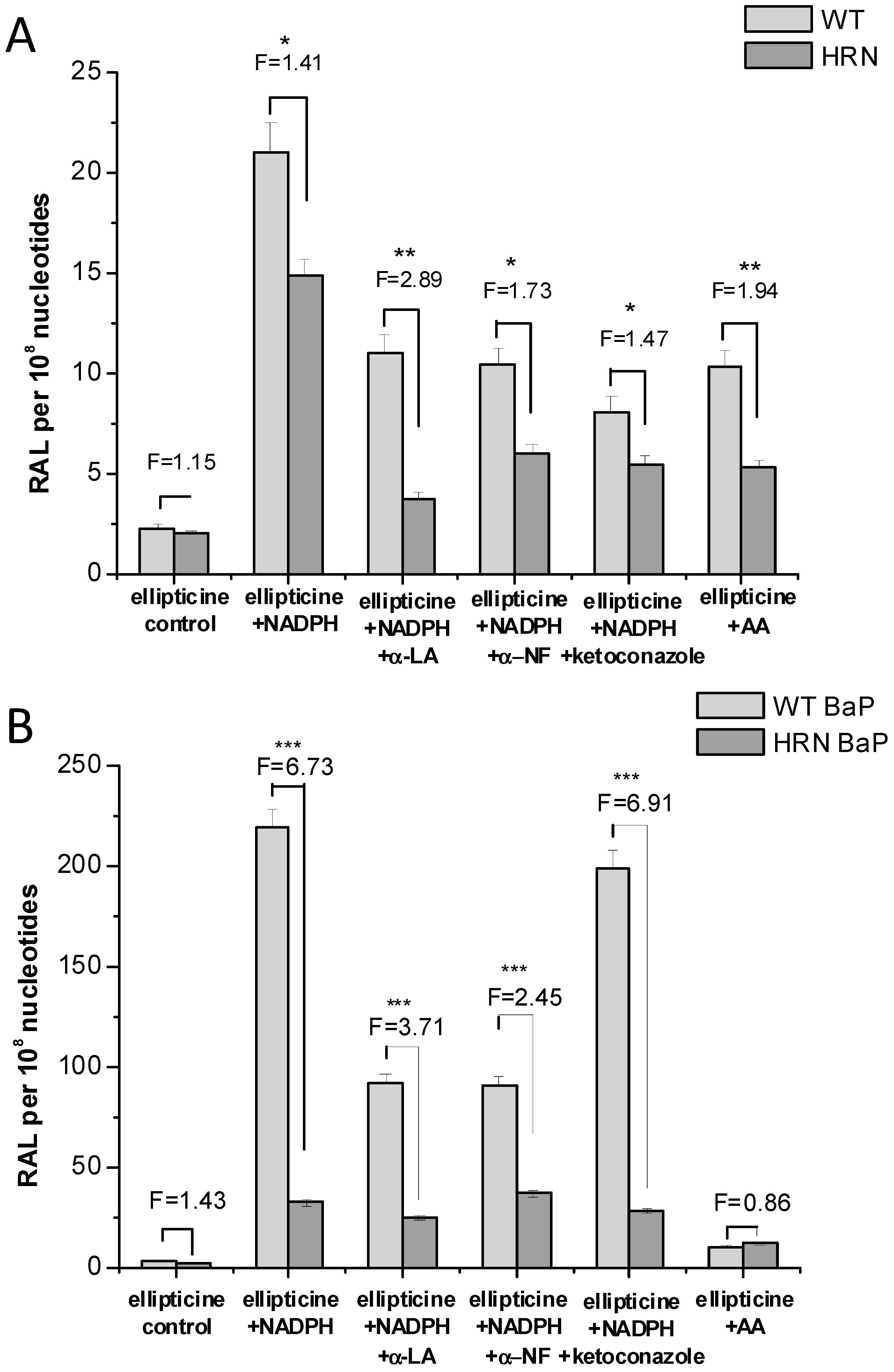
4.3. Studies with Human Microsomes and Cancer Cells

Because in the studies described above with isolated enzymes the ratios of the various partners of CYP are generated experimentally, a more physiological model to identify human enzymes responsible for ellipticine activation was used, namely, human hepatic microsomes [31,39]. These microsomal fractions contain a mixture of human CYPs, POR, cytochrome b5 and its reductase (NADH:cytochrome b5 reductase). Thus, they comprise the essential components of the enzymatic system metabolizing drugs, mimicking well a situation in human liver, where a majority of drug metabolism occurs [78,95]. Human hepatic microsomes oxidize ellipticine mainly to 13-hydroxy- and 12-hydroxyellipticine, whereas 7-hydroxy-, 9-hydroxyellipticine and ellipticine N2-oxide are generated at more than 10-fold lower amounts (see table 2 in [39]). Similar results were found in hepatic microsomes of rats [60]. As a consequence, high levels of both major ellipticine-DNA adducts are formed when DNA is added to the microsomal incubations. The amounts formed correlated with the activity of the major CYPs found previously to form the metabolites generating these DNA adducts (see above) [31,39].
All these results explained why the CYP1A enzymes are more important in ellipticine activation in vivo; their activity is modulated by cytochrome b5. These results also demonstrated that not only the expression levels of CYP1A and 3A in several species including human, but also the amounts of expressed cytochrome b5 dictate the oxidative activation and detoxification of ellipticine in vivo. Moreover, because ellipticine itself is capable of inducing expression of CYP1A (predominantly CYP1A1), POR, and cytochrome b5 [51,96], it increases its own metabolism leading predominantly to activation of this drug to reactive species forming DNA adducts [34,35], thereby modulating its own pharmacological potential.
The ellipticine-derived DNA adducts formed by enzymatic activation of ellipticine in vitro and in vivo were also found in human tumor cells, in which CYP enzymes are expressed. The adducts were found in human adenocarcinoma MCF-7 [41], neuroblastoma IMR-32, UKF-NB-3, and UKF-NB-4 [43,44], glioblastoma U87MG [46], and BHT-101, B-CPAP and 8505-C thyroid cancer cells [45] exposed to ellipticine. Cytotoxicity of ellipticine corresponded to the amounts of ellipticine-DNA adducts formed in the specific cancer cells and depended on expression levels of CYP enzymes metabolizing ellipticine (CYP1A1, 1B1, and 3A4) and/or cytochrome b5 in these cells [36,41,43,44,45,46]. High expression levels of cytochrome b5 together with those of CYP1A1 and 3A4 lead to more ellipticine-DNA adducts and higher cytotoxicity of ellipticine predominantly in neuroblastoma UKF-NB-4, glioblastoma U87MG and thyroid cancer cells [14,36,41,43,44,45]. These findings again demonstrate the importance of expression of CYPs, but also of cytochrome b5, in the tumor and normal tissue, because the ratios of these enzymes determine the pharmacological effects of ellipticine.

5. Conclusions

The data summarized in this review demonstrate that the DNA-damaging anticancer alkaloid ellipticine might be considered a prodrug, whose major mechanism of action is mediated by an enzymatic activation leading to formation of covalent DNA adducts in target tissues. These ellipticine-DNA adducts are formed in both healthy and tumor tissues and cells, but they do not persist in healthy tissues. The data show that cytotoxic effects of ellipticine in tumor tissues are dictated by (I) levels of CYP expression (and/or peroxidase expression); (II) levels of cytochrome b5 expression; and (III) its own potency to induce CYP1A1, CYP3A and cytochrome b5 in tumor tissues and cells. The results also demonstrate that animal models, where levels of biotransformation enzymes either knocked out or induced, are appropriate tools to identify enzymes responsible for the metabolic activation and detoxification of ellipticine. They also demonstrate that extrapolation from in vitro data to the situation in vivo is not always possible, confirming the need for these animal models.
Even though the role of cytochrome b5 in modulation of ellipticine metabolism in vitro was clearly shown, its effect in vivo is still quite enigmatic. Two mouse lines, one with a conditional hepatic deletion of cytochrome b5 (HBN, Hepatic cytochrome b5 Null) [97] and a double conditional mutant, HBRN (Hepatic cytochrome b5/P450 Reductase Null), in which both enzymes are deleted specifically in the liver [98], which were recently developed in the laboratory of Wolf and coworkers [97,98], may help to resolve the role of cytochrome b5 in CYP-mediated metabolism of ellipticine in vivo.
The results summarized in this review form the basis to further predict the susceptibility of human cancers to ellipticine and suggest this alkaloid for treatment in combination with CYP gene transfer (CYP-gene-directed enzyme-prodrug therapy) [99,100], which has the potential to provide efficient activation of ellipticine in target tumor tissue, thereby increasing the anticancer potential of this prodrug. Furthermore, two of the ellipticine metabolites formed by oxidation with CYPs in combination with cytochrome b5, 13-hydroxy- and 12-hydroxyellipticine, are reactive enough to decompose spontaneously to the carbenium ions forming DNA adducts that are predominantly responsible for killing cancer cells. Both these ellipticine metabolites are, therefore, excellent candidates for tumor-specific targeting by appropriate derivatives, including their encapsulated forms into nanocarriers. Research into such targeted carrier systems for active ellipticine metabolites is a major research aim on the path to clinical application of ellipticine in tumor therapy.

Acknowledgments

This work was supported by the Grant Agency of the Czech Republic (grant 14-18344S in panel P301) and Charles University (UNCE204025/2012). Work at King’s College London was supported by the Cancer Research UK and the Wellcome Trust. Volker M Arlt is a member of the Wellcome-funded COMSIG (Causes of Mutational SIGnatures) consortium.

Author Contributions

Conception and design: Marie Stiborová, Věra Černá, Michaela Moserová, Iveta Mrízová, Eva Frei. Analysis and interpretation of the data: Marie Stiborová, Volker M. Arlt, Eva Frei. Drafting of the article: Marie Stiborová, Eva Frei. Critical revision of the article for important intellectual content: Marie Stiborová, Volker M. Arlt, Eva Frei.

Conflicts of Interest

The authors declare no conflict of interest.

References

  1. Stiborova, M.; Bieler, C.A.; Wiessler, M.; Frei, E. The anticancer agent ellipticine on activation by cytochrome P450 forms covalent DNA adducts. Biochem. Pharmacol. 2001, 62, 1675–1684. [Google Scholar] [CrossRef] [PubMed]
  2. Stiborova, M.; Rupertova, M.; Schmeiser, H.H.; Frei, E. Molecular mechanisms of antineoplastic action of an anticancer drug ellipticine. Biomed. Pap. Med. Fac. Univ. Palacky Olomouc Czech Repub. 2006, 150, 13–23. [Google Scholar] [CrossRef] [PubMed]
  3. Stiborova, M.; Rupertova, M.; Frei, E. Cytochrome P450- and peroxidase-mediated oxidation of anticancer alkaloid ellipticine dictates its anti-tumor efficiency. Biochim. Biophys. Acta 2011, 1814, 175–185. [Google Scholar] [CrossRef] [PubMed]
  4. Kizek, R.; Adam, V.; Hrabeta, J.; Eckschlager, T.; Smutny, S.; Burda, J.V.; Frei, E.; Stiborova, M. Anthracyclines and ellipticines as DNA-damaging anticancer drugs: Recent advances. Pharmacol. Ther. 2012, 133, 26–39. [Google Scholar] [CrossRef] [PubMed]
  5. Auclair, C. Multimodal action of antitumor agents on DNA: The ellipticine series. Arch. Biochem. Biophys. 1987, 259, 1–14. [Google Scholar] [CrossRef] [PubMed]
  6. Garbett, N.C.; Graves, D.E. Extending nature’s leads: The anticancer agent ellipticine. Curr. Med. Chem. Anti-Cancer Agents 2004, 4, 149–172. [Google Scholar] [CrossRef]
  7. Kuo, P.L.; Hsu, Y.L.; Chang, C.H.; Lin, C.C. The antiproliferative inhibition of ellipticine in human breast mda-mb-231 cancer cells is through cell cycle arrest and apoptosis induction. Anti-Cancer Drugs 2005, 16, 789–795. [Google Scholar] [CrossRef] [PubMed]
  8. Russell, E.G.; O’Sullivan, E.C.; Miller, C.M.; Stanicka, J.; McCarthy, F.O.; Cotter, T.G. Ellipticine derivative induces potent cytostatic effect in acute myeloid leukaemia cells. Investig. New Drugs 2014, 32, 1113–1122. [Google Scholar] [CrossRef]
  9. Kim, J.Y.; Lee, S.G.; Chung, J.Y.; Kim, Y.J.; Park, J.E.; Koh, H.; Han, M.S.; Park, Y.C.; Yoo, Y.H.; Kim, J.M. Ellipticine induces apoptosis in human endometrial cancer cells: The potential involvement of reactive oxygen species and mitogen-activated protein kinases. Toxicology 2011, 289, 91–102. [Google Scholar] [CrossRef] [PubMed]
  10. Kuo, P.L.; Hsu, Y.L.; Chang, C.H.; Lin, C.C. The mechanism of ellipticine-induced apoptosis and cell cycle arrest in human breast MCF-7 cancer cells. Cancer Lett. 2005, 223, 293–301. [Google Scholar] [CrossRef] [PubMed]
  11. Ohashi, M.; Sugikawa, E.; Nakanishi, N. Inhibition of p53 protein phosphorylation by 9-hydroxyellipticine: A possible anticancer mechanism. Jpn. J. Cancer Res. 1995, 86, 819–829. [Google Scholar] [CrossRef] [PubMed]
  12. Shi, L.M.; Myers, T.G.; Fan, Y.; O’Conno, R.P.M.; Paull, K.D.; Friend, S.H.; Weinstein, J.N. Mining the National Cancer Institute Anticancer Drug Discovery Database: Cluster analysis of ellipticine analogs with p53-inverse and central nervous system-selective patterns of activity. Mol. Pharmacol. 1998, 53, 241–251. [Google Scholar] [PubMed]
  13. Kuo, P.L.; Kuo, Y.C.; Hsu, Y.L.; Cho, C.Y.; Lin, C.C. Ellipticine induced apoptosis through p53-dependent pathway in human hepatocellular carcinoma HepG2 cells. Life Sci. 2006, 78, 2550–2557. [Google Scholar]
  14. Martinkova, E.; Maglott, A.; Leger, D.Y.; Bonnet, D.; Stiborova, M.; Takeda, K.; Martin, S.; Dontenwill, M. α5β1 integrin antagonists reduce chemotherapy-induced premature senescence and facilitate apoptosis in human glioblastoma cells. Int. J. Cancer 2010, 127, 1240–1248. [Google Scholar] [CrossRef] [PubMed]
  15. Sugikawa, E.; Hosoi, T.; Yazaki, N.; Gamanuma, N.; Nakanishi, N.; Ohashi, M. Mutant p53 mediated induction of cell cycle arrest and apoptosis at G1 phase by 9-hydroxyellipticine. Anticancer Res. 1999, 19, 3099–3108. [Google Scholar] [PubMed]
  16. Peng, Y.; Li, C.; Chen, L.; Sebti, S.; Chen, J. Rescue of mutant p53 transcription function by ellipticine. Oncogene 2003, 22, 4478–4487. [Google Scholar] [CrossRef] [PubMed]
  17. Savorani, C.; Manfé, V.; Biskup, E.; Gniadecki, R. Ellipticine induces apoptosis in T-cell lymphoma via oxidative DNA damage. Leuk. Lymphoma 2014, 4, 1–9. [Google Scholar] [CrossRef]
  18. Fang, K.; Chen, S.P.; Lin, C.W.; Cheng, W.C.; Huang, H.T. Ellipticine-induced apoptosis depends on Akt translocation and signaling in lung epithelial cancer cells. Lung Cancer 2009, 63, 227–234. [Google Scholar] [CrossRef]
  19. Wang, F.; Liu, J.; Robbins, D.; Morris, K.; Sit, A.; Liu, Y.Y.; Zhao, Y. Mutant p53 exhibits trivial effects on mitochondrial functions which can be reactivated by ellipticine in lymphoma cells. Apoptosis 2011, 16, 301–310. [Google Scholar] [CrossRef]
  20. Miller, C.M.; McCarthy, F.O. Isolation, biological activity and synthesis of the natural product ellipticine and related pyridocarbazoles. RSC Adv. 2012, 2, 8883–8918. [Google Scholar] [CrossRef]
  21. Fritsche, M.; Haessler, C.; Brandner, G. Induction of nuclear accumulation of the tumor-suppressor protein p53 by DNA-damaging agents. Oncogene 1993, 8, 307–318. [Google Scholar] [PubMed]
  22. Xu, G.W.; Mawji, I.A.; Macrae, C.J.; Koch, C.A.; Datti, A.; Wrana, J.L.; Dennis, J.W.; Schimmer, A.D. A high-content chemical screen identifies ellipticine as a modulator of p53 nuclear localization. Apoptosis 2008, 13, 413–422. [Google Scholar] [CrossRef] [PubMed]
  23. Kohn, K.W.; Waring, M.J.; Glaubiger, D.; Friedman, C.A. Intercalative binding of ellipticine to DNA. Cancer Res. 1975, 35, 71–76. [Google Scholar] [PubMed]
  24. Patel, N.; Bergman, J.; Gräslund, A. 1H-NMR studies of the interaction between a self-complementary deoxyoligonucleotide duplex and indolo[2,3-b]quinoxaline derivatives active against herpes virus. Eur. J. Biochem. 1991, 197, 597–604. [Google Scholar] [PubMed]
  25. Chu, Y.; Hsu, M.T. Ellipticine increases the superhelical density of intracellular SV40 DNA by intercalation. Nucleic Acids Res. 1992, 20, 4033–4038. [Google Scholar] [CrossRef] [PubMed]
  26. Singh, M.P.; Hill, G.C.; Peoch, D.; Rayner, B.; Inabach, J.L.; Lown, J.W. High-field NMR and restrained molecular modeling studies on a DNA heteroduplex containing a modified apurinic abasic site in the form of covalently linked 9-aminoellipticine. Biochemistry 1994, 33, 10271–10285. [Google Scholar] [CrossRef]
  27. Belehradek, J., Jr.; Fermandjian, S. DNA-drug recognition and effects on topoisomerase II-mediated cytotoxicity. A three-mode binding model for ellipticine derivatives. J. Biol. Chem. 1991, 266, 1820–1829. [Google Scholar] [PubMed]
  28. Froelich-Ammon, S.J.; Patchan, M.W.; Osheroff, N.; Thompson, R.B. Topoisomerase II binds to ellipticine in the absence or presence of DNA. Characterization of enzyme-drug interactions by fluorescence spectroscopy. J. Biol. Chem. 1995, 270, 14998–5004. [Google Scholar] [CrossRef] [PubMed]
  29. Andrews, W.J.; Panova, T.; Normand, C.; Gadal, O.; Tikhonova, I.G.; Panov, K.I. Old drug, new target: Ellipticines selectively inhibit RNA polymerase I transcription. J. Biol. Chem. 2013, 288, 4567–4582. [Google Scholar] [CrossRef] [PubMed]
  30. Ghosh, S.; Kar, A.; Chowdhury, S.; Dasgupta, D. Ellipticine binds to a human telomere sequence: An additional mode of action as a putative anticancer agent? Biochemistry 2013, 52, 4127–4137. [Google Scholar] [PubMed]
  31. Stiborová, M.; Sejbal, J.; Borek-Dohalská, L.; Aimová, D.; Poljaková, J.; Forsterová, K.; Rupertová, M.; Wiesner, J.; Hudecek, J.; Wiessler, M.; et al. The anticancer drug ellipticine forms covalent DNA adducts, mediated by human cytochromes P450, through metabolism to 13-hydroxyellipticine and ellipticine N2-oxide. Cancer Res. 2004, 64, 8374–8380. [Google Scholar] [CrossRef] [PubMed]
  32. Stiborová, M.; Poljaková, J.; Ryslavá, H.; Dracínský, M.; Eckschlager, T.; Frei, E. Mammalian peroxidases activate anticancer drug ellipticine to intermediates forming deoxyguanosine adducts in DNA identical to those found in vivo and generated from 12-hydroxyellipticine and 13-hydroxyellipticine. Int. J. Cancer 2007, 120, 243–251. [Google Scholar] [CrossRef] [PubMed]
  33. Stiborová, M.; Rupertová, M.; Aimová, D.; Ryslavá, H.; Frei, E. Formation and persistence of DNA adducts of anticancer drug ellipticine in rats. Toxicology 2007, 236, 50–60. [Google Scholar] [CrossRef] [PubMed]
  34. Stiborová, M.; Indra, R.; Moserová, M.; Cerná, V.; Rupertová, M.; Martínek, V.; Eckschlager, T.; Kizek, R.; Frei, E. Cytochrome b5 increases cytochrome P450 3A4-mediated activation of anticancer drug ellipticine to 13-hydroxyellipticine whose covalent binding to DNA is elevated by sulfotransferases and N,O-acetyltransferases. Chem. Res. Toxicol. 2012, 25, 1075–1085. [Google Scholar] [CrossRef] [PubMed]
  35. Kotrbová, V.; Mrázová, B.; Moserová, M.; Martínek, V.; Hodek, P.; Hudeček, J.; Frei, E.; Stiborová, M. Cytochrome b5 shifts oxidation of the anticancer drug ellipticine by cytochromes P450 1A1 and 1A2 from its detoxication to activation, thereby modulating its pharmacological efficacy. Biochem. Pharmacol. 2011, 82, 669–680. [Google Scholar] [CrossRef] [PubMed]
  36. Stiborova, M.; Frei, E. Ellipticines as DNA-targeted chemotherapeutics. Curr. Med. Chem. 2014, 21, 575–591. [Google Scholar] [CrossRef] [PubMed]
  37. Stiborová, M.; Breuer, A.; Aimová, D.; Stiborová-Rupertová, M.; Wiessler, M.; Frei, E. DNA adduct formation by the anticancer drug ellipticine in rats determined by 32P-postlabeling. Int. J. Cancer 2003, 107, 885–890. [Google Scholar] [CrossRef] [PubMed]
  38. Stiborová, M.; Stiborová-Rupertová, M.; Bořek-Dohalská, L.; Wiessler, M.; Frei, E. Rat microsomes activating the anticancer drug ellipticine to species covalently binding to deoxyguanosine in DNA are a suitable model mimicking ellipticine bioactivation in humans. Chem. Res. Toxicol. 2003, 16, 38–47. [Google Scholar] [CrossRef] [PubMed]
  39. Stiborová, M.; Poljakova, J.; Martínková, E.; Ulrichová, J.; Šimánek, V.; Dvořák, Z.; Frei, E. Ellipticine oxidation and DNA adduct formation in human hepatocytes is catalyzed by human cytochromes P450 and enhanced by cytochrome b5. Toxicology 2012, 302, 233–241. [Google Scholar] [CrossRef] [PubMed]
  40. Kotrbová, V.; Aimová, D.; Březinová, A.; Janouchová, K.; Poljaková, J.; Hodek, P.; Frei, E.; Stiborová, M. Cytochromes P450 reconstituted with NADPH:P450 reductase mimic the activating and detoxicating metabolism of the anticancer drug ellipticine in microsomes. Neuro Endocrinol. Lett. 2006, 27 (Suppl. 2), 18–20. [Google Scholar]
  41. Stiborova, M.; Poljakova, J.; Mrizova, I.; Borek-Dohalska, L.; Eckschlager, T.; Adam, V.; Kizek, R.; Frei, E. Expression levels of enzymes metabolizing an anticancer drug ellipticine determined by electromigration assays influence its cytotoxicity to cancer cells—A comparative study. Int. J. Electrochem. Sci. 2014, 9, 5675–5689. [Google Scholar]
  42. Poljaková, J.; Frei, E.; Gomez, J.E.; Aimová, D.; Eckschlager, T.; Hraběta, J.; Stiborová, M. DNA adduct formation by the anticancer drug ellipticine in human leukemia HL-60 and CCRF-CEM cells. Cancer Lett. 2007, 252, 270–279. [Google Scholar] [CrossRef]
  43. Poljaková, J.; Eckschlager, T.; Hraběta, J.; Hřebačková, J.; Smutný, S.; Frei, E.; Martínek, V.; Kizek, R.; Stiborová, M. The mechanism of cytotoxicity and DNA adduct formation by the anticancer drug ellipticine in human neuroblastoma cells. Biochem. Pharmacol. 2009, 77, 1466–1479. [Google Scholar] [CrossRef] [PubMed]
  44. Poljakova, J.; Hrebackova, J.; Dvořákova, M.; Moserova, M.; Eckschlager, T.; Hrabeta, J.; Göttlicherova, M.; Kope Jtkova, B.; Frei, E.; Kizek, R.; et al. Anticancer agent ellipticine combined with histone deacetylase inhibitors, valproic acid and trichostatin A, is an effective DNA damage strategy in human neuroblastoma. Neuro Endocrinol. Lett. 2011, 32 (Suppl. 1), 101–116. [Google Scholar] [PubMed]
  45. Poljaková, J.; Eckschlager, T.; Kizek, R.; Frei, E.; Stiborová, M. Electrochemical determination of enzymes metabolizing ellipticine in thyroid cancer cells—A tool to explain the mechanism of ellipticine toxicity to these cells. Int. J. Electrochem. Sci. 2013, 8, 1573–1585. [Google Scholar]
  46. Martinkova, E.; Dontenwill, M.; Frei, E.; Stiborová, M. Cytotoxicity of and DNA adduct formation by ellipticine in human U87MG glioblastoma cancer cells. Neuro Endocrinol. Lett. 2009, 30 (Suppl. 1), 60–66. [Google Scholar] [PubMed]
  47. Poljaková, J.; Dračínský, M.; Frei, E.; Hudeček, J.; Stiborová, M. The effect of pH on peroxidase-mediated oxidation of and DNA-adduct formation by ellipticine. Collect. Czech Chem. Commun. 2006, 71, 1169–1185. [Google Scholar] [CrossRef]
  48. Stiborová, M.; Arlt, V.M.; Henderson, C.J.; Wolf, C.R.; Kotrbová, V.; Moserová, M.; Hudecek, J.; Phillips, D.H.; Frei, E. Role of hepatic cytochromes P450 in bioactivation of the anticancer drug ellipticine: Studies with the hepatic NADPH:Cytochrome P450 reductase null mouse. Toxicol. Appl. Pharmacol. 2008, 226, 318–327. [Google Scholar] [CrossRef] [PubMed]
  49. Stiborová, M.; Moserová, M.; Mrázová, B.; Kotrbová, V.; Frei, E. Role of cytochromes P450 and peroxidases in metabolism of the anticancer drug ellipticine: Additional evidence of their contribution to ellipticine activation in rat liver, lung and kidney. Neuro Endocrinol. Lett. 2010, 31 (Suppl. 2), 26–35. [Google Scholar]
  50. Stiborová, M.; Eckschlager, T.; Poljaková, J.; Hraběta, J.; Adam, V.; Kizek, R.; Frei, E. The synergistic effects of DNA-targeted chemotherapeutics and histone deacetylase inhibitors as therapeutic strategies for cancer treatment. Curr. Med. Chem. 2012, 19, 4218–4238. [Google Scholar] [CrossRef] [PubMed]
  51. Vranová, I.; Moserová, M.; Hodek, P.; Kizek, R.; Frei, E.; Stiborová, M. The anticancer drug ellipticine induces cytochromes P450 1A1, 1A2 and 3A, cytochrome b5 and NADPH:Cytochrome P450 in rat liver, kidney and lung. Int. J. Electrochem. Sci. 2013, 8, 1586–1597. [Google Scholar]
  52. Chadwick, M.; Silveira, D.M.; Platz, B.R.; Hayes, D. Comparative physiological disposition of ellipticine in several animal species after intravenous administration. Drug Metab. Dispos. 1978, 6, 528–541. [Google Scholar] [PubMed]
  53. Branfam, A.R.; Bruni, R.J.; Reihold, V.N.; Silveira, D.M.; Chadwick, M.; Yesair, D.W. Characterization of metabolites of ellipticine in rat bile. Drug Metab. Dispos. 1978, 6, 542–548. [Google Scholar] [PubMed]
  54. Ismail, M.A.; Sanders, K.J.; Fennell, G.C.; Latham, H.C.; Wormell, P.; Rodger, A. Spectroscopic studies of 9-hydroxyellipticine binding to DNA. Biopolymers 1998, 46, 127–143. [Google Scholar] [CrossRef] [PubMed]
  55. Fossé, P.; René, B.; le Bret, M.; Paoletti, C.; Saucier, J.M. Sequence requirements for mammalian topoisomerase II mediated DNA cleavage stimulated by an ellipticine derivative. Nucleic Acids Res. 1991, 19, 2861–2868. [Google Scholar] [CrossRef] [PubMed]
  56. Fossé, P.; René, B.; Charra, M.; Paoletti, C.; Saucier, J.M. Stimulation of topoisomerase II-mediated DNA cleavage by ellipticine derivatives: Structure-activity relationships. Mol. Pharmacol. 1992, 42, 590–595. [Google Scholar] [PubMed]
  57. Hofle, G.; Glase, N.; Leibold, T.; Sefkow, M. Epothilone A–D and their thiazole-modified analogs as novel anticancer agents. Pure Appl. Chem. 1999, 71, 2019–2024. [Google Scholar] [CrossRef]
  58. Moserova, M.; Kotrbova, V.; Rupertova, M.; Naiman, K.; Hudecek, J.; Hodek, P.; Frei, E.; Stiborova, M. Isolation and partial characterization of the adduct formed by 13-hydroxyellipticine with deoxyguanosine in DNA. Neuro Endocrinol. Lett. 2008, 29, 728–732. [Google Scholar] [PubMed]
  59. Martínek, V.; Sklenár, J.; Dracínsky, M.; Sulc, M.; Hofbauerová, K.; Bezouska, K.; Frei, E.; Stiborová, M. Glycosylation protects proteins against free radicals generated from toxic xenobiotics. Toxicol. Sci. 2010, 117, 59–74. [Google Scholar]
  60. Stiborová, M.; Borek-Dohalská, L.; Aimová, D.; Kotrbová, V.; Kukacková, K.; Janouchová, K.; Rupertová, M.; Ryslavá, H.; Hudecek, J.; Frei, E. Oxidation pattern of the anticancer drug ellipticine by hepatic microsomes—Similarity between human and rat systems. Gen. Physiol. Biophys. 2006, 25, 245–261. [Google Scholar]
  61. Stiborova, M.; Cerna, V.; Moserova, M.; Arlt, V.M.; Frei, E. The effect of benzo[a]pyrene on metabolic activation of anticancer drug ellipticine in mice. Neuro Endocrinol. Lett. 2013, 34 (Suppl. 2), 43–54. [Google Scholar]
  62. Henderson, C.J.; Otto, D.M.; Carrie, D.; Magnuson, M.A.; McLaren, A.W.; Rosewell, I.; Wolf, C.R. Inactivation of the hepatic cytochrome P450 system by conditional deletion of hepatic cytochrome P450 reductase. J. Biol. Chem. 2003, 278, 13480–13486. [Google Scholar] [CrossRef]
  63. Arlt, V.M.; Stiborova, M.; Henderson, C.J.; Osborne, M.R.; Bieler, C.A.; Frei, E.; Martinek, V.; Sopko, B.; Wolf, C.R.; Schmeiser, H.H.; et al. Environmental pollutant and potent mutagen 3-nitrobenzanthrone forms DNA adducts after reduction by NAD(P)H:quinone oxidoreductase and conjugation by acetyltransferases and sulfotransferases in human hepatic cytosols. Cancer Res. 2005, 65, 2644–2652. [Google Scholar] [CrossRef]
  64. Arlt, V.M.; Henderson, C.J.; Wolf, C.R.; Schmeiser, H.H.; Phillips, D.H.; Stiborova, M. Bioactivation of 3-aminobenzanthrone, a human metabolite of the environmental pollutant 3-nitrobenzanthrone: Evidence for DNA adduct formation mediated by cytochrome P450 enzymes and peroxidases. Cancer Lett. 2006, 234, 220–2231. [Google Scholar] [CrossRef]
  65. Arlt, V.M.; Stiborová, M.; Henderson, C.J.; Thiemann, M.; Frei, E.; Aimová, D.; Singh, R.; Gamboa da Costa, G.; Schmitz, O.J.; Farmer, P.B.; et al. Metabolic activation of benzo[a]pyrene in vitro by hepatic cytochrome P450 contrasts with detoxification in vivo: Experiments with hepatic cytochrome P450 reductase null mice. Carcinogenesis 2008, 29, 656–665. [Google Scholar] [CrossRef]
  66. Arlt, V.M.; Poirier, M.C.; Sykes, S.E.; John, K.; Moserova, M.; Stiborova, M.; Wolf, C.R.; Henderson, C.J.; Phillips, D.H. Exposure to benzo[a]pyrene of Hepatic Cytochrome P450 Reductase Null (HRN) and P450 Reductase Conditional Null (RCN) mice: Detection of benzo[a]pyrene diol epoxide-DNA adducts by immunohistochemistry and 32P-postlabelling. Toxicol. Lett. 2012, 213, 160–166. [Google Scholar] [CrossRef] [PubMed]
  67. Pass, G.J.; Carrie, D.; Boylan, M.; Lorimore, S.; Wright, E.; Houston, B.; Henderson, C.J.; Wolf, C.R. Role of hepatic cytochrome P450s in the pharmacokinetics and toxicity of cyclophosphamide: Studies with the hepatic cytochrome P450 reductase null mouse. Cancer Res. 2005, 65, 4211–4217. [Google Scholar] [CrossRef] [PubMed]
  68. Xiao, Y.; Ge, M.; Xue, X.; Wang, H.; Wu, X.; Li, L.; Liu, L.; Qi, X.; Zhang, Y.; Li, Y.; et al. Detoxication role of hepatic cytochrome P450s in the kidney toxicity induced by aristolochic acid. Kidney Int. 2008, 73, 1231–1239. [Google Scholar] [CrossRef] [PubMed]
  69. Levová, K.; Moserová, M.; Kotrbová, V.; Šulc, M.; Henderson, C.J.; Wolf, C.R.; Phillips, D.H.; Frei, E.; Schmeiser, H.H.; Mareš, J.; et al. Role of cytochromes P450 1A1/2 in detoxication and activation of carcinogenic aristolochic acid I: Studies with the hepatic NADPH:cytochrome P450 reductase null (HRN) mouse model. Toxicol. Sci. 2011, 121, 43–56. [Google Scholar] [CrossRef] [PubMed]
  70. Eling, T.E.; Thompson, D.C.; Foureman, G.L.; Curtis, J.F.; Hughes, M.F. Prostaglandin H synthase and xenobiotic oxidation. Annu. Rev. Pharmacol. Toxicol. 1990, 30, 1–45. [Google Scholar] [CrossRef] [PubMed]
  71. Eling, T.E.; Curtis, J.F. Xenobiotic metabolism by prostaglandin H synthase. Pharm. Ther. 1992, 53, 261–273. [Google Scholar] [CrossRef]
  72. Stiborova, M.; Frei, E.; Hodek, P.; Wiessler, M.; Schmeiser, H.H. Human hepatic and renal microsomes, cytochromes P450 1A1/2, NADPH:cytochrome P450 reductase and prostaglandin H synthase mediate the formation of aristolochic acid-DNA adducts found in patients with urothelial cancer. Int. J. Cancer 2005, 113, 189–197. [Google Scholar] [CrossRef] [PubMed]
  73. Downie, D.; McFadyen, M.C.; Rooney, P.H.; Cruickshank, M.E.; Parkin, D.E.; Miller, I.D.; Telfer, C.; Melvin, W.T.; Murray, G.I. Profiling cytochrome P450 expression in ovarian cancer: Identification of prognostic markers. Clin. Cancer Res. 2005, 11, 7369–7735. [Google Scholar] [CrossRef] [PubMed]
  74. Saarikoski, T.; Rivera, S.P.; Hankinson, O.; Husgafvel-Pursiainen, K. CYP2S1: A short review. Toxicol. Appl. Pharmacol. 2005, 207, 62–69. [Google Scholar] [CrossRef] [PubMed]
  75. Bui, P.H.; Hankinson, O. Functional characterization of human cytochrome P450 2S1 using a synthetic gene-expressed protein in Escherichia coli. Mol. Pharmacol. 2009, 76, 1031–1043. [Google Scholar] [CrossRef] [PubMed]
  76. Bui, P.H.; Hsu, E.L.; Hankinson, O. Fatty acid hydroperoxides support cytochrome P450 2S1-mediated bioactivation of benzo[a]pyrene-7,8-dihydrodiol. Mol. Pharmacol. 2009, 76, 1044–1052. [Google Scholar] [CrossRef] [PubMed]
  77. Mrizová, I.; Moserová, M.; Milichovsky, J.; Šulc, M.; Guengerich, F.P.; Stiborová, M. Heterologous expression of cytochrome P450 2S1 in Escherichia coli. Interdisc. Toxicol. 2014, 7 (Suppl. 1), 66. [Google Scholar]
  78. Stiborova, M.; Moserova, M.; Černá, V.; Indra, R.; Dračínský, M.; Šulc, M.; Henderson, C.J.; Wolf, C.R.; Schmeiser, H.H.; Phillips, D.H.; et al. Cytochrome b5 and epoxide hydrolase contribute to benzo[a]pyrene-DNA adduct formation catalyzed by cytochrome P450 1A1 under low NADPH:P450 oxidoreductase conditions. Toxicology 2014, 318, 1–12. [Google Scholar] [CrossRef] [PubMed]
  79. Rendic, S.; DiCarlo, F.J. Human cytochrome P450 enzymes: A status report summarizing their reactions, substrates, inducers, and inhibitors. Drug Metab. Rev. 1997, 29, 413–480. [Google Scholar] [CrossRef] [PubMed]
  80. Ueng, Y.-F.; Kuwabara, T.; Chun, Y.-J.; Guengerich, F.P. Cooperativity in oxidation catalyzed by cytochrome P450 3A4. Biochemistry 1997, 36, 370–381. [Google Scholar] [CrossRef] [PubMed]
  81. Patterson, L.H.; McKeown, S.R.; Robson, T.; Gallagher, R.; Raleigh, S.M.; Orr, S. Antitumor prodrug development using cytochrome P450 (CYP) mediated activation. Anti-Cancer Drug Des. 1999, 14, 473–486. [Google Scholar]
  82. Murray, G.I.; Melvin, W.T.; Burke, M. Cytochrome P450 expression in tumors. J. Pathol. 1995, 176, 323–324. [Google Scholar]
  83. El-Rayes, B.F.; Ali, S.; Heilbrun, L.K.; Lababidi, S.; Bouwman, S.; Vischer, D.; Philip, P.A. Cytochrome P450 and glutathione transferase expression in human breast cancer. Clin. Cancer Res. 2003, 9, 1705–1709. [Google Scholar]
  84. Godschalk, R.W.L.; Moonen, E.J.C.; Schilderman, P.A.E.L.; Broekmans, W.M.R.; Kleinjans, J.C.S.; van Schooten, F.J. Exposure-route-dependent DNA adduct formation by polycyclic aromatic hydrocarbons. Carcinogenesis 2000, 21, 87–92. [Google Scholar] [PubMed]
  85. Poirier, M.C. Chemical-induced DNA damage and human cancer risk. Nat. Rev. 2004, 4, 630–637. [Google Scholar] [CrossRef]
  86. Randerath, K.; Haglund, R.E.; Phillips, D.H.; Reddy, M.V. 32P-post-labelling analysis of DNA adducts formed in the livers of animals treated with safrole, estragole and other naturally-occurring alkenylbenzenes. I. Adult female CD-1 mice. Carcinogenesis 1984, 5, 1613–1622. [Google Scholar] [CrossRef] [PubMed]
  87. Ross, J.A.; Nelson, G.B.; Wilson, K.H.; Rabinowitz, J.R.; Galati, A.; Stoner, G.D.; Nesnow, S.; Mass, M.J. Adenomas induced by polycyclic aromatic hydrocarbons in strain A/J mouse lung correlate with time-integrated DNA adduct levels. Cancer Res. 1995, 55, 1039–1044. [Google Scholar] [PubMed]
  88. Smith, B.A.; Fullerton, N.F.; Heflich, R.H.; Beland, F.A. DNA adduct formation and T-lymphocyte mutation induction in F344 rats implanted with tumorigenic doses of 1,6-dinitropyrene. Cancer Res. 1995, 55, 2316–2324. [Google Scholar] [PubMed]
  89. Yamazaki, H.; Gillam, E.M.; Dong, M.S.; Johnson, W.W.; Guengerich, F.P.; Shimada, T. Reconstitution of recombinant cytochrome P450 2C10(2C9) and comparison with cytochrome P450 3A4 and other forms: Effects of cytochrome P450–P450 and cytochrome P450–b5 interactions. Arch. Biochem. Biophys. 1997, 342, 329–337. [Google Scholar] [CrossRef] [PubMed]
  90. Yamazaki, H.; Shimada, T.; Martin, M.V.; Guengerich, F.P. Stimulation of cytochrome P450 reactions by apo-cytochrome b5: Evidence against transfer of heme from cytochrome P450 3A4 to apo-cytochrome b5 or heme oxygenase. J. Biol. Chem. 2001, 276, 30885–30891. [Google Scholar] [CrossRef] [PubMed]
  91. Schenkman, J.B.; Jansson, I. The many roles of cytochrome b5. Pharmacol. Ther. 2003, 97, 139–152. [Google Scholar] [CrossRef] [PubMed]
  92. Zhang, H.; Myshkin, E.; Waskell, L. Role of cytochrome b5 in catalysis by cytochrome P450 2B4. Biochem. Biophys. Res. Commun. 2005, 338, 499–506. [Google Scholar] [CrossRef] [PubMed]
  93. Zhang, H.; Im, S.C.; Waskell, L. Cytochrome b5 increases the rate of product formation by cytochrome P450 2B4 and competes with cytochrome P450 reductase for a binding site on cytochrome P450 2B4. J. Biol. Chem. 2007, 282, 29766–29776. [Google Scholar] [CrossRef] [PubMed]
  94. Kotrbová, V.; Aimová, D.; Ingr, M.; Borek-Dohalská, L.; Martínek, V.; Stiborová, M. Preparation of a biologically active apo-cytochrome b5 via heterologous expression in Escherichia coli. Protein Expr. Purif. 2009, 66, 203–209. [Google Scholar] [CrossRef] [PubMed]
  95. Rendic, S.; Guengerich, F.P. Contributions of human enzymes in carcinogen metabolism. Chem. Res. Toxicol. 2012, 25, 1316–1383. [Google Scholar] [CrossRef] [PubMed]
  96. Aimová, D.; Svobodová, L.; Kotrbová, V.; Mrázová, B.; Hodek, P.; Hudeček, J.; Václavíková, R.; Frei, E.; Stiborová, M. The anticancer drug ellipticine is a potent inducer of rat cytochromes P450 1A1 and 1A2, thereby modulating its own metabolism. Drug Metab. Dispos. 2007, 35, 1926–1934. [Google Scholar] [CrossRef] [PubMed]
  97. Finn, R.D.; McLaughlin, L.A.; Ronseaux, S.; Rosewell, I.; Houston, J.B.; Henderson, C.J.; Wolf, C.R. Defining the in vivo role for cytochrome b5 in cytochrome P450 function through the conditional hepatic deletion of microsomal cytochrome b5. J. Biol. Chem. 2008, 283, 31385–31393. [Google Scholar] [CrossRef] [PubMed]
  98. Henderson, C.J.; McLaughlin, L.A.; Wolf, C.R. Evidence that cytochrome b5 and cytochrome b5 reductase can act as sole electron donors to the hepatic cytochrome P450 system. Mol. Pharmacol. 2013, 83, 1209–1217. [Google Scholar] [CrossRef] [PubMed]
  99. Ma, J.; Waxman, D.J. Collaboration between hepatic and intratumoral prodrug activation in a P450 prodrug-activation gene therapy model for cancer treatment. Mol. Cancer Ther. 2007, 6, 2879–2890. [Google Scholar] [CrossRef] [PubMed]
  100. Lu, H.; Chen, C.S.; Waxman, D.J. Potentiation of methoxymorpholinyl doxorubicin antitumor activity by P450 3A4 gene transfer. Cancer Gene Ther. 2009, 16, 393–404. [Google Scholar] [CrossRef] [PubMed]

