Interferons and Their Receptors in Birds: A Comparison of Gene Structure, Phylogenetic Analysis, and Cross Modulation


Abstract
:1. Introduction

| Taxonomy | Gene Name | Species | Accession Number | Reference |
|---|---|---|---|---|
| Type I IFN | IFNα | Mouse | X01969 | [15] |
| Giant panda | DQ392967 | [20] | ||
| IFNβ | Ferret | KJ831215 | [18] | |
| IFNε | Canine | KC527684 | [23] | |
| Type II IFN | IFNγ | Mouse | K00083 | [16] |
| Asian elephant | EF203241 | [19] | ||
| Ferret | Y11647 | [21] | ||
| Porcine | X53085 | [17] | ||
| Type III IFN | IFNλ | Bovine | XM002695050 | [22] |
| Taxonomy | Gene name | Species | Ligand | Accession Number | Reference |
|---|---|---|---|---|---|
| Type I IFN receptor | IFNAR1 | Woodchuck | IFNα/IFNβ | JN379357 | [28] |
| Ovis aries | IFNα/IFNβ | U65978 | [27] | ||
| IFNAR2 | Bos taurus | IFNα/IFNβ | U75304 | [27] | |
| Ovis aries | IFNα/IFNβ | U65979 | [27] | ||
| Feline | IFNα/IFNβ | JN797630 | [26] | ||
| Woodchuck | IFNα/IFNβ | JN379359 | [28] | ||
| Type II IFN receptor | IFNGR1 | Mouse | IFNγ | NM010511 | [24] |
| IFNGR2 | Mouse | IFNγ | NM008338 | [25] | |
| Type III IFN receptor | IFNLR1 | Mouse | IFNλ | NM174851 | [3] |
2. Interferon
| Taxonomy | Gene Name | Species | Accession Number | Reference |
|---|---|---|---|---|
| Type I IFN | IFNα | Chicken | U07868 | [29] |
| Duck | X84764 | [30] | ||
| Goose | AY524422 | [31] | ||
| Turkey | U28140 | [32] | ||
| IFNβ | Chicken | X92479 | [33] | |
| Type II IFN | IFNγ | Chicken | U27465 | [34] |
| Duck | AF087134 | [35] | ||
| Goose | AY524421 | [36] | ||
| Turkey | AJ000725 | [37] | ||
| Pigeon | DQ479967 | [38] | ||
| Pheasant | AJ001289 | [37] | ||
| Quail | AJ001678 | [37] | ||
| Guinea Fowl | AJ001263 | [37] | ||
| Type III IFN | IFNλ | Chicken | EF587763 | [39] |
| Duck | KJ206897 | [40] |
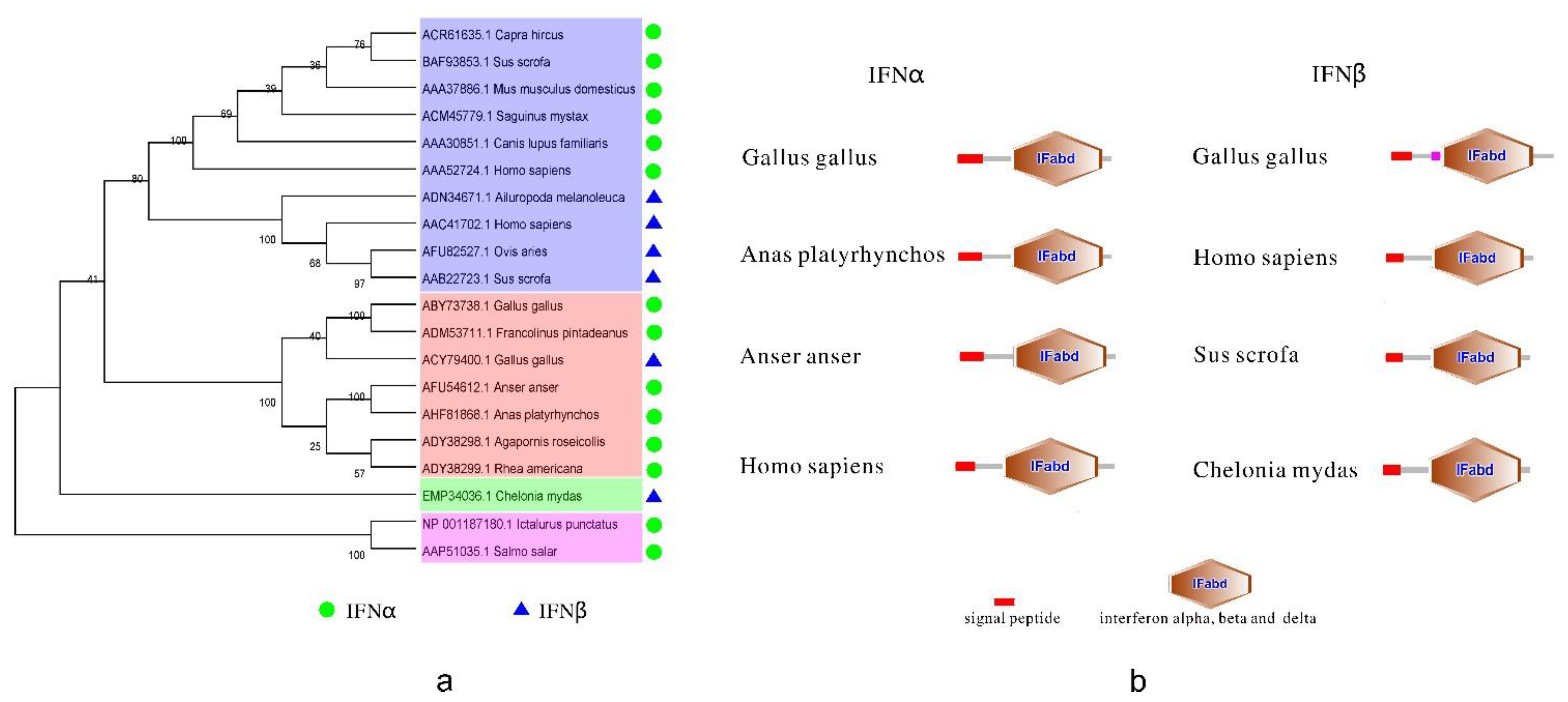
2.1. Type I Interferon (IFN)
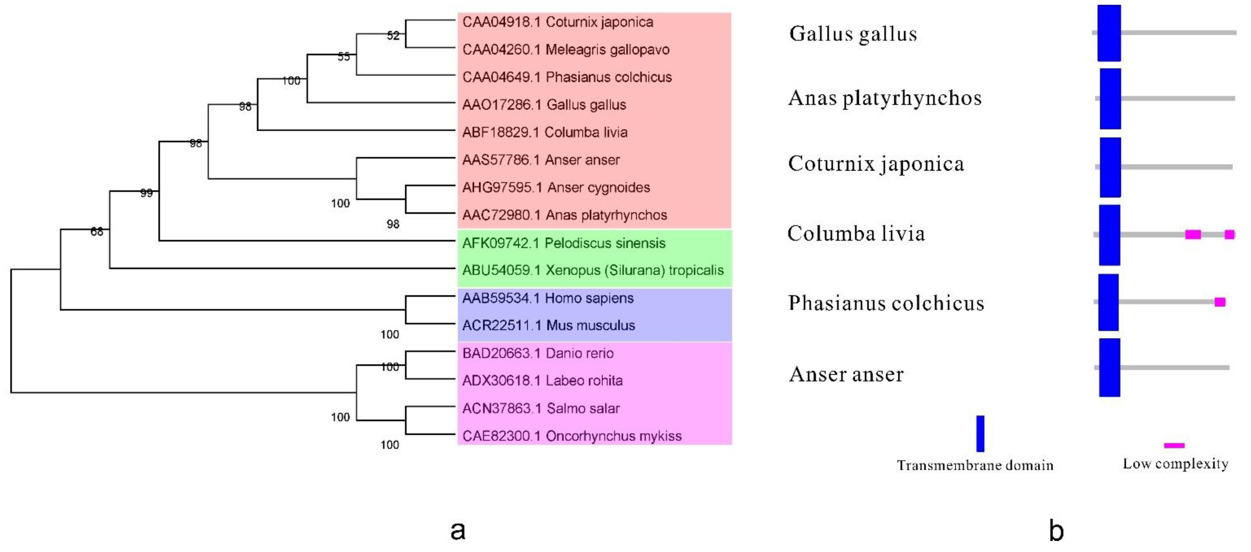
2.2. Type II IFN
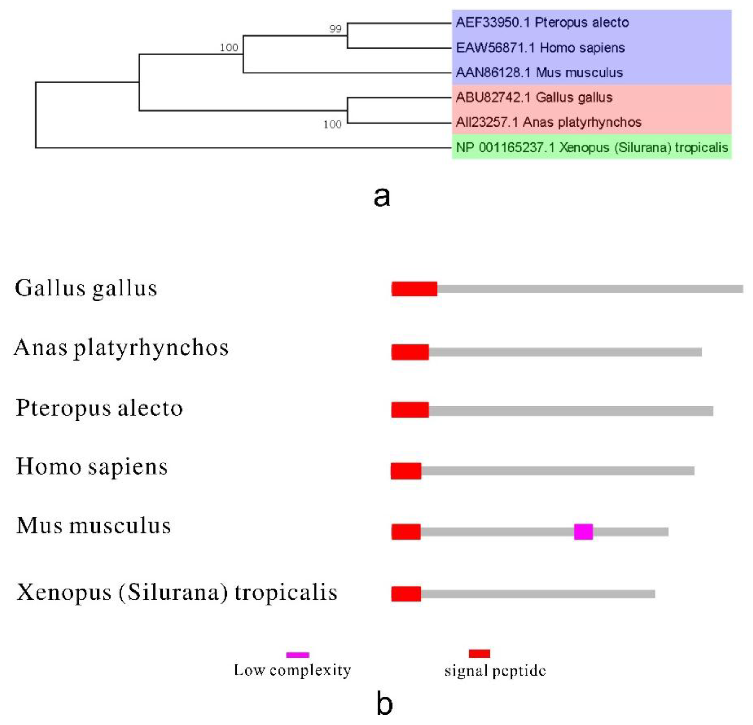
2.3. Type III IFN
2.4. Ontogeny of Avian Interferons
3. Interferon Receptors
| Taxonomy | Gene Name | Species | Ligand | Accession Number | Reference |
|---|---|---|---|---|---|
| Type I IFN receptor | IFNAR1 | Chicken | IFNα/IFNβ | AF082664 | [76] |
| IFNAR2 | Chicken | IFNα/IFNβ | AF082665 | [76] | |
| Type II IFN receptor | IFNGR1 | Chicken | IFNγ | EU057149 | [77] |
| IFNGR2 | Chicken | IFNγ | AY957508 | [78] | |
| Type III IFN receptor | IFNLR1 | Chicken | IFNλ | 419694(Gene ID) | [79] |
| IL10R2 | Chicken | IFNλ | AF082666 | [76] |
3.1. Type I IFN Receptors
3.2. Type II IFN Receptors
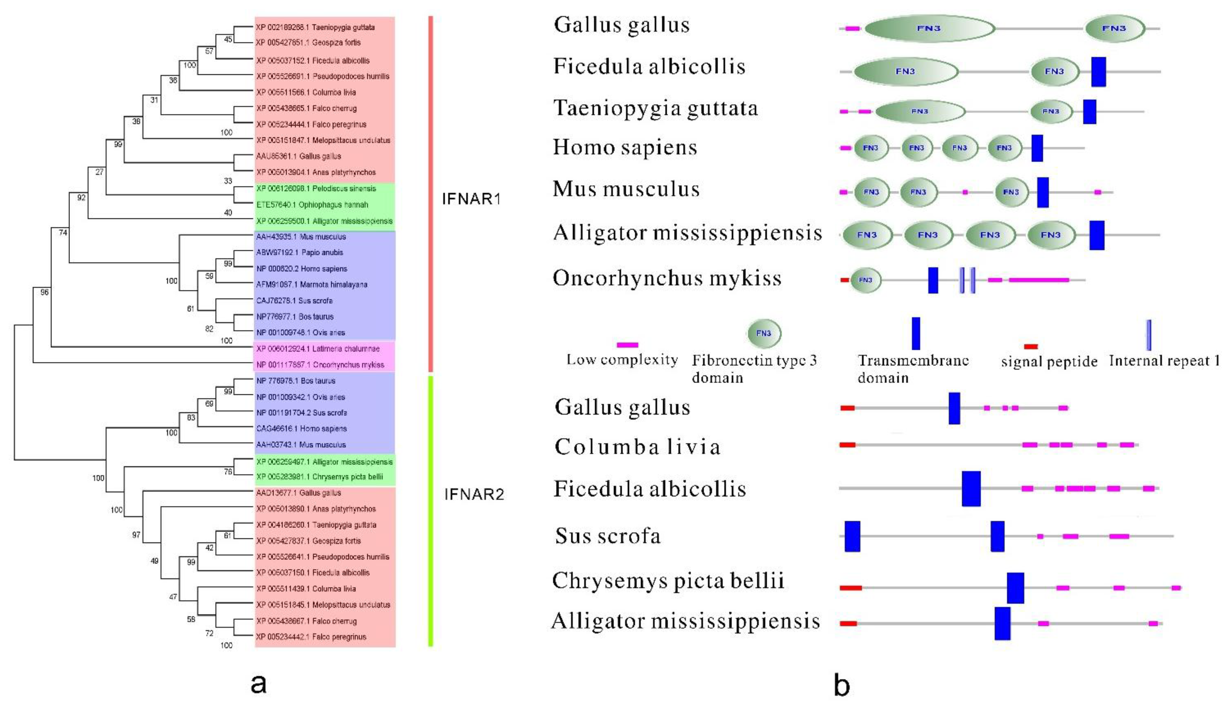
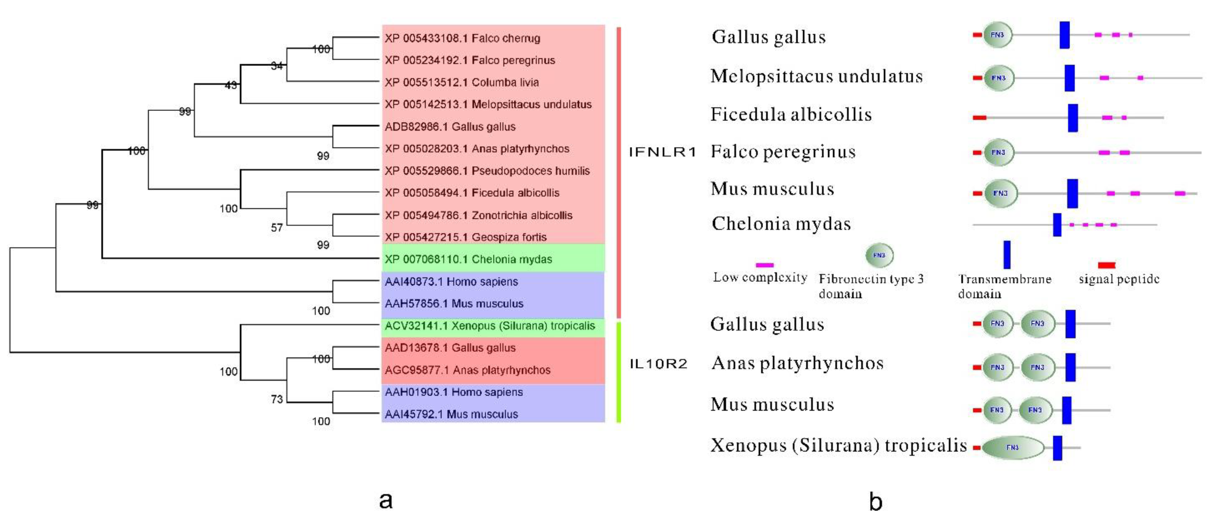
3.3. Type III IFN Receptors

3.4. Ontogeny of Avian Interferon Receptors
4. Antiviral Molecular Mechanism of Interferon and Interferon Receptor Activity
4.1. Interferon Functions as the Master Key
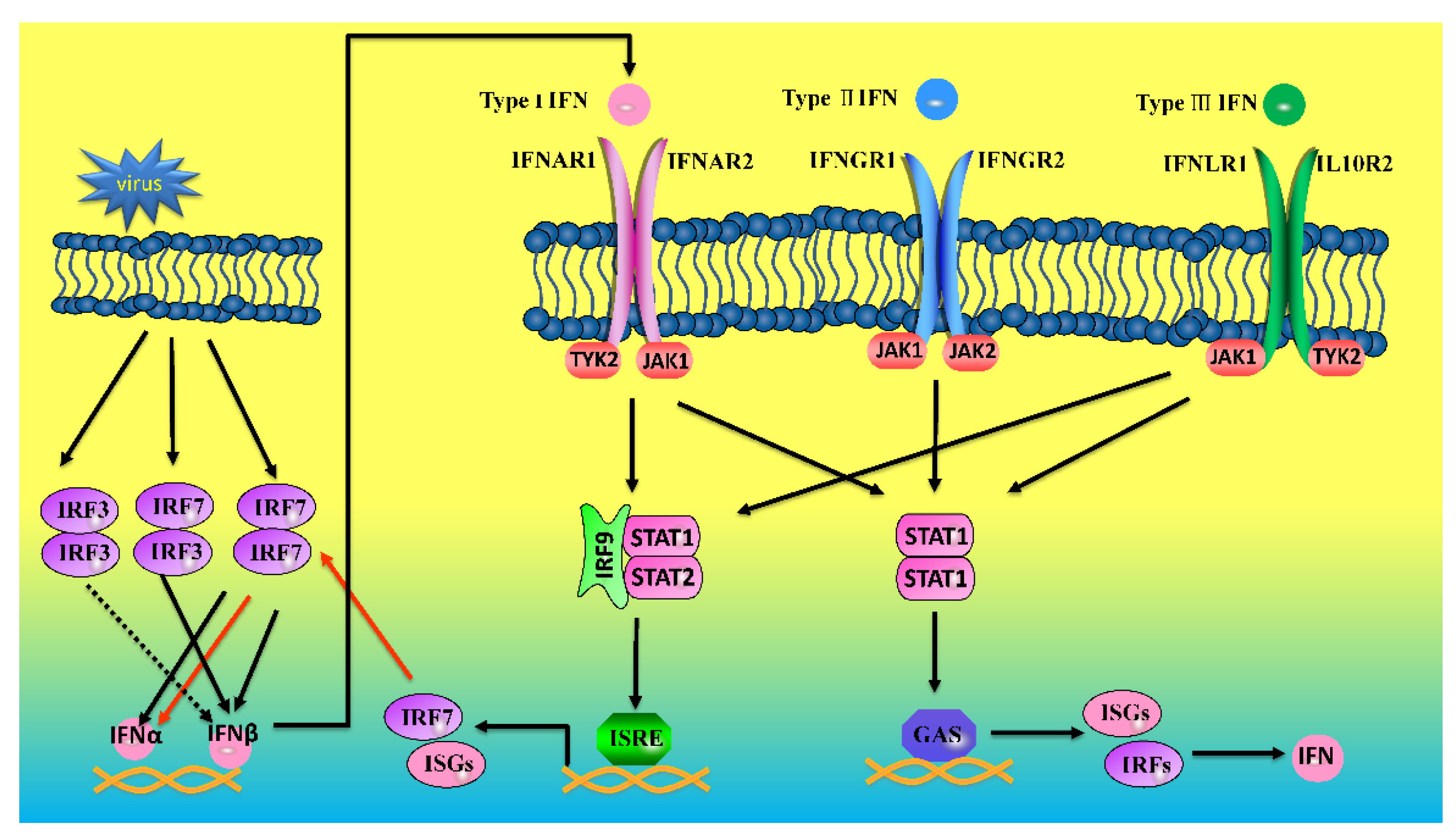
4.2. Interferon Receptors Function as the Signal Locks
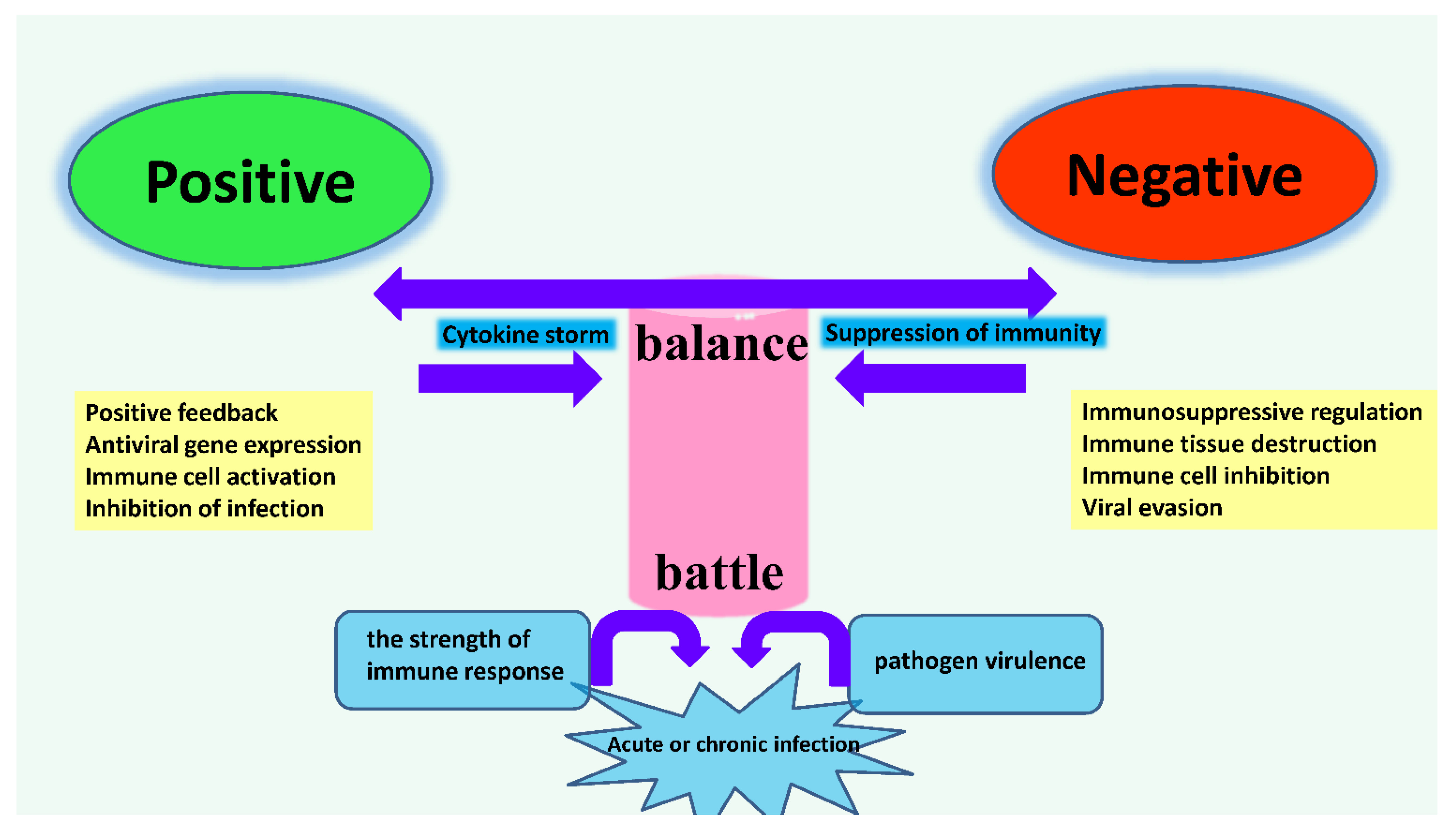
4.4. The Dynamic Balance of Immune Regulation in Vivo
5. Conclusions
Acknowledgments
Author Contributions
Conflicts of Interest
References
- Sadler, A.J.; Williams, B.R. Interferon-inducible antiviral effectors. Nat. Rev. Immunol. 2008, 8, 559–568. [Google Scholar] [CrossRef] [PubMed]
- Sheppard, P.; Kindsvogel, W.; Xu, W.; Henderson, K.; Schlutsmeyer, S.; Whitmore, T.E.; Kuestner, R.; Garrigues, U.; Birks, C.; Roraback, J.; et al. IL-28, IL-29 and their class II cytokine receptor IL-28R. Nat. Immunol. 2003, 4, 63–68. [Google Scholar] [CrossRef] [PubMed]
- Kotenko, S.V.; Gallagher, G.; Baurin, V.V.; Lewis-Antes, A.; Shen, M.; Shah, N.K.; Langer, J.A.; Sheikh, F.; Dickensheets, H.; Donnelly, R.P. IFN-λs mediate antiviral protection through a distinct class II cytokine receptor complex. Nat. Immunol. 2003, 4, 69–77. [Google Scholar] [CrossRef] [PubMed]
- De Weerd, N.A.; Nguyen, T. The interferons and their receptors—Distribution and regulation. Immunol. Cell. Biol. 2012, 90, 483–491. [Google Scholar] [CrossRef] [PubMed]
- Langer, J.A.; Cutrone, E.C.; Kotenko, S. The Class II cytokine receptor (CRF2) family: Overview and patterns of receptor-ligand interactions. Cytokine Growth Factor Rev. 2004, 15, 33–48. [Google Scholar] [CrossRef] [PubMed]
- Chelbi-Alix, M.K.; Wietzerbin, J. Interferon, a growing cytokine family: Fifty years of interferon research. Biochimie 2007, 89, 713–718. [Google Scholar] [CrossRef] [PubMed]
- Shoji, M.; Tachibana, M.; Katayama, K.; Tomita, K.; Tsuzuki, S.; Sakurai, F.; Kawabata, K.; Ishii, K.J.; Akira, S.; Mizuguchi, H. Type-I IFN signaling is required for the induction of antigen-specific CD8+ T cell responses by adenovirus vector vaccine in the gut-mucosa. Biochem. Biophys. Res. Commun. 2012, 425, 89–93. [Google Scholar] [CrossRef] [PubMed]
- Stark, G.R.; Darnell, J.E., Jr. The JAK-STAT pathway at twenty. Immunity 2012, 36, 503–514. [Google Scholar] [CrossRef] [PubMed]
- Zhou, Z.; Hamming, O.J.; Ank, N.; Paludan, S.R.; Nielsen, A.L.; Hartmann, R. Type III interferon (IFN) induces a type I IFN-like response in a restricted subset of cells through signaling pathways involving both the JAK-STAT pathway and the mitogen-activated protein kinases. J. Virol. 2007, 81, 7749–7758. [Google Scholar] [CrossRef] [PubMed]
- Ank, N.; West, H.; Bartholdy, C.; Eriksson, K.; Thomsen, A.R.; Paludan, S.R. λ Interferon (IFN-λ), a type III IFN, is induced by viruses and IFNs and displays potent antiviral activity against select virus infections in vivo. J. Virol. 2006, 80, 4501–4509. [Google Scholar] [CrossRef] [PubMed]
- Farrar, M.A.; Schreiber, R.D. The molecular cell biology of interferon-γ and its receptor. Annu. Rev. Immunol. 1993, 11, 571–611. [Google Scholar] [CrossRef] [PubMed]
- Odorizzi, P.M.; Wherry, E.J. Immunology an interferon paradox. Science 2013, 340, 155–1556. [Google Scholar] [CrossRef] [PubMed]
- Takaoka, A.; Yanai, H. Interferon signalling network in innate defence. Cell Microbiol. 2006, 8, 907–922. [Google Scholar] [CrossRef] [PubMed]
- Levy, D.E.; Marie, I.; Smith, E.; Prakash, A. Enhancement and diversification of IFN induction by IRF-7-mediated positive feedback. J. Interferon Cytokine Res. 2002, 22, 87–93. [Google Scholar] [CrossRef] [PubMed]
- Shaw, G.D.; Boll, W.; Taira, H.; Mantei, N.; Lengyel, P.; Weissmann, C. Structure and expression of cloned murine IFN-α genes. Nucl. Acids Res. 1983, 11, 555–573. [Google Scholar] [CrossRef] [PubMed]
- Gray, P.W.; Goeddel, D.V. Cloning and expression of murine immune interferon cDNA. Proc. Natl. Acad. Sci. USA 1983, 80, 5842–5846. [Google Scholar] [CrossRef] [PubMed]
- Dijkmans, R.; Vandenbroeck, K.; Beuken, E.; Billiau, A. Sequence of the porcine interferon-γ (IFN-γ) gene. Nucl. Acids Res. 1990, 18, 4259. [Google Scholar] [CrossRef] [PubMed]
- Carolan, L.A.; Butler, J.; Rockman, S.; Guarnaccia, T.; Hurt, A.C.; Reading, P.; Kelso, A.; Barr, I.; Laurie, K.L. TaqMan real time RT-PCR assays for detecting ferret innate and adaptive immune responses. J. Virol. Methods 2014, 205C, 38–52. [Google Scholar] [CrossRef]
- Sreekumar, E.; Janki, M.B.; Arathy, D.S.; Hariharan, R.; Premraj, C.A.; Rasool, T.J. Molecular characterization and expression of interferon-gamma of Asian elephant (Elephas maximus). Vet. Immunol. Immunopathol. 2007, 118, 75–83. [Google Scholar] [CrossRef] [PubMed]
- Tan, X.M.; Tang, Y.; Yang, Y.F.; Song, H.M.; Zhang, Y.Z. Gene cloning, sequencing, expression and biological activity of giant panda (Ailuropoda melanoleuca) interferon-α. Mol. Immunol. 2007, 44, 3061–3069. [Google Scholar] [CrossRef] [PubMed]
- Ochi, A.; Danesh, A.; Seneviratne, C.; Banner, D.; Devries, M.E.; Rowe, T.; Xu, L.; Ran, L.; Czub, M.; Bosinger, S.E.; et al. Cloning, expression and immunoassay detection of ferret IFN-γ. Dev. Comp. Immunol. 2008, 32, 890–897. [Google Scholar] [CrossRef] [PubMed]
- Diaz-San Segundo, F.; Weiss, M.; Perez-Martin, E.; Koster, M.J.; Zhu, J.; Grubman, M.J.; de los Santos, T. Antiviral activity of bovine type III interferon against foot-and-mouth disease virus. Virology 2011, 413, 283–292. [Google Scholar]
- Yang, L.; Xu, L.; Li, Y.; Li, J.; Bi, Y.; Liu, W. Molecular and functional characterization of canine interferon-ε. J. Interferon Cytokine Res. 2013, 33, 760–768. [Google Scholar] [CrossRef] [PubMed]
- Kumar, C.S.; Muthukumaran, G.; Frost, L.J.; Noe, M.; Ahn, Y.H.; Mariano, T.M.; Pestka, S. Molecular characterization of the murine interferon γ receptor cDNA. J. Biol. Chem. 1989, 264, 17939–17946. [Google Scholar] [PubMed]
- Hibino, Y.; Mariano, T.M.; Kumar, C.S.; Kozak, C.A.; Pestka, S. Expression and reconstitution of a biologically active mouse interferon γ receptor in hamster cells. Chromosomal location of an accessory factor. J. Biol. Chem. 1991, 266, 6948–6951. [Google Scholar] [PubMed]
- Xue, Q.; Yang, L.; Liu, X.; Liu, W. Molecular characterization of feline type I interferon receptor 2. J. Interferon Cytokine Res. 2010, 30, 81–88. [Google Scholar] [CrossRef] [PubMed]
- Han, C.S.; Mathialagan, N.; Klemann, S.W.; Roberts, R.M. Molecular cloning of ovine and bovine type I interferon receptor subunits from uteri, and endometrial expression of messenger ribonucleic acid for ovine receptors during the estrous cycle and pregnancy. Endocrinology 1997, 138, 4757–4767. [Google Scholar] [PubMed]
- Fan, H.; Zhu, Z.; Wang, Y.; Zhang, X.; Lu, Y.; Tao, Y.; Fan, W.; Wang, Z.; Wang, H.; Roggendorf, M.; et al. Molecular characterization of the type I IFN receptor in two woodchuck species and detection of its expression in liver samples from woodchucks infected with woodchuck hepatitis virus (WHV). Cytokine 2012, 60, 179–185. [Google Scholar] [CrossRef] [PubMed]
- Sekellick, M.J.; Ferrandino, A.F.; Hopkins, D.A.; Marcus, P.I. Chicken interferon gene: Cloning, expression, and analysis. J. Interferon Cytokine Res. 1994, 14, 71–79. [Google Scholar] [CrossRef]
- Schultz, U.; Kock, J.; Schlicht, H.J.; Staeheli, P. Recombinant duck interferon: A new reagent for studying the mode of interferon action against hepatitis B virus. Virology 1995, 212, 641–649. [Google Scholar] [CrossRef] [PubMed]
- Li, H.-T.; Ma, B.; Mi, J.-W.; Jin, H.-Y.; Xu, L.-N.; Wang, J.-W. Cloning, in vitro expression and bioactivity of goose interferon-α. Cytokine 2006, 34, 177–183. [Google Scholar] [CrossRef] [PubMed]
- Suresh, M.; Karaca, K.; Foster, D.; Sharma, J.M. Molecular and functional characterization of turkey interferon. J. Virol. 1995, 69, 8159–8163. [Google Scholar] [PubMed]
- Sick, C.; Schultz, U.; Staeheli, P. A family of genes coding for two serologically distinct chicken interferons. J. Biol. Chem. 1996, 271, 7635–7639. [Google Scholar] [CrossRef] [PubMed]
- Digby, M.R.; Lowenthal, J.W. Cloning and expression of the chicken interferon-γ gene. J. Interferon Cytokine Res. 1995, 15, 939–945. [Google Scholar] [CrossRef] [PubMed]
- Schultz, U.; Chisari, F.V. Recombinant duck interferon γ inhibits duck hepatitis B virus replication in primary hepatocytes. J. Virol. 1999, 73, 3162–3168. [Google Scholar] [PubMed]
- Li, H.T.; Ma, B.; Mi, J.W.; Jin, H.Y.; Xu, L.N.; Wang, J.W. Molecular cloning and functional analysis of goose interferon gamma. Vet. Immunol. Immunopathol. 2007, 117, 67–74. [Google Scholar] [CrossRef] [PubMed]
- Kaiser, P.; Sonnemans, D.; Smith, L.M. Avian IFN-γ genes: Sequence analysis suggests probable cross-species reactivity among galliforms. J. Interferon Cytokine Res. 1998, 18, 711–719. [Google Scholar] [CrossRef] [PubMed]
- Fringuelli, E.; Urbanelli, L.; Tharuni, O.; Proietti, P.C.; Bietta, A.; Davidson, I.; Franciosini, M.P. Cloning and expression of pigeon IFN-γ gene. Res. Vet. Sci. 2010, 89, 367–372. [Google Scholar] [CrossRef] [PubMed]
- Karpala, A.J.; Morris, K.R.; Broadway, M.M.; McWaters, P.G.; OʼNeil, T.E.; Goossens, K.E.; Lowenthal, J.W.; Bean, A.G. Molecular cloning, expression, and characterization of chicken IFN-λ. J. Interferon Cytokine Res. 2008, 28, 341–350. [Google Scholar] [CrossRef] [PubMed]
- Yao, Q.; Fischer, K.P.; Arnesen, K.; Tyrrell, D.L.; Gutfreund, K.S. Molecular cloning, expression and characterization of Pekin duck interferon-λ. Gene 2014, 548, 29–38. [Google Scholar] [CrossRef] [PubMed]
- Pestka, S.; Krause, C.D.; Walter, M.R. Interferons, interferon-like cytokines, and their receptors. Immunol. Rev. 2004, 202, 8–32. [Google Scholar] [CrossRef] [PubMed]
- Hertzog, P.J.; Williams, B.R. Fine tuning type I interferon responses. Cytokine Growth Factor Rev. 2013, 24, 217–225. [Google Scholar] [CrossRef] [PubMed]
- Meng, S.; Yang, L.; Xu, C.; Qin, Z.; Xu, H.; Wang, Y.; Sun, L.; Liu, W. Recombinant chicken interferon-α inhibits H9N2 avian influenza virus replication in vivo by oral administration. J. Interferon Cytokine Res. 2011, 31, 533–538. [Google Scholar] [CrossRef] [PubMed]
- Qu, H.; Yang, L.; Meng, S.; Xu, L.; Bi, Y.; Jia, X.; Li, J.; Sun, L.; Liu, W. The differential antiviral activities of chicken interferon α (ChIFN-α) and ChIFN-β are related to distinct interferon-stimulated gene expression. PLoS One 2013, 8, e59307. [Google Scholar] [PubMed]
- Ziegler, R.E.; Joklik, W.K. Effect of interferon on multiplication of Avian sarcoma virus B77 in duck embryo fibroblasts. J. Interferon Cytokine Res. 1981, 1, 521–538. [Google Scholar] [CrossRef]
- Xu, L.; Yang, L.; Liu, W. Distinct evolution process among type I interferon in mammals. Protein Cell 2013, 4, 383–392. [Google Scholar] [CrossRef] [PubMed]
- Woelk, C.H.; Frost, S.D.; Richman, D.D.; Higley, P.E.; Kosakovsky Pond, S.L. Evolution of the interferon α gene family in eutherian mammals. Gene 2007, 397, 38–50. [Google Scholar] [CrossRef] [PubMed]
- Radhakrishnan, R.; Walter, L.J.; Subramaniam, P.S.; Johnson, H.M.; Walter, M.R. Crystal structure of ovine interferon-т at 2.1 Å resolution. J. Mol. Biol. 1999, 286, 151–162. [Google Scholar] [CrossRef] [PubMed]
- Karpusas, M.; Nolte, M.; Benton, C.B.; Meier, W.; Lipscomb, W.N.; Goelz, S. The crystal structure of human interferon β at 2.2-Å resolution. Proc. Natl. Acad. Sci. USA 1997, 94, 11813–11818. [Google Scholar] [CrossRef] [PubMed]
- Senda, T.; Shimazu, T.; Matsuda, S.; Kawano, G.; Shimizu, H.; Nakamura, K.T.; Mitsui, Y. Three-dimensional crystal structure of recombinant murine interferon-β. EMBO J. 1992, 11, 3193–3201. [Google Scholar] [PubMed]
- Schroder, K.; Hertzog, P.J.; Ravasi, T.; Hume, D.A. Interferon-γ: An overview of signals, mechanisms and functions. J. Leukoc. Biol. 2004, 75, 163–189. [Google Scholar] [CrossRef] [PubMed]
- He, T.; Tang, C.; Xu, S.; Moyana, T.; Xiang, J. Interferon γ stimulates cellular maturation of dendritic cell line DC2.4 leading to induction of efficient cytotoxic T cell responses and antitumor immunity. Cell Mol. Immunol. 2007, 4, 105–111. [Google Scholar] [PubMed]
- Boehm, U.; Klamp, T.; Groot, M.; Howard, J.C. Cellular responses to interferon-γ. Annu Rev. Immunol. 1997, 15, 749–795. [Google Scholar] [CrossRef] [PubMed]
- Young, H.A.; Hardy, K.J. Role of interferon-gamma in immune cell regulation. J. Leukoc Biol. 1995, 58, 373–381. [Google Scholar] [PubMed]
- Szabo, S.J.; Sullivan, B.M.; Peng, S.L.; Glimcher, L.H. Molecular mechanisms regulating Th1 immune responses. Annu. Rev. Immunol. 2003, 21, 713–758. [Google Scholar] [CrossRef] [PubMed]
- Susta, L.; Cornax, I.; Diel, D.G.; Garcia, S.C.; Miller, P.J.; Liu, X.; Hu, S.; Brown, C.C.; Afonso, C.L. Expression of interferon γ by a highly virulent strain of Newcastle disease virus decreases its pathogenicity in chickens. Microb. Pathog. 2013, 61–62, 73–83. [Google Scholar] [CrossRef] [PubMed]
- Haq, K.; Wootton, S.K.; Barjesteh, N.; St Paul, M.; Golovan, S.; Bendall, A.J.; Sharif, S. Small interfering RNA-mediated knockdown of chicken interferon-γ expression. J. Interferon Cytokine Res. 2013, 33, 319–327. [Google Scholar] [CrossRef] [PubMed]
- Lawson, S.; Rothwell, L.; Lambrecht, B.; Howes, K.; Venugopal, K.; Kaiser, P. Turkey and chicken interferon-γ, which share high sequence identity, are biologically cross-reactive. Dev. Comp. Immunol. 2001, 25, 69–82. [Google Scholar] [CrossRef] [PubMed]
- Savan, R.; Ravichandran, S.; Collins, J.R.; Sakai, M.; Young, H.A. Structural conservation of interferon γ among vertebrates. Cytokine Growth Factor Rev. 2009, 20, 115–124. [Google Scholar] [CrossRef] [PubMed]
- Chen, S.N.; Huang, B.; Zhang, X.W.; Li, Y.; Zhao, L.J.; Li, N.; Gao, Q.; Nie, P. IFN-γ and its receptors in a reptile reveal the evolutionary conservation of type II IFNs in vertebrates. Dev. Comp. Immunol. 2013, 41, 587–596. [Google Scholar] [PubMed]
- Kotenko, S.V. IFN-λs. Curr. Opin. Immunol. 2011, 23, 583–590. [Google Scholar] [CrossRef] [PubMed]
- Kempuraj, D.; Donelan, J.; Frydas, S.; Iezzi, T.; Conti, F.; Boucher, W.; Papadopoulou, N.G.; Madhappan, B.; Letourneau, L.; Cao, J.; et al. Interleukin-28 and 29 (IL-28 and IL-29): New cytokines with anti-viral activities. Int. J. Immunopathol. Pharmacol. 2004, 17, 103–106. [Google Scholar] [PubMed]
- Lasfar, A.; Abushahba, W.; Balan, M.; Cohen-Solal, K.A. Interferon λ: A new sword in cancer immunotherapy. Clin. Dev. Immunol. 2011, 2011, 349575. [Google Scholar] [CrossRef] [PubMed]
- Reuter, A.; Soubies, S.; Hartle, S.; Schusser, B.; Kaspers, B.; Staeheli, P.; Rubbenstroth, D. Antiviral activity of λ interferon in chickens. J. Virol. 2014, 88, 2835–2843. [Google Scholar] [CrossRef]
- Matsuyama, T.; Nakayasu, C.; Fujiwara, A.; Kurita, J.; Takano, T.; Ito, T.; Sano, M. Ontogeny of anti-viral hemorrhagic septicemia virus (VHSV) immunity in developing Japanese flounder. Dev. Comp. Immunol. 2012, 37, 313–322. [Google Scholar] [CrossRef] [PubMed]
- Levraud, J.P.; Boudinot, P.; Colin, I.; Benmansour, A.; Peyrieras, N.; Herbomel, P.; Lutfalla, G. Identification of the zebrafish IFN receptor: Implications for the origin of the vertebrate IFN system. J. Immunol. 2007, 178, 4385–4394. [Google Scholar] [CrossRef] [PubMed]
- Sinkora, M.; Butler, J.E. The ontogeny of the porcine immune system. Dev. Comp. Immunol. 2009, 33, 273–283. [Google Scholar] [CrossRef] [PubMed]
- Solana, R.; Tarazona, R.; Gayoso, I.; Lesur, O.; Dupuis, G.; Fulop, T. Innate immunosenescence: Effect of aging on cells and receptors of the innate immune system in humans. Semin Immunol. 2012, 24, 331–341. [Google Scholar] [CrossRef]
- Karpala, A.J.; Bagnaud-Baule, A.; Goossens, K.E.; Lowenthal, J.W.; Bean, A.G. Ontogeny of the interferon system in chickens. J. Reprod. Immunol. 2012, 94, 169–174. [Google Scholar] [CrossRef] [PubMed]
- Abdul-Careem, M.F.; Hunter, D.B.; Lambourne, M.D.; Barta, J.; Sharif, S. Ontogeny of cytokine gene expression in the chicken spleen. Poult. Sci. 2007, 86, 1351–1355. [Google Scholar] [CrossRef] [PubMed]
- Song, C.; Yu, S.; Duan, Y.; Hu, Y.; Qiu, X.; Tan, L.; Sun, Y.; Wang, M.; Cheng, A.; Ding, C. Effect of age on the pathogenesis of DHV-1 in Pekin ducks and on the innate immune responses of ducks to infection. Arch. Virol. 2013, 159, 905–914. [Google Scholar] [CrossRef] [PubMed]
- Renauld, J.C. Class II cytokine receptors and their ligands: Key antiviral and inflammatory modulators. Nat. Rev. Immunol. 2003, 3, 667–676. [Google Scholar] [CrossRef] [PubMed]
- De Weerd, N.A.; Samarajiwa, S.A.; Hertzog, P.J. Type I interferon receptors: Biochemistry and biological functions. J. Biol. Chem. 2007, 282, 20053–20057. [Google Scholar]
- Jabbar, T.K.; Calvo-Pinilla, E.; Mateos, F.; Gubbins, S.; Bin-Tarif, A.; Bachanek-Bankowska, K.; Alpar, O.; Ortego, J.; Takamatsu, H.H.; Mertens, P.P.; et al. Protection of IFNAR (−/−) mice against bluetongue virus serotype 8, by heterologous (DNA/rMVA) and homologous (rMVA/rMVA) vaccination, expressing outer-capsid protein VP2. PLoS One 2013, 8, e60574. [Google Scholar] [CrossRef]
- De la Poza, F.; Calvo-Pinilla, E.; Lopez-Gil, E.; Marin-Lopez, A.; Mateos, F.; Castillo-Olivares, J.; Lorenzo, G.; Ortego, J. Ns1 is a key protein in the vaccine composition to protect IFNAR (−/−) mice against infection with multiple serotypes of African horse sickness virus. PLoS One 2013, 8, e70197. [Google Scholar] [CrossRef] [PubMed]
- Reboul, J.; Gardiner, K.; Monneron, D.; Uzé, G.; Lutfalla, G. Comparative genomic analysis of the interferon/interleukin-10 receptor gene cluster. Genome Res. 1999, 9, 242–250. [Google Scholar] [PubMed]
- Han, X.; Chen, T.; Wang, M. Molecular cloning and characterization of chicken interferon-γ receptor α-chain. J. Interferon Cytokine Res. 2008, 28, 445–454. [Google Scholar] [CrossRef] [PubMed]
- Han, C.L.; Zhang, W.; Dong, H.T.; Han, X.; Wang, M. A novel gene of β chain of the IFN-γ receptor of Huiyang chicken: Cloning, distribution, and CD assay. J. Interferon Cytokine Res. 2006, 26, 441–448. [Google Scholar] [CrossRef] [PubMed]
- Hillier, L.W.; Miller, W.; Birney, E.; Warren, W.; Hardison, R.C.; Ponting, C.P.; Bork, P.; Burt, D.W.; Groenen, M.A.M.; et al. Sequence and comparative analysis of the chicken genome provide unique perspectives on vertebrate evolution. Nature 2004, 432, 695–716. [Google Scholar]
- Skorstengaard, K.; Jensen, M.S.; Sahl, P.; Petersen, T.E.; Magnusson, S. Complete primary structure of bovine plasma fibronectin. Eur. J. Biochem. 1986, 161, 441–453. [Google Scholar] [CrossRef] [PubMed]
- Karpala, A.J.; Lowenthal, J.W.; Bean, A.G. Identifying innate immune pathways of the chicken may lead to new antiviral therapies. Vet. Immunol. Immunopathol. 2012, 148, 100–109. [Google Scholar] [CrossRef] [PubMed]
- Chen, S.; Cheng, A.; Wang, M. Innate sensing of viruses by pattern recognition receptors in birds. Vet. Res. 2013, 44, 82. [Google Scholar] [CrossRef]
- Ivashkiv, L.B.; Donlin, L.T. Regulation of type I interferon responses. Nat. Rev. Immunol. 2014, 14, 36–49. [Google Scholar] [CrossRef] [PubMed]
- Schoggins, J.W.; Wilson, S.J.; Panis, M.; Murphy, M.Y.; Jones, C.T.; Bieniasz, P.; Rice, C.M. A diverse range of gene products are effectors of the type I interferon antiviral response. Nature 2011, 472, 481–485. [Google Scholar] [CrossRef] [PubMed]
- Van Boxel-Dezaire, A.H.; Rani, M.R.; Stark, G.R. Complex modulation of cell type-specific signaling in response to type I interferons. Immunity 2006, 25, 361–372. [Google Scholar]
- Stark, G.R.; Kerr, I.M.; Williams, B.R.; Silverman, R.H.; Schreiber, R.D. How cells respond to interferons. Annu. Rev. Biochem. 1998, 67, 227–264. [Google Scholar] [CrossRef] [PubMed]
- Shuai, K.; Liu, B. Regulation of JAK-STAT signalling in the immune system. Nat. Rev. Immunol. 2003, 3, 900–911. [Google Scholar] [CrossRef] [PubMed]
- Owczarek, C.M.; Hwang, S.Y.; Holland, K.A.; Gulluyan, L.M.; Tavaria, M.; Weaver, B.; Reich, N.C.; Kola, I.; Hertzog, P.J. Cloning and characterization of soluble and transmembrane isoforms of a novel component of the murine type I interferon receptor, IFNAR 2. J. Biol. Chem. 1997, 272, 23865–23870. [Google Scholar] [CrossRef] [PubMed]
- Goodman, A.G.; Zeng, H.; Proll, S.C.; Peng, X.; Cilloniz, C.; Carter, V.S.; Korth, M.J.; Tumpey, T.M.; Katze, M.G. The α/β interferon receptor provides protection against influenza virus replication but is dispensable for inflammatory response signaling. J. Virol. 2010, 84, 2027–2037. [Google Scholar] [CrossRef] [PubMed]
- Piganis, R.A.; de Weerd, N.A.; Gould, J.A.; Schindler, C.W.; Mansell, A.; Nicholson, S.E.; Hertzog, P.J. Suppressor of cytokine signaling (SOCS) 1 inhibits type I interferon (IFN) signaling via the interferon α receptor (IFNAR1)-associated tyrosine kinase TYK2. J. Biol Chem. 2011, 286, 33811–33818. [Google Scholar] [CrossRef] [PubMed]
- Liu, B.S.; Janssen, H.L.; Boonstra, A. IL-29 and IFNα differ in their ability to modulate IL-12 production by TLR-activated human macrophages and exhibit differential regulation of the IFNγ receptor expression. Blood 2011, 117, 2385–2395. [Google Scholar] [CrossRef] [PubMed]
- Magor, K.E.; Miranzo Navarro, D.; Barber, M.R.; Petkau, K.; Fleming-Canepa, X.; Blyth, G.A.; Blaine, A.H. Defense genes missing from the flight division. Dev. Comp. Immunol 2013, 41, 377–388. [Google Scholar] [CrossRef] [PubMed]
- Huang, B.; Qi, Z.T.; Xu, Z.; Nie, P. Global characterization of interferon regulatory factor (IRF) genes in vertebrates: Glimpse of the diversification in evolution. BMC Immunol. 2010, 11, 22. [Google Scholar] [CrossRef] [PubMed]
- Gordien, E.; Rosmorduc, O.; Peltekian, C.; Garreau, F.; Brechot, C.; Kremsdorf, D. Inhibition of hepatitis B virus replication by the interferon-inducible MxA protein. J. Virol. 2001, 75, 2684–2691. [Google Scholar] [CrossRef] [PubMed]
- Stojdl, D.F.; Abraham, N.; Knowles, S.; Marius, R.; Brasey, A.; Lichty, B.D.; Brown, E.G.; Sonenberg, N.; Bell, J.C. The murine double-stranded RNA-dependent protein kinase PKR is required for resistance to vesicular stomatitis virus. J. Virol. 2000, 74, 9580–9585. [Google Scholar] [CrossRef] [PubMed]
- Samuel, M.A.; Whitby, K.; Keller, B.C.; Marri, A.; Barchet, W.; Williams, B.R.; Silverman, R.H.; Gale, M., Jr.; Diamond, M.S. PKR and RNase L contribute to protection against lethal West Nile virus infection by controlling early viral spread in the periphery and replication in neurons. J. Virol. 2006, 80, 7009–7019. [Google Scholar] [CrossRef]
- Turan, K.; Mibayashi, M.; Sugiyama, K.; Saito, S.; Numajiri, A.; Nagata, K. Nuclear MxA proteins form a complex with influenza virus NP and inhibit the transcription of the engineered influenza virus genome. Nucl. Acids Res. 2004, 32, 643–652. [Google Scholar] [CrossRef] [PubMed]
- Kochs, G.; Haller, O. Interferon-induced human MxA GTPase blocks nuclear import of Thogoto virus nucleocapsids. Proc. Natl. Acad. Sci. USA 1999, 96, 2082–2086. [Google Scholar] [CrossRef] [PubMed]
- Munir, M.; Berg, M. The multiple faces of proteinkinase R in antiviral defense. Virulence 2013, 4, 85–89. [Google Scholar] [CrossRef] [PubMed]
- Liu, S.Y.; Sanchez, D.J.; Cheng, G. New developments in the induction and antiviral effectors of type I interferon. Curr. Opin. Immunol. 2011, 23, 57–64. [Google Scholar] [CrossRef] [PubMed]
- Daviet, S.; van Borm, S.; Habyarimana, A.; Ahanda, M.L.; Morin, V.; Oudin, A.; van Den Berg, T.; Zoorob, R. Induction of Mx and PKR failed to protect chickens from H5N1 infection. Viral Immunol. 2009, 22, 467–472. [Google Scholar] [CrossRef] [PubMed]
- Smith, S.E.; Gibson, M.S.; Wash, R.S.; Ferrara, F.; Wright, E.; Temperton, N.; Kellam, P.; Fife, M. Chicken interferon-inducible transmembrane protein 3 restricts influenza viruses and lyssaviruses in vitro. J. Virol. 2013, 87, 12957–12966. [Google Scholar] [CrossRef] [PubMed]
- Marie, I.; Durbin, J.E.; Levy, D.E. Differential viral induction of distinct interferon-α genes by positive feedback through interferon regulatory factor-7. EMBO J. 1998, 17, 6660–6669. [Google Scholar] [CrossRef] [PubMed]
- Ank, N.; Iversen, M.B.; Bartholdy, C.; Staeheli, P.; Hartmann, R.; Jensen, U.B.; Dagnaes-Hansen, F.; Thomsen, A.R.; Chen, Z.; Haugen, H.; et al. An important role for type III interferon (IFN-λ/IL-28) in TLR-induced antiviral activity. J. Immunol. 2008, 180, 2474–2485. [Google Scholar] [CrossRef] [PubMed]
- Thanthrige-Don, N.; Read, L.R.; Abdul-Careem, M.F.; Mohammadi, H.; Mallick, A.I.; Sharif, S. Marekʼs disease virus influences the expression of genes associated with IFN-γ-inducible MHC class II expression. Viral Immunol. 2010, 23, 227–232. [Google Scholar] [CrossRef] [PubMed]
- Zhou, P.; Cowled, C.; Todd, S.; Crameri, G.; Virtue, E.R.; Marsh, G.A.; Klein, R.; Shi, Z.; Wang, L.F.; Baker, M.L. Type III IFNs in pteropid bats: Differential expression patterns provide evidence for distinct roles in antiviral immunity. J. Immunol. 2011, 186, 3138–3147. [Google Scholar] [CrossRef] [PubMed]
- Wilson, E.B.; Brooks, D.G. Interfering with type I interferon: A novel approach to purge persistent viral infection. Cell Cycle 2013, 12, 2919–2920. [Google Scholar] [CrossRef] [PubMed]
- Guo, P. Suppression of interferon-mediated antiviral immunity by hepatitis B virus: An overview of research progress. Scand. J. Immunol. 2013, 78, 230–237. [Google Scholar] [CrossRef] [PubMed]
© 2014 by the authors; licensee MDPI, Basel, Switzerland. This article is an open access article distributed under the terms and conditions of the Creative Commons Attribution license (http://creativecommons.org/licenses/by/4.0/).
Share and Cite
Zhou, H.; Chen, S.; Wang, M.; Cheng, A. Interferons and Their Receptors in Birds: A Comparison of Gene Structure, Phylogenetic Analysis, and Cross Modulation. Int. J. Mol. Sci. 2014, 15, 21045-21068. https://doi.org/10.3390/ijms151121045
Zhou H, Chen S, Wang M, Cheng A. Interferons and Their Receptors in Birds: A Comparison of Gene Structure, Phylogenetic Analysis, and Cross Modulation. International Journal of Molecular Sciences. 2014; 15(11):21045-21068. https://doi.org/10.3390/ijms151121045
Chicago/Turabian StyleZhou, Hao, Shun Chen, Mingshu Wang, and Anchun Cheng. 2014. "Interferons and Their Receptors in Birds: A Comparison of Gene Structure, Phylogenetic Analysis, and Cross Modulation" International Journal of Molecular Sciences 15, no. 11: 21045-21068. https://doi.org/10.3390/ijms151121045