Article

Identification of a Genomic Region Containing a Novel Promoter Resistant to Glucose Repression and Over-Expression of β-Glucosidase Gene in Hypocrea orientalis EU7-22

School of Energy Research, Xiamen University, Xiamen 361102, China
*
Author to whom correspondence should be addressed.
These authors contributed equally to this work.
Int. J. Mol. Sci. 2013, 14(4), 8479-8490; https://doi.org/10.3390/ijms14048479
Submission received: 6 January 2013 / Revised: 8 March 2013 / Accepted: 12 April 2013 / Published: 17 April 2013
(This article belongs to the Section Biochemistry)

Abstract

:
A high concentration of glucose in the medium could greatly inhibit the expression of cellulase in filamentous fungi. The aspartic protease from fungus Hypocrea orientalis EU7-22 could efficiently express under both induction condition and glucose repression condition. Based on the sequence of structure gene of aspartic protease, the upstream sequence harboring the putative promoter proA for driving the expression of aspartic protease was obtained by genome walking. The upstream sequence contained the typical promoter motifs “TATA” and “CAAT”. The β-glucosidase gene (Bgl1) from H. orientalis was cloned and recombined with promoter proA and terminator trpC. The expression cassette was ligated to the binary vector to form pUR5750-Bgl1, and then transferred into the host strain EU7-22 via Agrobacterium tumefaciens mediated transformation (ATMT), using hygromycin B resistance gene as the screening marker. Four transformants Bgl-1, Bgl-2, Bgl-3 and Bgl-4 were screened. Compared with the host strain EU7-22, the enzyme activities of filter paper (FPA) and β-glucosidase (BG) of transformant Bgl-2 increased by 10.6% and 19.1% under induction condition, respectively. The FPA and BG activities were enhanced by 22.2% and 700% under 2% glucose repression condition, respectively, compared with the host strain. The results showed that the putative promoter proA has successfully driven the over-expression of Bgl1 gene in H. orientalis under glucose repression condition.

1. Introduction

Lignocellulosic biomass represents abundant polysaccharides on the earth and a renewable resource, which can be used to produce environment-friendly biofuels, chemicals, polymers and materials [1]. Conversion of lignocellulosic biomass into fermentable sugars mainly depends on the degradation of cellulolytic enzymes produced by many filamentous fungi, including Trichoderma, Aspergillus, Penicillium and Acremonium[2,3]. The cellulolytic enzyme complex consists of three basic categories of enzymes: cellobiohydrolases (EC 3.2.1.91), endoglucanases (EC 3.2.1.4) and β-glucosidases (EC3.2.1.21), which act synergistically to degrade insoluble cellulose to glucose [4]. However, when filamentous fungi yielded large amounts of biomass-degrade enzymes protein, it was regulated by carbon catabolite repression (CCR), which mainly controls carbon assimilation [5]. The C2H2 type transcription factor CreI/CreA, has been shown to act as a repressor mediating CCR, which binds to the promoter of target genes via the consensus motif 5′-SYGGRG-3′, whose function in vivo has been shown in both Trichoderma reesei[5,6] and Aspergillus nidulans[7]. Cellulase production could be induced by cellulose, but strongly repressed by the major end-product hydrolysate-glucose.
For efficient expression gene in filamentous fungi, it is essential that the expression of the target gene would be enhanced by use of an active promoter either derived from the host or a related species. The cellobiohydrolase I gene (cbh1) promoter from T. reesei has been considered the strongest promoter, and has generally been used to construct high-efficient expression vectors to yield homologous and heterologous proteins [8]. However, the cbh1 promoter is repressed by glucose and negatively regulated by Cre I/CreA [5,9]. Therefore, cbh1 promoter couldn’t express the target gene efficiently in the high concentration of glucose-containing medium. Bando et al.[10] found a novel promoter from a hemolysin-like gene (hlyA), which was not repressed by glucose and could efficiently over-express protein in Aspergillus oryzae grown in solid-state culture.
In the present study, a novel putative promoter proA regulating the expression of aspartic protease gene was isolated from Hypocrea orientalis EU7-22 by genome walking technology. The over-expression of β-glucosidases gene (Bgl1) in strain EU7-22 driven by putative promoter proA was studied.

2. Results and Discussion

2.1. Promoter proA Cloning and Sequence Analysis

The extension-based genome walking (E-GW) method was used to amplify the unknown 5′-upstream sequences of the proA gene. The three nested PCR products were analyzed by agarose gel electrophoresis (Figure 1). Following the Genome Walking Kit instructions, the DNA fragment (approximately 1200 bp) was obtained (Figure 1, lane 12, indicated by the arrow) with the primer AP4 (supplied by the kit) and reverse primers SP3, which was excised, purified and directly sequenced.
The upstream sequences of proA gene were successfully cloned and deposited at the GenBank database with accession no. JQ728540. Homology analysis with NCBI Blast search showed 91% nucleotide identity to the proA gene upstream sequence (only 438 bp in length, from the start codon ATG) from Hypocrea jecorina (GenBank accession No. AM168137.1). A putative TATA box (TATAAA) was found at position −116 (relative to the ATG), and three putative CAAT boxes appeared at positions −172, −291 and −653 (Figure 2). Both motifs are related with the initiation of transcription [11]. The sequence contained multiple copies of the 5′-HGATAR-3′ motif, which corresponds to the nitrogen regulator AreA [12,13], located at −151, −511 (negative strand), and −738. Two 5′-GCCARG-3′ motifs for the PacC protein, which mediates pH regulation in Aspergillus[11], were also present at positions −1055 and −1163 (negative strand). A single copy of the motif 5′-SYGGRG-3′, the consensus sequence for the carbon catabolite repressor Cre I [5,9], was located at position −299. This was different from the promoter of papA gege encoding for aspartic protease from Trichoderma harzianum CECT 2413 which lacked potential CreA sites for carbon regulation [14]. The secreted protein aspartic protease was strongly expressed even under the presence of 3% glucose in the medium (data not shown). It suggested that the putative promoter proA was resistant to glucose repression, which could be applied for constructing high-efficiency cellulase expression system.

2.2. Over-Expression of β-Glucosidase Gene in H. orientalis

To investigate the resistance to glucose repression of the putative proA promoter, β-glucosidase gene (Bgl1) from H. orientalis was used as a reporter. The binary expression vector (pUR5750-Bgl1) was constructed, and transfered into H. orientalis by Agrobacterium tumefaciens mediated transformation (ATMT). After cultivated 3–6 days on PDA selecting plates, four putative transformants were visible. When inoculating the transformants to new plate containing 100 μg/mL hygromycin B, they all grew well. The mitotic stability of the transformants was tested by growing them on PDA plates. After successively repeating three generations, the transformants were inoculated on PDA plates containing 100 μg/mL hygromycin B. All the transformants grew well, which showed that it was mitotically stable. The four stable hygromycin B-resistant transformants were obtained and named as Bgl-1, Bgl-2, Bgl-3 and Bgl-4. PCR assay with primer hph-F&R amplified an 811 bp fragment, which suggested that the hph gene was successfully integrated into the genomes of the four transformants (Figure 3A). Another PCR with primer PproA-F&TtrpC-R for amplifying the 4194 bp gene expression cassette (PproA-Bgl1-TtrpC) fragment was performed to testify whether the putative proA promoter, bgl1 gene and trpC terminator were synchronously integrated into the genomes. Data indicated only transformants Bgl-2 and Bgl-3 were successfully integrated into the genomes (Figure 3B, lanes 3 and 4). The host strain EU7-22 was used as the control, which no gene expression cassette fragment was amplified (Figure 3B, lane 1). The ATMT method was an effective and simple technique for fungal transformation, but ATMT mediating T-DNA integration the host genome appeared to be a random event [15,16], the truncation of the inserted T-DNA would be possible. The phenomena also occurred in Fusarium oxysporum[17] and Aspergillus awamori[18].

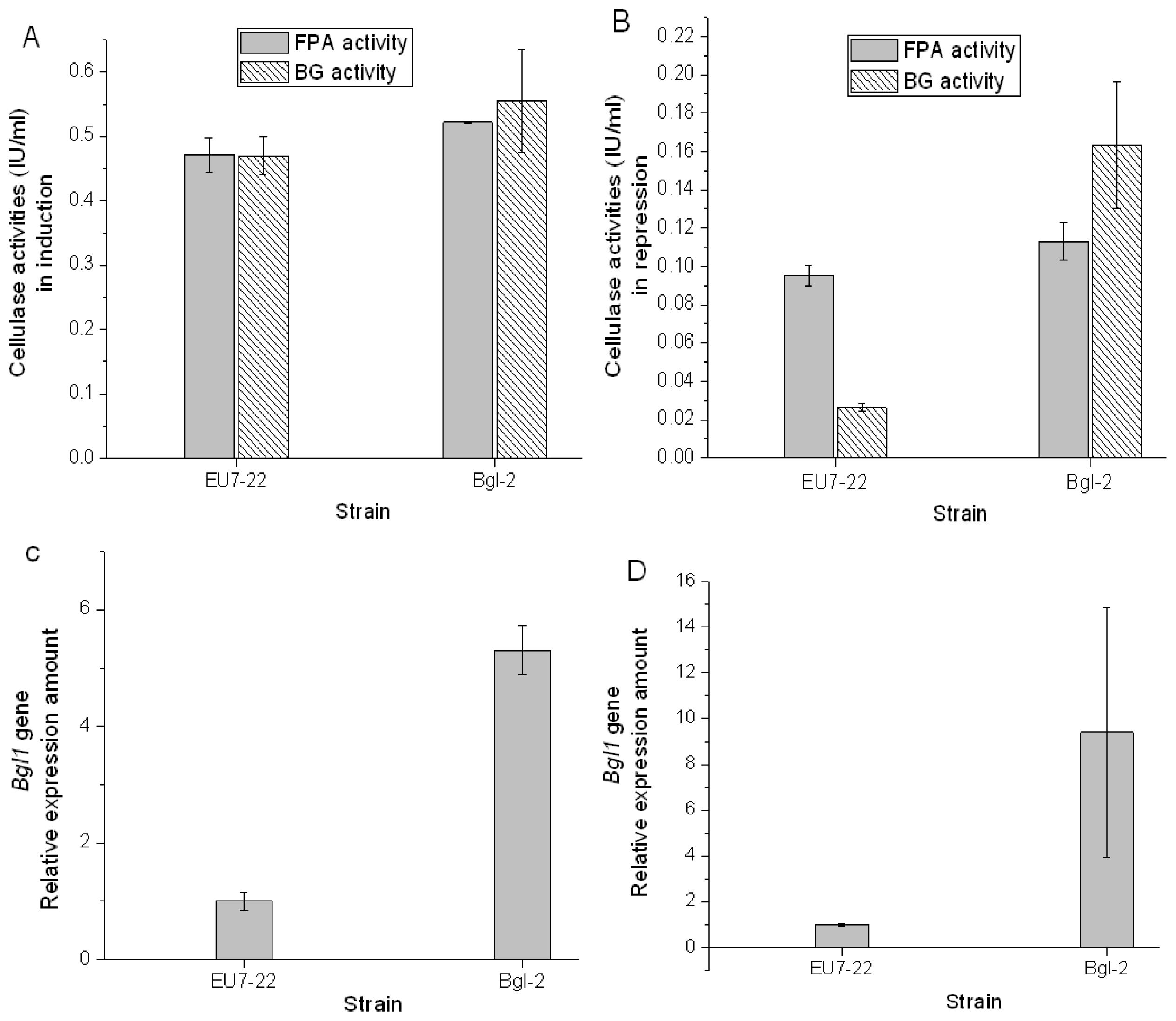
2.3. Comparison Analysis of Cellulase Production of Transformants and Host Strain EU7-22

According to cellulase production analysis (data not shown), the transformant Bgl-2 was selected as the work strain. The pretreated Miscanthus cellulose was used as inducer to produce cellulase, the filter paper activity (FPA) and β-glucosidase (BG) activity of host strain EU7-22 were 0.47 IU/mL and 0.47 IU/mL, respectively; the FPA and BG activities of strain Bgl-2 were 0.52 IU/mL and 0.56 IU/mL, respectively (Figure 4A). In comparison with strain EU7-22, the FPA and BG activities of transformant Bgl-2 increased by 10.6% and 19.1% under induction conditions, respectively, which owed to the over-expression β-glucosidase gene. While a final concentration of 2% glucose was used as repressor and supplemented in the induction medium, the FPA and BG activities of host strain EU7-22 were 0.09 IU/mL and 0.02 IU/mL, respectively; the FPA and BG activities of strain Bgl-2 were 0.11 IU/mL and 0.16 IU/mL, respectively (Figure 4B). In comparison with host strain EU7-22, the FPA and BG activities of transformant Bgl-2 increased by 22.2% and 700%, respectively.
Both FPA activity and BG activity would decrease under glucose repression condition. FPA activity reflected the total activity of cellulase system. BG activity represented the enzyme activity of β-glucosidase. In comparison with the induction condition, the BG activities of host strain EU7-22 decreased 95.7% under 2% glucose repression, only 4.3% BG activity was remained, which means that the expression of Bgl1 gene was almost been inhibited. However, the BG activity of transformant Bgl-2 only declined 71.4% under glucose repression. There are two β-glucosidase expression systems, one was driven by putative promoter proA, and another was driven by the original endogenous promoter of Bgl1 gene from H. orientalis, which was repressed in the presence of glucose. The BG activity of strain Bgl-2 reached 0.16 IU/mL under 2% glucose repression, which equals 34.0% activity of host strain under induction condition, and 800% activity of host strain under repression condition. It suggested that the transferred bgl1 gene expression system promoted by the putative promoter proA was not inhibited by 2% glucose repression. The over-expression system could express a quantity of 22%–34% β-glucosidase that produced by host strain EU7-22 under induction condition.
The transcription level of bgl1 gene was analyzed by qRT-PCR. In the induced cellulase production, the amount of bgl1 transcripts in strain EU7-22 was set as 1. The relative mRNA expression of bgl1 transcripts in strain Bgl-2 increased by 5.3 fold (Figure 4C). Under glucose repression condition, the amount of bgl1 transcripts in strain EU7-22 was also set as 1. The relative mRNA expression of bgl1 transcripts in strain Bgl-2 increased by 9.4 fold (Fig 4D). The results showed that the expression of β-glucosidase promoted by the proA promoter and was not inhibited by 2% glucose. The putative promoter proA was an idea promoter for expressing cellulase genes under glucose repression condition.

3. Experimental Section

3.1. Microorganism Strains and Culture Conditions

The cellulase-producing strain Hypocrea orientalis EU7-22 was identified according to its ITS (GenBank accession no. KC751873) and tef1 sequence (GenBank accession No. KC751874), and were analysed by TrichOKey v. 2.0: The Molecular Barcode Program ( http://www.isth.info/index.php).The strain was screened by this laboratory, and was used as a host cell for over-expression of Bgl1 gene. It was preserved at −80 °C in a 20% glycerol solution. Before inoculation preparation, it was activated on potato dextrose agar (PDA) slants, and incubated in liquid PDA on a rotary shaker (30 °C, 34–36 h, 180 rpm), and then transferred into submerged fermentation medium with 10% (v/v) inoculation quantity (106 spores mL−1). Escherichia coli DH5a was used for vector construction and propagation. The Agrobacterium tumefaciens AGL1 was used to mediate transformation and was grown either on Luria-Bertani (LB) broth (50 μg/mL kanamycin) or induction medium (IM) [19] supplemented with 0.2 mM acetosyringone (IMAS). Transformants were selected on PDA medium supplemented with hygromycin B (100 μg/ml), 0.2 mM cefotaxime and 20% Triton-X100.

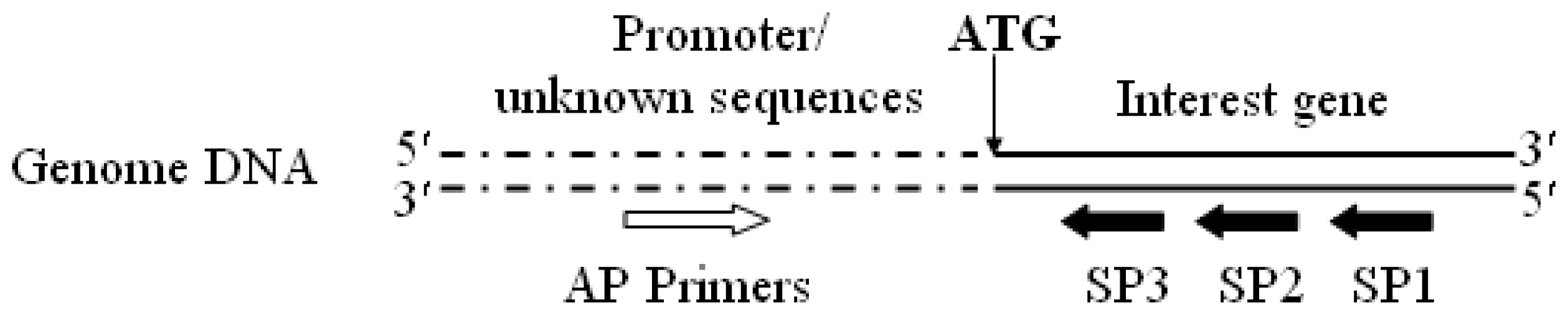
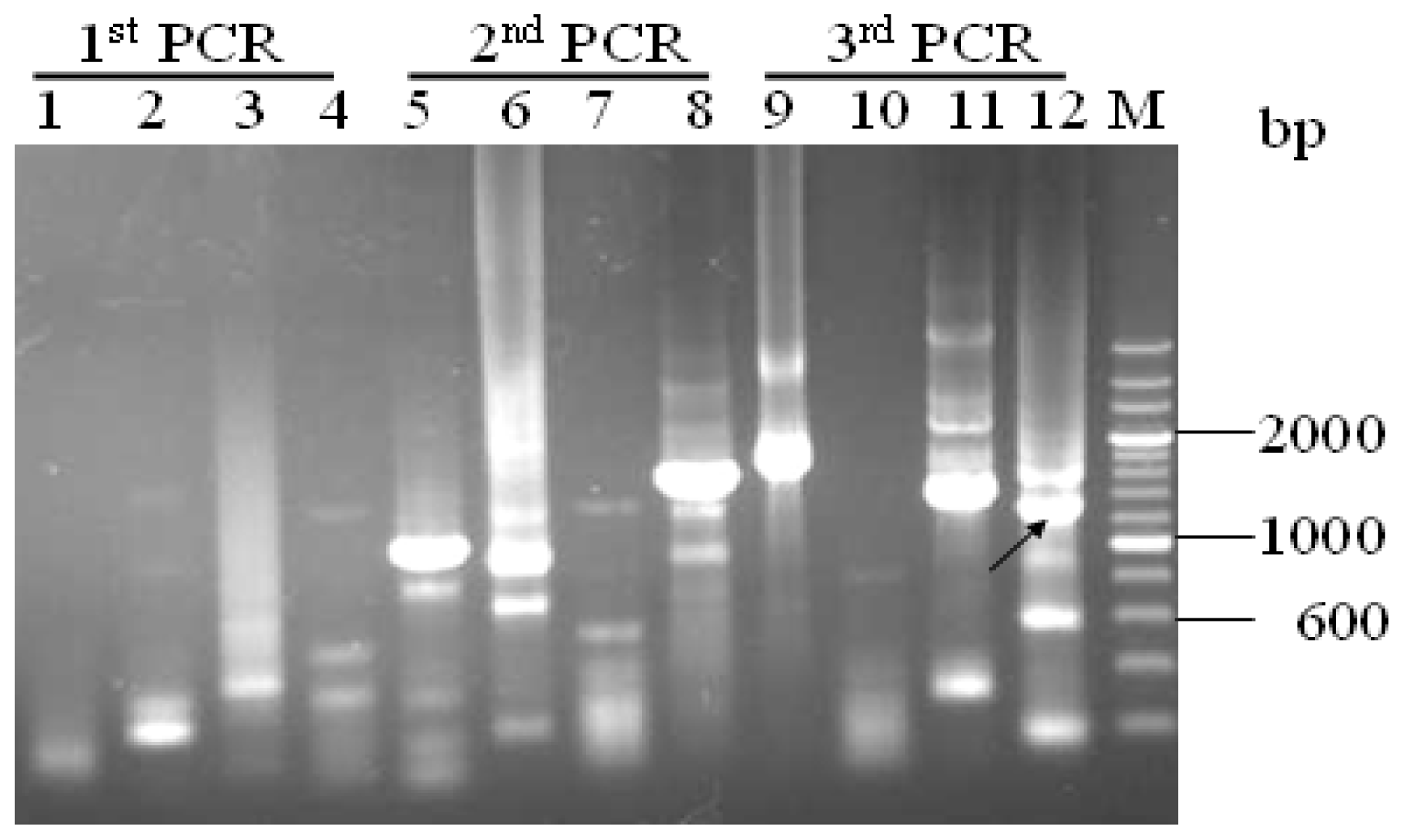
3.2. Promoter proA Cloning and Analysis

The aspartic protease could express at high level under glucose repression condition. The gene proA encoding for aspartic protease has been cloned, and the gene sequence was deposited at the GenBank database with accession no. JQ728540. The putative promoter proA of H. orientalis was expected as the potential enhanced promoter for expressing cellulase genes under glucose repression condition. The genome walking technology [20,21] was used to amplify the unknown region sequences based on the identified gene sequences. Genomic DNA of H. orientalis EU7-22 was extracted from the available mycelia according to the method of Penttilä et al.[22]. Specific reverse primers, SP1, SP2 and SP3 (Table 1; Figure 5), were designed according to the cloned proA gene sequence from H. orientalis. The forward primers AP1, AP2, AP3 and AP4 were supplied in the Genome Walking Kit (Takara, Japan).
The first nested PCR reaction was applied by using genomic DNA as the template, with specific primer SP1 which was paired with AP1, AP2, AP3 and AP4 in four separate reactions). The PCR condition was designed as: 94 °C for 1 min, 98 °C for 1 min, then 5 cycles of amplification (94 °C for 30 s, 62 °C for 1 min, 72 °C for 2 min), 94 °C for 30 s, 25 °C for 3 min, 72 °C for 2 min, then 15 cycles of amplification (94 °C for 30 s, 62 °C for 1 min, 72 °C for 2 min, 94 °C for 30 s, 62 °C for 1 min, 72 °C for 2 min, 94 °C for 30 s, 44 °C for 1 min, 72 °C for 2 min), and finally 72 °C for 10 min. The PCR products were shown in Figure 1 (lanes 1–4).
The second nested PCR was carried out by using the first nested PCR products as templates, with specific primer SP2 and primer AP1, AP2, AP3 and AP4, respectively. The PCR condition was designed as: 15 cycles of amplification (94 °C for 30 s, 62 °C for 1 min, 72 °C for 2 min, 94 °C for 30 s, 62 °C for 1 min, 72 °C for 2 min, 94 °C for 30 s, 44 °C for 1 min, 72 °C for 2 min), then 72 °C for 10 min. The PCR products were shown in Figure 1 (lanes 5–8).
The third nested PCR was carried out by using the second nested PCR products as templates, with specific primer SP3 and primer AP1, AP2, AP3 and AP4, respectively. The PCR condition was designed as: 15 cycles of amplification (94 °C for 30 s, 62 °C for 1 min, 72 °C for 2 min, 94 °C for 30 s, 62 °C for 1 min, 72 °C for 2 min, 94 °C for 30 s, 44 °C for 1 min, 72 °C for 2 min), then 72 °C for 10 min. The PCR products were shown in Figure 1 (lanes 9–12). The third round PCR products were purified, directly sequenced and validated. The resulting putative promoter sequence was then analyzed.

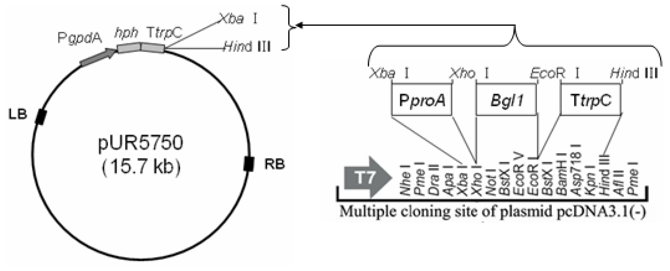
3.3. Construction of Binary Expression Vectors pUR5750-Bgl1

The putative proA promoter (1200 bp) was amplified by PCR with primer PproA-F&R (contain Xba I and Xho I site). The bgl1 gene (2235 bp) was amplified from H. orientalis mRNA by RT-PCR with primer Bgl1-F&R (contain Xho I and EcoR I site), The trpC terminator (740bp) was amplified from plasmid pUR5750 with primer TtrpC-F&R (contain EcoR I and Hind III site). The plasmid pUR5750 was used as a backbone to construct the binary expression vector, and a hygromycin B resistant gene (hph) was adopted as a selecting marker for screening H. orientalis transformats. The dual expression vector pUR5750-Bgl1 was built by means of the intermediate plasmid pcDNA3.1 (−) (Figure 6). The primers used in this study were listed in Table 1.

3.4. Agrobacterium-Tumefaciens-Mediated-Transformation (ATMT) in H. orientalis EU7-22

A.tumefaciens AGL-1, containing a binary vector (pUR5750-Bgl1), was grown at 28 °C for 2 days in LB supplemented with kanamycin (50 μg/mL). The AGL-1 cells were diluted to (optical density) OD600 = 0.15 in induction medium (IMAS), and were grown for an additional 8 h until an optical density at 660 nm of 0.4 before mixing them with an equal volume of a conidial suspension from strain EU7-22 (1 × 10⁶ conidia per mL). This mix (200 μL per plate) was spread onto an IMAS agar plate for co-cultivation (same as IMAS except containing 5 mM glucose instead of 10 mM glucose). Following incubation at 25 °C for 48 h, then place the PDA medium supplemented with hygromycin B (100 μg/mL), 0.2 mM cefotaxime and 20% Triton-X100 onto IMAS agar plate, sequentially incubated at 30 °C for 3–6 days, and the visible putative transformants were chosen.

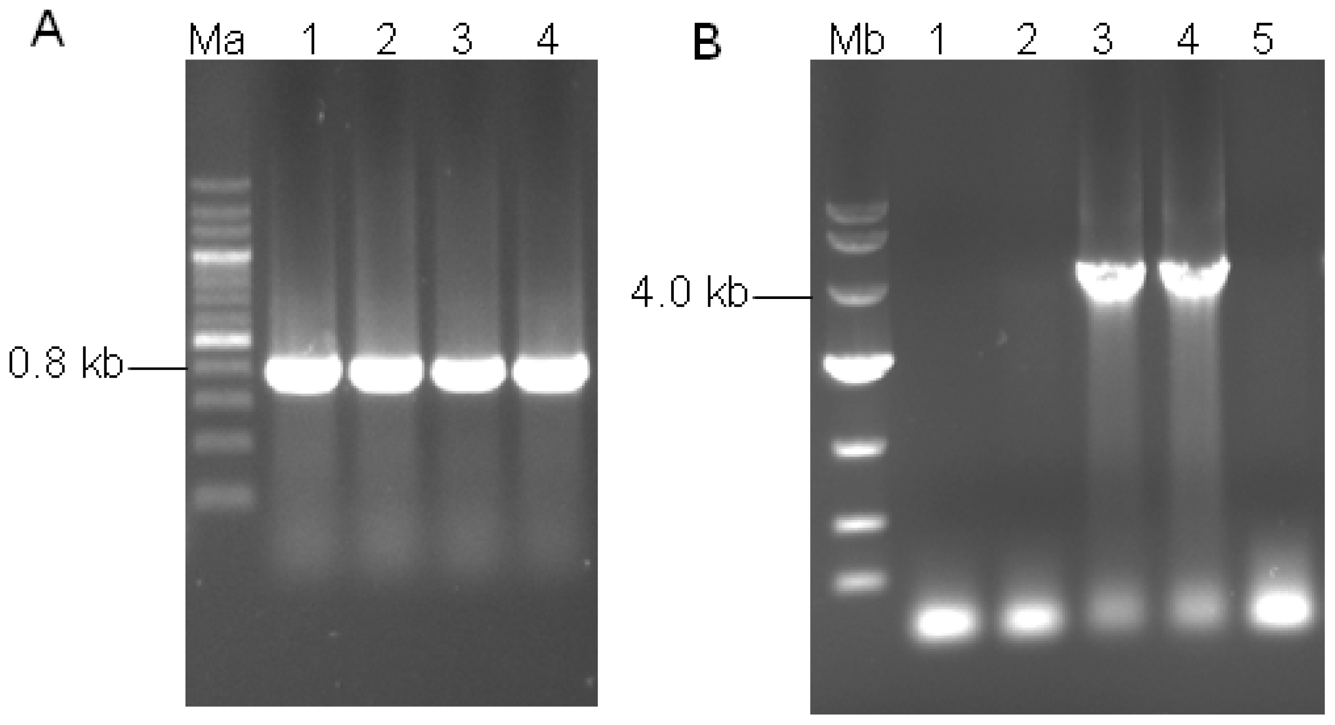
3.5. Transformant Stability and Molecular Analysis of the Transformants

The putative visible transformants was firstly picked up and transferred to a PDA agar plate containing hygromycin B. The transformants were then cultured on PDA agar plate without hygromycin B by three times. Then the monoconidial cultures were transferred to PDA plates containing 100 μg/mL hygromycin B for determining the stability of the transformant. Genomic DNA of transformant was extracted from all available mycelia according to the method of Penttilä et al.[22]. The gene fragments were analysed by PCR amplification using primer hph-F&R (for 811 bp) and PproA-F &TtrpC-R (for 4194 bp).

3.6. Enzymatic Analysis

Experiments were conducted in the 250-mL Erlenmeyer flasks. The induction medium (pH 5.2) for cellulase production was 2% pretreated Miscanthus cellulose (the dry Miscanthus straw was crashed into powder, extracted with 2% NaOH and 1% H2O2 for 2h at 60 °C, then extracted with 2% NaOH for 2 h at 100 °C, wash to neutral and drying), 1% wheat bran, 0.5% peptone, 0.05% CaCl2, 0.05% MgSO4, 0.4% Tween 80 and 2.5 g/L KH2PO4. The repression medium was supplemented glucose (final concentration 2%) to the induction medium to act as a repressor. They were incubated on a rotary shaker (30 °C, 4 days, 180 rpm). Crude enzyme was firstly centrifugated (6000 rpm, 10 min) to remove the cells and solid material. The enzyme activity of the supernatant was then determined. Filter paper activity (FPA) was measured as described by Ghose [23]. A standard curve of d-glucose was used as a reference. One unit of enzyme activity was defined as the amount of enzyme required to liberate 1 μM of reducing sugar per minute, and expressed as IU mL−1. β-glucosidase (BG) activity were assayed as described by Saha [24]. A standard curve of p-nitrophenol (pNP) was used as a reference. One unit of enzyme activity was defined as the amount of enzyme that released 1 μmol pNP per minute in the reaction, and expressed as IU mL−1. The protein concentration of crude enzyme was measured with a Bradford protein assay kit (Sangon Biotech Co. Ltd., Shanghai, China).

3.7. Transcription Analysis

Approximately 200 mg of mycelium was ground to a fine powder under liquid nitrogen and transferred to a 50 mL Corning tube on ice. RNA was extracted from the mycelium of H. orientalis and transformants by Trizol reagent (Takara). The quality of the extracted total RNA was identified by agarose gel electrophoresis and the concentration of mRNA was measured by spectroscopy. Reverse transcription was carried out using the PrimeScript® RT reagent Kit (Takara). Relative expression levels of bgl1 were calculated in comparison with the expression of 18S rRNA gene by Real-Time PCR (ABI StepOnePlus) with primers BGLYG-F&R, 18s-F&R.

4. Conclusions

A novel putative promoter proA, which was resistant to glucose repression, was successfully isolated by genome walking. The binary vector pUR5750-Bgl1 for expressing bgl1 gene with promoter proA was constructed and transferred into H. orientalis via ATMT. Under the induction condition, the FPA and BG activity of transformant strain Bgl-2 increased by 10.6% and 19.1% compare with the host strain EU7-22, respectively. Under the repression condition with a final concentration of 2% glucose, the FPA and BG activity of strain Bgl-2 increased by 22.2% and 700% compare to host strain EU7-22, respectively. The bgl1 gene expression system promoted by promoter proA in strain Bgl-2 could highly express β-glucosidase under both induction and glucose repression. The over-expression system could express a quantity of 22%–34% β-glucosidase, which produced by host strain EU7-22 under the induction condition.

Acknowledgments

This work was supported by the National Basic Research Program of China (973 Program) (Grant No. 2010CB732201), the Fundamental Research Funds for the Central Universities of China (Grant No. 201112G026), and the National Natural Science Foundation of China (Grant No. 31170067).

Conflict of Interest

The authors declare that they have no conflicts of interest.

References

  1. Kamm, B.; Kamm, M. Biorefineries–Multi product processes. Adv. Biochem. Eng. Biotechnol 2007, 105, 175–204. [Google Scholar]
  2. Kumar, R.; Singh, S.; Singh, O.V. Bioconversion of lignocellulosic biomass: Biochemical and molecular perspectives. J. Ind. Microbiol. Biotechnol 2008, 35, 377–391. [Google Scholar]
  3. Kovács, K.; Szakacs, G.; Zacchi, G. Comparative enzymatic hydrolysis of pretreated spruce by supernatants, whole fermentation broths and washed mycelia of Trichoderma reesei and Trichoderma atroviride. Bioresour. Technol 2009, 100, 1350–1357. [Google Scholar]
  4. Sipos, B.; Benko, Z.; Dienes, D.; Réczey, K.; Viikari, L.; Siika-aho, M. Characterisation of specific activities and hydrolytic properties of cell-wall degrading enzymes produced by Trichoderma reesei Rut C30 on different carbon sources. Appl. Biochem. Biotechnol 2010, 161, 347–364. [Google Scholar]
  5. Portnoy, T.; Margeot, A.; Linke, R.; Atanasova, L.; Fekete, E.; Sándor, E.; Hartl, L.; Karaffa, L.; Druzhinina, I.S.; Seiboth, B.; et al. The CRE1 carbon catabolite repressor of the fungus Trichoderma reesei: A master regulator of carbon assimilation. BMC Genomics 2011, 12, 269. [Google Scholar]
  6. Takashima, S.; Iikura, H.; Nakamura, A.; Masaki, H.; Uozumi, T. Analysis of Cre1 binding sites in the Trichoderma reesei cbh1 upstream region. FEMS Microbiol. Lett 1996, 145, 361–366. [Google Scholar]
  7. Cubero, B.; Scazzocchio, C. Two different, adjacent and divergent zinc finger binding sites are necessary for CREA-mediated carbon catabolite repression in the proline gene cluster of Aspergillus nidulans. EMBO J 1994, 13, 407–415. [Google Scholar]
  8. Liu, T.; Wang, TH.; Li, X.; Liu, X. Improved heterologous gene expression in Trichoderma reesei by cellobiohydrolase I gene (cbh1) promoter optimization. Acta Biochim. Biophys. Sin 2008, 40, 158–165. [Google Scholar]
  9. Kubicek, C.P.; Mikus, M.; Schuster, A.; Schmoll, M.; Seiboth, B. Metabolic engineering strategies for the improvement of cellulose production by Hypocrea jecorina. Biotechnol. Biofuels 2009, 2, 1–14. [Google Scholar]
  10. Bando, H.; Hisada, H.; Ishida, H.; Hata, Y.; Katakura, Y.; Kondo, A. Isolation of a novel promoter for efficient protein expression by Aspergillus oryzae in solid-state culture. Appl. Microbiol. Biotechnol 2011, 92, 561–569. [Google Scholar]
  11. Tilburn, J.; Sarkar, S.; Widdick, D.A.; Espeso, E.A.; Orejas, M.; Mungroo, J.; Peñalva, M.A. The Aspergillus PacC zinc finger transcription factor mediates regulation of both acid- and alkaline-expressed genes by ambient pH. EMBO J 1995, 14, 779–790. [Google Scholar]
  12. Ravagnani, A.; Gorfinkiel, L.; Langdon, T.; Diallinas, G.; Adjadj, E.; Demais, S.; Gorton, D.; Arst, H.N.; Scazzocchio, C. Subtle hydrophobic interactions between the seventh residue of the zinc finger loop and the first base of an HGATAR sequence determine promoter-specific recognition by the Aspergillus nidulans GATA factor AreA. EMBO J 1997, 16, 3974–3986. [Google Scholar]
  13. Marzluf, G.A. Genetic regulation of nitrogen metabolism in the fungi. Microbiol. Mol. Biol. Rev 1997, 61, 17–32. [Google Scholar]
  14. Delgado-Jarana, J.; Rincón, A.M.; Benítez, T. Aspartyl protease from Trichoderma harzianum CECT 2413: Cloning and characterization. Microbiology 2002, 148, 1305–1315. [Google Scholar]
  15. Lee, L.Y.; Gelvin, S.B. T-DNA binary vectors and systems. Plant Physiol 2008, 146, 325–332. [Google Scholar]
  16. Michielse, C.B.; Hooykaas, P.J.J.; van den Hondel, C.; Ram, A.F.J. Agrobacterium-mediated transformation as a tool for functional genomics in fungi. Curr. Genet 2005, 48, 1–17. [Google Scholar]
  17. Mullins, E.D.; Chen, X.; Romaine, P.; Raina, R.; Geiser, D.M.; Kang, S. Agrobacterium-mediated transformation of Fusarium oxysporum: An efficient tool for insertional mutagenesis and gene transfer. Phytopathology 2001, 91, 173–180. [Google Scholar]
  18. de Groot, M.J.; Bundock, P.; Hooykaas, P.J.; Beijersbergen, A.G. Agrobacterium tumefaciens-mediated transformation of filamentous fungi. Nat. Biotechnol 1998, 16, 839–842. [Google Scholar]
  19. Bundock, P.; den Dulk-Ras, A.; Beijersbergen, A.; Hooykaas, P.J. Trans-kingdom T-DNA transfer from Agrobacterium tumefaciens to Saccharomyces cerevisiae. EMBO J 1995, 14, 3206–3214. [Google Scholar]
  20. Reddy, P.S.; Mahanty, S.; Kaul, T.; Nair, S.; Sopory, S.K.; Reddy, M.K. A high-throughput genome-walking method and its use for cloning unknown flanking sequences. Anal. Biochem 2008, 381, 248–253. [Google Scholar]
  21. Leoni, C.; Volpicella, M.; Leo, F.D.; Gallerani, R.; Ceci, L.R. Genome walking in eukaryotes. FEBS J 2011, 278, 3953–3977. [Google Scholar]
  22. Penttilä, M.; Nevalainen, H.; Rättö, M.; Salminen, E.; Knowles, J. A versatile transformation system for the cellulolytic filamentous fungus Trichoderma reesei. Gene 1987, 61, 155–164. [Google Scholar]
  23. Ghose, T.K. Measurement of cellulase activities. Pure. Appl. Chem 1987, 59, 257–268. [Google Scholar]
  24. Saha, B.C. Production, purification and properties of endoglucanase from a newly isolated strain of Mucor circinelloides. Process. Biochem 2004, 39, 1871–1876. [Google Scholar]
Figure 1. Gel electrophoresis of amplified products after the three nested genome walking PCRs. Lanes 1–4 were the first nested PCR products with the genome DNA as template using primer SP1 and AP1, SP1 andAP2, SP1 and AP3, SP1 and AP4, respectively; Lanes 5–8 were the second nested PCR products with the firstly nested PCR products as template using primer SP2 and AP1, SP2 and AP2, SP2 and AP3, SP2 and AP4, respectively; Lanes 9–12 were the third nested PCR products with the secondly nested PCR products as template using primer SP3 and AP1, SP3 and AP2, SP3 and AP3, SP3 and AP4, respectively. The arrow indicates the target amplified fragment of the unknown region. Lane M represents the DNA size standard markers (200 bp DNA Ladder Marker). The numbers on the right represent the DNA fragment length in bp.
Ijms 14 08479f1
Figure 2. The nucleotide sequence of upstream of proA gene. The putative TATA (Indicated by “——”) and CAAT (Indicated by “– –”) boxes are indicated. The consensus binding sequences for the fungal transcription factors CRE I (5′-SYGGRG-3′, indicated by “······”), AREA (5′-HGATAR-3′, indicated by “——”), and PACC (5′-GCCARG-3′, indicated by “ Ijms 14 08479f7”) involved in carbon, nitrogen, and pH regulations, respectively. Arrow underline the sequences indicated the reverse primer SP3. M = A or C; S = G or C; Y = C or T; R = A or G; H = A, C or T.
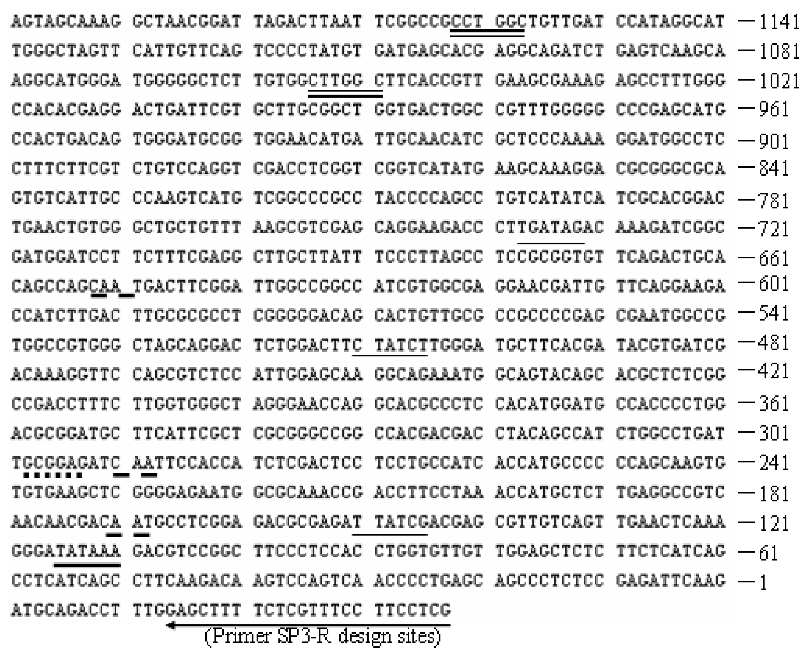
Ijms 14 08479f2
Figure 3. Molecular analysis of integration of T-DNA into H.orientalis. (A) PCR analysis of the phosphotransferase gene (hph) using specific primers hph-F&R to amplify an 811 bp fragment of transformants Lanes 1-4: transformants Bgl-1, Bgl-2, Bgl-3 and Bgl-4, respectively; Ma: 200 bp DNA Marker; (B) PCR analysis of the gene expression cassette (PproA-Bgl1-TtrpC) using specific primers PproA-F&TtrpC-R to amplify a 4194 bp fragment of transformants. Lane 1: host strain EU7-22; Lanes 2–5: transformants Bgl-1, Bgl-2, Bgl-3 and Bgl-4. Mb: DL10 000 DNA Marker.
Ijms 14 08479f3
Figure 4. Analysis of FPA and BG activities and bgl1 gene relative expression amount of host strain EU7-22 and transformant Bgl-2. (A) FPA and BG activities under induction condition; (B) FPA and BG activities under repression condition; (C) relative expression amount of Bgl1 gene under induction condition; (D) relative expression amount of Bgl1 gene under repression condition.
Ijms 14 08479f4
Figure 5. Sketch map of designed reverse primers sites for SP1, SP2 and SP3. The reverse primers (SP1, SP2 and SP3) were designed according to the interest gene (proA) sequence from H. orientalis EU7-22.
Ijms 14 08479f5
Figure 6. Schematic illustration of the binary expression vectors pUR5750-Bgl1 construction.
Ijms 14 08479f6
Table 1. Primers used in this study for polymerase chain reaction.
Primer namePrimer sequence (5′–3′)
SP1-RbAGACGGAGGTGATGTACTCCGAATC
SP2-RbCTTCTTGGCCTTGAAGAGAGCAGTAG
SP3-RbCGAGGAAGGAAACGAGAAAAGCTC
M13-FaCGCCAGGGTTTTCCCAGTCACGAC
M13-RbGAGCGGATAACAATTTCACACAGG
PproA-FaCTAGTCTAGAAGTAGCAAAGGCTAACGGATTAG
PproA-RbCCGCTCGAGCTTGAATCTCGGAGAGGGCTG
Bgl1-FaCCGCTCGAGATGCGTTACCGAACAGCAG
Bgl1-RbCCGGAATTCCTACGCTACCGACAGAGTGCT
TtrpC-FaCCGGAATTCAGTAGATGCCGACCGCG
TtrpC-RbCCCAAGCTTAAGAAGGATTACCTCTAAACAAG
hph-FaCGACAGCGTCTCCGACCTGA
hph-RbCGCCCAAGCTGCATCATCGAA
BGLYG-FaATCACCTACCCGCCTTCA
BGLYG-RbTCTCGTCGTCGGATGTTG
18s-FaAGGCGCGCAAATTACCCAATCC
18s-RbGCCCTCCAATTGTTCCTCGTTAAG
Fa: forward primer; Rb: reverse primer.

