<?xml version="1.0" encoding="UTF-8"?>
<!DOCTYPE article PUBLIC "-//NLM//DTD Journal Publishing DTD v2.3 20070202//EN" "journalpublishing.dtd">
<article xmlns:mml="http://www.w3.org/1998/Math/MathML" xmlns:xlink="http://www.w3.org/1999/xlink" xml:lang="en" article-type="review-article">
<front>
<journal-meta>
<journal-id journal-id-type="publisher-id">ijms</journal-id>
<journal-title>International Journal of Molecular Sciences</journal-title>
<abbrev-journal-title>Int. J. Mol. Sci.</abbrev-journal-title>
<issn pub-type="epub">1422-0067</issn>
<publisher>
<publisher-name>Molecular Diversity Preservation International (MDPI)</publisher-name></publisher></journal-meta>
<article-meta>
<article-id pub-id-type="doi">10.3390/ijms130911610</article-id>
<article-id pub-id-type="publisher-id">ijms-13-11610</article-id>
<article-categories>
<subj-group>
<subject>Review</subject></subj-group></article-categories>
<title-group>
<article-title>Experimental Aspects of Colloidal Interactions in Mixed Systems of Liposome and Inorganic Nanoparticle and Their Applications</article-title></title-group>
<contrib-group>
<contrib contrib-type="author">
<name><surname>Michel*</surname><given-names>Raphael</given-names></name></contrib>
<contrib contrib-type="author">
<name><surname>Gradzielski*</surname><given-names>Michael</given-names></name></contrib>
<aff id="af1-ijms-13-11610">Stranski-Laboratorium für Physikalische und Theoretische Chemie, Institut für Chemie, Technische Universität Berlin, Berlin D-10623, Germany; E-Mails: <email>raphael.michel@mailbox.tu-berlin.de</email> (R.M.); <email>michael.gradzielski@tu-berlin.de</email> (M.G.); Tel.: +49-30-314-22822 (R.M.); +49-30-314-24934 (M.G.); Fax: +49-30-314-26602 (M.G.).</aff></contrib-group>
<pub-date pub-type="collection">
<year>2012</year></pub-date>
<pub-date pub-type="epub">
<day>17</day>
<month>09</month>
<year>2012</year></pub-date>
<volume>13</volume>
<issue>9</issue>
<fpage>11610</fpage>
<lpage>11642</lpage>
<history>
<date date-type="received">
<day>07</day>
<month>08</month>
<year>2012</year></date>
<date date-type="rev-recd">
<day>01</day>
<month>09</month>
<year>2012</year></date>
<date date-type="accepted">
<day>06</day>
<month>09</month>
<year>2012</year></date></history>
<permissions>
<copyright-statement>© 2012 by the authors; licensee Molecular Diversity Preservation International, Basel, Switzerland.</copyright-statement>
<copyright-year>2012</copyright-year>
<license license-type="open-access" xlink:href="http://creativecommons.org/licenses/by/3.0">
<p>This article is an open-access article distributed under the terms and conditions of the Creative Commons Attribution license (http://creativecommons.org/licenses/by/3.0/).</p></license></permissions>
<abstract>
<p>In the past few years, growing attention has been devoted to the study of the interactions taking place in mixed systems of phospholipid membranes (for instance in the form of vesicles) and hard nanoparticles (NPs). In this context liposomes (vesicles) may serve as versatile carriers or as a model system for biological membranes. Research on these systems has led to the observation of novel hybrid structures whose morphology strongly depends on the charge, composition and size of the interacting colloidal species as well as on the nature (pH, ionic strength) of their dispersing medium. A central role is played by the phase behaviour of phospholipid bilayers which have a tremendous influence on the liposome properties. Another central aspect is the incorporation of nanoparticles into vesicles, which is intimately linked to the conditions required for transporting a nanoparticle through a membrane. Herein, we review recent progress made on the investigations of the interactions in liposome/nanoparticle systems focusing on the particularly interesting structures that are formed in these hybrid systems as well as their potential applications.</p></abstract>
<kwd-group>
<kwd>liposome</kwd>
<kwd>bilayer</kwd>
<kwd>nanoparticle</kwd>
<kwd>encapsulation</kwd>
<kwd>decorated vesicles</kwd>
<kwd>internalization</kwd>
<kwd>supported lipid bilayer</kwd>
<kwd>small unilamellar vesicle (SUV)</kwd>
<kwd>colloidal stabilization</kwd></kwd-group></article-meta></front>
<body>
<sec sec-type="intro">
<title>1. Introduction</title>
<p>Nowadays, the use of nanoparticles in biotechnology is of major interest due to the amazing potential displayed by mixed systems combining the properties of materials and the specific architectures and functions of the biological world. In particular, nanoparticles are attractive as they exhibit strongly size-dependent properties [<xref ref-type="bibr" rid="b1-ijms-13-11610">1</xref>–<xref ref-type="bibr" rid="b3-ijms-13-11610">3</xref>], which allows their use as functional system components, as for instance in sensor applications or as delivery vehicles [<xref ref-type="bibr" rid="b4-ijms-13-11610">4</xref>–<xref ref-type="bibr" rid="b6-ijms-13-11610">6</xref>]. However, they are now also in abundant use as antimicrobial agents (e.g., Ag nanoparticles) [<xref ref-type="bibr" rid="b7-ijms-13-11610">7</xref>,<xref ref-type="bibr" rid="b8-ijms-13-11610">8</xref>] or in the case of silica nanoparticles for various purposes, such as biomarkers for controlled release [<xref ref-type="bibr" rid="b9-ijms-13-11610">9</xref>–<xref ref-type="bibr" rid="b11-ijms-13-11610">11</xref>].</p>
<p>In the past few years, intensive research activities have been devoted to the characterization of the interaction between hard nanoparticles and biomolecules among which are proteins [<xref ref-type="bibr" rid="b12-ijms-13-11610">12</xref>], DNA molecules [<xref ref-type="bibr" rid="b13-ijms-13-11610">13</xref>] and phospholipid molecules (typically in the form of bilayers) [<xref ref-type="bibr" rid="b14-ijms-13-11610">14</xref>]. This is an important issue in the context of using nanoparticles in nanomedicine but also in the related field of nanotoxicology [<xref ref-type="bibr" rid="b15-ijms-13-11610">15</xref>–<xref ref-type="bibr" rid="b18-ijms-13-11610">18</xref>]. Accordingly, fundamental studies in this field are necessary in order to gain a systematic understanding of the relevant aspects in the interactions between nanoparticles and lipid bilayers.</p>
<sec>
<title>1.1. Properties of Phospholipid Bilayers</title>
<p>Phospholipids are amphiphilic molecules composed of a hydrophilic headgroup (phosphate) and two hydrocarbon chains thereby yielding to a packing parameter value <italic>p</italic> [<xref ref-type="bibr" rid="b19-ijms-13-11610">19</xref>–<xref ref-type="bibr" rid="b21-ijms-13-11610">21</xref>] between 0.5 and 1, granting them a truncated cone form (<italic>p</italic> being defined as: <italic>p = v</italic>/(<italic>al</italic>); with <italic>v</italic> being the volume of the hydrophobic chains, <italic>a</italic> the head group area of the lipid and <italic>l</italic> its stretched length [<xref ref-type="bibr" rid="b19-ijms-13-11610">19</xref>]). Thus, phospholipids have a tendency for the formation of bilayers [<xref ref-type="bibr" rid="b22-ijms-13-11610">22</xref>], also in the form of closed bilayers, <italic>i.e.</italic>, vesicles [<xref ref-type="bibr" rid="b23-ijms-13-11610">23</xref>,<xref ref-type="bibr" rid="b24-ijms-13-11610">24</xref>].</p>
<p>Phospholipid vesicles often called liposomes are frequently used in cosmetic and pharmaceutical formulations. This is mostly due to their ability to solubilise hydrophobic compounds and to allow for rheological control of the formulations [<xref ref-type="bibr" rid="b25-ijms-13-11610">25</xref>,<xref ref-type="bibr" rid="b26-ijms-13-11610">26</xref>]. Liposomes are also known to have a significant potential as drug carriers [<xref ref-type="bibr" rid="b27-ijms-13-11610">27</xref>,<xref ref-type="bibr" rid="b28-ijms-13-11610">28</xref>]. This is the case as they are able to transport hydrophobic molecules in their bilayer and/or hydrophilic molecules in their interior, thereby being very flexible carrier systems. In addition, having cell membrane mimetic surfaces, they facilitate the transport through membranes while being non cytotoxic and may serve as model systems for experimental and theoretical studies on cell membranes.</p>
<p>Pure phospholipid bilayer membranes exhibit a sharp main phase transition [<xref ref-type="bibr" rid="b29-ijms-13-11610">29</xref>] which is lipid specific and corresponds to the melting of their hydrocarbon chains. Below the chain melting temperature (<italic>T</italic><sub>m</sub>), the phospholipid molecules are locked in place [<xref ref-type="bibr" rid="b30-ijms-13-11610">30</xref>], bound tightly together by the van der Waals forces between their hydrocarbon chains, so that the bilayer is in a “solid-state” (gel phase), leading to the appearance of characteristic angular shapes in the case of vesicles [<xref ref-type="bibr" rid="b31-ijms-13-11610">31</xref>]. On the contrary, at temperatures above <italic>T</italic><sub>m</sub>, the lipids exhibit lateral as well as inter-layer mobility (flip-flop [<xref ref-type="bibr" rid="b32-ijms-13-11610">32</xref>]) responsible for the fluidity of the membrane. When passing through its phase transition, the bilayer vesicle membrane experiences a temporary permeability allowing the release of molecules from its interior [<xref ref-type="bibr" rid="b33-ijms-13-11610">33</xref>]. This last property being one other reason why liposomes are used as nanocontainers in drug delivery technologies, since they enable the achievement of on-demand release of encapsulated molecules by passing through the phase transition temperature.</p></sec>
<sec sec-type="methods">
<title>1.2. Methods of Vesicle Formation</title>
<p>The formation of liposomes occurs naturally but in many circumstances is done using appropriate procedures. Phospholipids dispersed in water “spontaneously” form giant multilamellar vesicles (MLVs) which are huge enough to be assimilated to a lamellar phase (it should be noted that typically during dispersion shear is applied, which is mainly responsible for the formation of the vesicles from these lamellar dispersions). However, starting from an MLV suspension, the preparation of well-defined small unilamellar vesicle (SUV) dispersions of specific composition, size and polydispersity can be relatively easily achieved using one or a combination of the different available techniques among which are sonication, rehydration of lipid film, or extrusion [<xref ref-type="bibr" rid="b24-ijms-13-11610">24</xref>,<xref ref-type="bibr" rid="b25-ijms-13-11610">25</xref>,<xref ref-type="bibr" rid="b28-ijms-13-11610">28</xref>,<xref ref-type="bibr" rid="b34-ijms-13-11610">34</xref>].</p>
<p>These preparation processes are required since the normal equilibrium state of phospholipids is a lamellar phase, whereas the vesicles are only metastable [<xref ref-type="bibr" rid="b35-ijms-13-11610">35</xref>]. Hence, this imposes one limitation to the possible applications of phospholipid vesicles as they are intrinsically unstable, while it might be noted that in some surfactant systems spontaneous vesicle formation and thermodynamic stability are present, especially in the case of cationic surfactants or mixtures of surfactants [<xref ref-type="bibr" rid="b36-ijms-13-11610">36</xref>–<xref ref-type="bibr" rid="b39-ijms-13-11610">39</xref>].</p>
<p>Thus, the investigation on mixed systems nanoparticles/liposomes is not only of interest from a fundamental point of view, as their interaction leads to novel colloidal structures such as decorated vesicles [<xref ref-type="bibr" rid="b40-ijms-13-11610">40</xref>,<xref ref-type="bibr" rid="b41-ijms-13-11610">41</xref>] and internalized particles [<xref ref-type="bibr" rid="b42-ijms-13-11610">42</xref>] but also due to their innovative potential applications for particle transport, cytotoxicity evaluation and liposome stabilization [<xref ref-type="bibr" rid="b40-ijms-13-11610">40</xref>,<xref ref-type="bibr" rid="b43-ijms-13-11610">43</xref>].</p>
<p>This review is organized as follows. We first focus on the study of the interactions present in mixed systems liposome/nanoparticle and the various colloidal structures they generate. Particular attention is given to the influence of the phase behaviour of the lipid bilayer (fluid/gel) on the nature of its interaction with colloidal particles. In the second part we present some interesting examples of applications and innovative technologies originating from specific features of these mixed systems. In the last part a particular emphasis is put on the use of silica, gold and silver nanoparticles, as being relevant examples for the case of oxides and pure metals.</p></sec></sec>
<sec>
<title>2. Experimental Investigations on Mixed Systems of Liposome and Nanoparticle</title>
<sec>
<title>2.1. Role of the Lipid Bilayer Phase Behaviour</title>
<p>An important aspect for the understanding of the interactions of lipid bilayers with nanoparticles is the phase behaviour of the lipids. In this context it is important to note that the properties of a given bilayer membrane in the gel phase (L<sub>β</sub>) are extremely different from those it exhibits in the fluid phase (L<sub>α</sub>).</p>
<p>First of all the structural arrangement of the phospholipid molecules is different. As described earlier, in the gel phase lipid-acyl chains are conformationally highly ordered while in the fluid phase the bilayer possesses both translational disorder and a high degree of lipid-acyl-chain disorder. This implies that as the bilayer is heated from the gel to the fluid phase, its thickness decreases substantially due to the conformational changes experienced by the acyl-chains [<xref ref-type="bibr" rid="b30-ijms-13-11610">30</xref>]. This decrease was analysed using both the Gibbs-Luzzati thickness (<italic>D</italic><sub>B</sub>) model [<xref ref-type="bibr" rid="b44-ijms-13-11610">44</xref>] (which partitions the overall bilayer thickness <italic>D</italic> into a lipid region <italic>D</italic><sub>B</sub> and a water region D<sub>W</sub> thereby ignoring details of the interfacial region) as well as with the steric bilayer thickness (<italic>D</italic><sub>B</sub>’) model (which takes into account the presence of water molecules mixed with the polar heads in the interfacial region) for the case of dipalmitoylphosphatidylcholine (DPPC) (<xref ref-type="table" rid="t1-ijms-13-11610">Table 1</xref>). This work confirmed that this decrease is not model-dependent and typically in the range of 10% to 20% [<xref ref-type="bibr" rid="b45-ijms-13-11610">45</xref>]. A concomitant increase in the bilayer area in the fluid state occurs, coming along with the corresponding decrease of the bilayer thickness, and that, of course, means one has then an enhanced hydration of the head group area. Similarly the state of the phospholipid chains has an important effect on the vesicle properties, in particular, when it comes to the deposition of supported lipid bilayers on nanoparticles where the surface ratio liposome/nanoparticle is of main interest. It can also be noted that the L<sub>α</sub>-phase shows a much larger swelling, which can be attributed to the undulation forces that are much more pronounced compared to the L<sub>β</sub>-phase (as the L<sub>α</sub>-phase possesses much lower bending moduli, see <xref ref-type="table" rid="t2-ijms-13-11610">Table 2</xref>.), where the Helfrich undulation repulsion between two bilayers can be described by [<xref ref-type="bibr" rid="b46-ijms-13-11610">46</xref>]:</p>
<disp-formula id="FD1">
<label>(1)</label>
<mml:math id="mm1" display="block">
<mml:semantics id="sm1">
<mml:mrow>
<mml:msub>
<mml:mrow>
<mml:mi>E</mml:mi></mml:mrow>
<mml:mrow>
<mml:mi>u</mml:mi>
<mml:mi>n</mml:mi>
<mml:mi>d</mml:mi></mml:mrow></mml:msub>
<mml:mo stretchy="false">(</mml:mo>
<mml:mi>D</mml:mi>
<mml:mo stretchy="false">)</mml:mo>
<mml:mo>=</mml:mo>
<mml:mfrac>
<mml:mrow>
<mml:mn>3</mml:mn>
<mml:msup>
<mml:mrow>
<mml:mi>π</mml:mi></mml:mrow>
<mml:mn>2</mml:mn></mml:msup></mml:mrow>
<mml:mrow>
<mml:mn>128</mml:mn></mml:mrow></mml:mfrac>
<mml:mfrac>
<mml:mrow>
<mml:msup>
<mml:mrow>
<mml:mrow>
<mml:mo stretchy="false">(</mml:mo>
<mml:mi>k</mml:mi>
<mml:mi>T</mml:mi>
<mml:mo stretchy="false">)</mml:mo></mml:mrow></mml:mrow>
<mml:mn>2</mml:mn></mml:msup></mml:mrow>
<mml:mrow>
<mml:msub>
<mml:mrow>
<mml:mi>κ</mml:mi></mml:mrow>
<mml:mi>b</mml:mi></mml:msub>
<mml:msup>
<mml:mrow>
<mml:mi>D</mml:mi></mml:mrow>
<mml:mn>2</mml:mn></mml:msup></mml:mrow></mml:mfrac></mml:mrow></mml:semantics></mml:math></disp-formula>
<p>which gives the free energy of the steric interaction per unit area of membrane (<italic>κ</italic><italic><sub>b</sub></italic>: mean bending modulus of one bilayer, <italic>D</italic>: distance between two adjacent membranes).</p>
<p>More importantly the fluidity of the membrane, as characterized by the bending elasticity, is dramatically altered by the phase transition. The bending elasticity (κ), described by the Helfrich theory [<xref ref-type="bibr" rid="b47-ijms-13-11610">47</xref>] as the energy required to bend a bilayer away from its spontaneous curvature, determines the balance between adhesive and elastic forces responsible for the bending (and potential wrapping) of the bilayer around a particle [<xref ref-type="bibr" rid="b42-ijms-13-11610">42</xref>,<xref ref-type="bibr" rid="b48-ijms-13-11610">48</xref>]. Values of the bending elasticity for a given bilayer have been found to decrease drastically when the bilayer is heated through the phase transition to its fluid phase [<xref ref-type="bibr" rid="b49-ijms-13-11610">49</xref>–<xref ref-type="bibr" rid="b51-ijms-13-11610">51</xref>] (<xref ref-type="table" rid="t2-ijms-13-11610">Table 2</xref>). This observation is related to the inability of the gel membrane to bend around particles or to fuse on flat surfaces, as a result of its high rigidity [<xref ref-type="bibr" rid="b52-ijms-13-11610">52</xref>].</p>
<p>Finally, in the case of zwitterionic phospholipids, the electrostatic properties of the bilayer are also altered by the conformational changes occurring during the main phase transition. The dielectric permittivity of lipid bilayer, as well as its conductivity in aqueous solution, have been found to increase while passing from the gel to the fluid phase [<xref ref-type="bibr" rid="b53-ijms-13-11610">53</xref>,<xref ref-type="bibr" rid="b54-ijms-13-11610">54</xref>]. These changes, resulting from the decrease of the dipolar correlation taking place between the head group dipoles in the hydrophilic part of the bilayer, have a non-negligible influence on the surface interactions taking place between lipid bilayers and nanoparticles.</p></sec>
<sec>
<title>2.2. Nature of the Interaction in Mixed System Liposome/Nanoparticle</title>
<p>The interaction between lipid bilayers and solid surfaces is rather complex and includes van der Waals, double layer, hydration, hydrophobic, thermal undulation and protrusion forces [<xref ref-type="bibr" rid="b52-ijms-13-11610">52</xref>]. The precise nature of the interaction, as well as the structure it creates, is in all cases related to the properties of the lipid and the solid surface used as well as the nature of the dispersing medium.</p>
<sec>
<title>2.2.1. Van der Waals Interaction</title>
<p>Describing the strength of van der Waals forces in mixed liposome/nanoparticle systems in a quantitative manner is challenging due to the geometry and nature of the system components. In the ideal case of a sphere (medium 1: nanoparticle) interacting with a shell (medium 3: liposome) in an aqueous medium (medium 2), the van der Waals energy is given by the equation [<xref ref-type="bibr" rid="b55-ijms-13-11610">55</xref>,<xref ref-type="bibr" rid="b56-ijms-13-11610">56</xref>]:</p>
<disp-formula id="FD2">
<label>(2)</label>
<mml:math id="mm2" display="block">
<mml:semantics id="sm2">
<mml:mrow>
<mml:msub>
<mml:mrow>
<mml:mi>E</mml:mi></mml:mrow>
<mml:mrow>
<mml:mi>v</mml:mi>
<mml:mi>d</mml:mi>
<mml:mi>w</mml:mi></mml:mrow></mml:msub>
<mml:mo>=</mml:mo>
<mml:mo>-</mml:mo>
<mml:msub>
<mml:mrow>
<mml:mi>A</mml:mi></mml:mrow>
<mml:mrow>
<mml:mn>123</mml:mn></mml:mrow></mml:msub>
<mml:mfrac>
<mml:mrow>
<mml:msub>
<mml:mrow>
<mml:mi>R</mml:mi></mml:mrow>
<mml:mn>1</mml:mn></mml:msub>
<mml:msub>
<mml:mrow>
<mml:mi>R</mml:mi></mml:mrow>
<mml:mn>2</mml:mn></mml:msub></mml:mrow>
<mml:mrow>
<mml:mn>6</mml:mn>
<mml:mo stretchy="false">(</mml:mo>
<mml:msub>
<mml:mrow>
<mml:mi>R</mml:mi></mml:mrow>
<mml:mn>1</mml:mn></mml:msub>
<mml:mo>+</mml:mo>
<mml:msub>
<mml:mrow>
<mml:mi>R</mml:mi></mml:mrow>
<mml:mn>2</mml:mn></mml:msub>
<mml:mo stretchy="false">)</mml:mo></mml:mrow></mml:mfrac>
<mml:mrow>
<mml:mo>(</mml:mo>
<mml:mrow>
<mml:mfrac>
<mml:mn>1</mml:mn>
<mml:mi>d</mml:mi></mml:mfrac>
<mml:mo>-</mml:mo>
<mml:mfrac>
<mml:mn>1</mml:mn>
<mml:mrow>
<mml:mo stretchy="false">(</mml:mo>
<mml:mi>d</mml:mi>
<mml:mo>+</mml:mo>
<mml:mi>h</mml:mi>
<mml:mo stretchy="false">)</mml:mo></mml:mrow></mml:mfrac></mml:mrow>
<mml:mo>)</mml:mo></mml:mrow>
<mml:mo>-</mml:mo>
<mml:mfrac>
<mml:mi>A</mml:mi>
<mml:mn>6</mml:mn></mml:mfrac>
<mml:mtext>ln</mml:mtext>
<mml:mrow>
<mml:mo>(</mml:mo>
<mml:mrow>
<mml:mfrac>
<mml:mi>d</mml:mi>
<mml:mrow>
<mml:mi>d</mml:mi>
<mml:mo>+</mml:mo>
<mml:mi>h</mml:mi></mml:mrow></mml:mfrac></mml:mrow>
<mml:mo>)</mml:mo></mml:mrow></mml:mrow></mml:semantics></mml:math></disp-formula>
<p>where <italic>R</italic><sub>1</sub> is the radius of the sphere (particle), <italic>R</italic><sub>2</sub> the outer radius of the shell, <italic>h</italic> the thickness of the shell, <italic>d</italic> the distance between the surfaces of the two objects and <italic>A</italic><sub>123</sub> the Hamaker constant of the system. An approximate value of <italic>A</italic><sub>123</sub> may be given in terms of the Hamaker constants of the individual media as follows [<xref ref-type="bibr" rid="b55-ijms-13-11610">55</xref>,<xref ref-type="bibr" rid="b57-ijms-13-11610">57</xref>]:</p>
<disp-formula id="FD3">
<label>(3)</label>
<mml:math id="mm3" display="block">
<mml:semantics id="sm3">
<mml:mrow>
<mml:msub>
<mml:mrow>
<mml:mi>A</mml:mi></mml:mrow>
<mml:mrow>
<mml:mn>123</mml:mn></mml:mrow></mml:msub>
<mml:mo>≈</mml:mo>
<mml:mo stretchy="false">(</mml:mo>
<mml:msqrt>
<mml:mrow>
<mml:msub>
<mml:mrow>
<mml:mi>A</mml:mi></mml:mrow>
<mml:mrow>
<mml:mn>11</mml:mn></mml:mrow></mml:msub></mml:mrow></mml:msqrt>
<mml:mo>-</mml:mo>
<mml:msqrt>
<mml:mrow>
<mml:msub>
<mml:mrow>
<mml:mi>A</mml:mi></mml:mrow>
<mml:mrow>
<mml:mn>22</mml:mn></mml:mrow></mml:msub></mml:mrow></mml:msqrt>
<mml:mo stretchy="false">)</mml:mo>
<mml:mo stretchy="false">(</mml:mo>
<mml:msqrt>
<mml:mrow>
<mml:msub>
<mml:mrow>
<mml:mi>A</mml:mi></mml:mrow>
<mml:mrow>
<mml:mn>33</mml:mn></mml:mrow></mml:msub></mml:mrow></mml:msqrt>
<mml:mo>-</mml:mo>
<mml:msqrt>
<mml:mrow>
<mml:msub>
<mml:mrow>
<mml:mi>A</mml:mi></mml:mrow>
<mml:mrow>
<mml:mn>22</mml:mn></mml:mrow></mml:msub></mml:mrow></mml:msqrt>
<mml:mo stretchy="false">)</mml:mo></mml:mrow></mml:semantics></mml:math></disp-formula>
<p>where <italic>A</italic><italic><sub>xx</sub></italic> is the Hamaker constant between two semi-infinite planes of medium <italic>x</italic> in vacuum.</p>
<p>For “symmetrical” systems, the Hamaker constant is always positive, leading to attractive forces, but between dissimilar surfaces, as in the case of mixed systems liposome/nanoparticle, <italic>A</italic><sub>123</sub> can be either positive or negative leading respectively to attractive or repulsive forces. For phospholipid bilayer interacting with oxide particles (SiO<sub>2</sub>, TiO<sub>2</sub>) the Hamaker constant is typically (3 − 4) × 10<sup>−21</sup> J ≈ 0.75 to 1 <italic>kT</italic> (for <italic>T</italic> = 25 °C) [<xref ref-type="bibr" rid="b52-ijms-13-11610">52</xref>,<xref ref-type="bibr" rid="b58-ijms-13-11610">58</xref>,<xref ref-type="bibr" rid="b59-ijms-13-11610">59</xref>].</p></sec>
<sec>
<title>2.2.2. Electrostatic Interaction</title>
<p>In general, the electrostatic double-layer interaction energy between two identical planar surfaces decreases exponentially with the distance. However, for two surfaces of different charge densities or potentials, which is the case in mixed system liposome/nanoparticle, this interaction energy can exhibit a minimum or a maximum at some finite distance [<xref ref-type="bibr" rid="b55-ijms-13-11610">55</xref>]. The study on these asymmetric systems has led to different approximate equations for the interaction energy of two surfaces of unequal but constant potentials [<xref ref-type="bibr" rid="b60-ijms-13-11610">60</xref>–<xref ref-type="bibr" rid="b62-ijms-13-11610">62</xref>]. The “Hogg–Healy–Fuerstenau” equation (HHF) [<xref ref-type="bibr" rid="b55-ijms-13-11610">55</xref>,<xref ref-type="bibr" rid="b60-ijms-13-11610">60</xref>] for two different planar surfaces of low constant potentials <italic>ψ</italic><sub>1</sub> and <italic>ψ</italic><sub>2</sub> in 1:1 electrolyte is given by:</p>
<disp-formula id="FD4">
<label>(4)</label>
<mml:math id="mm4" display="block">
<mml:semantics id="sm4">
<mml:mrow>
<mml:msub>
<mml:mrow>
<mml:mi>W</mml:mi></mml:mrow>
<mml:mrow>
<mml:mi>D</mml:mi>
<mml:mi>L</mml:mi></mml:mrow></mml:msub>
<mml:mo stretchy="false">(</mml:mo>
<mml:mi>D</mml:mi>
<mml:mo stretchy="false">)</mml:mo>
<mml:mo>=</mml:mo>
<mml:mfrac>
<mml:mrow>
<mml:msub>
<mml:mrow>
<mml:mi>ɛ</mml:mi></mml:mrow>
<mml:mn>0</mml:mn></mml:msub>
<mml:mi>ɛ</mml:mi>
<mml:mi>κ</mml:mi>
<mml:mo stretchy="false">[</mml:mo>
<mml:mn>2</mml:mn>
<mml:msub>
<mml:mrow>
<mml:mi>ψ</mml:mi></mml:mrow>
<mml:mn>1</mml:mn></mml:msub>
<mml:msub>
<mml:mrow>
<mml:mi>ψ</mml:mi></mml:mrow>
<mml:mn>2</mml:mn></mml:msub>
<mml:mo>-</mml:mo>
<mml:mo stretchy="false">(</mml:mo>
<mml:msubsup>
<mml:mrow>
<mml:mi>ψ</mml:mi></mml:mrow>
<mml:mn>1</mml:mn>
<mml:mn>2</mml:mn></mml:msubsup>
<mml:mo>+</mml:mo>
<mml:msubsup>
<mml:mrow>
<mml:mi>ψ</mml:mi></mml:mrow>
<mml:mn>2</mml:mn>
<mml:mn>2</mml:mn></mml:msubsup>
<mml:mo stretchy="false">)</mml:mo>
<mml:msup>
<mml:mrow>
<mml:mi>e</mml:mi></mml:mrow>
<mml:mrow>
<mml:mo>-</mml:mo>
<mml:mi>κ</mml:mi>
<mml:mi>D</mml:mi></mml:mrow></mml:msup>
<mml:mo stretchy="false">]</mml:mo></mml:mrow>
<mml:mrow>
<mml:mo stretchy="false">(</mml:mo>
<mml:msup>
<mml:mrow>
<mml:mi>e</mml:mi></mml:mrow>
<mml:mrow>
<mml:mo>+</mml:mo>
<mml:mi>κ</mml:mi>
<mml:mi>D</mml:mi></mml:mrow></mml:msup>
<mml:mo>-</mml:mo>
<mml:msup>
<mml:mrow>
<mml:mi>e</mml:mi></mml:mrow>
<mml:mrow>
<mml:mo>-</mml:mo>
<mml:mi>κ</mml:mi>
<mml:mi>D</mml:mi></mml:mrow></mml:msup>
<mml:mo stretchy="false">)</mml:mo></mml:mrow></mml:mfrac></mml:mrow></mml:semantics></mml:math></disp-formula>
<p>where <italic>D</italic> is the distance between the surfaces and κ their Debye length. This equation has recently been successfully implemented to characterize the double layer interaction between lipid bilayer and silica substrates [<xref ref-type="bibr" rid="b52-ijms-13-11610">52</xref>].</p>
<p>Nevertheless, when working with nanoparticles and vesicles one often has to take into account the curvature of the interacting surfaces. To this aim, applying the Derjaguin approximation on <xref rid="FD4" ref-type="disp-formula">equation 4</xref> leads to the following formula for the approximate interaction energy between dissimilar double layers on two spherical particles with radii <italic>R</italic><sub>1</sub> and <italic>R</italic><sub>2</sub> [<xref ref-type="bibr" rid="b60-ijms-13-11610">60</xref>]:</p>
<disp-formula id="FD5">
<label>(5)</label>
<mml:math id="mm5" display="block">
<mml:semantics id="sm5">
<mml:mrow>
<mml:msub>
<mml:mrow>
<mml:mi>W</mml:mi></mml:mrow>
<mml:mrow>
<mml:mi>D</mml:mi>
<mml:mi>L</mml:mi></mml:mrow></mml:msub>
<mml:mo stretchy="false">(</mml:mo>
<mml:mi>D</mml:mi>
<mml:mo stretchy="false">)</mml:mo>
<mml:mo>=</mml:mo>
<mml:mfrac>
<mml:mrow>
<mml:mi>ɛ</mml:mi>
<mml:msub>
<mml:mrow>
<mml:mi>R</mml:mi></mml:mrow>
<mml:mn>1</mml:mn></mml:msub>
<mml:msub>
<mml:mrow>
<mml:mi>R</mml:mi></mml:mrow>
<mml:mn>2</mml:mn></mml:msub>
<mml:mo stretchy="false">(</mml:mo>
<mml:msubsup>
<mml:mrow>
<mml:mi>ψ</mml:mi></mml:mrow>
<mml:mn>1</mml:mn>
<mml:mn>2</mml:mn></mml:msubsup>
<mml:mo>+</mml:mo>
<mml:msubsup>
<mml:mrow>
<mml:mi>ψ</mml:mi></mml:mrow>
<mml:mn>2</mml:mn>
<mml:mn>2</mml:mn></mml:msubsup>
<mml:mo stretchy="false">)</mml:mo></mml:mrow>
<mml:mrow>
<mml:mn>4</mml:mn>
<mml:mo stretchy="false">(</mml:mo>
<mml:msub>
<mml:mrow>
<mml:mi>R</mml:mi></mml:mrow>
<mml:mn>1</mml:mn></mml:msub>
<mml:mo>+</mml:mo>
<mml:msub>
<mml:mrow>
<mml:mi>R</mml:mi></mml:mrow>
<mml:mn>2</mml:mn></mml:msub>
<mml:mo stretchy="false">)</mml:mo></mml:mrow></mml:mfrac>
<mml:mrow>
<mml:mo>[</mml:mo>
<mml:mrow>
<mml:mfrac>
<mml:mrow>
<mml:mn>2</mml:mn>
<mml:msub>
<mml:mrow>
<mml:mi>ψ</mml:mi></mml:mrow>
<mml:mn>1</mml:mn></mml:msub>
<mml:msub>
<mml:mrow>
<mml:mi>ψ</mml:mi></mml:mrow>
<mml:mn>2</mml:mn></mml:msub></mml:mrow>
<mml:mrow>
<mml:mo stretchy="false">(</mml:mo>
<mml:msubsup>
<mml:mrow>
<mml:mi>ψ</mml:mi></mml:mrow>
<mml:mn>1</mml:mn>
<mml:mn>2</mml:mn></mml:msubsup>
<mml:mo>+</mml:mo>
<mml:msubsup>
<mml:mrow>
<mml:mi>ψ</mml:mi></mml:mrow>
<mml:mn>2</mml:mn>
<mml:mn>2</mml:mn></mml:msubsup>
<mml:mo stretchy="false">)</mml:mo></mml:mrow></mml:mfrac>
<mml:mtext>ln</mml:mtext>
<mml:mrow>
<mml:mo>(</mml:mo>
<mml:mrow>
<mml:mfrac>
<mml:mrow>
<mml:mn>1</mml:mn>
<mml:mo>+</mml:mo>
<mml:msup>
<mml:mrow>
<mml:mi>e</mml:mi></mml:mrow>
<mml:mrow>
<mml:mo>-</mml:mo>
<mml:mi>κ</mml:mi>
<mml:mi>D</mml:mi></mml:mrow></mml:msup></mml:mrow>
<mml:mrow>
<mml:mn>1</mml:mn>
<mml:mo>-</mml:mo>
<mml:msup>
<mml:mrow>
<mml:mi>e</mml:mi></mml:mrow>
<mml:mrow>
<mml:mo>-</mml:mo>
<mml:mi>κ</mml:mi>
<mml:mi>D</mml:mi></mml:mrow></mml:msup></mml:mrow></mml:mfrac></mml:mrow>
<mml:mo>)</mml:mo></mml:mrow>
<mml:mo>+</mml:mo>
<mml:mtext>ln</mml:mtext>
<mml:mo stretchy="false">(</mml:mo>
<mml:mn>1</mml:mn>
<mml:mo>-</mml:mo>
<mml:msup>
<mml:mrow>
<mml:mi>e</mml:mi></mml:mrow>
<mml:mrow>
<mml:mo>-</mml:mo>
<mml:mn>2</mml:mn>
<mml:mi>κ</mml:mi>
<mml:mi>D</mml:mi></mml:mrow></mml:msup>
<mml:mo stretchy="false">)</mml:mo></mml:mrow>
<mml:mo>]</mml:mo></mml:mrow></mml:mrow></mml:semantics></mml:math></disp-formula>
<p>It is important to note that the HHF formula is based on the Debye-Hückel linear approximation. This approximation is thus applicable for sufficiently low potentials (commonly <italic>ψ</italic> &lt; 25 mV). More recent work using the Poisson-Boltzmann expression for the potential has led to more accurate analytical approximations [<xref ref-type="bibr" rid="b61-ijms-13-11610">61</xref>].</p>
<p>Alternatively, The Gouy-Chapman Theory has also been used to describe the electrostatic interaction energy between a flat substrate with surface potential <italic>ψ</italic><sub>sub</sub> and a flat bilayer of surface potential <italic>ψ</italic><sub>bil</sub> in monovalent salt solution [<xref ref-type="bibr" rid="b63-ijms-13-11610">63</xref>,<xref ref-type="bibr" rid="b64-ijms-13-11610">64</xref>] leading to the following formula:</p>
<disp-formula id="FD6">
<label>(6)</label>
<mml:math id="mm6" display="block">
<mml:semantics id="sm6">
<mml:mrow>
<mml:msub>
<mml:mrow>
<mml:mi>W</mml:mi></mml:mrow>
<mml:mrow>
<mml:mi>D</mml:mi>
<mml:mi>L</mml:mi></mml:mrow></mml:msub>
<mml:mo stretchy="false">(</mml:mo>
<mml:mi>D</mml:mi>
<mml:mo stretchy="false">)</mml:mo>
<mml:mo>=</mml:mo>
<mml:mn>64</mml:mn>
<mml:mi>k</mml:mi>
<mml:mi>T</mml:mi>
<mml:msub>
<mml:mrow>
<mml:mi>ρ</mml:mi></mml:mrow>
<mml:mo>∞</mml:mo></mml:msub>
<mml:mi> </mml:mi>
<mml:mtext>tanh</mml:mtext>
<mml:mi> </mml:mi>
<mml:mrow>
<mml:mo>(</mml:mo>
<mml:mrow>
<mml:mfrac>
<mml:mrow>
<mml:mi>e</mml:mi>
<mml:msub>
<mml:mrow>
<mml:mi>ψ</mml:mi></mml:mrow>
<mml:mrow>
<mml:mi>s</mml:mi>
<mml:mi>u</mml:mi>
<mml:mi>b</mml:mi></mml:mrow></mml:msub></mml:mrow>
<mml:mrow>
<mml:mn>4</mml:mn>
<mml:mi>k</mml:mi>
<mml:mi>T</mml:mi></mml:mrow></mml:mfrac></mml:mrow>
<mml:mo>)</mml:mo></mml:mrow>
<mml:mi> </mml:mi>
<mml:mtext>tanh</mml:mtext>
<mml:mi> </mml:mi>
<mml:mrow>
<mml:mo>(</mml:mo>
<mml:mrow>
<mml:mfrac>
<mml:mrow>
<mml:mi>e</mml:mi>
<mml:msub>
<mml:mrow>
<mml:mi>ψ</mml:mi></mml:mrow>
<mml:mrow>
<mml:mi>b</mml:mi>
<mml:mi>i</mml:mi>
<mml:mi>l</mml:mi></mml:mrow></mml:msub></mml:mrow>
<mml:mrow>
<mml:mn>4</mml:mn>
<mml:mi>k</mml:mi>
<mml:mi>T</mml:mi></mml:mrow></mml:mfrac></mml:mrow>
<mml:mo>)</mml:mo></mml:mrow>
<mml:mfrac>
<mml:mrow>
<mml:msup>
<mml:mrow>
<mml:mi>e</mml:mi></mml:mrow>
<mml:mrow>
<mml:mo>-</mml:mo>
<mml:mi>κ</mml:mi>
<mml:mi>D</mml:mi></mml:mrow></mml:msup></mml:mrow>
<mml:mi>κ</mml:mi></mml:mfrac></mml:mrow></mml:semantics></mml:math></disp-formula>
<p>where <italic>T</italic> is the temperature, <italic>k</italic> the Boltzmann constant and <italic>ρ</italic><italic><sub>∞</sub></italic> the ions concentration. This equation also assumes a constant surface potential at any distance <italic>D</italic>.</p></sec>
<sec>
<title>2.2.3. Hydration Forces</title>
<p>In systems containing phospholipid bilayers, van der Waals and double layer interactions, as jointly expressed by the DLVO theory, often fail to precisely describe the phenomena taking place at short distances due to the presence of repulsive hydration forces [<xref ref-type="bibr" rid="b65-ijms-13-11610">65</xref>,<xref ref-type="bibr" rid="b66-ijms-13-11610">66</xref>], as has been observed experimentally by means of the surface force apparatus. These repulsive forces are believed to arise from the presence of a layer of water molecules, strongly bound to the hydrophilic surfaces and preventing them from approaching any closer than the thickness of two water molecules [<xref ref-type="bibr" rid="b55-ijms-13-11610">55</xref>,<xref ref-type="bibr" rid="b65-ijms-13-11610">65</xref>,<xref ref-type="bibr" rid="b67-ijms-13-11610">67</xref>].</p>
<p>The full nature of this interaction is still an active field of research. However, in the case of hydrophilic surfaces in aqueous solution, the hydration energy is attributed to stable structured water layers hydrogen bonded to the solid surfaces, while the hydration repulsion between two lipid membranes is believed to be dominated by entropic factors (microscopic thermal fluctuations).</p>
<p>Tero <italic>et al</italic>. [<xref ref-type="bibr" rid="b64-ijms-13-11610">64</xref>] were the first to propose an expression for the calculation of the hydration energy between lipid bilayer and hydrophilic surfaces, by assuming that the hydration between lipid membrane and solid substrate (<italic>W</italic><sub>hyd</sub>) is the average of each hydration energy, giving:</p>
<disp-formula id="FD7">
<label>(7)</label>
<mml:math id="mm7" display="block">
<mml:semantics id="sm7">
<mml:mrow>
<mml:msub>
<mml:mrow>
<mml:mi>W</mml:mi></mml:mrow>
<mml:mrow>
<mml:mi>h</mml:mi>
<mml:mi>y</mml:mi>
<mml:mi>d</mml:mi></mml:mrow></mml:msub>
<mml:mo>=</mml:mo>
<mml:mfrac>
<mml:mrow>
<mml:msub>
<mml:mrow>
<mml:mi>W</mml:mi></mml:mrow>
<mml:mrow>
<mml:mi>s</mml:mi>
<mml:mi>o</mml:mi>
<mml:mi>l</mml:mi>
<mml:mi>i</mml:mi>
<mml:mi>d</mml:mi></mml:mrow></mml:msub>
<mml:mo>+</mml:mo>
<mml:msub>
<mml:mrow>
<mml:mi>W</mml:mi></mml:mrow>
<mml:mrow>
<mml:mi>l</mml:mi>
<mml:mi>i</mml:mi>
<mml:mi>p</mml:mi>
<mml:mi>i</mml:mi>
<mml:mi>d</mml:mi></mml:mrow></mml:msub></mml:mrow>
<mml:mn>2</mml:mn></mml:mfrac></mml:mrow></mml:semantics></mml:math></disp-formula>
<p>where <italic>W</italic><sub>solid</sub> is the hydration force between hydrophilic substrates, depending on the nature of the substrate and its medium. Empirically, <italic>W</italic><sub>solid</sub> has been found to decay exponentially with the distance (<italic>D</italic>) between the surfaces [<xref ref-type="bibr" rid="b55-ijms-13-11610">55</xref>,<xref ref-type="bibr" rid="b63-ijms-13-11610">63</xref>,<xref ref-type="bibr" rid="b64-ijms-13-11610">64</xref>]:</p>
<disp-formula id="FD8">
<label>(8)</label>
<mml:math id="mm8" display="block">
<mml:semantics id="sm8">
<mml:mrow>
<mml:msub>
<mml:mrow>
<mml:mi>W</mml:mi></mml:mrow>
<mml:mrow>
<mml:mi>s</mml:mi>
<mml:mi>o</mml:mi>
<mml:mi>l</mml:mi>
<mml:mi>i</mml:mi>
<mml:mi>d</mml:mi></mml:mrow></mml:msub>
<mml:mo>=</mml:mo>
<mml:msub>
<mml:mrow>
<mml:mi>W</mml:mi></mml:mrow>
<mml:mn>0</mml:mn></mml:msub>
<mml:msup>
<mml:mrow>
<mml:mi>e</mml:mi></mml:mrow>
<mml:mrow>
<mml:mo>-</mml:mo>
<mml:mi>D</mml:mi>
<mml:mo>/</mml:mo>
<mml:msub>
<mml:mrow>
<mml:mi>λ</mml:mi></mml:mrow>
<mml:mn>0</mml:mn></mml:msub></mml:mrow></mml:msup></mml:mrow></mml:semantics></mml:math></disp-formula>
<p>where <italic>λ</italic><sub>0</sub> is on the order of 1 nm and <italic>W</italic><italic><sub>0</sub></italic> depends on the hydration of the surfaces but is usually in the range 3–30 mJ·m<sup>−2</sup> ≈ 0.7 to 7 <italic>kT</italic>·nm<sup>−2</sup> (for <italic>T</italic> = 25 °C).</p>
<p>On the other hand, <italic>W</italic><sub>lipid</sub> is believed to arise from thermal fluctuations [<xref ref-type="bibr" rid="b68-ijms-13-11610">68</xref>] and thus depends on the three different types of thermal motion that a membrane can experience: protrusion, peristalsis and undulation. The energetic contribution of these different motions can be separately expressed as follows [<xref ref-type="bibr" rid="b55-ijms-13-11610">55</xref>,<xref ref-type="bibr" rid="b64-ijms-13-11610">64</xref>]:</p>
<disp-formula id="FD9">
<label>(9)</label>
<mml:math id="mm9" display="block">
<mml:semantics id="sm9">
<mml:mrow>
<mml:msub>
<mml:mrow>
<mml:mi>W</mml:mi></mml:mrow>
<mml:mrow>
<mml:mi>p</mml:mi>
<mml:mi>r</mml:mi>
<mml:mi>o</mml:mi>
<mml:mi>t</mml:mi>
<mml:mi>r</mml:mi>
<mml:mi>u</mml:mi>
<mml:mi>s</mml:mi>
<mml:mi>i</mml:mi>
<mml:mi>o</mml:mi>
<mml:mi>n</mml:mi></mml:mrow></mml:msub>
<mml:mo>=</mml:mo>
<mml:mn>2.7</mml:mn>
<mml:mi mathvariant="normal">Γ</mml:mi>
<mml:msup>
<mml:mrow>
<mml:mi>k</mml:mi>
<mml:mi>T</mml:mi>
<mml:mi>e</mml:mi></mml:mrow>
<mml:mrow>
<mml:mo>-</mml:mo>
<mml:mo stretchy="false">(</mml:mo>
<mml:msub>
<mml:mrow>
<mml:mi>α</mml:mi></mml:mrow>
<mml:mi>p</mml:mi></mml:msub>
<mml:mi>D</mml:mi>
<mml:mo>/</mml:mo>
<mml:mi>k</mml:mi>
<mml:mi>t</mml:mi>
<mml:mo stretchy="false">)</mml:mo></mml:mrow></mml:msup></mml:mrow></mml:semantics></mml:math></disp-formula>
<disp-formula id="FD10">
<label>(10)</label>
<mml:math id="mm10" display="block">
<mml:semantics id="sm10">
<mml:mrow>
<mml:msub>
<mml:mrow>
<mml:mi>W</mml:mi></mml:mrow>
<mml:mrow>
<mml:mi>p</mml:mi>
<mml:mi>e</mml:mi>
<mml:mi>r</mml:mi>
<mml:mi>i</mml:mi>
<mml:mi>s</mml:mi>
<mml:mi>t</mml:mi>
<mml:mi>a</mml:mi>
<mml:mi>l</mml:mi>
<mml:mi>s</mml:mi>
<mml:mi>i</mml:mi>
<mml:mi>s</mml:mi></mml:mrow></mml:msub>
<mml:mo>=</mml:mo>
<mml:mfrac>
<mml:mrow>
<mml:msup>
<mml:mrow>
<mml:mrow>
<mml:mo stretchy="false">(</mml:mo>
<mml:mi>k</mml:mi>
<mml:mi>T</mml:mi>
<mml:mo stretchy="false">)</mml:mo></mml:mrow></mml:mrow>
<mml:mn>2</mml:mn></mml:msup></mml:mrow>
<mml:mrow>
<mml:mn>20</mml:mn>
<mml:msub>
<mml:mrow>
<mml:mi>k</mml:mi></mml:mrow>
<mml:mi>a</mml:mi></mml:msub>
<mml:msup>
<mml:mrow>
<mml:mi>D</mml:mi></mml:mrow>
<mml:mn>4</mml:mn></mml:msup></mml:mrow></mml:mfrac></mml:mrow></mml:semantics></mml:math></disp-formula>
<disp-formula id="FD11">
<label>(11)</label>
<mml:math id="mm11" display="block">
<mml:semantics id="sm11">
<mml:mrow>
<mml:msub>
<mml:mrow>
<mml:mi>W</mml:mi></mml:mrow>
<mml:mrow>
<mml:mi>u</mml:mi>
<mml:mi>n</mml:mi>
<mml:mi>d</mml:mi>
<mml:mi>u</mml:mi>
<mml:mi>l</mml:mi>
<mml:mi>a</mml:mi>
<mml:mi>t</mml:mi>
<mml:mi>i</mml:mi>
<mml:mi>o</mml:mi>
<mml:mi>n</mml:mi></mml:mrow></mml:msub>
<mml:mo>=</mml:mo>
<mml:mfrac>
<mml:mrow>
<mml:mn>3</mml:mn>
<mml:msup>
<mml:mrow>
<mml:mi>π</mml:mi></mml:mrow>
<mml:mn>2</mml:mn></mml:msup>
<mml:msup>
<mml:mrow>
<mml:mrow>
<mml:mo stretchy="false">(</mml:mo>
<mml:mi>k</mml:mi>
<mml:mi>T</mml:mi>
<mml:mo stretchy="false">)</mml:mo></mml:mrow></mml:mrow>
<mml:mn>2</mml:mn></mml:msup></mml:mrow>
<mml:mrow>
<mml:mn>64</mml:mn>
<mml:msub>
<mml:mrow>
<mml:mi>k</mml:mi></mml:mrow>
<mml:mi>b</mml:mi></mml:msub>
<mml:msup>
<mml:mrow>
<mml:mi>D</mml:mi></mml:mrow>
<mml:mn>3</mml:mn></mml:msup></mml:mrow></mml:mfrac></mml:mrow></mml:semantics></mml:math></disp-formula>
<p>where Γ is the surface density of protruding head groups, <italic>α</italic><italic><sub>p</sub></italic> the (hydrophobic) protrusion energy per unit length<sub>,</sub> <italic>k</italic><italic><sub>a</sub></italic> the area expansion modulus and <italic>k</italic><italic><sub>b</sub></italic> the bending modulus. Typical values for these parameters are: <italic>α</italic><italic><sub>p</sub></italic> = 2.5 × 10<sup>−11</sup> J·m<sup>−1</sup>, <italic>k</italic><italic><sub>a</sub></italic> = 0.15 J·m<sup>−2</sup> and <italic>k</italic><italic><sub>b</sub></italic> = 10<sup>−19</sup> J [<xref ref-type="bibr" rid="b55-ijms-13-11610">55</xref>,<xref ref-type="bibr" rid="b64-ijms-13-11610">64</xref>] while Γ depends on the molecular occupying area of the chosen lipid.</p>
<p>It should be noted that undulation forces may be neglected in the case of membranes carrying unscreened surface charges or being under tension. Nevertheless, when considering every contribution, the hydration energy between a bilayer and a flat hydrophilic substrate is given by the expression:</p>
<disp-formula id="FD12">
<label>(12)</label>
<mml:math id="mm12" display="block">
<mml:semantics id="sm12">
<mml:mrow>
<mml:msub>
<mml:mrow>
<mml:mi>W</mml:mi></mml:mrow>
<mml:mrow>
<mml:mi>h</mml:mi>
<mml:mi>y</mml:mi>
<mml:mi>d</mml:mi></mml:mrow></mml:msub>
<mml:mo>=</mml:mo>
<mml:mfrac>
<mml:mn>1</mml:mn>
<mml:mn>2</mml:mn></mml:mfrac>
<mml:mrow>
<mml:mo>{</mml:mo>
<mml:mrow>
<mml:msub>
<mml:mrow>
<mml:mi>W</mml:mi></mml:mrow>
<mml:mn>0</mml:mn></mml:msub>
<mml:msup>
<mml:mrow>
<mml:mi>e</mml:mi></mml:mrow>
<mml:mrow>
<mml:mo>-</mml:mo>
<mml:mi>D</mml:mi>
<mml:mo>/</mml:mo>
<mml:msub>
<mml:mrow>
<mml:mi>λ</mml:mi></mml:mrow>
<mml:mn>0</mml:mn></mml:msub></mml:mrow></mml:msup>
<mml:mo>+</mml:mo>
<mml:mfrac>
<mml:mrow>
<mml:mn>3</mml:mn>
<mml:msup>
<mml:mrow>
<mml:mi>π</mml:mi></mml:mrow>
<mml:mn>2</mml:mn></mml:msup>
<mml:msup>
<mml:mrow>
<mml:mrow>
<mml:mo stretchy="false">(</mml:mo>
<mml:mi>k</mml:mi>
<mml:mi>T</mml:mi>
<mml:mo stretchy="false">)</mml:mo></mml:mrow></mml:mrow>
<mml:mn>2</mml:mn></mml:msup></mml:mrow>
<mml:mrow>
<mml:mn>64</mml:mn>
<mml:mi> </mml:mi>
<mml:msub>
<mml:mrow>
<mml:mi>k</mml:mi></mml:mrow>
<mml:mi>b</mml:mi></mml:msub>
<mml:mi> </mml:mi>
<mml:msup>
<mml:mrow>
<mml:mi>D</mml:mi></mml:mrow>
<mml:mn>3</mml:mn></mml:msup></mml:mrow></mml:mfrac>
<mml:mo>+</mml:mo>
<mml:mfrac>
<mml:mrow>
<mml:msup>
<mml:mrow>
<mml:mrow>
<mml:mo stretchy="false">(</mml:mo>
<mml:mi>k</mml:mi>
<mml:mi>T</mml:mi>
<mml:mo stretchy="false">)</mml:mo></mml:mrow></mml:mrow>
<mml:mn>2</mml:mn></mml:msup></mml:mrow>
<mml:mrow>
<mml:mn>20</mml:mn>
<mml:mi> </mml:mi>
<mml:msub>
<mml:mrow>
<mml:mi>k</mml:mi></mml:mrow>
<mml:mi>a</mml:mi></mml:msub>
<mml:mi> </mml:mi>
<mml:msup>
<mml:mrow>
<mml:mi>D</mml:mi></mml:mrow>
<mml:mn>4</mml:mn></mml:msup></mml:mrow></mml:mfrac>
<mml:mo>+</mml:mo>
<mml:mn>2.7</mml:mn>
<mml:mi mathvariant="normal">Γ</mml:mi>
<mml:msup>
<mml:mrow>
<mml:mi>k</mml:mi>
<mml:mi>T</mml:mi>
<mml:mi>e</mml:mi></mml:mrow>
<mml:mrow>
<mml:mo>-</mml:mo>
<mml:mo stretchy="false">(</mml:mo>
<mml:msub>
<mml:mrow>
<mml:mi>α</mml:mi></mml:mrow>
<mml:mn>0</mml:mn></mml:msub>
<mml:mi>D</mml:mi>
<mml:mo>/</mml:mo>
<mml:mi>k</mml:mi>
<mml:mi>T</mml:mi>
<mml:mo stretchy="false">)</mml:mo></mml:mrow></mml:msup></mml:mrow>
<mml:mo>}</mml:mo></mml:mrow></mml:mrow></mml:semantics></mml:math></disp-formula>
<p>Additionally, it should be mentioned that the geometry of a curved surface tends to smother short range interaction (hydration) and emphasizes longer range van der Waals and electrostatic double layer forces [<xref ref-type="bibr" rid="b65-ijms-13-11610">65</xref>,<xref ref-type="bibr" rid="b69-ijms-13-11610">69</xref>,<xref ref-type="bibr" rid="b70-ijms-13-11610">70</xref>]. This is of course of crucial importance when studying the balance of forces in mixed systems liposomes/nanoparticles where both species often exhibit a curved surface.</p></sec>
<sec>
<title>2.2.4. Hydrophobic Interaction</title>
<p>Hydrophobic interactions may arise when phospholipid bilayers, subjected to a stretching force or stress, expand laterally and expose areas of their hydrophobic interior to the aqueous solvent.</p>
<p>Hydrophobic attraction plays an important role in the mechanisms of vesicle fusion or particle embedding, when hydrophobic particles are internalized into the hydrophobic interior of the membrane. In the case of vesicle-vesicle interaction, the increase in stress experienced by the membrane would be directly proportional to the increased adhesion force between two vesicles [<xref ref-type="bibr" rid="b55-ijms-13-11610">55</xref>]. In the case of particle embedding, hydrophobic attraction can be expressed as the balance between the free energy change to move a hydrophobic sphere from pure water into a hydrophobic membrane (<italic>ΔG</italic><sub>emb</sub>) and the energy penalty to deform the bilayer (<italic>ΔG</italic><sub>def</sub>) [<xref ref-type="bibr" rid="b71-ijms-13-11610">71</xref>]. Where <italic>ΔG</italic><sub>emb</sub> is given by [<xref ref-type="bibr" rid="b71-ijms-13-11610">71</xref>,<xref ref-type="bibr" rid="b72-ijms-13-11610">72</xref>]:</p>
<disp-formula id="FD13">
<label>(13)</label>
<mml:math id="mm13" display="block">
<mml:semantics id="sm13">
<mml:mrow>
<mml:mi mathvariant="normal">Δ</mml:mi>
<mml:msub>
<mml:mrow>
<mml:mi>G</mml:mi></mml:mrow>
<mml:mrow>
<mml:mi>e</mml:mi>
<mml:mi>m</mml:mi>
<mml:mi>b</mml:mi></mml:mrow></mml:msub>
<mml:mo>=</mml:mo>
<mml:mi>π</mml:mi>
<mml:msup>
<mml:mrow>
<mml:mi>D</mml:mi></mml:mrow>
<mml:mn>2</mml:mn></mml:msup>
<mml:mi>γ</mml:mi></mml:mrow></mml:semantics></mml:math></disp-formula>
<p>where <italic>D</italic> is the nanoparticle diameter and γ the liquid-vapor surface tension of water.</p>
<p>Ultimately, the interplay between Van der Waals, double layer, hydration and hydrophobic interactions can be tuned by playing on numerous parameters, among which are pH, ionic strength and temperature, in order to obtain various structures such as supported lipid bilayers, internalized particles and decorated vesicles.</p></sec></sec>
<sec>
<title>2.3. Structures Encountered in Liposome/Nanoparticle Systems</title>
<p>In the following we present selected different structures that can be obtained in membrane/nanoparticle systems and describe the balance of force contributions responsible for their formation and stabilization.</p>
<sec>
<title>
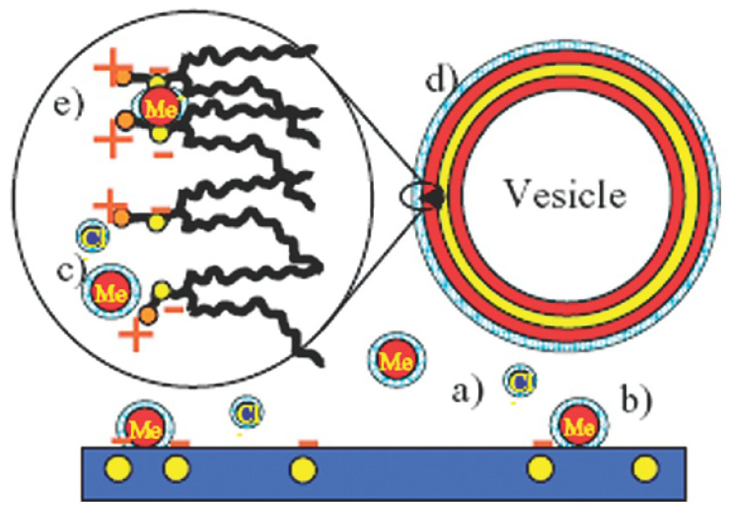
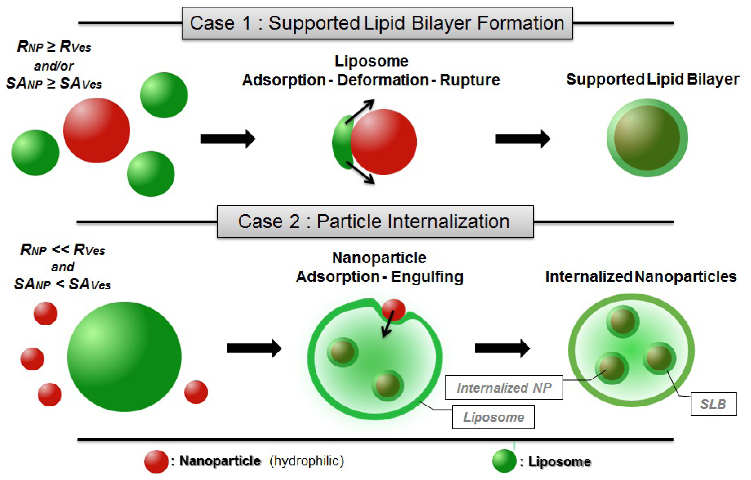
2.3.1. Supported Lipid Bilayer (SLB) Formation &amp; Particle Internalization</title>
<p>Supported lipid bilayers (SLB) are continuous fluid lipid membranes (<italic>T &gt; T</italic><sub>m</sub>) adsorbed on a solid substrate, and separated from this substrate by a thin water layer (1–3 nm) [<xref ref-type="bibr" rid="b73-ijms-13-11610">73</xref>]. It is important to note the necessity of having a bilayer in the fluid phase (to have a membrane of sufficiently low elasticity [<xref ref-type="bibr" rid="b49-ijms-13-11610">49</xref>–<xref ref-type="bibr" rid="b51-ijms-13-11610">51</xref>]) and a hydrophilic substrate (to bind the supporting water layer) to achieve the deposition of SLB.</p>
<p>SLB formation has been studied first for the case of planar substrate (glass, silica, mica) as the basis to produce innovative catalytic surfaces or immobilized protein arrays [<xref ref-type="bibr" rid="b52-ijms-13-11610">52</xref>,<xref ref-type="bibr" rid="b58-ijms-13-11610">58</xref>,<xref ref-type="bibr" rid="b73-ijms-13-11610">73</xref>–<xref ref-type="bibr" rid="b82-ijms-13-11610">82</xref>], as well as to study bilayer-bilayer interactions, as they are important in biology, under well-defined conditions. Nowadays, this field of research has been extended to the case of nanoparticles, with the intention of designing nanovectors by rendering nanoparticles biocompatible through the deposition of a lipid bilayer onto their surface [<xref ref-type="bibr" rid="b83-ijms-13-11610">83</xref>–<xref ref-type="bibr" rid="b85-ijms-13-11610">85</xref>]. Such studies on well-defined membranes are important in order to control the interactions and thereby optimize the design of the delivery systems.</p>
<p>The formation of SLB, as has been investigated on both planar surfaces and nanoparticles, can be achieved by depositing vesicles from the solution [<xref ref-type="bibr" rid="b52-ijms-13-11610">52</xref>,<xref ref-type="bibr" rid="b75-ijms-13-11610">75</xref>,<xref ref-type="bibr" rid="b79-ijms-13-11610">79</xref>,<xref ref-type="bibr" rid="b83-ijms-13-11610">83</xref>–<xref ref-type="bibr" rid="b87-ijms-13-11610">87</xref>] where the liposomes adhere, fuse and spread on the solid surface. In the case of a planar substrate, this process has even largely replaced the Langmuir-Blodget deposition due to the wider availability of more convenient methods for unilamellar liposomes preparation [<xref ref-type="bibr" rid="b76-ijms-13-11610">76</xref>]. Despite the considerable work on vesicle adsorption, there is still no complete picture of the forces responsible for the adhesion, fusion and rupture of the liposomes on solid surfaces. However, the SLB formation is believed to be governed by three types of interactions [<xref ref-type="bibr" rid="b79-ijms-13-11610">79</xref>,<xref ref-type="bibr" rid="b88-ijms-13-11610">88</xref>]:</p>
<list list-type="bullet">
<list-item>
<p>vesicle-surface interaction;</p></list-item>
<list-item>
<p>vesicle-vesicle interaction, as the lateral interaction between neighbouring vesicles adsorbed on a surface plays an important role in the process of vesicle fusion leading to SLB formation;</p></list-item>
<list-item>
<p>cohesive strength of the vesicle (vesicle stability).</p></list-item></list>
<p>Note that the latter has been found to be of less importance in this process [<xref ref-type="bibr" rid="b88-ijms-13-11610">88</xref>].</p>
<p>Experimentally, one can play with several parameters (ionic strength, addition of specific ions, pH, surface properties of the substrate, nature of the phospholipid(s) used) in order to monitor the strength of these three different interactions.</p>
<p>Changing the ionic strength and/or the pH is a way to alter the electrostatic properties of the substrate as well as the phospholipid net charge leading to different adsorption behaviors depending on the nature of the substrate and on the type of lipid used [<xref ref-type="bibr" rid="b52-ijms-13-11610">52</xref>,<xref ref-type="bibr" rid="b75-ijms-13-11610">75</xref>,<xref ref-type="bibr" rid="b84-ijms-13-11610">84</xref>,<xref ref-type="bibr" rid="b85-ijms-13-11610">85</xref>]. More precisely, the use of specific ions (in particular divalent cations) has been found to have a surprisingly strong effect on SLB formation, when bridging negatively charged or zwitterionic lipids and negatively charged substrates (silica-<xref ref-type="fig" rid="f1-ijms-13-11610">Figure 1</xref>) [<xref ref-type="bibr" rid="b79-ijms-13-11610">79</xref>,<xref ref-type="bibr" rid="b86-ijms-13-11610">86</xref>–<xref ref-type="bibr" rid="b89-ijms-13-11610">89</xref>]. This is not surprising given the known affinity of Ca<sup>2+</sup> and phosphate. The case of zwitterionic phospholipids is particularly interesting, as the cations convert the headgroup from a dipole toward a positive monopole thus strengthening the interaction with negatively charged substrate (<xref ref-type="fig" rid="f2-ijms-13-11610">Figure 2</xref>) [<xref ref-type="bibr" rid="b85-ijms-13-11610">85</xref>,<xref ref-type="bibr" rid="b88-ijms-13-11610">88</xref>].</p>
<p>However, even if the deposition mechanism is believed to be mainly driven by electrostatic attractions between vesicle membranes and the substrate surfaces, other forces are also non negligible. As an example, SLB formation or vesicle adsorption have been reported in systems where the electrostatic interactions were clearly repulsive [<xref ref-type="bibr" rid="b52-ijms-13-11610">52</xref>] or minimized by experimental conditions (pH [<xref ref-type="bibr" rid="b75-ijms-13-11610">75</xref>], substrate surface properties [<xref ref-type="bibr" rid="b90-ijms-13-11610">90</xref>]) showing the importance of van der Waals attractive interactions as well as of hydration and steric forces in the deposition mechanism.</p>
<p>Previous studies on the formation of SLB on hard nanoparticles can be divided in two types that are depicted in <xref ref-type="fig" rid="f2-ijms-13-11610">Figure 2</xref>.</p>
<list list-type="order">
<list-item>
<p>In the first case, the adsorption of liposomes onto nanoparticles leads to the formation of continuous SLB on the particle surface (<xref ref-type="fig" rid="f2-ijms-13-11610">Figure 2</xref>). This is the case when either the radius of the particle (<italic>R</italic><italic><sub>NP</sub></italic>) is larger than the vesicle radius (<italic>R</italic><italic><sub>Ves</sub></italic>) or when the surface area of all particles is equal or larger than the total surface of the potentially formed SLB (<italic>SA</italic><italic><sub>NP</sub></italic> ≥ <italic>SA</italic><italic><sub>Ves</sub></italic>—taking into account that when <italic>SA</italic><italic><sub>NP</sub></italic> ≫ <italic>SA</italic><italic><sub>Ves</sub></italic> nanoparticles are believed to be partially covered by lipid bilayer patches [<xref ref-type="bibr" rid="b91-ijms-13-11610">91</xref>]).</p></list-item>
<list-item>
<p>
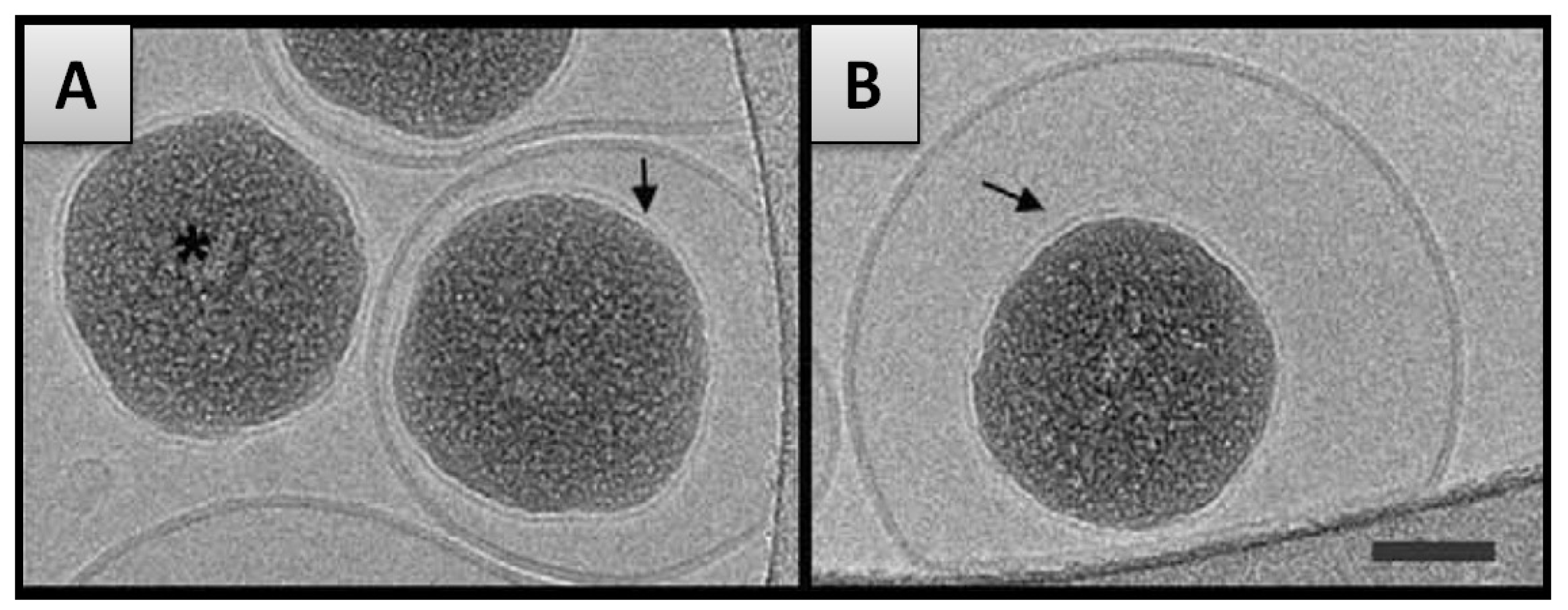
In the second case, the formation of SLB on a particle is an intermediate step of the internalization mechanism in which particles covered with SLB become encapsulated within liposomes [<xref ref-type="bibr" rid="b42-ijms-13-11610">42</xref>]. This may occur when <italic>R</italic><italic><sub>Ves</sub></italic> is considerably larger than <italic>R</italic><italic><sub>NP</sub></italic> and <italic>SA</italic><italic><sub>NP</sub></italic> inferior to <italic>SA</italic><italic><sub>Ves</sub></italic> (<xref ref-type="fig" rid="f2-ijms-13-11610">Figure 2</xref>). The internalization of the nanoparticles is firstly driven by the attractive interaction between membrane and nanoparticle but in addition, a thermodynamic driving force is the release of water from the vesicle interior and the concomitant entropy gain that occurs during the process.</p></list-item></list>
<p>(1) In the first case mentioned above, the mechanism of SLB formation proceeds via subsequent adsorption, deformation and rupture of liposomes on the nanoparticle surface [<xref ref-type="bibr" rid="b83-ijms-13-11610">83</xref>] much like in the case of flat substrates [<xref ref-type="bibr" rid="b52-ijms-13-11610">52</xref>,<xref ref-type="bibr" rid="b79-ijms-13-11610">79</xref>,<xref ref-type="bibr" rid="b80-ijms-13-11610">80</xref>,<xref ref-type="bibr" rid="b89-ijms-13-11610">89</xref>]. After adsorption of the liposomes, the contact area between the lipid surface and the substrate increases, due to adhesion forces until the deformation of the vesicle reaches a limit above which rupture occurs, resulting in the appearance of patches on the particle surface. It is to be noted that this process has been found to occur either independently or dependently of the presence of preformed lipid patches in the vicinity of the adsorbed vesicle. As a matter of fact, the edges of bilayer patches are likely to activate the decomposition of adsorbed vesicles [<xref ref-type="bibr" rid="b79-ijms-13-11610">79</xref>]. Hence, the formation of a SLB covering an entire particle often involves the coalescence of neighbouring liposome patches via active edge effects [<xref ref-type="bibr" rid="b89-ijms-13-11610">89</xref>]. In any case the binding of a nanoparticle to a bilayer will lead to a loss of configurational entropy of the membrane as it suppresses undulations of the bilayers.</p>
<p>However, the process of SLB formation depends highly on the ability of the membrane to curve around the particle and adopt its shape [<xref ref-type="bibr" rid="b92-ijms-13-11610">92</xref>,<xref ref-type="bibr" rid="b93-ijms-13-11610">93</xref>]. The curvature of the lipid bilayer is depicted in the literature as the result of the interplay between the (favourable) adhesion energy between the membrane and its substrate and the (unfavourable) elastic energy (or bending energy) needed to bend the bilayer around the particle [<xref ref-type="bibr" rid="b48-ijms-13-11610">48</xref>,<xref ref-type="bibr" rid="b80-ijms-13-11610">80</xref>,<xref ref-type="bibr" rid="b89-ijms-13-11610">89</xref>,<xref ref-type="bibr" rid="b92-ijms-13-11610">92</xref>–<xref ref-type="bibr" rid="b94-ijms-13-11610">94</xref>]. Corresponding theoretical studies have led to structural phase diagrams demarking the regions of fully enveloped, partially wrapped or freely dispersed colloids [<xref ref-type="bibr" rid="b95-ijms-13-11610">95</xref>–<xref ref-type="bibr" rid="b97-ijms-13-11610">97</xref>]. The investigations on the transition steps between the wrapped and the free state of particles allow the definition of a critical radius for the particle (<italic>R</italic><italic><sub>C</sub></italic>), meaning that the formation of SLB is energetically favourable on particles having a radius above this critical value. In the case of a tensionless membrane, the value of <italic>R</italic><italic><sub>C</sub></italic> may be extrapolated from the expression of the energetic balance when the adhesion energy is compensated by the bending energy [<xref ref-type="bibr" rid="b48-ijms-13-11610">48</xref>,<xref ref-type="bibr" rid="b98-ijms-13-11610">98</xref>], giving:</p>
<disp-formula id="FD14">
<label>(14)</label>
<mml:math id="mm14" display="block">
<mml:semantics id="sm14">
<mml:mrow>
<mml:msub>
<mml:mrow>
<mml:mi>R</mml:mi></mml:mrow>
<mml:mi>C</mml:mi></mml:msub>
<mml:mo>≡</mml:mo>
<mml:msqrt>
<mml:mrow>
<mml:mn>2</mml:mn>
<mml:mi>κ</mml:mi>
<mml:mo>/</mml:mo>
<mml:mo>|</mml:mo>
<mml:mi>W</mml:mi>
<mml:mo>|</mml:mo></mml:mrow></mml:msqrt></mml:mrow></mml:semantics></mml:math></disp-formula>
<p>where <italic>R</italic><italic><sub>C</sub></italic> depends on the strength of the adhesion energy per unit area (<italic>W</italic>) as well as on the value of the bilayer mean bending modulus (κ), thus depending on the nature of both particle and lipid bilayer. Consequently, one can modify this critical value by changing experimental conditions such as the nature of the lipids, the properties of the substrate surface [<xref ref-type="bibr" rid="b90-ijms-13-11610">90</xref>] as well as the properties of the medium such as pH [<xref ref-type="bibr" rid="b84-ijms-13-11610">84</xref>] and ionic strength [<xref ref-type="bibr" rid="b85-ijms-13-11610">85</xref>]. For instance the bending modulus (κ) is proportional to the third power of the bilayer thickness [<xref ref-type="bibr" rid="b99-ijms-13-11610">99</xref>–<xref ref-type="bibr" rid="b101-ijms-13-11610">101</xref>]. Accordingly, the values of <italic>R</italic><italic><sub>C</sub></italic> determined experimentally have been found to vary with the nature of the system. In the case of silica particles interacting with zwitterionic lipid bilayers, the critical radius has been found to be around 10 nm [<xref ref-type="bibr" rid="b42-ijms-13-11610">42</xref>,<xref ref-type="bibr" rid="b92-ijms-13-11610">92</xref>,<xref ref-type="bibr" rid="b93-ijms-13-11610">93</xref>] corresponding to an adhesive strength <italic>W</italic> of a few mJ/m<sup>2</sup>—a value comparable to usual interfacial energies [<xref ref-type="bibr" rid="b55-ijms-13-11610">55</xref>].</p>
<p>Interestingly, supported lipid bilayers adsorbed on small particles (<italic>R</italic><italic><sub>C</sub></italic> &lt; 100 nm) exhibit morphology changes due to the high curvature they experience. Shifts of the <italic>T</italic><sub>m</sub> value [<xref ref-type="bibr" rid="b102-ijms-13-11610">102</xref>] as well as a broadening and/or splitting of the transition peak obtained by differential scanning calorimetry (DSC) [<xref ref-type="bibr" rid="b102-ijms-13-11610">102</xref>,<xref ref-type="bibr" rid="b103-ijms-13-11610">103</xref>] suggest a curvature-dependent decoupling between the inner and outer leaflet of the bilayer as has also been observed in the case of small SUVs [<xref ref-type="bibr" rid="b104-ijms-13-11610">104</xref>,<xref ref-type="bibr" rid="b105-ijms-13-11610">105</xref>]. In extreme cases (<italic>R</italic><italic><sub>P</sub></italic> ≤ 10 nm) the high curvature induces the creation of a large free volume between the acyl-chains of the supported bilayer [<xref ref-type="bibr" rid="b102-ijms-13-11610">102</xref>,<xref ref-type="bibr" rid="b106-ijms-13-11610">106</xref>] (<xref ref-type="fig" rid="f3-ijms-13-11610">Figure 3</xref>, left). In this case, interdigitation of the acyl-chains occurs in order to optimize hydrophobic interaction while avoiding the exposure of hydrophobic chains to an aqueous environment (<xref ref-type="fig" rid="f3-ijms-13-11610">Figure 3</xref>, right) [<xref ref-type="bibr" rid="b107-ijms-13-11610">107</xref>].</p>
<p>(2) In the second case mentioned previously, the SLB formation on smaller particles is accompanied by the internalization of the nanoparticle into the vesicle interior, as has for instance been observed for the case of silica NPs interacting with DOPC vesicles (<xref ref-type="fig" rid="f4-ijms-13-11610">Figure 4</xref>). As described by Le Bihan <italic>et al</italic>. [<xref ref-type="bibr" rid="b42-ijms-13-11610">42</xref>], the proposed invagination mechanism predicts the adsorption of the particle on the liposome outer surface, followed by the spreading of the membrane around the particle finally leading to the engulfing and the full internalization of the nanoparticle. This process of nanoparticle transmigration is similar to that of cellular uptake (or endocytosis) [<xref ref-type="bibr" rid="b108-ijms-13-11610">108</xref>,<xref ref-type="bibr" rid="b109-ijms-13-11610">109</xref>]. Simulations on similar systems [<xref ref-type="bibr" rid="b110-ijms-13-11610">110</xref>] have been found to consistently reproduce the invagination mechanism described above.</p>
<p>The invagination mechanism is governed by the same energy balance as in the previously discussed case of SLB formation [<xref ref-type="bibr" rid="b48-ijms-13-11610">48</xref>,<xref ref-type="bibr" rid="b49-ijms-13-11610">49</xref>,<xref ref-type="bibr" rid="b96-ijms-13-11610">96</xref>,<xref ref-type="bibr" rid="b111-ijms-13-11610">111</xref>]. Accordingly, the same expression (<xref rid="FD14" ref-type="disp-formula">Equation 14</xref>) arises for the calculation of the critical particle radius value below which the particle remains adsorbed on the liposome outer surface. In this case, the total adhesion energy does not overcome the energy cost associated with the bending of the membrane, thus preventing the invagination mechanism to proceed further. Nevertheless, the experimental findings [<xref ref-type="bibr" rid="b42-ijms-13-11610">42</xref>] seem to contradict some of the requirements expressed by theoretical studies such as the necessity of working with large liposomes (<italic>R</italic><italic><sub>Ves</sub></italic> &gt; 300 nm) [<xref ref-type="bibr" rid="b95-ijms-13-11610">95</xref>] as well as the required presence of rafts which are phase separated from the rest of the membrane in order to promote the fission process allowing the detachment of the internalized particle from the membrane [<xref ref-type="bibr" rid="b111-ijms-13-11610">111</xref>]. However, related to the latter, it may not be necessary to tune the membrane to obtain phase separated domains since the adsorption of particles has been found, in some cases, to induce local phase separation on its own [<xref ref-type="bibr" rid="b112-ijms-13-11610">112</xref>,<xref ref-type="bibr" rid="b113-ijms-13-11610">113</xref>].</p></sec>
<sec>
<title>2.3.2. Decoration and Aggregate Formation</title>
<p>Vesicle decoration results from the adsorption of hydrophilic NPs onto the surface of liposome membranes in systems where the nanoparticle radius is below the critical value <italic>R</italic><italic><sub>C</sub></italic> (<xref rid="FD14" ref-type="disp-formula">Equation 14</xref>) [<xref ref-type="bibr" rid="b40-ijms-13-11610">40</xref>,<xref ref-type="bibr" rid="b43-ijms-13-11610">43</xref>,<xref ref-type="bibr" rid="b114-ijms-13-11610">114</xref>] (keeping in mind that this value can be controlled by tuning other parameters such as pH or ionic strength of the solution) [<xref ref-type="bibr" rid="b43-ijms-13-11610">43</xref>]. In this case, the balance between adhesion and bending energy is therefore shifted to a range where it is unfavourable to the invagination process. Thus, the membrane is “incapable” of internalizing the particles, leaving them on its outer surface (<xref ref-type="fig" rid="f5-ijms-13-11610">Figure 5A</xref>). Due to the attractive interaction between membrane and the nanoparticles there will be indentations by the NPs into the membrane surface which depend on the balance between attractive interaction and the bending energy required for this partial wrapping. This does not only lead to a deformation of the membrane but affects also its local stiffness and will moreover cause an additional interaction force between the different NPs attached. Furthermore it has to be considered, that in some cases, where the particle radius is close to the critical value, the kinetic of the invagination process is slowed down, allowing the observation of decorated vesicle structures which, in these cases, are only intermediate structures [<xref ref-type="bibr" rid="b115-ijms-13-11610">115</xref>].</p>
<p>For a number of cases of charged nanoparticles, the decoration of liposomes by particles (zwitterionic lipids/charged nanoparticles) has been found to increase the absolute Zeta potential value of the dispersion by introducing charges on the liposome surface [<xref ref-type="bibr" rid="b40-ijms-13-11610">40</xref>,<xref ref-type="bibr" rid="b43-ijms-13-11610">43</xref>]. The appearance of these charges on the vesicle surface gives rise to repulsive electrostatic forces between the decorated vesicle structures and thus stabilizes the vesicle dispersion [<xref ref-type="bibr" rid="b40-ijms-13-11610">40</xref>,<xref ref-type="bibr" rid="b41-ijms-13-11610">41</xref>,<xref ref-type="bibr" rid="b43-ijms-13-11610">43</xref>]. This repulsion can be quantified by means of measurements of the ζ-potential and such experiments have shown a decrease of the ζ-potential and concomitant stability increase which becomes larger with increasing nanoparticle sizes [<xref ref-type="bibr" rid="b43-ijms-13-11610">43</xref>]. This result is of great interest given the metastable nature of many liposome dispersions [<xref ref-type="bibr" rid="b35-ijms-13-11610">35</xref>] which often leads to rather fast phase separation and which represents a major disadvantage for their potential applications. Accordingly, the addition of charged silica NPs is a way to regulate the stability of liposomes.</p>
<p>However, in these systems, the colloidal stabilization is only achieved upon the adsorption of a minimum amount of nanoparticles, which corresponds to a sufficient surface coverage of the liposomes to ensure the repulsion between the decorated vesicle structures (<xref ref-type="fig" rid="f5-ijms-13-11610">Figure 5B</xref>) [<xref ref-type="bibr" rid="b40-ijms-13-11610">40</xref>,<xref ref-type="bibr" rid="b41-ijms-13-11610">41</xref>].</p>
<p>This has similarly been observed in recent work by us as documented by the change of particle size as a function of time (<xref ref-type="fig" rid="f6-ijms-13-11610">Figure 6</xref>). This study on the stability of unilamellar DPPC vesicles (R ~ 50 nm) below their phase transition temperature by means of dynamic light scattering showed that the addition of small amounts of small silica NPs (<italic>R</italic><sub>h</sub> ≈ 8 nm) leads to a rapid destabilization of the vesicle dispersion [<xref ref-type="bibr" rid="b40-ijms-13-11610">40</xref>]. Only the adsorption of a sufficiently high amount of silica particles onto the liposome surface, here about 14 NPs per vesicle, is able to prevent the fusion of neighbouring liposomes and ensure their colloidal stability for several months. This is an enormous enhancement compared to the situation of the unmodified vesicles and such long-time stability is very important for the storage of liposome dispersions and also for having controlled release properties.</p>
<p>On the contrary, if the NP concentration is too low, nanoparticles would bridge between adjacent liposomes, thereby introducing interactions and thus accelerating their fusion [<xref ref-type="bibr" rid="b40-ijms-13-11610">40</xref>,<xref ref-type="bibr" rid="b41-ijms-13-11610">41</xref>], as has been observed in nanoparticle-microsphere systems [<xref ref-type="bibr" rid="b116-ijms-13-11610">116</xref>]. In order to limit the magnitude of this effect, one may change the sign and magnitude of the particle charge. As has been found for PC liposomes, the use of cationic particles allows colloidal stabilization at lower surface coverage as compared to anionic ones [<xref ref-type="bibr" rid="b114-ijms-13-11610">114</xref>]. In fact, cationic particles adsorb weakly on the membrane due to the geometry of the P<sup>−</sup>–N<sup>+</sup> dipole of the lecithin head group (<xref ref-type="fig" rid="f7-ijms-13-11610">Figure 7</xref>), and consequently, they are less apt to bridge between neighbouring liposomes. These results confirm the predictions made by computer simulations on analogous systems [<xref ref-type="bibr" rid="b117-ijms-13-11610">117</xref>,<xref ref-type="bibr" rid="b118-ijms-13-11610">118</xref>].</p>
<p>Specific studies on these nanoparticle-stabilized liposomes have led to interesting findings on their properties. First and in opposition to other stabilization methods (PEG coating [<xref ref-type="bibr" rid="b119-ijms-13-11610">119</xref>–<xref ref-type="bibr" rid="b121-ijms-13-11610">121</xref>]), the adsorption of nanoparticles neither prevents nor reduces the binding between liposome immobilized ligand (biotin) and free receptor proteins (streptavidin) in the bulk: at low surface coverage (but still sufficient to ensure colloidal stabilization), the outer surface of decorated liposomes has been found to be well accessible and biofunctional despite the presence of NPs [<xref ref-type="bibr" rid="b122-ijms-13-11610">122</xref>]. Furthermore, the particle decoration does not seem to prevent its permeability; on the contrary, the release of molecules (proteins) loaded in the aqueous interior of the liposome may be more accurately controlled by appropriately engineering the hydrophilic nanoparticle layer on the liposome outer surface [<xref ref-type="bibr" rid="b123-ijms-13-11610">123</xref>]. This is important for potential applications, since it allows for the stabilization of liposomes while retaining their other (biological) functionalities. Additionally, decorated vesicles are easily immobilized on a planar substrate by tuning the substrate charges to create an attractive electrostatic interaction between the substrate and the adsorbed nanoparticles [<xref ref-type="bibr" rid="b124-ijms-13-11610">124</xref>]. Such immobilized liposomes have an amazing potential as nanocontainers for the study of biomolecules: they allow the prolonged observation of molecules loaded in their inside with lesser surface perturbation than in the bulk.</p></sec>
<sec>
<title>2.3.3. Internalization within the Membrane</title>
<p>The incorporation of nanoparticles within the bilayer membrane occurs when using hydrophobic nanoparticles which have a higher affinity to the acyl-chains confined in the hydrophobic interior of the phospholipid bilayer. The most common preparation pathway to form such vesicle-nanoparticle hybrids is to dry a mixture of hydrophobic particles and phospholipid MLVs in chloroform, thus obtaining a NP-lipid film which is hydrated and further extruded or sonicated to obtain the final Vesicle-NP hybrid dispersion—provided the nanoparticles are sufficiently small [<xref ref-type="bibr" rid="b125-ijms-13-11610">125</xref>].</p>
<p>There are numerous studies on such loaded vesicles using different hydrophobic particles including Au [<xref ref-type="bibr" rid="b71-ijms-13-11610">71</xref>,<xref ref-type="bibr" rid="b126-ijms-13-11610">126</xref>–<xref ref-type="bibr" rid="b129-ijms-13-11610">129</xref>], Ag [<xref ref-type="bibr" rid="b130-ijms-13-11610">130</xref>,<xref ref-type="bibr" rid="b131-ijms-13-11610">131</xref>], Si [<xref ref-type="bibr" rid="b125-ijms-13-11610">125</xref>], Fe<sub>3</sub>O<sub>4</sub> [<xref ref-type="bibr" rid="b132-ijms-13-11610">132</xref>,<xref ref-type="bibr" rid="b133-ijms-13-11610">133</xref>], and quantum dots (CdSe, ZnS) [<xref ref-type="bibr" rid="b128-ijms-13-11610">128</xref>,<xref ref-type="bibr" rid="b130-ijms-13-11610">130</xref>,<xref ref-type="bibr" rid="b134-ijms-13-11610">134</xref>,<xref ref-type="bibr" rid="b135-ijms-13-11610">135</xref>]. In most cases, the incorporation of nanoparticles does not appear to have a significant influence on the vesicle size or stability [<xref ref-type="bibr" rid="b125-ijms-13-11610">125</xref>,<xref ref-type="bibr" rid="b136-ijms-13-11610">136</xref>] even though there has been mention of a decrease of the liposome size upon particle embedding for which no explanation has yet been provided [<xref ref-type="bibr" rid="b128-ijms-13-11610">128</xref>,<xref ref-type="bibr" rid="b133-ijms-13-11610">133</xref>]. However, the internalized NPs will induce local disruption of the bilayer structure, reduce lipid ordering and thereby grant a higher fluidity to the membrane [<xref ref-type="bibr" rid="b126-ijms-13-11610">126</xref>,<xref ref-type="bibr" rid="b131-ijms-13-11610">131</xref>,<xref ref-type="bibr" rid="b136-ijms-13-11610">136</xref>] at the same time changing the thermodynamics of the lipid bilayer phase transition (merging of the pretransition–transition from the ordered gel (L<sub>β</sub>) to the rippled gel phase (P<sub>β</sub>)–with the main phase transition, and a broadening of the melting region and shifts in <italic>T</italic><sub>m</sub>) [<xref ref-type="bibr" rid="b129-ijms-13-11610">129</xref>,<xref ref-type="bibr" rid="b130-ijms-13-11610">130</xref>,<xref ref-type="bibr" rid="b136-ijms-13-11610">136</xref>,<xref ref-type="bibr" rid="b137-ijms-13-11610">137</xref>].</p>
<p>Theoretical studies have defined the mechanism of particle embedding as the result of the dominance of the favourable free energy change caused by moving a hydrophobic sphere from pure water into the hydrophobic membrane over the energy penalty needed to deform the bilayer [<xref ref-type="bibr" rid="b138-ijms-13-11610">138</xref>,<xref ref-type="bibr" rid="b139-ijms-13-11610">139</xref>]. In that context, it might be noted that the gain in interfacial energy (Δλ·A) is about 1000 k<italic>T</italic> for a nanoparticle of 4 nm radius (for a change of interfacial tension of 20 mN/m). Nevertheless, the incorporation within the membrane is only energetically favourable if the particle size is inferior to a critical size depending on the type of lipid used and being close to the value of the membrane thickness [<xref ref-type="bibr" rid="b71-ijms-13-11610">71</xref>,<xref ref-type="bibr" rid="b125-ijms-13-11610">125</xref>,<xref ref-type="bibr" rid="b138-ijms-13-11610">138</xref>,<xref ref-type="bibr" rid="b139-ijms-13-11610">139</xref>]. Larger particles cannot become accommodated within the lipid bilayer due to the high elastic energy penalty, which then would suppress the membrane fluctuations, and the geometric difficulties of having a membrane surrounding such a large nanoparticle. Yet the interaction of liposomes or phospholipid molecules with large and highly hydrophobic particles (as well as with flat hydrophobic substrates) has been reported to lead to the formation of a phospholipid monolayer on the hydrophobic surfaces [<xref ref-type="bibr" rid="b140-ijms-13-11610">140</xref>–<xref ref-type="bibr" rid="b143-ijms-13-11610">143</xref>]. The resulting structures may be used as biocompatible particles for drug delivery [<xref ref-type="bibr" rid="b141-ijms-13-11610">141</xref>,<xref ref-type="bibr" rid="b144-ijms-13-11610">144</xref>,<xref ref-type="bibr" rid="b145-ijms-13-11610">145</xref>], as column materials for high-pressure liquid chromatography [<xref ref-type="bibr" rid="b146-ijms-13-11610">146</xref>] or else as a way of investigating membrane-like assembly in an immobilized state [<xref ref-type="bibr" rid="b140-ijms-13-11610">140</xref>,<xref ref-type="bibr" rid="b142-ijms-13-11610">142</xref>,<xref ref-type="bibr" rid="b147-ijms-13-11610">147</xref>].</p>
<p>However, in the case of the internalization of small NPs in the hydrophobic interior of the phospholipid bilayer, the phospholipid membrane must “unzip”, <italic>i.e.</italic>, dissociate the acyl-chains of both layers (<xref ref-type="fig" rid="f8-ijms-13-11610">Figure 8C</xref>). The unzipping creates void spaces in the vicinity of the incorporated NP, an energetically unfavourable situation. In order to minimize this void space as well as the free energy of deformation, incorporated NPs have been found to cluster within the bilayer (<xref ref-type="fig" rid="f8-ijms-13-11610">Figure 8C</xref>) [<xref ref-type="bibr" rid="b71-ijms-13-11610">71</xref>,<xref ref-type="bibr" rid="b129-ijms-13-11610">129</xref>] resulting in the appearance of either a few fully loaded liposomes (vesicle in the center with dark rim) within a “normal” SUV dispersion (<xref ref-type="fig" rid="f8-ijms-13-11610">Figure 8A</xref>) of vesicles without NPs contained in the membrane. In other specific cases, Janus-type vesicle structures are formed where internalized NPs are concentrated on one side of the vesicle (<xref ref-type="fig" rid="f8-ijms-13-11610">Figure 8B</xref>) [<xref ref-type="bibr" rid="b71-ijms-13-11610">71</xref>]. Very recent work on this phenomenon has underlined the importance of the bilayer phase behaviour in this process of particle aggregation. Nanoparticle clustering is observed upon lipid melting, and believed to be driven by greater hydrophobic mismatch between embedded particles in the fluid phase coupled with lipid mediated forces driven by lateral capillarity [<xref ref-type="bibr" rid="b129-ijms-13-11610">129</xref>]. Nonetheless, although the argument justifying the clustering of particles is quite convincing, the preferential embedding of particles in a few vesicles within a liposome dispersion or in one side of a vesicle (case of Janus-vesicles) has not been reported in other similar works [<xref ref-type="bibr" rid="b130-ijms-13-11610">130</xref>,<xref ref-type="bibr" rid="b133-ijms-13-11610">133</xref>,<xref ref-type="bibr" rid="b135-ijms-13-11610">135</xref>] and therefore is still a topic open for discussion.</p>
<p>Such nanoparticle-vesicle hybrids are promising tools in nanobiotechnology. For example, the use of quantum dot-vesicles allows the controlled vesicle fusion on cell surfaces while at the same time enabling the visualization of the resulting patches on the cell membrane by fluorescence [<xref ref-type="bibr" rid="b135-ijms-13-11610">135</xref>]. Also, embedded paramagnetic nanoparticles, when heated using an electromagnetic field, have been found to provide a useful means of controlling the liposome membrane permeability by inducing bilayer phase transition [<xref ref-type="bibr" rid="b133-ijms-13-11610">133</xref>,<xref ref-type="bibr" rid="b137-ijms-13-11610">137</xref>,<xref ref-type="bibr" rid="b148-ijms-13-11610">148</xref>]. One important challenge of this novel technology is to achieve the phase transition locally, without changing the temperature of the medium which, <italic>in vivo</italic>, may damage the biological structures in the vicinity of the NP loaded liposome (this is also an important challenge in the case of the light actuation of gold NP, see Section 3.2.). To this end, unlike the case of SPIO (super paramagnetic iron oxide) particles [<xref ref-type="bibr" rid="b137-ijms-13-11610">137</xref>], the use of individually stabilized iron oxide particles, has been reported to allow local heating of the membrane without major influences on its environment [<xref ref-type="bibr" rid="b133-ijms-13-11610">133</xref>]. This system then allows for directed and controlled delivery of active agents from vesicle systems. In addition, of course, this approach allows for a much lower and controlled energy input, which is a desired effect, especially when considering potential medical applications.</p></sec></sec></sec>
<sec>
<title>3. Applications</title>
<p>Combining the properties of phospholipid vesicles and nanoparticles as well as observing the influence of nanoparticles on biological membranes for which liposomes may serve as a model system leads to innovative approaches for novel medical applications. In addition, they are a relevant model system for investigations regarding nanotoxicity [<xref ref-type="bibr" rid="b149-ijms-13-11610">149</xref>], since nanoparticles have received increased scrutiny and attention with respect to their potential effects on health issues. Furthermore nanoparticles can serve also as formulating agents to control the properties of membranes since they are frequently employed in vesicle formulations as applied in pharmacy or cosmetics.</p>
<sec>
<title>3.1. Oxide Nanoparticles (Silica)</title>
<p>Before being used for medical purposes, NPs were subjected to numerous preventive studies about their cytotoxicity [<xref ref-type="bibr" rid="b150-ijms-13-11610">150</xref>–<xref ref-type="bibr" rid="b153-ijms-13-11610">153</xref>]. In this respect, silica particles appear to be a preferential choice since silica is considered as a biocompatible material due to its low cytotoxicity [<xref ref-type="bibr" rid="b150-ijms-13-11610">150</xref>] and is furthermore required for the production of structural material of many living organisms [<xref ref-type="bibr" rid="b154-ijms-13-11610">154</xref>]. However, more than its low toxicity, the versatility of silica in synthesis as well as in surface functionalization is the main argument for the use of silica particles in biotechnology [<xref ref-type="bibr" rid="b10-ijms-13-11610">10</xref>]. As a matter of fact, silica particles can be easily synthesized in various ways [<xref ref-type="bibr" rid="b9-ijms-13-11610">9</xref>,<xref ref-type="bibr" rid="b155-ijms-13-11610">155</xref>,<xref ref-type="bibr" rid="b156-ijms-13-11610">156</xref>] and the silanol groups on their surface can react with various compounds to form amine, carboxyl, thiol, or other groups, <italic>i.e.</italic>, there exists a large versatility of possible chemical surface modifications. However, silica surface modification can also be performed by passive adsorption of molecules such as proteins, [<xref ref-type="bibr" rid="b10-ijms-13-11610">10</xref>,<xref ref-type="bibr" rid="b12-ijms-13-11610">12</xref>] granting silica particles an amazing potential as very versatile drug carriers.</p>
<p>More specifically, mesoporous silica nanoparticles (MSN) have been found very attractive for drug delivery applications due to their high surface area and their tuneable pores [<xref ref-type="bibr" rid="b157-ijms-13-11610">157</xref>–<xref ref-type="bibr" rid="b160-ijms-13-11610">160</xref>]. In such particles, the loading of drugs can be achieved up to very high concentrations through simple electrostatic interactions. Nevertheless, once in the body fluids, drugs in accessible pores may be displaced by metabolites/ions leading to premature release. To avoid this phenomenon, gating devices have been designed using polymers [<xref ref-type="bibr" rid="b161-ijms-13-11610">161</xref>] or nanoparticles [<xref ref-type="bibr" rid="b162-ijms-13-11610">162</xref>,<xref ref-type="bibr" rid="b163-ijms-13-11610">163</xref>] to regulate the release of guest molecules. In this precise matter, supported lipid bilayer (SLB) adsorbed on mesoporous silica particles has been found to be very efficient as a gating agent while at the same time ensuring the biocompatibility of the particles and facilitating its cellular uptake [<xref ref-type="bibr" rid="b164-ijms-13-11610">164</xref>]. Furthermore the surface charges of such protocells are easily tuned by lipid exchange when introducing free liposomes of different composition in the bulk [<xref ref-type="bibr" rid="b164-ijms-13-11610">164</xref>].</p>
<p>Similarly, in the case of plain silica particles, lipid bilayers supported on silica NPs have been found to be a useful transfection agent for gene delivery [<xref ref-type="bibr" rid="b165-ijms-13-11610">165</xref>]. In this case, SLBs present many advantages over empty liposomes: SLBs exhibit a higher stability, since the lipid layers are unlikely to fuse due to the presence of a solid core, while allowing the monitoring of the SLB size by changing the size of the silica core.</p>
<p>Decorated vesicle structures based on silica particles have also been shown to have interesting features for medical application. In fact, adsorption of negatively charged silica nanoparticles on the vesicle membrane stabilizes the liposome dispersion by introducing electrostatic repulsion between the particle/vesicle complexes while retaining the fundamental properties of free liposomes [<xref ref-type="bibr" rid="b41-ijms-13-11610">41</xref>,<xref ref-type="bibr" rid="b123-ijms-13-11610">123</xref>]. Furthermore, when adsorbed on drug loaded liposomes travelling in a biological media, the nanoparticulate outer layer protects the liposome against enzymatic degradation and at the same time allows for the controlled release in gastric conditions [<xref ref-type="bibr" rid="b123-ijms-13-11610">123</xref>]. Similar results have been obtained with liposil (silica/phospholipid composite) dispersions, where the liposome is used as template for the hydrolysis and condensation of TEOS leading to the synthesis of a protecting silica nanocapsule around the liposome [<xref ref-type="bibr" rid="b166-ijms-13-11610">166</xref>–<xref ref-type="bibr" rid="b168-ijms-13-11610">168</xref>]. Further studies have been carried out on the deposition of supported lipid bilayers on liposils leading to liposome-silica-liposome structures where the two bilayers are separated from each other by a sol-gel derived silica layer [<xref ref-type="bibr" rid="b169-ijms-13-11610">169</xref>]. These hybrid structures present a significant interest since they allow the separated formulation of each of the two membranes to be optimized for their given purposes increasing the control over the drug release mechanism.</p></sec>
<sec>
<title>3.2. Metallic Nanoparticles (Gold and Silver)</title>
<p>Metallic nanoparticles, for instance made from gold and silver are rather frequently employed, where for Au NPs a high potential for catalytic properties exists and Ag NPs are employed in many applications due to their antimicrobial properties. Accordingly, the impact of such metal NPs on lipid bilayer presents many interesting aspects with respect to their nanotoxicity and also for further medical applications of such metal NPs. One interesting feature of metallic particles (especially Au nanoparticles) is their ability to efficiently convert electromagnetic radiation into heat. Hence, the irradiation of Au nanoparticles staying in the vicinity of a lipid membrane has been found to induce locally reversible phase transition in lipid bilayer systems by means of a laser heating. As shown in <xref ref-type="fig" rid="f9-ijms-13-11610">Figure 9</xref> this phase transition can be observed optically as it may lead from facetted vesicles in the gel state to spherical vesicle in the fluid state [<xref ref-type="bibr" rid="b170-ijms-13-11610">170</xref>]. By monitoring the size of the nanoparticles and the energy input by irradiation one can control the exact particle temperature while at the same time controlling the size of the melted footprint on the bilayer around the irradiated particle [<xref ref-type="bibr" rid="b171-ijms-13-11610">171</xref>]. Such an optical control over the gel-fluid phase transition has also been reported to allow the guiding of a nanoparticle along the membrane plane [<xref ref-type="bibr" rid="b170-ijms-13-11610">170</xref>]. In fact, although Au nanoparticles coated with CTAB are immobile when attached to a lipid bilayer in the gel phase, when laser heated, the particle induces the melting of the membrane in its vicinity, granting the lipids a much higher mobility thereby re-enabling its own motion. By tuning the properties of the irradiation one can achieve an effective guiding of the particle along the bilayer [<xref ref-type="bibr" rid="b170-ijms-13-11610">170</xref>], thus enhancing the potential of single nanoparticles as thermally controlled nanotools.</p>
<p>These features of Au nanoparticles may have a great impact in medical applications as is the case for the remote-release of molecules from the interior of the liposomes [<xref ref-type="bibr" rid="b172-ijms-13-11610">172</xref>]. In this instance, optical heating on liposome-NP assemblies, with adsorbed [<xref ref-type="bibr" rid="b172-ijms-13-11610">172</xref>] or embedded particles [<xref ref-type="bibr" rid="b127-ijms-13-11610">127</xref>], has been found to remotely release dye molecules from the interior of the liposomes by inducing local phase transition. An extensive use of the technique of optical heating has led to the development of a new approach in cancer therapy: the so-called selective nanophotothermolysis [<xref ref-type="bibr" rid="b173-ijms-13-11610">173</xref>,<xref ref-type="bibr" rid="b174-ijms-13-11610">174</xref>]. Here, the selective attachment of metallic nanoparticle on cancer cells is achieved by tuning the surface properties of the NPs (coating with antibodies) [<xref ref-type="bibr" rid="b175-ijms-13-11610">175</xref>] in order to achieve the selective delivery properties. The irradiation is then tuned so that the temperature of the NP exceeds the threshold of bubble formation in the surrounding medium, thereby creating vapour bubbles which disrupt and destroy the cell membrane.</p>
<p>One of the main remaining challenges of these medical applications is the ability to perform the irradiation without damaging the environmental tissues and cells. This could be achieved by choosing a wavelength in the near-IR part of the spectrum [<xref ref-type="bibr" rid="b172-ijms-13-11610">172</xref>,<xref ref-type="bibr" rid="b176-ijms-13-11610">176</xref>], where light is only absorbed weakly by biological molecules and thus exhibits a deep tissue penetration. However, small single Au NPs (with diameter inferior to 200 nm) exhibit a surface plasmon resonance in the visible part of the spectrum (≈530 nm) [<xref ref-type="bibr" rid="b170-ijms-13-11610">170</xref>,<xref ref-type="bibr" rid="b172-ijms-13-11610">172</xref>] while not absorbing substantially at wavelengths closer to the IR-domain (illumination at 633 nm) [<xref ref-type="bibr" rid="b170-ijms-13-11610">170</xref>] thereby preventing the use of optical heating in the near IR-domain. In this precise case the use of larger gold particles or gold NP aggregates as well as gold nanorods, which have been found to lead to higher near-IR absorption [<xref ref-type="bibr" rid="b172-ijms-13-11610">172</xref>,<xref ref-type="bibr" rid="b177-ijms-13-11610">177</xref>], is a way to efficiently achieve selective optical heating.</p>
<p>It is also of major interest to consider the case of silver particles due to their increasing mass production and domestic use [<xref ref-type="bibr" rid="b178-ijms-13-11610">178</xref>].</p>
<p>Silver nanoparticles (Ag-NPs) may be used as biosensor material [<xref ref-type="bibr" rid="b179-ijms-13-11610">179</xref>] but are mostly interesting for their antibacterial [<xref ref-type="bibr" rid="b7-ijms-13-11610">7</xref>,<xref ref-type="bibr" rid="b180-ijms-13-11610">180</xref>,<xref ref-type="bibr" rid="b181-ijms-13-11610">181</xref>] and antiviral properties, as they have been found to inhibit the HIV-1 virus infectivity <italic>in vitro</italic> [<xref ref-type="bibr" rid="b182-ijms-13-11610">182</xref>,<xref ref-type="bibr" rid="b183-ijms-13-11610">183</xref>]. Silver nanoparticles are thus intensively used in a wide range of applications. For example in surface treatment, when deposited on polymer and metal surfaces [<xref ref-type="bibr" rid="b184-ijms-13-11610">184</xref>] as well as in the textile industry [<xref ref-type="bibr" rid="b185-ijms-13-11610">185</xref>] where wool textiles treated with nanosilver colloidal solution develop various functionalities such as mothproofing, antibacterial and antistatic properties. However, applications of Ag-NPs as an anti-proliferative agent could be limited by the fact that they are equally toxic to human cells and DNA [<xref ref-type="bibr" rid="b186-ijms-13-11610">186</xref>–<xref ref-type="bibr" rid="b188-ijms-13-11610">188</xref>]. In this context hybrid lipid/nanoparticle conjugates provide a biologically inspired route of designing therapeutic agents and a means of reducing and controlling nanoparticle toxicity.</p>
<p>In one pathway of producing such hybrid materials, liposomes are used as nanoreactors, where the nucleation and growth of Ag-NP takes place, as well as a stabilizing agent forming an SLB around the silver particle surface and preventing their aggregation [<xref ref-type="bibr" rid="b189-ijms-13-11610">189</xref>]. This stabilization occurs without dramatically altering antibacterial properties of the nanoparticles. Nevertheless, an excessive increase of the lipid concentration may lead to the presence of multiple lipid bilayers on the particle surface which then results in a lower bacterial killing rate [<xref ref-type="bibr" rid="b189-ijms-13-11610">189</xref>,<xref ref-type="bibr" rid="b190-ijms-13-11610">190</xref>]. Similar work has been carried out using nanotubes consisting of Ag-NP embedded bilayer membranes where the spacing between bilayer membranes is used as scaffold for the growth of Ag-NPs. Similarly, in this case, the bilayer membrane contributes to stabilizing the NPs against aggregation and air oxidation processes while controlling their size [<xref ref-type="bibr" rid="b191-ijms-13-11610">191</xref>].</p>
<p>Another route arises when using hydrophobic decane-thiol or stearylamine modified Ag-NPs. Due to their higher affinity to the hydrophobic interior of the lipid bilayer, these hydrophobic NPs are embedded into the membrane provided their size is equal or less than the bilayer thickness (See Section 2.3.3.). Similar to the case of embedded gold or SPIO NPs [<xref ref-type="bibr" rid="b126-ijms-13-11610">126</xref>,<xref ref-type="bibr" rid="b137-ijms-13-11610">137</xref>] the presence of Ag-NPs within the membrane can induce changes in lipid packing and may disrupt the lipid-lipid interactions leading to changes in the bilayer phase behavior and viscosity. Studies on such systems have revealed a suppression of the lipid pretransition, a broadening of the main phase transition region [<xref ref-type="bibr" rid="b136-ijms-13-11610">136</xref>] as well as an increase in membrane fluidity above the <italic>T</italic><sub>m</sub> [<xref ref-type="bibr" rid="b131-ijms-13-11610">131</xref>] when an important amount of Ag-NPs is internalized into the membrane. Thus, depending on their size and surface chemistry, embedded NPs may have an influence on the stability and function of hybrid vesicles providing innovative routes to designing drug delivery systems.</p></sec></sec>
<sec sec-type="conclusions">
<title>4. Conclusions</title>
<p>The investigations of the interaction of nanoparticles with vesicles (liposomes) as model systems for membranes cover a very variable field of recent research activities, as this is one of the key aspects for understanding nanotoxicity. This is important as nanoparticles have recently been employed increasingly in a variety of different applications due to their specific properties. Furthermore nanoparticles can also be utilized as transport vehicles for selectively delivering active agents into cells where their interaction with the membrane is a crucial aspect in controlling the delivery properties. Nanoparticles can bind strongly to the membrane surface but also become incorporated into bilayers or transported through the bilayers. The latter process is an essential step in the context of nanotoxicology as the incorporation of nanoparticles in cells is an elementary stage in this process, and may depend largely on pH, ionic strength and, of course, on the detailed structure of the nanoparticles, that can be changed by chemical or physical modifications. Alternatively the selective incorporation of nanoparticles into membranes is one way to introduce a sensor into the system or to obtain a means of controlling properties such as permeability of membranes via light induced heating. In summary, it can be stated that nanoparticle/liposome interactions are the key for understanding many developments in nanomedical research.</p></sec></body>
<back>
<ack>
<title>Acknowledgments</title>
<p>Financial support by the German Research Foundation (DFG) in the framework of IGRTG 1524 is gratefully acknowledged. Furthermore we would like to acknowledge fruitful discussions with D. Danino (Technion, Haifa, Israel) and T. Weikl (MPIKG, Golm, Germany).</p></ack>
<ref-list>
<title>References</title>
<ref id="b1-ijms-13-11610"><label>1</label><citation citation-type="journal"><person-group person-group-type="author"><name><surname>El-Sayed</surname><given-names>M.A.</given-names></name></person-group><article-title>Small is different: Shape-, size-, and composition-dependent properties of some colloidal semiconductor nanocrystals</article-title><source>Acc. Chem. Res</source><year>2004</year><volume>37</volume><fpage>326</fpage><lpage>333</lpage><pub-id pub-id-type="doi">10.1021/ar020204f</pub-id><pub-id pub-id-type="pmid">15147173</pub-id></citation></ref>
<ref id="b2-ijms-13-11610"><label>2</label><citation citation-type="journal"><person-group person-group-type="author"><name><surname>Rao</surname><given-names>C.N.R.</given-names></name><name><surname>Kulkarni</surname><given-names>G.U.</given-names></name><name><surname>Thomas</surname><given-names>P.J.</given-names></name><name><surname>Edwards</surname><given-names>P.P.</given-names></name></person-group><article-title>Size-dependent chemistry: Properties of nanocrystals</article-title><source>Chemistry (Weinheim an der Bergstrasse, Germany)</source><year>2002</year><volume>8</volume><fpage>28</fpage><lpage>35</lpage><pub-id pub-id-type="doi">10.1002/1521-3765(20020104)8:1&lt;28::AID-CHEM28&gt;3.0.CO;2-B</pub-id></citation></ref>
<ref id="b3-ijms-13-11610"><label>3</label><citation citation-type="journal"><person-group person-group-type="author"><name><surname>Seo</surname><given-names>W.S.</given-names></name><name><surname>Jo</surname><given-names>H.H.</given-names></name><name><surname>Lee</surname><given-names>K.</given-names></name><name><surname>Kim</surname><given-names>B.</given-names></name><name><surname>Oh</surname><given-names>S.J.</given-names></name><name><surname>Park</surname><given-names>J.T.</given-names></name></person-group><article-title>Size-dependent magnetic properties of colloidal Mn<sub>3</sub>O<sub>4</sub> and MnO nanoparticles</article-title><source>Angew. Chem. Int. Ed</source><year>2004</year><volume>43</volume><fpage>1115</fpage><lpage>1117</lpage><pub-id pub-id-type="doi">10.1002/anie.200352400</pub-id></citation></ref>
<ref id="b4-ijms-13-11610"><label>4</label><citation citation-type="journal"><person-group person-group-type="author"><name><surname>Elghanian</surname><given-names>R.</given-names></name><name><surname>Storhoff</surname><given-names>J.J.</given-names></name><name><surname>Mucic</surname><given-names>R.C.</given-names></name><name><surname>Letsinger</surname><given-names>R.L.</given-names></name><name><surname>Mirkin</surname><given-names>C.A.</given-names></name></person-group><article-title>Selective colorimetric detection of polynucleotides based on the distance-dependent optical properties of gold nanoparticles</article-title><source>Science</source><year>1997</year><volume>277</volume><fpage>1078</fpage><lpage>1081</lpage><pub-id pub-id-type="doi">10.1126/science.277.5329.1078</pub-id><pub-id pub-id-type="pmid">9262471</pub-id></citation></ref>
<ref id="b5-ijms-13-11610"><label>5</label><citation citation-type="journal"><person-group person-group-type="author"><name><surname>McFarland</surname><given-names>A.D.</given-names></name><name><surname>van Duyne</surname><given-names>R.P.</given-names></name></person-group><article-title>Single silver nanoparticles as real-time optical sensors with zeptomole sensitivity</article-title><source>Nano Lett</source><year>2003</year><volume>3</volume><fpage>1057</fpage><lpage>1062</lpage><pub-id pub-id-type="doi">10.1021/nl034372s</pub-id></citation></ref>
<ref id="b6-ijms-13-11610"><label>6</label><citation citation-type="journal"><person-group person-group-type="author"><name><surname>Shipway</surname><given-names>A.N.</given-names></name><name><surname>Katz</surname><given-names>E.</given-names></name><name><surname>Willner</surname><given-names>I.</given-names></name></person-group><article-title>Nanoparticle arrays on surfaces for electronic, optical, and sensor applications</article-title><source>ChemPhysChem</source><year>2000</year><volume>1</volume><fpage>18</fpage><lpage>52</lpage><pub-id pub-id-type="doi">10.1002/1439-7641(20000804)1:1&lt;18::AID-CPHC18&gt;3.0.CO;2-L</pub-id></citation></ref>
<ref id="b7-ijms-13-11610"><label>7</label><citation citation-type="journal"><person-group person-group-type="author"><name><surname>Kim</surname><given-names>J.S.</given-names></name><name><surname>Kuk</surname><given-names>E.</given-names></name><name><surname>Yu</surname><given-names>K.N.</given-names></name><name><surname>Kim</surname><given-names>J.H.</given-names></name><name><surname>Park</surname><given-names>S.J.</given-names></name><name><surname>Lee</surname><given-names>H.J.</given-names></name><name><surname>Kim</surname><given-names>S.H.</given-names></name><name><surname>Park</surname><given-names>Y.K.</given-names></name><name><surname>Park</surname><given-names>Y.H.</given-names></name><name><surname>Hwang</surname><given-names>C.Y.</given-names></name><etal/></person-group><article-title>Antimicrobial effects of silver nanoparticles</article-title><source>Nanomed. Nanotechnol. Biol. Med</source><year>2007</year><volume>3</volume><fpage>95</fpage><lpage>101</lpage><pub-id pub-id-type="doi">10.1016/j.nano.2006.12.001</pub-id></citation></ref>
<ref id="b8-ijms-13-11610"><label>8</label><citation citation-type="journal"><person-group person-group-type="author"><name><surname>Kumar</surname><given-names>A.</given-names></name><name><surname>Vemula</surname><given-names>P.K.</given-names></name><name><surname>Ajayan</surname><given-names>P.M.</given-names></name><name><surname>John</surname><given-names>G.</given-names></name></person-group><article-title>Silver-nanoparticle-embedded antimicrobial paints based on vegetable oil</article-title><source>Nat. Mater</source><year>2008</year><volume>7</volume><fpage>236</fpage><lpage>241</lpage><pub-id pub-id-type="doi">10.1038/nmat2099</pub-id><pub-id pub-id-type="pmid">18204453</pub-id></citation></ref>
<ref id="b9-ijms-13-11610"><label>9</label><citation citation-type="journal"><person-group person-group-type="author"><name><surname>Santra</surname><given-names>S.</given-names></name><name><surname>Wang</surname><given-names>K.M.</given-names></name><name><surname>Tapec</surname><given-names>R.</given-names></name><name><surname>Tan</surname><given-names>W.H.</given-names></name></person-group><article-title>Development of novel dye-doped silica nanoparticles for biomarker application</article-title><source>J. Biomed. Opt</source><year>2001</year><volume>6</volume><fpage>160</fpage><lpage>166</lpage><pub-id pub-id-type="doi">10.1117/1.1353590</pub-id><pub-id pub-id-type="pmid">11375725</pub-id></citation></ref>
<ref id="b10-ijms-13-11610"><label>10</label><citation citation-type="journal"><person-group person-group-type="author"><name><surname>Tan</surname><given-names>W.H.</given-names></name><name><surname>Wang</surname><given-names>K.M.</given-names></name><name><surname>He</surname><given-names>X.X.</given-names></name><name><surname>Zhao</surname><given-names>X.J.</given-names></name><name><surname>Drake</surname><given-names>T.</given-names></name><name><surname>Wang</surname><given-names>L.</given-names></name><name><surname>Bagwe</surname><given-names>R.P.</given-names></name></person-group><article-title>Bionanotechnology based on silica nanoparticles</article-title><source>Med. Res. Rev</source><year>2004</year><volume>24</volume><fpage>621</fpage><lpage>638</lpage><pub-id pub-id-type="doi">10.1002/med.20003</pub-id><pub-id pub-id-type="pmid">15224383</pub-id></citation></ref>
<ref id="b11-ijms-13-11610"><label>11</label><citation citation-type="journal"><person-group person-group-type="author"><name><surname>Trewyn</surname><given-names>B.G.</given-names></name><name><surname>Slowing</surname><given-names>II</given-names></name><name><surname>Giri</surname><given-names>S.</given-names></name><name><surname>Chen</surname><given-names>H.T.</given-names></name><name><surname>Lin</surname><given-names>V.S.Y.</given-names></name></person-group><article-title>Synthesis and functionalization of a mesoporous silica nanoparticle based on the sol-gel process and applications in controlled release</article-title><source>Acc. Chem. Res.</source><year>2007</year><volume>40</volume><fpage>846</fpage><lpage>853</lpage><pub-id pub-id-type="doi">10.1021/ar600032u</pub-id><pub-id pub-id-type="pmid">17645305</pub-id></citation></ref>
<ref id="b12-ijms-13-11610"><label>12</label><citation citation-type="journal"><person-group person-group-type="author"><name><surname>Bharti</surname><given-names>B.</given-names></name><name><surname>Meissner</surname><given-names>J.</given-names></name><name><surname>Findenegg</surname><given-names>G.H.</given-names></name></person-group><article-title>Aggregation of Silica Nanoparticles Directed by Adsorption of Lysozyme</article-title><source>Langmuir</source><year>2011</year><volume>27</volume><fpage>9823</fpage><lpage>9833</lpage><pub-id pub-id-type="doi">10.1021/la201898v</pub-id><pub-id pub-id-type="pmid">21728288</pub-id></citation></ref>
<ref id="b13-ijms-13-11610"><label>13</label><citation citation-type="journal"><person-group person-group-type="author"><name><surname>Fujita</surname><given-names>M.</given-names></name><name><surname>Katafuchi</surname><given-names>Y.</given-names></name><name><surname>Ito</surname><given-names>K.</given-names></name><name><surname>Kanayama</surname><given-names>N.</given-names></name><name><surname>Takarada</surname><given-names>T.</given-names></name><name><surname>Maeda</surname><given-names>M.</given-names></name></person-group><article-title>Structural study on gold nanoparticle functionalized with DNA and its non-cross-linking aggregation</article-title><source>J. Colloid Interface Sci</source><year>2012</year><volume>368</volume><fpage>629</fpage><lpage>635</lpage><pub-id pub-id-type="doi">10.1016/j.jcis.2011.11.021</pub-id><pub-id pub-id-type="pmid">22143000</pub-id></citation></ref>
<ref id="b14-ijms-13-11610"><label>14</label><citation citation-type="journal"><person-group person-group-type="author"><name><surname>Lin</surname><given-names>Y.F.</given-names></name><name><surname>Chen</surname><given-names>J.H.</given-names></name><name><surname>Hsu</surname><given-names>S.H.</given-names></name><name><surname>Hsiao</surname><given-names>H.C.</given-names></name><name><surname>Chung</surname><given-names>T.W.</given-names></name><name><surname>Tung</surname><given-names>K.L.</given-names></name></person-group><article-title>The synthesis of Lewis acid ZrO<sub>2</sub> nanoparticles and their applications in phospholipid adsorption from Jatropha oil used for biofuel</article-title><source>J. Colloid Interface Sci</source><year>2012</year><volume>368</volume><fpage>660</fpage><lpage>662</lpage><pub-id pub-id-type="doi">10.1016/j.jcis.2011.11.055</pub-id><pub-id pub-id-type="pmid">22183260</pub-id></citation></ref>
<ref id="b15-ijms-13-11610"><label>15</label><citation citation-type="journal"><person-group person-group-type="author"><name><surname>Fischer</surname><given-names>H.C.</given-names></name><name><surname>Chan</surname><given-names>W.C.W.</given-names></name></person-group><article-title>Nanotoxicity: The growing need for <italic>in vivo</italic> study</article-title><source>Curr. Opin. Biotechnol</source><year>2007</year><volume>18</volume><fpage>565</fpage><lpage>571</lpage><pub-id pub-id-type="doi">10.1016/j.copbio.2007.11.008</pub-id><pub-id pub-id-type="pmid">18160274</pub-id></citation></ref>
<ref id="b16-ijms-13-11610"><label>16</label><citation citation-type="journal"><person-group person-group-type="author"><name><surname>Kagan</surname><given-names>V.E.</given-names></name><name><surname>Bayir</surname><given-names>H.</given-names></name><name><surname>Shvedova</surname><given-names>A.A.</given-names></name></person-group><article-title>Nanomedicine and nanotoxicology: Two sides of the same coin</article-title><source>Nanomed. Nanotechnol. Biol. Med</source><year>2005</year><volume>1</volume><fpage>313</fpage><lpage>316</lpage><pub-id pub-id-type="doi">10.1016/j.nano.2005.10.003</pub-id></citation></ref>
<ref id="b17-ijms-13-11610"><label>17</label><citation citation-type="journal"><person-group person-group-type="author"><name><surname>Li</surname><given-names>Y.F.</given-names></name><name><surname>Chen</surname><given-names>C.Y.</given-names></name></person-group><article-title>Fate and toxicity of metallic and metal-containing nanoparticles for biomedical applications</article-title><source>Small</source><year>2011</year><volume>7</volume><fpage>2965</fpage><lpage>2980</lpage><pub-id pub-id-type="doi">10.1002/smll.201101059</pub-id><pub-id pub-id-type="pmid">21932238</pub-id></citation></ref>
<ref id="b18-ijms-13-11610"><label>18</label><citation citation-type="journal"><person-group person-group-type="author"><name><surname>Oberdorster</surname><given-names>G.</given-names></name><name><surname>Stone</surname><given-names>V.</given-names></name><name><surname>Donaldson</surname><given-names>K.</given-names></name></person-group><article-title>Toxicology of nanoparticles: A historical perspective</article-title><source>Nanotoxicology</source><year>2007</year><volume>1</volume><fpage>2</fpage><lpage>25</lpage><pub-id pub-id-type="doi">10.1080/17435390701314761</pub-id></citation></ref>
<ref id="b19-ijms-13-11610"><label>19</label><citation citation-type="journal"><person-group person-group-type="author"><name><surname>Israelachvili</surname><given-names>J.N.</given-names></name><name><surname>Mitchell</surname><given-names>D.J.</given-names></name><name><surname>Ninham</surname><given-names>B.W.</given-names></name></person-group><article-title>Theory of Self-Assembly of hydrocarbon amphiphiles into micelles and bilayers</article-title><source>J. Chem. Soc. Faraday Trans. 2 Mol. Chem. Phys</source><year>1976</year><volume>72</volume><fpage>1525</fpage><lpage>1568</lpage><pub-id pub-id-type="doi">10.1039/f29767201525</pub-id></citation></ref>
<ref id="b20-ijms-13-11610"><label>20</label><citation citation-type="journal"><person-group person-group-type="author"><name><surname>Kunz</surname><given-names>W.</given-names></name><name><surname>Testard</surname><given-names>F.</given-names></name><name><surname>Zemb</surname><given-names>T.</given-names></name></person-group><article-title>Correspondence between curvature, packing parameter, and hydrophilic-lipophilic deviation scales around the phase-inversion temperature</article-title><source>Langmuir</source><year>2009</year><volume>25</volume><fpage>112</fpage><lpage>115</lpage><pub-id pub-id-type="doi">10.1021/la8028879</pub-id><pub-id pub-id-type="pmid">19072017</pub-id></citation></ref>
<ref id="b21-ijms-13-11610"><label>21</label><citation citation-type="journal"><person-group person-group-type="author"><name><surname>Segota</surname><given-names>S.</given-names></name><name><surname>Tezak</surname><given-names>D.</given-names></name></person-group><article-title>Spontaneous formation of vesicles</article-title><source>Adv. Colloid Interface Sci</source><year>2006</year><volume>121</volume><fpage>51</fpage><lpage>75</lpage><pub-id pub-id-type="doi">10.1016/j.cis.2006.01.002</pub-id><pub-id pub-id-type="pmid">16769012</pub-id></citation></ref>
<ref id="b22-ijms-13-11610"><label>22</label><citation citation-type="journal"><person-group person-group-type="author"><name><surname>Marsh</surname><given-names>D.</given-names></name></person-group><article-title>General features of phospholipid phase-transitions</article-title><source>Chem. Phys. Lipids</source><year>1991</year><volume>57</volume><fpage>109</fpage><lpage>120</lpage><pub-id pub-id-type="doi">10.1016/0009-3084(91)90071-I</pub-id><pub-id pub-id-type="pmid">2054899</pub-id></citation></ref>
<ref id="b23-ijms-13-11610"><label>23</label><citation citation-type="journal"><person-group person-group-type="author"><name><surname>Bangham</surname><given-names>A.D.</given-names></name><name><surname>Standish</surname><given-names>M.M.</given-names></name><name><surname>Watkins</surname><given-names>J.C.</given-names></name></person-group><article-title>Diffusion of univalent ions across the lamellae of swollen phospholipids</article-title><source>J. Mol. Biol</source><year>1965</year><volume>13</volume><fpage>238</fpage><lpage>252</lpage><pub-id pub-id-type="doi">10.1016/S0022-2836(65)80093-6</pub-id><pub-id pub-id-type="pmid">5859039</pub-id></citation></ref>
<ref id="b24-ijms-13-11610"><label>24</label><citation citation-type="journal"><person-group person-group-type="author"><name><surname>Gradzielski</surname><given-names>M.</given-names></name></person-group><article-title>Vesicles and vesicle gels-structure and dynamics of formation</article-title><source>J. Phys. Condes. Matter</source><year>2003</year><volume>15</volume><fpage>R655</fpage><lpage>R697</lpage><pub-id pub-id-type="doi">10.1088/0953-8984/15/19/202</pub-id></citation></ref>
<ref id="b25-ijms-13-11610"><label>25</label><citation citation-type="journal"><person-group person-group-type="author"><name><surname>Lasic</surname><given-names>D.D.</given-names></name></person-group><article-title>Novel applications of liposomes</article-title><source>Trends Biotechnol</source><year>1998</year><volume>16</volume><fpage>307</fpage><lpage>321</lpage><pub-id pub-id-type="doi">10.1016/S0167-7799(98)01220-7</pub-id><pub-id pub-id-type="pmid">9675915</pub-id></citation></ref>
<ref id="b26-ijms-13-11610"><label>26</label><citation citation-type="book"><person-group person-group-type="author"><name><surname>Lasic</surname><given-names>D.D.</given-names></name><name><surname>Papahadjopoulos</surname><given-names>D</given-names></name></person-group><source>Medical Applications of Liposomes-Foreword</source><publisher-name>Elsevier Science, Bv</publisher-name><publisher-loc>Amsterdam, The Netherlands</publisher-loc><year>1998</year></citation></ref>
<ref id="b27-ijms-13-11610"><label>27</label><citation citation-type="journal"><person-group person-group-type="author"><name><surname>Cevc</surname><given-names>G.</given-names></name></person-group><article-title>Lipid vesicles and other colloids as drug carriers on the skin</article-title><source>Adv. Drug Deliv. Rev</source><year>2004</year><volume>56</volume><fpage>675</fpage><lpage>711</lpage><pub-id pub-id-type="doi">10.1016/j.addr.2003.10.028</pub-id><pub-id pub-id-type="pmid">15019752</pub-id></citation></ref>
<ref id="b28-ijms-13-11610"><label>28</label><citation citation-type="book"><person-group person-group-type="author"><name><surname>Jesorka</surname><given-names>A.</given-names></name><name><surname>Orwar</surname><given-names>O</given-names></name></person-group><article-title>Liposomes: Technologies and Analytical Applications</article-title><source>Annual Review of Analytical Chemistry</source><publisher-name>Annual Reviews</publisher-name><publisher-loc>Palo Alto, CA, USA</publisher-loc><year>2008</year><volume>1</volume><fpage>801</fpage><lpage>832</lpage></citation></ref>
<ref id="b29-ijms-13-11610"><label>29</label><citation citation-type="journal"><person-group person-group-type="author"><name><surname>Mabrey</surname><given-names>S.</given-names></name><name><surname>Sturtevant</surname><given-names>J.M.</given-names></name></person-group><article-title>Investigation of phase-transitions of lipids and lipid mixtures by high sensitivity differential scanning calorimetry</article-title><source>Proc. Natl. Acad. Sci. USA</source><year>1976</year><volume>73</volume><fpage>3862</fpage><lpage>3866</lpage><pub-id pub-id-type="doi">10.1073/pnas.73.11.3862</pub-id><pub-id pub-id-type="pmid">1069270</pub-id></citation></ref>
<ref id="b30-ijms-13-11610"><label>30</label><citation citation-type="journal"><person-group person-group-type="author"><name><surname>Janiak</surname><given-names>M.J.</given-names></name><name><surname>Small</surname><given-names>D.M.</given-names></name><name><surname>Shipley</surname><given-names>G.G.</given-names></name></person-group><article-title>Nature of thermal pre-transition of synthetic phospholipids - dimyristoyllecithin and dipalmitoyllecithin</article-title><source>Biochemistry</source><year>1976</year><volume>15</volume><fpage>4575</fpage><lpage>4580</lpage><pub-id pub-id-type="doi">10.1021/bi00666a005</pub-id><pub-id pub-id-type="pmid">974077</pub-id></citation></ref>
<ref id="b31-ijms-13-11610"><label>31</label><citation citation-type="journal"><person-group person-group-type="author"><name><surname>Ickenstein</surname><given-names>L.M.</given-names></name><name><surname>Arfvidsson</surname><given-names>M.C.</given-names></name><name><surname>Needham</surname><given-names>D.</given-names></name><name><surname>Mayer</surname><given-names>L.D.</given-names></name><name><surname>Edwards</surname><given-names>K.</given-names></name></person-group><article-title>Disc formation in cholesterol-free lippsornes during phase transition</article-title><source>Biochim. Biophys. Acta Biomembr</source><year>2003</year><volume>1614</volume><fpage>135</fpage><lpage>138</lpage><pub-id pub-id-type="doi">10.1016/S0005-2736(03)00196-2</pub-id></citation></ref>
<ref id="b32-ijms-13-11610"><label>32</label><citation citation-type="journal"><person-group person-group-type="author"><name><surname>Kornberg</surname><given-names>R.D.</given-names></name><name><surname>McConnel</surname><given-names>H.M.</given-names></name></person-group><article-title>Inside-outside transitions of phospholipids in vesicle membranes</article-title><source>Biochemistry</source><year>1971</year><volume>10</volume><fpage>1111</fpage><lpage>1120</lpage><pub-id pub-id-type="doi">10.1021/bi00783a003</pub-id><pub-id pub-id-type="pmid">4324203</pub-id></citation></ref>
<ref id="b33-ijms-13-11610"><label>33</label><citation citation-type="journal"><person-group person-group-type="author"><name><surname>Bolinger</surname><given-names>P.Y.</given-names></name><name><surname>Stamou</surname><given-names>D.</given-names></name><name><surname>Vogel</surname><given-names>H.</given-names></name></person-group><article-title>Integrated nanoreactor systems: Triggering the release and mixing of compounds inside single vesicles</article-title><source>J. Am. Chem. Soc</source><year>2004</year><volume>126</volume><fpage>8594</fpage><lpage>8595</lpage><pub-id pub-id-type="doi">10.1021/ja049023u</pub-id><pub-id pub-id-type="pmid">15250679</pub-id></citation></ref>
<ref id="b34-ijms-13-11610"><label>34</label><citation citation-type="journal"><person-group person-group-type="author"><name><surname>Watwe</surname><given-names>R.M.</given-names></name><name><surname>Bellare</surname><given-names>J.R.</given-names></name></person-group><article-title>Manufacture of liposomes—A review</article-title><source>Curr. Sci</source><year>1995</year><volume>68</volume><fpage>715</fpage><lpage>724</lpage></citation></ref>
<ref id="b35-ijms-13-11610"><label>35</label><citation citation-type="journal"><person-group person-group-type="author"><name><surname>Laughlin</surname><given-names>R.G.</given-names></name></person-group><article-title>Equilibrium vesicles: Fact or fiction?</article-title><source>Colloid Surf. A Physicochem. Eng. Asp</source><year>1997</year><volume>128</volume><fpage>27</fpage><lpage>38</lpage><pub-id pub-id-type="doi">10.1016/S0927-7757(97)03899-5</pub-id></citation></ref>
<ref id="b36-ijms-13-11610"><label>36</label><citation citation-type="journal"><person-group person-group-type="author"><name><surname>Gradzielski</surname><given-names>M.</given-names></name><name><surname>Bergmeier</surname><given-names>M.</given-names></name><name><surname>Hoffmann</surname><given-names>H.</given-names></name><name><surname>Muller</surname><given-names>M.</given-names></name><name><surname>Grillo</surname><given-names>I.</given-names></name></person-group><article-title>Vesicle gel formed by a self-organization process</article-title><source>J. Phys. Chem. B</source><year>2000</year><volume>104</volume><fpage>11594</fpage><lpage>11597</lpage><pub-id pub-id-type="doi">10.1021/jp0028913</pub-id></citation></ref>
<ref id="b37-ijms-13-11610"><label>37</label><citation citation-type="journal"><person-group person-group-type="author"><name><surname>Jung</surname><given-names>H.T.</given-names></name><name><surname>Lee</surname><given-names>S.Y.</given-names></name><name><surname>Kaler</surname><given-names>E.W.</given-names></name><name><surname>Coldren</surname><given-names>B.</given-names></name><name><surname>Zasadzinski</surname><given-names>J.A.</given-names></name></person-group><article-title>Gaussian curvature and the equilibrium among bilayer cylinders, spheres, and discs</article-title><source>Proc. Natl. Acad. Sci. USA</source><year>2002</year><volume>99</volume><fpage>15318</fpage><lpage>15322</lpage><pub-id pub-id-type="doi">10.1073/pnas.242374499</pub-id><pub-id pub-id-type="pmid">12444257</pub-id></citation></ref>
<ref id="b38-ijms-13-11610"><label>38</label><citation citation-type="journal"><person-group person-group-type="author"><name><surname>Ninham</surname><given-names>B.W.</given-names></name><name><surname>Evans</surname><given-names>D.F.</given-names></name><name><surname>Wei</surname><given-names>G.J.</given-names></name></person-group><article-title>The curious world of hydroxide surfactants-spontaneous vesicles and anomalous micelles</article-title><source>J. Phys. Chem</source><year>1983</year><volume>87</volume><fpage>5020</fpage><lpage>5025</lpage><pub-id pub-id-type="doi">10.1021/j150642a049</pub-id></citation></ref>
<ref id="b39-ijms-13-11610"><label>39</label><citation citation-type="journal"><person-group person-group-type="author"><name><surname>Schmolzer</surname><given-names>S.</given-names></name><name><surname>Grabner</surname><given-names>D.</given-names></name><name><surname>Gradzielski</surname><given-names>M.</given-names></name><name><surname>Narayanan</surname><given-names>T</given-names></name></person-group><article-title>Millisecond-range time-resolved small-angle x-ray scattering studies of micellar transformations</article-title><source>Phys. Rev. Lett.</source><year>2002</year><volume>88</volume><pub-id pub-id-type="doi">10.1103/PhysRevLett.88.258301</pub-id></citation></ref>
<ref id="b40-ijms-13-11610"><label>40</label><citation citation-type="book"><person-group person-group-type="author"><name><surname>Michel</surname><given-names>R.</given-names></name><name><surname>Plostica</surname><given-names>T.</given-names></name><name><surname>Danino</surname><given-names>D.</given-names></name><name><surname>Almasy</surname><given-names>L.</given-names></name><name><surname>Gradzielski</surname><given-names>M</given-names></name></person-group><source>Detailed Structure and Electrostatic Properties of Decorated Liposome</source><comment>Unpublished work</comment><year>2012</year></citation></ref>
<ref id="b41-ijms-13-11610"><label>41</label><citation citation-type="journal"><person-group person-group-type="author"><name><surname>Zhang</surname><given-names>L.F.</given-names></name><name><surname>Granick</surname><given-names>S.</given-names></name></person-group><article-title>How to stabilize phospholipid liposomes (using nanoparticles)</article-title><source>Nano Lett</source><year>2006</year><volume>6</volume><fpage>694</fpage><lpage>698</lpage><pub-id pub-id-type="doi">10.1021/nl052455y</pub-id><pub-id pub-id-type="pmid">16608266</pub-id></citation></ref>
<ref id="b42-ijms-13-11610"><label>42</label><citation citation-type="journal"><person-group person-group-type="author"><name><surname>Lambert</surname><given-names>O.</given-names></name><name><surname>Le Bihan</surname><given-names>O.</given-names></name><name><surname>Bonnafous</surname><given-names>P.</given-names></name><name><surname>Marak</surname><given-names>L.</given-names></name><name><surname>Bickel</surname><given-names>T.</given-names></name><name><surname>Trepout</surname><given-names>S.</given-names></name><name><surname>Mornet</surname><given-names>S.</given-names></name><name><surname>de Haas</surname><given-names>F.</given-names></name><name><surname>Talbot</surname><given-names>H.</given-names></name><name><surname>Taveau</surname><given-names>J.C.</given-names></name></person-group><article-title>Cryo-electron tomography of nanoparticle transmigration into liposome</article-title><source>J. Struct. Biol</source><year>2009</year><volume>168</volume><fpage>419</fpage><lpage>425</lpage><pub-id pub-id-type="doi">10.1016/j.jsb.2009.07.006</pub-id><pub-id pub-id-type="pmid">19596070</pub-id></citation></ref>
<ref id="b43-ijms-13-11610"><label>43</label><citation citation-type="journal"><person-group person-group-type="author"><name><surname>Savarala</surname><given-names>S.</given-names></name><name><surname>Ahmed</surname><given-names>S.</given-names></name><name><surname>Ilies</surname><given-names>M.A.</given-names></name><name><surname>Wunder</surname><given-names>S.L.</given-names></name></person-group><article-title>Stabilization of soft lipid colloids: Competing effects of nanoparticle decoration and supported lipid bilayer formation</article-title><source>ACS Nano</source><year>2011</year><volume>5</volume><fpage>2619</fpage><lpage>2628</lpage><pub-id pub-id-type="doi">10.1021/nn1025884</pub-id><pub-id pub-id-type="pmid">21381770</pub-id></citation></ref>
<ref id="b44-ijms-13-11610"><label>44</label><citation citation-type="journal"><person-group person-group-type="author"><name><surname>Luzzati</surname><given-names>V.</given-names></name><name><surname>Husson</surname><given-names>F.</given-names></name></person-group><article-title>Structure of liquid-crystalline phases of lipid-water systems</article-title><source>J. Cell Biol</source><year>1962</year><volume>12</volume><fpage>207</fpage><lpage>219</lpage><pub-id pub-id-type="doi">10.1083/jcb.12.2.207</pub-id><pub-id pub-id-type="pmid">14467542</pub-id></citation></ref>
<ref id="b45-ijms-13-11610"><label>45</label><citation citation-type="journal"><person-group person-group-type="author"><name><surname>Nagle</surname><given-names>J.F.</given-names></name><name><surname>Tristram-Nagle</surname><given-names>S.</given-names></name></person-group><article-title>Structure of lipid bilayers</article-title><source>Biochim. Biophys. Acta Rev. Biomembr</source><year>2000</year><volume>1469</volume><fpage>159</fpage><lpage>195</lpage><pub-id pub-id-type="doi">10.1016/S0304-4157(00)00016-2</pub-id></citation></ref>
<ref id="b46-ijms-13-11610"><label>46</label><citation citation-type="journal"><person-group person-group-type="author"><name><surname>Helfrich</surname><given-names>W.</given-names></name></person-group><article-title>Steric interaction of fluid membranes in multilayer systems</article-title><source>Z. Naturfors. Sect. A J. Phys. Sci</source><year>1978</year><volume>33</volume><fpage>305</fpage><lpage>315</lpage></citation></ref>
<ref id="b47-ijms-13-11610"><label>47</label><citation citation-type="journal"><person-group person-group-type="author"><name><surname>Helfrich</surname><given-names>W.</given-names></name></person-group><article-title>Elastic properties of lipid bilayers-theory and possible experiments</article-title><source>Z.Naturforsch. (C)</source><year>1973</year><volume>28</volume><fpage>693</fpage><lpage>703</lpage></citation></ref>
<ref id="b48-ijms-13-11610"><label>48</label><citation citation-type="journal"><person-group person-group-type="author"><name><surname>Dobereiner</surname><given-names>H.G.</given-names></name><name><surname>Lipowsky</surname><given-names>R.</given-names></name></person-group><article-title>Vesicles in contact with nanoparticles and colloids</article-title><source>Europhys. Lett</source><year>1998</year><volume>43</volume><fpage>219</fpage><lpage>225</lpage><pub-id pub-id-type="doi">10.1209/epl/i1998-00343-4</pub-id></citation></ref>
<ref id="b49-ijms-13-11610"><label>49</label><citation citation-type="journal"><person-group person-group-type="author"><name><surname>Daillant</surname><given-names>J.</given-names></name><name><surname>Bellet-Amalric</surname><given-names>E.</given-names></name><name><surname>Braslau</surname><given-names>A.</given-names></name><name><surname>Charitat</surname><given-names>T.</given-names></name><name><surname>Fragneto</surname><given-names>G.</given-names></name><name><surname>Graner</surname><given-names>F.</given-names></name><name><surname>Mora</surname><given-names>S.</given-names></name><name><surname>Rieutord</surname><given-names>F.</given-names></name><name><surname>Stidder</surname><given-names>B.</given-names></name></person-group><article-title>Structure and fluctuations of a single floating lipid bilayer</article-title><source>Proc. Natl. Acad. Sci. USA</source><year>2005</year><volume>102</volume><fpage>11639</fpage><lpage>11644</lpage><pub-id pub-id-type="doi">10.1073/pnas.0504588102</pub-id><pub-id pub-id-type="pmid">16087874</pub-id></citation></ref>
<ref id="b50-ijms-13-11610"><label>50</label><citation citation-type="journal"><person-group person-group-type="author"><name><surname>Lemmich</surname><given-names>J.</given-names></name><name><surname>Mortensen</surname><given-names>K.</given-names></name><name><surname>Ipsen</surname><given-names>J.H.</given-names></name><name><surname>Honger</surname><given-names>T.</given-names></name><name><surname>Bauer</surname><given-names>R.</given-names></name><name><surname>Mouritsen</surname><given-names>O.G.</given-names></name></person-group><article-title>Small-angle neutron scattering from multilamellar lipid bilayers: Theory, model, and experiment</article-title><source>Phys. Rev. E</source><year>1996</year><volume>53</volume><fpage>5169</fpage><lpage>5180</lpage><pub-id pub-id-type="doi">10.1103/PhysRevE.53.5169</pub-id></citation></ref>
<ref id="b51-ijms-13-11610"><label>51</label><citation citation-type="journal"><person-group person-group-type="author"><name><surname>Yi</surname><given-names>Z.</given-names></name><name><surname>Nagao</surname><given-names>M.</given-names></name><name><surname>Bossev</surname><given-names>D.P.</given-names></name></person-group><article-title>Bending elasticity of saturated and monounsaturated phospholipid membranes studied by the neutron spin echo technique</article-title><source>J. Phys. Condens. Matter Inst. Phys. J</source><year>2009</year><volume>21</volume><fpage>155104</fpage><pub-id pub-id-type="doi">10.1088/0953-8984/21/15/155104</pub-id></citation></ref>
<ref id="b52-ijms-13-11610"><label>52</label><citation citation-type="journal"><person-group person-group-type="author"><name><surname>Anderson</surname><given-names>T.H.</given-names></name><name><surname>Min</surname><given-names>Y.J.</given-names></name><name><surname>Weirich</surname><given-names>K.L.</given-names></name><name><surname>Zeng</surname><given-names>H.B.</given-names></name><name><surname>Fygenson</surname><given-names>D.</given-names></name><name><surname>Israelachvili</surname><given-names>J.N.</given-names></name></person-group><article-title>Formation of supported bilayers on silica substrates</article-title><source>Langmuir</source><year>2009</year><volume>25</volume><fpage>6997</fpage><lpage>7005</lpage><pub-id pub-id-type="doi">10.1021/la900181c</pub-id><pub-id pub-id-type="pmid">19354208</pub-id></citation></ref>
<ref id="b53-ijms-13-11610"><label>53</label><citation citation-type="journal"><person-group person-group-type="author"><name><surname>Briganti</surname><given-names>G.</given-names></name><name><surname>Cametti</surname><given-names>C.</given-names></name><name><surname>Castelli</surname><given-names>F.</given-names></name><name><surname>Raudino</surname><given-names>A.</given-names></name></person-group><article-title>Dielectric behavior of lipid vesicles: The case of L-alpha-Dipalmitoylphosphatidylcholine vesicles as a function of size and temperature</article-title><source>Langmuir</source><year>2007</year><volume>23</volume><fpage>7518</fpage><lpage>7525</lpage><pub-id pub-id-type="doi">10.1021/la700314d</pub-id><pub-id pub-id-type="pmid">17539665</pub-id></citation></ref>
<ref id="b54-ijms-13-11610"><label>54</label><citation citation-type="journal"><person-group person-group-type="author"><name><surname>Schrader</surname><given-names>W.</given-names></name><name><surname>Halstenberg</surname><given-names>S.</given-names></name><name><surname>Behrends</surname><given-names>R.</given-names></name><name><surname>Kaatze</surname><given-names>U.</given-names></name></person-group><article-title>Critical slowing in lipid bilayers</article-title><source>J. Phys. Chem. B</source><year>2003</year><volume>107</volume><fpage>14457</fpage><lpage>14463</lpage><pub-id pub-id-type="doi">10.1021/jp0306489</pub-id></citation></ref>
<ref id="b55-ijms-13-11610"><label>55</label><citation citation-type="book"><person-group person-group-type="author"><name><surname>Israelachvili</surname><given-names>J</given-names></name></person-group><source>Intermolecular and Surface Forces</source><edition>3rd ed</edition><publisher-name>Academic Press</publisher-name><publisher-loc>London, UK</publisher-loc><fpage>2011</fpage></citation></ref>
<ref id="b56-ijms-13-11610"><label>56</label><citation citation-type="journal"><person-group person-group-type="author"><name><surname>Tadmor</surname><given-names>R.</given-names></name></person-group><article-title>The London-van der Waals interaction energy between objects of various geometries</article-title><source>J. Phys. Condes. Matter</source><year>2001</year><volume>13</volume><fpage>L195</fpage><lpage>L202</lpage><pub-id pub-id-type="doi">10.1088/0953-8984/13/9/101</pub-id></citation></ref>
<ref id="b57-ijms-13-11610"><label>57</label><citation citation-type="journal"><person-group person-group-type="author"><name><surname>Israelachvili</surname><given-names>J.N.</given-names></name></person-group><article-title>Calculation of vanderwaals dispersion forces between macroscopic bodies</article-title><source>Proc. R. Soc. Lond. Ser. A Math. Phys. Sci</source><year>1972</year><volume>331</volume><fpage>39</fpage><lpage>55</lpage><pub-id pub-id-type="doi">10.1098/rspa.1972.0163</pub-id></citation></ref>
<ref id="b58-ijms-13-11610"><label>58</label><citation citation-type="journal"><person-group person-group-type="author"><name><surname>Cremer</surname><given-names>P.S.</given-names></name><name><surname>Boxer</surname><given-names>S.G.</given-names></name></person-group><article-title>Formation and spreading of lipid bilayers on planar glass supports</article-title><source>J. Phys. Chem. B</source><year>1999</year><volume>103</volume><fpage>2554</fpage><lpage>2559</lpage><pub-id pub-id-type="doi">10.1021/jp983996x</pub-id></citation></ref>
<ref id="b59-ijms-13-11610"><label>59</label><citation citation-type="journal"><person-group person-group-type="author"><name><surname>Radler</surname><given-names>J.</given-names></name><name><surname>Strey</surname><given-names>H.</given-names></name><name><surname>Sackmann</surname><given-names>E.</given-names></name></person-group><article-title>Phenomenology and kinetics of lipid bilayer spreading on hydrophilic surfaces</article-title><source>Langmuir</source><year>1995</year><volume>11</volume><fpage>4539</fpage><lpage>4548</lpage><pub-id pub-id-type="doi">10.1021/la00011a058</pub-id></citation></ref>
<ref id="b60-ijms-13-11610"><label>60</label><citation citation-type="journal"><person-group person-group-type="author"><name><surname>Hogg</surname><given-names>R.</given-names></name><name><surname>Healy</surname><given-names>T.W.</given-names></name><name><surname>Fuersten</surname><given-names>D.W.</given-names></name></person-group><article-title>Mutual coagulation of colloidal dispersions</article-title><source>Trans. Faraday Soc</source><year>1966</year><volume>62</volume><fpage>1638</fpage><lpage>1651</lpage><pub-id pub-id-type="doi">10.1039/tf9666201638</pub-id></citation></ref>
<ref id="b61-ijms-13-11610"><label>61</label><citation citation-type="journal"><person-group person-group-type="author"><name><surname>Ohshima</surname><given-names>H.</given-names></name><name><surname>Healy</surname><given-names>T.W.</given-names></name><name><surname>White</surname><given-names>L.R.</given-names></name></person-group><article-title>Improvement on the Hogg-Healy-Fuerstenau Formulas for the Interaction of Dissimilar Double-Layers.1. 2nd and 3rd Approximations for Moderate Potentials</article-title><source>J. Colloid Interface Sci</source><year>1982</year><volume>89</volume><fpage>484</fpage><lpage>493</lpage><pub-id pub-id-type="doi">10.1016/0021-9797(82)90199-0</pub-id></citation></ref>
<ref id="b62-ijms-13-11610"><label>62</label><citation citation-type="journal"><person-group person-group-type="author"><name><surname>Parsegian</surname><given-names>V.A.</given-names></name><name><surname>Gingell</surname><given-names>D.</given-names></name></person-group><article-title>Electrostatic interaction across a salt solution between 2 bodies bearing unequal charges</article-title><source>Biophys. J.</source><year>1972</year><volume>12</volume><fpage>1192</fpage><lpage>1204</lpage><pub-id pub-id-type="doi">10.1016/S0006-3495(72)86155-1</pub-id><pub-id pub-id-type="pmid">5056963</pub-id></citation></ref>
<ref id="b63-ijms-13-11610"><label>63</label><citation citation-type="journal"><person-group person-group-type="author"><name><surname>Oleson</surname><given-names>T.A.</given-names></name><name><surname>Sahai</surname><given-names>N.</given-names></name></person-group><article-title>Interaction energies between oxide surfaces and multiple phosphatidylcholine bilayers from extended-DLVO theory</article-title><source>J. Colloid Interface Sci</source><year>2010</year><volume>352</volume><fpage>316</fpage><lpage>326</lpage><pub-id pub-id-type="doi">10.1016/j.jcis.2010.08.056</pub-id><pub-id pub-id-type="pmid">20869066</pub-id></citation></ref>
<ref id="b64-ijms-13-11610"><label>64</label><citation citation-type="journal"><person-group person-group-type="author"><name><surname>Tero</surname><given-names>R.</given-names></name><name><surname>Ujihara</surname><given-names>T.</given-names></name><name><surname>Urisut</surname><given-names>T.</given-names></name></person-group><article-title>Lipid bilayer membrane with atomic step structure: Supported bilayer on a step-and-terrace TiO2(100) surface</article-title><source>Langmuir</source><year>2008</year><volume>24</volume><fpage>11567</fpage><lpage>11576</lpage><pub-id pub-id-type="doi">10.1021/la801080f</pub-id><pub-id pub-id-type="pmid">18785710</pub-id></citation></ref>
<ref id="b65-ijms-13-11610"><label>65</label><citation citation-type="journal"><person-group person-group-type="author"><name><surname>Leikin</surname><given-names>S.</given-names></name><name><surname>Parsegian</surname><given-names>V.A.</given-names></name><name><surname>Rau</surname><given-names>D.C.</given-names></name><name><surname>Rand</surname><given-names>R.P.</given-names></name></person-group><article-title>Hydration forces</article-title><source>Annu. Rev. Phys. Chem</source><year>1993</year><volume>44</volume><fpage>369</fpage><lpage>395</lpage><pub-id pub-id-type="doi">10.1146/annurev.pc.44.100193.002101</pub-id><pub-id pub-id-type="pmid">8257560</pub-id></citation></ref>
<ref id="b66-ijms-13-11610"><label>66</label><citation citation-type="journal"><person-group person-group-type="author"><name><surname>Ohki</surname><given-names>S.</given-names></name><name><surname>Ohshima</surname><given-names>H.</given-names></name></person-group><article-title>Interaction and aggregation of lipid vesicles (DLVO theory versus modified DLVO theory)</article-title><source>Colloids Surf. B</source><year>1999</year><volume>14</volume><fpage>27</fpage><lpage>45</lpage><pub-id pub-id-type="doi">10.1016/S0927-7765(99)00022-3</pub-id></citation></ref>
<ref id="b67-ijms-13-11610"><label>67</label><citation citation-type="journal"><person-group person-group-type="author"><name><surname>Israelachvili</surname><given-names>J.</given-names></name><name><surname>Wennerstrom</surname><given-names>H.</given-names></name></person-group><article-title>Role of hydration and water structure in biological and colloidal interactions</article-title><source>Nature</source><year>1996</year><volume>379</volume><fpage>219</fpage><lpage>225</lpage><pub-id pub-id-type="doi">10.1038/379219a0</pub-id><pub-id pub-id-type="pmid">8538786</pub-id></citation></ref>
<ref id="b68-ijms-13-11610"><label>68</label><citation citation-type="journal"><person-group person-group-type="author"><name><surname>Ip</surname><given-names>S.</given-names></name><name><surname>MacLaughlin</surname><given-names>C.M.</given-names></name><name><surname>Gunari</surname><given-names>N.</given-names></name><name><surname>Walker</surname><given-names>G.C.</given-names></name></person-group><article-title>Phospholipid membrane encapsulation of nanoparticles for surface-enhanced raman scattering</article-title><source>Langmuir</source><year>2011</year><volume>27</volume><fpage>7024</fpage><lpage>7033</lpage><pub-id pub-id-type="doi">10.1021/la200212c</pub-id><pub-id pub-id-type="pmid">21528851</pub-id></citation></ref>
<ref id="b69-ijms-13-11610"><label>69</label><citation citation-type="journal"><person-group person-group-type="author"><name><surname>Horn</surname><given-names>R.G.</given-names></name><name><surname>Israelachvili</surname><given-names>J.N.</given-names></name><name><surname>Marra</surname><given-names>J.</given-names></name><name><surname>Parsegian</surname><given-names>V.A.</given-names></name><name><surname>Rand</surname><given-names>R.P.</given-names></name></person-group><article-title>Comparison of forces measured between phosphatidylcholine bilayers</article-title><source>Biophys. J</source><year>1988</year><volume>54</volume><fpage>1185</fpage><lpage>1186</lpage><pub-id pub-id-type="doi">10.1016/S0006-3495(88)83055-8</pub-id><pub-id pub-id-type="pmid">3233273</pub-id></citation></ref>
<ref id="b70-ijms-13-11610"><label>70</label><citation citation-type="journal"><person-group person-group-type="author"><name><surname>Parsegian</surname><given-names>V.A.</given-names></name><name><surname>Rand</surname><given-names>R.P.</given-names></name><name><surname>Fuller</surname><given-names>N.L.</given-names></name></person-group><article-title>Direct osmotic-stress measurements of hydration and electrostatic double-layer forces between bilayers of double-chained ammonium acetate surfactants</article-title><source>J. Phys. Chem</source><year>1991</year><volume>95</volume><fpage>4777</fpage><lpage>4782</lpage><pub-id pub-id-type="doi">10.1021/j100165a034</pub-id></citation></ref>
<ref id="b71-ijms-13-11610"><label>71</label><citation citation-type="journal"><person-group person-group-type="author"><name><surname>Rasch</surname><given-names>M.R.</given-names></name><name><surname>Rossinyol</surname><given-names>E.</given-names></name><name><surname>Hueso</surname><given-names>J.L.</given-names></name><name><surname>Goodfellow</surname><given-names>B.W.</given-names></name><name><surname>Arbiol</surname><given-names>J.</given-names></name><name><surname>Korgel</surname><given-names>B.A.</given-names></name></person-group><article-title>hydrophobic gold nanoparticle self-assembly with phosphatidylcholine lipid: Membrane-loaded and janus vesicles</article-title><source>Nano Lett</source><year>2010</year><volume>10</volume><fpage>3733</fpage><lpage>3739</lpage><pub-id pub-id-type="doi">10.1021/nl102387n</pub-id><pub-id pub-id-type="pmid">20731366</pub-id></citation></ref>
<ref id="b72-ijms-13-11610"><label>72</label><citation citation-type="journal"><person-group person-group-type="author"><name><surname>Chandler</surname><given-names>D.</given-names></name></person-group><article-title>Interfaces and the driving force of hydrophobic assembly</article-title><source>Nature</source><year>2005</year><volume>437</volume><fpage>640</fpage><lpage>647</lpage><pub-id pub-id-type="doi">10.1038/nature04162</pub-id><pub-id pub-id-type="pmid">16193038</pub-id></citation></ref>
<ref id="b73-ijms-13-11610"><label>73</label><citation citation-type="journal"><person-group person-group-type="author"><name><surname>Bayerl</surname><given-names>T.M.</given-names></name><name><surname>Bloom</surname><given-names>M.</given-names></name></person-group><article-title>Physical-properties of single phospholipid-bilayers adsorbed to micro glass-beads—A new vesicular model system studied by h-2-nuclear magnetic-resonance</article-title><source>Biophys. J</source><year>1990</year><volume>58</volume><fpage>357</fpage><lpage>362</lpage><pub-id pub-id-type="doi">10.1016/S0006-3495(90)82382-1</pub-id><pub-id pub-id-type="pmid">2207243</pub-id></citation></ref>
<ref id="b74-ijms-13-11610"><label>74</label><citation citation-type="journal"><person-group person-group-type="author"><name><surname>Cha</surname><given-names>T.</given-names></name><name><surname>Guo</surname><given-names>A.</given-names></name><name><surname>Zhu</surname><given-names>X.Y.</given-names></name></person-group><article-title>Formation of supported phospholipid bilayers on molecular surfaces: Role of surface charge density and electrostatic interaction</article-title><source>Biophys. J</source><year>2006</year><volume>90</volume><fpage>1270</fpage><lpage>1274</lpage><pub-id pub-id-type="doi">10.1529/biophysj.105.061432</pub-id><pub-id pub-id-type="pmid">16449198</pub-id></citation></ref>
<ref id="b75-ijms-13-11610"><label>75</label><citation citation-type="journal"><person-group person-group-type="author"><name><surname>Cho</surname><given-names>N.J.</given-names></name><name><surname>Jackman</surname><given-names>J.A.</given-names></name><name><surname>Liu</surname><given-names>M.</given-names></name><name><surname>Frank</surname><given-names>C.W.</given-names></name></person-group><article-title>pH-Driven assembly of various supported lipid platforms: A comparative study on silicon oxide and titanium oxide</article-title><source>Langmuir</source><year>2011</year><volume>27</volume><fpage>3739</fpage><lpage>3748</lpage><pub-id pub-id-type="doi">10.1021/la104348f</pub-id><pub-id pub-id-type="pmid">21366275</pub-id></citation></ref>
<ref id="b76-ijms-13-11610"><label>76</label><citation citation-type="journal"><person-group person-group-type="author"><name><surname>Czolkos</surname><given-names>I.</given-names></name><name><surname>Jesorka</surname><given-names>A.</given-names></name><name><surname>Orwar</surname><given-names>O.</given-names></name></person-group><article-title>Molecular phospholipid films on solid supports</article-title><source>Soft Matter</source><year>2011</year><volume>7</volume><fpage>4562</fpage><lpage>4576</lpage><pub-id pub-id-type="doi">10.1039/c0sm01212b</pub-id></citation></ref>
<ref id="b77-ijms-13-11610"><label>77</label><citation citation-type="journal"><person-group person-group-type="author"><name><surname>Helm</surname><given-names>C.A.</given-names></name><name><surname>Israelachvili</surname><given-names>J.N.</given-names></name><name><surname>McGuiggan</surname><given-names>P.M.</given-names></name></person-group><article-title>Molecular mechanisms and forces involved in the adhesion and fusion of amphiphilic bilayers</article-title><source>Science</source><year>1989</year><volume>246</volume><fpage>919</fpage><lpage>922</lpage><pub-id pub-id-type="doi">10.1126/science.2814514</pub-id><pub-id pub-id-type="pmid">2814514</pub-id></citation></ref>
<ref id="b78-ijms-13-11610"><label>78</label><citation citation-type="journal"><person-group person-group-type="author"><name><surname>Reimhult</surname><given-names>E.</given-names></name><name><surname>Hook</surname><given-names>F.</given-names></name><name><surname>Kasemo</surname><given-names>B.</given-names></name></person-group><article-title>Vesicle adsorption on SiO<sub>2</sub> and TiO<sub>2</sub>: Dependence on vesicle size</article-title><source>J. Chem. Phys</source><year>2002</year><volume>117</volume><fpage>7401</fpage><lpage>7404</lpage><pub-id pub-id-type="doi">10.1063/1.1515320</pub-id></citation></ref>
<ref id="b79-ijms-13-11610"><label>79</label><citation citation-type="journal"><person-group person-group-type="author"><name><surname>Richter</surname><given-names>R.</given-names></name><name><surname>Mukhopadhyay</surname><given-names>A.</given-names></name><name><surname>Brisson</surname><given-names>A.</given-names></name></person-group><article-title>Pathways of lipid vesicle deposition on solid surfaces: A combined QCM-D and AFM study</article-title><source>Biophys. J</source><year>2003</year><volume>85</volume><fpage>3035</fpage><lpage>3047</lpage><pub-id pub-id-type="doi">10.1016/S0006-3495(03)74722-5</pub-id><pub-id pub-id-type="pmid">14581204</pub-id></citation></ref>
<ref id="b80-ijms-13-11610"><label>80</label><citation citation-type="journal"><person-group person-group-type="author"><name><surname>Seifert</surname><given-names>U.</given-names></name><name><surname>Lipowsky</surname><given-names>R.</given-names></name></person-group><article-title>Adhesion of Vesicles</article-title><source>Phys. Rev. A</source><year>1990</year><volume>42</volume><fpage>4768</fpage><lpage>4771</lpage><pub-id pub-id-type="doi">10.1103/PhysRevA.42.4768</pub-id><pub-id pub-id-type="pmid">9904586</pub-id></citation></ref>
<ref id="b81-ijms-13-11610"><label>81</label><citation citation-type="journal"><person-group person-group-type="author"><name><surname>Min</surname><given-names>Y.J.</given-names></name><name><surname>Pesika</surname><given-names>N.</given-names></name><name><surname>Zasadzinski</surname><given-names>J.</given-names></name><name><surname>Israelachvili</surname><given-names>J.</given-names></name></person-group><article-title>Studies of bilayers and vesicle adsorption to solid substrates: Development of a miniature Streaming Potential Apparatus (SPA)</article-title><source>Langmuir</source><year>2010</year><volume>26</volume><fpage>8684</fpage><lpage>8689</lpage><pub-id pub-id-type="doi">10.1021/la904842v</pub-id><pub-id pub-id-type="pmid">20180570</pub-id></citation></ref>
<ref id="b82-ijms-13-11610"><label>82</label><citation citation-type="journal"><person-group person-group-type="author"><name><surname>Goksu</surname><given-names>E.I.</given-names></name><name><surname>Vanegas</surname><given-names>J.M.</given-names></name><name><surname>Blanchette</surname><given-names>C.D.</given-names></name><name><surname>Lin</surname><given-names>W.C.</given-names></name><name><surname>Longo</surname><given-names>M.L.</given-names></name></person-group><article-title>AFM for structure and dynamics of biomembranes</article-title><source>Biochim. Biophys. Acta Biomembr</source><year>2009</year><volume>1788</volume><fpage>254</fpage><lpage>266</lpage><pub-id pub-id-type="doi">10.1016/j.bbamem.2008.08.021</pub-id></citation></ref>
<ref id="b83-ijms-13-11610"><label>83</label><citation citation-type="journal"><person-group person-group-type="author"><name><surname>Mornet</surname><given-names>S.</given-names></name><name><surname>Lambert</surname><given-names>O.</given-names></name><name><surname>Duguet</surname><given-names>E.</given-names></name><name><surname>Brisson</surname><given-names>A.</given-names></name></person-group><article-title>The formation of supported lipid bilayers on silica nanoparticles revealed by cryoelectron microscopy</article-title><source>Nano Lett</source><year>2005</year><volume>5</volume><fpage>281</fpage><lpage>285</lpage><pub-id pub-id-type="doi">10.1021/nl048153y</pub-id><pub-id pub-id-type="pmid">15794611</pub-id></citation></ref>
<ref id="b84-ijms-13-11610"><label>84</label><citation citation-type="journal"><person-group person-group-type="author"><name><surname>Moura</surname><given-names>S.P.</given-names></name><name><surname>Carmona-Ribeiro</surname><given-names>A.M.</given-names></name></person-group><article-title>Biomimetic particles: Optimization of phospholipid bilayer coverage on silica and colloid stabilization</article-title><source>Langmuir</source><year>2005</year><volume>21</volume><fpage>10160</fpage><lpage>10164</lpage><pub-id pub-id-type="doi">10.1021/la0504614</pub-id><pub-id pub-id-type="pmid">16229540</pub-id></citation></ref>
<ref id="b85-ijms-13-11610"><label>85</label><citation citation-type="journal"><person-group person-group-type="author"><name><surname>Savarala</surname><given-names>S.</given-names></name><name><surname>Ahmed</surname><given-names>S.</given-names></name><name><surname>Ilies</surname><given-names>M.A.</given-names></name><name><surname>Wunder</surname><given-names>S.L.</given-names></name></person-group><article-title>Formation and colloidal stability of DMPC supported lipid bilayers on SiO(2) nanobeads</article-title><source>Langmuir</source><year>2010</year><volume>26</volume><fpage>12081</fpage><lpage>12088</lpage><pub-id pub-id-type="doi">10.1021/la101304v</pub-id><pub-id pub-id-type="pmid">20527833</pub-id></citation></ref>
<ref id="b86-ijms-13-11610"><label>86</label><citation citation-type="journal"><person-group person-group-type="author"><name><surname>Richter</surname><given-names>R.P.</given-names></name><name><surname>Berat</surname><given-names>R.</given-names></name><name><surname>Brisson</surname><given-names>A.R.</given-names></name></person-group><article-title>Formation of solid-supported lipid bilayers: An integrated view</article-title><source>Langmuir</source><year>2006</year><volume>22</volume><fpage>3497</fpage><lpage>3505</lpage><pub-id pub-id-type="doi">10.1021/la052687c</pub-id><pub-id pub-id-type="pmid">16584220</pub-id></citation></ref>
<ref id="b87-ijms-13-11610"><label>87</label><citation citation-type="journal"><person-group person-group-type="author"><name><surname>Richter</surname><given-names>R.P.</given-names></name><name><surname>Brisson</surname><given-names>A.R.</given-names></name></person-group><article-title>Following the formation of supported lipid bilayers on mica: A study combining AFM, QCM-D, and ellipsometry</article-title><source>Biophys. J</source><year>2005</year><volume>88</volume><fpage>3422</fpage><lpage>3433</lpage><pub-id pub-id-type="doi">10.1529/biophysj.104.053728</pub-id><pub-id pub-id-type="pmid">15731391</pub-id></citation></ref>
<ref id="b88-ijms-13-11610"><label>88</label><citation citation-type="journal"><person-group person-group-type="author"><name><surname>Seantier</surname><given-names>B.</given-names></name><name><surname>Kasemo</surname><given-names>B.</given-names></name></person-group><article-title>Influence of mono- and divalent ions on the formation of supported phospholipid bilayers via vesicle adsorption</article-title><source>Langmuir</source><year>2009</year><volume>25</volume><fpage>5767</fpage><lpage>5772</lpage><pub-id pub-id-type="doi">10.1021/la804172f</pub-id><pub-id pub-id-type="pmid">19358596</pub-id></citation></ref>
<ref id="b89-ijms-13-11610"><label>89</label><citation citation-type="journal"><person-group person-group-type="author"><name><surname>Reviakine</surname><given-names>I.</given-names></name><name><surname>Brisson</surname><given-names>A.</given-names></name></person-group><article-title>Formation of supported phospholipid bilayers from unilamellar vesicles investigated by atomic force microscopy</article-title><source>Langmuir</source><year>2000</year><volume>16</volume><fpage>1806</fpage><lpage>1815</lpage><pub-id pub-id-type="doi">10.1021/la9903043</pub-id></citation></ref>
<ref id="b90-ijms-13-11610"><label>90</label><citation citation-type="journal"><person-group person-group-type="author"><name><surname>Ahmed</surname><given-names>S.</given-names></name><name><surname>Madathingal</surname><given-names>R.R.</given-names></name><name><surname>Wunder</surname><given-names>S.L.</given-names></name><name><surname>Chen</surname><given-names>Y.J.</given-names></name><name><surname>Bothun</surname><given-names>G.</given-names></name></person-group><article-title>Hydration repulsion effects on the formation of supported lipid bilayers</article-title><source>Soft Matter</source><year>2011</year><volume>7</volume><fpage>1936</fpage><lpage>1947</lpage><pub-id pub-id-type="doi">10.1039/c0sm01045f</pub-id></citation></ref>
<ref id="b91-ijms-13-11610"><label>91</label><citation citation-type="journal"><person-group person-group-type="author"><name><surname>Savarala</surname><given-names>S.</given-names></name><name><surname>Monson</surname><given-names>F.</given-names></name><name><surname>Ilies</surname><given-names>M.A.</given-names></name><name><surname>Wunder</surname><given-names>S.L.</given-names></name></person-group><article-title>Supported lipid bilayer nanosystems: Stabilization by undulatory-protrusion forces and destabilization by lipid bridging</article-title><source>Langmuir</source><year>2011</year><volume>27</volume><fpage>5850</fpage><lpage>5861</lpage><pub-id pub-id-type="doi">10.1021/la200636k</pub-id><pub-id pub-id-type="pmid">21500811</pub-id></citation></ref>
<ref id="b92-ijms-13-11610"><label>92</label><citation citation-type="journal"><person-group person-group-type="author"><name><surname>Roiter</surname><given-names>Y.</given-names></name><name><surname>Ornatska</surname><given-names>M.</given-names></name><name><surname>Rammohan</surname><given-names>A.R.</given-names></name><name><surname>Balakrishnan</surname><given-names>J.</given-names></name><name><surname>Heine</surname><given-names>D.R.</given-names></name><name><surname>Minko</surname><given-names>S.</given-names></name></person-group><article-title>Interaction of nanoparticles with lipid membrane</article-title><source>Nano Lett</source><year>2008</year><volume>8</volume><fpage>941</fpage><lpage>944</lpage><pub-id pub-id-type="doi">10.1021/nl080080l</pub-id><pub-id pub-id-type="pmid">18254602</pub-id></citation></ref>
<ref id="b93-ijms-13-11610"><label>93</label><citation citation-type="journal"><person-group person-group-type="author"><name><surname>Roiter</surname><given-names>Y.</given-names></name><name><surname>Ornatska</surname><given-names>M.</given-names></name><name><surname>Rammohan</surname><given-names>A.R.</given-names></name><name><surname>Balakrishnan</surname><given-names>J.</given-names></name><name><surname>Heine</surname><given-names>D.R.</given-names></name><name><surname>Minko</surname><given-names>S.</given-names></name></person-group><article-title>Interaction of Lipid Membrane with Nanostructured Surfaces</article-title><source>Langmuir</source><year>2009</year><volume>25</volume><fpage>6287</fpage><lpage>6299</lpage><pub-id pub-id-type="doi">10.1021/la900119a</pub-id><pub-id pub-id-type="pmid">19466783</pub-id></citation></ref>
<ref id="b94-ijms-13-11610"><label>94</label><citation citation-type="journal"><person-group person-group-type="author"><name><surname>Seifert</surname><given-names>U.</given-names></name></person-group><article-title>Configurations of fluid membranes and vesicles</article-title><source>Adv. Phys</source><year>1997</year><volume>46</volume><fpage>13</fpage><lpage>137</lpage><pub-id pub-id-type="doi">10.1080/00018739700101488</pub-id></citation></ref>
<ref id="b95-ijms-13-11610"><label>95</label><citation citation-type="journal"><person-group person-group-type="author"><name><surname>Deserno</surname><given-names>M.</given-names></name><name><surname>Gelbart</surname><given-names>W.M.</given-names></name></person-group><article-title>Adhesion and wrapping in colloid-vesicle complexes</article-title><source>J. Phys. Chem. B</source><year>2002</year><volume>106</volume><fpage>5543</fpage><lpage>5552</lpage><pub-id pub-id-type="doi">10.1021/jp0138476</pub-id></citation></ref>
<ref id="b96-ijms-13-11610"><label>96</label><citation citation-type="journal"><person-group person-group-type="author"><name><surname>Fleck</surname><given-names>C.C.</given-names></name><name><surname>Netz</surname><given-names>R.R.</given-names></name></person-group><article-title>Electrostatic colloid-membrane binding</article-title><source>Europhys. Lett</source><year>2004</year><volume>67</volume><fpage>314</fpage><lpage>320</lpage><pub-id pub-id-type="doi">10.1209/epl/i2004-10068-x</pub-id></citation></ref>
<ref id="b97-ijms-13-11610"><label>97</label><citation citation-type="journal"><person-group person-group-type="author"><name><surname>Deserno</surname><given-names>M.</given-names></name></person-group><article-title>When do fluid membranes engulf sticky colloids?</article-title><source>J. Phys. Condes. Matter</source><year>2004</year><volume>16</volume><fpage>S2061</fpage><lpage>S2070</lpage><pub-id pub-id-type="doi">10.1088/0953-8984/16/22/004</pub-id></citation></ref>
<ref id="b98-ijms-13-11610"><label>98</label><citation citation-type="journal"><person-group person-group-type="author"><name><surname>Deserno</surname><given-names>M.</given-names></name><name><surname>Bickel</surname><given-names>T.</given-names></name></person-group><article-title>Wrapping of a spherical colloid by a fluid membrane</article-title><source>Europhys. Lett</source><year>2003</year><volume>62</volume><fpage>767</fpage><lpage>773</lpage><pub-id pub-id-type="doi">10.1209/epl/i2003-00438-4</pub-id></citation></ref>
<ref id="b99-ijms-13-11610"><label>99</label><citation citation-type="journal"><person-group person-group-type="author"><name><surname>Gradzielski</surname><given-names>M.</given-names></name><name><surname>Langevin</surname><given-names>D.</given-names></name><name><surname>Sottmann</surname><given-names>T.</given-names></name><name><surname>Strey</surname><given-names>R.</given-names></name></person-group><article-title>Droplet microemulsions at the emulsification boundary: The influence of the surfactant structure on the elastic constants of the amphiphillic film</article-title><source>J. Chem. Phys</source><year>1997</year><volume>106</volume><fpage>8232</fpage><lpage>8238</lpage><pub-id pub-id-type="doi">10.1063/1.473888</pub-id></citation></ref>
<ref id="b100-ijms-13-11610"><label>100</label><citation citation-type="journal"><person-group person-group-type="author"><name><surname>Safinya</surname><given-names>C.R.</given-names></name><name><surname>Sirota</surname><given-names>E.B.</given-names></name><name><surname>Roux</surname><given-names>D.</given-names></name><name><surname>Smith</surname><given-names>G.S.</given-names></name></person-group><article-title>Universality in interacting membranes—The effect of cosurfactants on the interfacial rigidity</article-title><source>Phys. Rev. Lett</source><year>1989</year><volume>62</volume><fpage>1134</fpage><lpage>1137</lpage><pub-id pub-id-type="doi">10.1103/PhysRevLett.62.1134</pub-id><pub-id pub-id-type="pmid">10039585</pub-id></citation></ref>
<ref id="b101-ijms-13-11610"><label>101</label><citation citation-type="journal"><person-group person-group-type="author"><name><surname>Szleifer</surname><given-names>I.</given-names></name><name><surname>Kramer</surname><given-names>D.</given-names></name><name><surname>Benshaul</surname><given-names>A.</given-names></name><name><surname>Gelbart</surname><given-names>W.M.</given-names></name><name><surname>Safran</surname><given-names>S.A.</given-names></name></person-group><article-title>Molecular theory of curvature elasticity in surfactant films</article-title><source>J. Chem. Phys</source><year>1990</year><volume>92</volume><fpage>6800</fpage><lpage>6817</lpage><pub-id pub-id-type="doi">10.1063/1.458267</pub-id></citation></ref>
<ref id="b102-ijms-13-11610"><label>102</label><citation citation-type="journal"><person-group person-group-type="author"><name><surname>Ahmed</surname><given-names>S.</given-names></name><name><surname>Wunder</surname><given-names>S.L.</given-names></name></person-group><article-title>Effect of high surface curvature on the main phase transition of supported phospholipid bilayers on SiO(2) nanoparticles</article-title><source>Langmuir</source><year>2009</year><volume>25</volume><fpage>3682</fpage><lpage>3691</lpage><pub-id pub-id-type="doi">10.1021/la803630m</pub-id><pub-id pub-id-type="pmid">19231878</pub-id></citation></ref>
<ref id="b103-ijms-13-11610"><label>103</label><citation citation-type="journal"><person-group person-group-type="author"><name><surname>Westerhausen</surname><given-names>C.</given-names></name><name><surname>Strobl</surname><given-names>F.G.</given-names></name><name><surname>Herrmann</surname><given-names>R.</given-names></name><name><surname>Bauer</surname><given-names>A.T.</given-names></name><name><surname>Schneider</surname><given-names>S.W.</given-names></name><name><surname>Reller</surname><given-names>A.</given-names></name><name><surname>Wixforth</surname><given-names>A.</given-names></name><name><surname>Schneider</surname><given-names>A.M.F.</given-names></name></person-group><article-title>Chemical and mechanical impact of silica nanoparticles on the phase transition behavior of phospholipid membranes in theory and experiment</article-title><source>Biophys. J</source><year>2012</year><volume>102</volume><fpage>1032</fpage><lpage>1038</lpage><pub-id pub-id-type="doi">10.1016/j.bpj.2011.12.004</pub-id><pub-id pub-id-type="pmid">22404925</pub-id></citation></ref>
<ref id="b104-ijms-13-11610"><label>104</label><citation citation-type="journal"><person-group person-group-type="author"><name><surname>Brocca</surname><given-names>P.</given-names></name><name><surname>Cantu</surname><given-names>L.</given-names></name><name><surname>Corti</surname><given-names>M.</given-names></name><name><surname>Del Favero</surname><given-names>E.</given-names></name><name><surname>Motta</surname><given-names>S.</given-names></name><name><surname>Nodari</surname><given-names>M.C.</given-names></name></person-group><article-title>Curved single-bilayers in the region of the anomalous swelling: Effect of curvature and chain length</article-title><source>Colloid Surf. A Physicochem. Eng. Asp</source><year>2006</year><volume>291</volume><fpage>63</fpage><lpage>68</lpage><pub-id pub-id-type="doi">10.1016/j.colsurfa.2006.08.014</pub-id></citation></ref>
<ref id="b105-ijms-13-11610"><label>105</label><citation citation-type="journal"><person-group person-group-type="author"><name><surname>Gaber</surname><given-names>B.P.</given-names></name><name><surname>Peticolas</surname><given-names>W.L.</given-names></name></person-group><article-title>On the quantitative interpretation of biomembrane structure by raman-spectroscopy</article-title><source>Biochim. Et Biophys. Acta</source><year>1977</year><volume>465</volume><fpage>260</fpage><lpage>274</lpage><pub-id pub-id-type="doi">10.1016/0005-2736(77)90078-5</pub-id></citation></ref>
<ref id="b106-ijms-13-11610"><label>106</label><citation citation-type="journal"><person-group person-group-type="author"><name><surname>Ahmed</surname><given-names>S.</given-names></name><name><surname>Nikolov</surname><given-names>Z.</given-names></name><name><surname>Wunder</surname><given-names>S.L.</given-names></name></person-group><article-title>Effect of curvature on nanoparticle supported lipid bilayers investigated by raman spectroscopy</article-title><source>J. Phys. Chem. B</source><year>2011</year><volume>115</volume><fpage>13181</fpage><lpage>13190</lpage><pub-id pub-id-type="doi">10.1021/jp205999p</pub-id><pub-id pub-id-type="pmid">21932795</pub-id></citation></ref>
<ref id="b107-ijms-13-11610"><label>107</label><citation citation-type="journal"><person-group person-group-type="author"><name><surname>Slater</surname><given-names>J.L.</given-names></name><name><surname>Huang</surname><given-names>C.H.</given-names></name></person-group><article-title>Interdigitated bilayer-membranes</article-title><source>Prog. Lipid. Res</source><year>1988</year><volume>27</volume><fpage>325</fpage><lpage>359</lpage><pub-id pub-id-type="doi">10.1016/0163-7827(88)90010-0</pub-id><pub-id pub-id-type="pmid">3076241</pub-id></citation></ref>
<ref id="b108-ijms-13-11610"><label>108</label><citation citation-type="journal"><person-group person-group-type="author"><name><surname>Mukherjee</surname><given-names>S.</given-names></name><name><surname>Ghosh</surname><given-names>R.N.</given-names></name><name><surname>Maxfield</surname><given-names>F.R.</given-names></name></person-group><article-title>Endocytosis</article-title><source>Physiol. Rev</source><year>1997</year><volume>77</volume><fpage>759</fpage><lpage>803</lpage><pub-id pub-id-type="pmid">9234965</pub-id></citation></ref>
<ref id="b109-ijms-13-11610"><label>109</label><citation citation-type="journal"><person-group person-group-type="author"><name><surname>Pelkmans</surname><given-names>L.</given-names></name><name><surname>Puntener</surname><given-names>D.</given-names></name><name><surname>Helenius</surname><given-names>A.</given-names></name></person-group><article-title>Local actin polymerization and dynamin recruitment in SV40-induced internalization of caveolae</article-title><source>Science</source><year>2002</year><volume>296</volume><fpage>535</fpage><lpage>539</lpage><pub-id pub-id-type="doi">10.1126/science.1069784</pub-id><pub-id pub-id-type="pmid">11964480</pub-id></citation></ref>
<ref id="b110-ijms-13-11610"><label>110</label><citation citation-type="journal"><person-group person-group-type="author"><name><surname>Vacha</surname><given-names>R.</given-names></name><name><surname>Martinez-Veracoechea</surname><given-names>F.J.</given-names></name><name><surname>Frenkel</surname><given-names>D.</given-names></name></person-group><article-title>Receptor-mediated endocytosis of nanoparticles of various shapes</article-title><source>Nano Lett</source><year>2011</year><volume>11</volume><fpage>5391</fpage><lpage>5395</lpage><pub-id pub-id-type="doi">10.1021/nl2030213</pub-id><pub-id pub-id-type="pmid">22047641</pub-id></citation></ref>
<ref id="b111-ijms-13-11610"><label>111</label><citation citation-type="journal"><person-group person-group-type="author"><name><surname>Smith</surname><given-names>K.A.</given-names></name><name><surname>Jasnow</surname><given-names>D.</given-names></name><name><surname>Balazs</surname><given-names>A.C.</given-names></name></person-group><article-title>Designing synthetic vesicles that engulf nanoscopic particles</article-title><source>J. Chem. Phys</source><year>2007</year><volume>127</volume><fpage>084703:1</fpage><lpage>084703:10</lpage></citation></ref>
<ref id="b112-ijms-13-11610"><label>112</label><citation citation-type="journal"><person-group person-group-type="author"><name><surname>Wang</surname><given-names>B.</given-names></name><name><surname>Zhang</surname><given-names>L.F.</given-names></name><name><surname>Bae</surname><given-names>S.C.</given-names></name><name><surname>Granick</surname><given-names>S.</given-names></name></person-group><article-title>Nanoparticle-induced surface reconstruction of phospholipid membranes</article-title><source>Proc. Natl. Acad. Sci. USA</source><year>2008</year><volume>105</volume><fpage>18171</fpage><lpage>18175</lpage><pub-id pub-id-type="doi">10.1073/pnas.0807296105</pub-id><pub-id pub-id-type="pmid">19011086</pub-id></citation></ref>
<ref id="b113-ijms-13-11610"><label>113</label><citation citation-type="journal"><person-group person-group-type="author"><name><surname>De Planque</surname><given-names>M.R.R.</given-names></name><name><surname>Aghdaei</surname><given-names>S.</given-names></name><name><surname>Roose</surname><given-names>T.</given-names></name><name><surname>Morgan</surname><given-names>H.</given-names></name></person-group><article-title>Electrophysiological Characterization of Membrane Disruption by Nanoparticles</article-title><source>ACS Nano</source><year>2011</year><volume>5</volume><fpage>3599</fpage><lpage>3606</lpage><pub-id pub-id-type="doi">10.1021/nn103320j</pub-id><pub-id pub-id-type="pmid">21517083</pub-id></citation></ref>
<ref id="b114-ijms-13-11610"><label>114</label><citation citation-type="journal"><person-group person-group-type="author"><name><surname>Yu</surname><given-names>Y.</given-names></name><name><surname>Anthony</surname><given-names>S.M.</given-names></name><name><surname>Zhang</surname><given-names>L.F.</given-names></name><name><surname>Bae</surname><given-names>S.C.</given-names></name><name><surname>Granick</surname><given-names>S.</given-names></name></person-group><article-title>Cationic nanoparticles stabilize zwitterionic liposomes better than anionic ones</article-title><source>J. Phys. Chem. C</source><year>2007</year><volume>111</volume><fpage>8233</fpage><lpage>8236</lpage><pub-id pub-id-type="doi">10.1021/jp072680z</pub-id></citation></ref>
<ref id="b115-ijms-13-11610"><label>115</label><citation citation-type="book"><person-group person-group-type="author"><name><surname>Michel</surname><given-names>R.</given-names></name><name><surname>Danino</surname><given-names>D.</given-names></name><name><surname>Gradzielski</surname><given-names>M</given-names></name></person-group><source>Internalisation of SiO<sub>2</sub> nanoparticles within phospholipid vesicles</source><comment>Unpublished work</comment><year>2012</year></citation></ref>
<ref id="b116-ijms-13-11610"><label>116</label><citation citation-type="journal"><person-group person-group-type="author"><name><surname>Tohver</surname><given-names>V.</given-names></name><name><surname>Smay</surname><given-names>J.E.</given-names></name><name><surname>Braem</surname><given-names>A.</given-names></name><name><surname>Braun</surname><given-names>P.V.</given-names></name><name><surname>Lewis</surname><given-names>J.A.</given-names></name></person-group><article-title>Nanoparticle halos: A new colloid stabilization mechanism</article-title><source>Proc. Natl. Acad. Sci. USA</source><year>2001</year><volume>98</volume><fpage>8950</fpage><lpage>8954</lpage><pub-id pub-id-type="doi">10.1073/pnas.151063098</pub-id><pub-id pub-id-type="pmid">11447264</pub-id></citation></ref>
<ref id="b117-ijms-13-11610"><label>117</label><citation citation-type="journal"><person-group person-group-type="author"><name><surname>Liu</surname><given-names>J.</given-names></name><name><surname>Luijten</surname><given-names>E</given-names></name></person-group><article-title>Colloidal stabilization via nanoparticle halo formation</article-title><source>Phys. Rev. E</source><year>2005</year><volume>72</volume><fpage>061401:1</fpage><lpage>061401:10</lpage></citation></ref>
<ref id="b118-ijms-13-11610"><label>118</label><citation citation-type="journal"><person-group person-group-type="author"><name><surname>Liu</surname><given-names>J.W.</given-names></name><name><surname>Luijten</surname><given-names>E</given-names></name></person-group><article-title>Stabilization of colloidal suspensions by means of highly charged nanoparticles</article-title><source>Phys. Rev. Lett.</source><year>2004</year><volume>93</volume><fpage>247802:1</fpage><lpage>247802:4</lpage></citation></ref>
<ref id="b119-ijms-13-11610"><label>119</label><citation citation-type="journal"><person-group person-group-type="author"><name><surname>Bartucci</surname><given-names>R.</given-names></name><name><surname>Pantusa</surname><given-names>M.</given-names></name><name><surname>Marsh</surname><given-names>D.</given-names></name><name><surname>Sportelli</surname><given-names>L.</given-names></name></person-group><article-title>Interaction of human serum albumin with membranes containing polymer-grafted lipids: Spin-label ESR studies in the mushroom and brush regimes</article-title><source>Biochim. Biophys. Acta Biomembr</source><year>2002</year><volume>1564</volume><fpage>237</fpage><lpage>242</lpage><pub-id pub-id-type="doi">10.1016/S0005-2736(02)00458-3</pub-id></citation></ref>
<ref id="b120-ijms-13-11610"><label>120</label><citation citation-type="journal"><person-group person-group-type="author"><name><surname>Ishihara</surname><given-names>A.</given-names></name><name><surname>Yamauchi</surname><given-names>M.</given-names></name><name><surname>Kusano</surname><given-names>H.</given-names></name><name><surname>Mimura</surname><given-names>Y.</given-names></name><name><surname>Nakakura</surname><given-names>M.</given-names></name><name><surname>Kamiya</surname><given-names>M.</given-names></name><name><surname>Katagiri</surname><given-names>A.</given-names></name><name><surname>Kawano</surname><given-names>M.</given-names></name><name><surname>Nemoto</surname><given-names>H.</given-names></name><name><surname>Suzawa</surname><given-names>T.</given-names></name><etal/></person-group><article-title>Preparation and properties of branched oligoglycerol modifiers for stabilization of liposomes</article-title><source>Int. J. Pharm</source><year>2010</year><volume>391</volume><fpage>237</fpage><lpage>243</lpage><pub-id pub-id-type="doi">10.1016/j.ijpharm.2010.03.031</pub-id><pub-id pub-id-type="pmid">20298767</pub-id></citation></ref>
<ref id="b121-ijms-13-11610"><label>121</label><citation citation-type="journal"><person-group person-group-type="author"><name><surname>Woodle</surname><given-names>M.C.</given-names></name><name><surname>Lasic</surname><given-names>D.D.</given-names></name></person-group><article-title>Sterically stabilized liposomes</article-title><source>Biochim. Et Biophys. Acta</source><year>1992</year><volume>1113</volume><fpage>171</fpage><lpage>199</lpage><pub-id pub-id-type="doi">10.1016/0304-4157(92)90038-C</pub-id></citation></ref>
<ref id="b122-ijms-13-11610"><label>122</label><citation citation-type="journal"><person-group person-group-type="author"><name><surname>Zhang</surname><given-names>L.F.</given-names></name><name><surname>Dammann</surname><given-names>K.</given-names></name><name><surname>Bae</surname><given-names>S.C.</given-names></name><name><surname>Granick</surname><given-names>S.</given-names></name></person-group><article-title>Ligand-receptor binding on nanoparticle-stabilized liposome surfaces</article-title><source>Soft Matter</source><year>2007</year><volume>3</volume><fpage>551</fpage><lpage>553</lpage><pub-id pub-id-type="doi">10.1039/b618172d</pub-id></citation></ref>
<ref id="b123-ijms-13-11610"><label>123</label><citation citation-type="journal"><person-group person-group-type="author"><name><surname>Mohanraj</surname><given-names>V.J.</given-names></name><name><surname>Barnes</surname><given-names>T.J.</given-names></name><name><surname>Prestidge</surname><given-names>C.A.</given-names></name></person-group><article-title>Silica nanoparticle coated liposomes: A new type of hybrid nanocapsule for proteins</article-title><source>Int. J. Pharm</source><year>2010</year><volume>392</volume><fpage>285</fpage><lpage>293</lpage><pub-id pub-id-type="doi">10.1016/j.ijpharm.2010.03.061</pub-id><pub-id pub-id-type="pmid">20363300</pub-id></citation></ref>
<ref id="b124-ijms-13-11610"><label>124</label><citation citation-type="journal"><person-group person-group-type="author"><name><surname>Zhang</surname><given-names>L.F.</given-names></name><name><surname>Hong</surname><given-names>L.</given-names></name><name><surname>Yu</surname><given-names>Y.</given-names></name><name><surname>Bae</surname><given-names>S.C.</given-names></name><name><surname>Granick</surname><given-names>S.</given-names></name></person-group><article-title>Nanoparticle-assisted surface immobilization of phospholipid liposomes</article-title><source>J. Am. Chem. Soc</source><year>2006</year><volume>128</volume><fpage>9026</fpage><lpage>9027</lpage><pub-id pub-id-type="doi">10.1021/ja062620r</pub-id><pub-id pub-id-type="pmid">16834363</pub-id></citation></ref>
<ref id="b125-ijms-13-11610"><label>125</label><citation citation-type="journal"><person-group person-group-type="author"><name><surname>Jang</surname><given-names>H.</given-names></name><name><surname>Pell</surname><given-names>L.E.</given-names></name><name><surname>Korgel</surname><given-names>B.A.</given-names></name><name><surname>English</surname><given-names>D.S.</given-names></name></person-group><article-title>Photoluminescence quenching of silicon nanoparticles in phospholipid vesicle bilayers</article-title><source>J. Photochem. Photobiol. A Chem</source><year>2003</year><volume>158</volume><fpage>111</fpage><lpage>117</lpage><pub-id pub-id-type="doi">10.1016/S1010-6030(03)00024-8</pub-id></citation></ref>
<ref id="b126-ijms-13-11610"><label>126</label><citation citation-type="journal"><person-group person-group-type="author"><name><surname>Park</surname><given-names>S.H.</given-names></name><name><surname>Oh</surname><given-names>S.G.</given-names></name><name><surname>Mun</surname><given-names>J.Y.</given-names></name><name><surname>Han</surname><given-names>S.S.</given-names></name></person-group><article-title>Loading of gold nanoparticles inside the DPPC bilayers of liposome and their effects on membrane fluidities</article-title><source>Colloid Surf. B Biointerfaces</source><year>2006</year><volume>48</volume><fpage>112</fpage><lpage>118</lpage><pub-id pub-id-type="doi">10.1016/j.colsurfb.2006.01.006</pub-id></citation></ref>
<ref id="b127-ijms-13-11610"><label>127</label><citation citation-type="journal"><person-group person-group-type="author"><name><surname>Paasonen</surname><given-names>L.</given-names></name><name><surname>Sipila</surname><given-names>T.</given-names></name><name><surname>Subrizi</surname><given-names>A.</given-names></name><name><surname>Laurinmaki</surname><given-names>P.</given-names></name><name><surname>Butcher</surname><given-names>S.J.</given-names></name><name><surname>Rappolt</surname><given-names>M.</given-names></name><name><surname>Yaghmur</surname><given-names>A.</given-names></name><name><surname>Urtti</surname><given-names>A.</given-names></name><name><surname>Yliperttula</surname><given-names>M.</given-names></name></person-group><article-title>Gold-embedded photosensitive liposomes for drug delivery: Triggering mechanism and intracellular release</article-title><source>J. Control. Release</source><year>2010</year><volume>147</volume><fpage>136</fpage><lpage>143</lpage><pub-id pub-id-type="doi">10.1016/j.jconrel.2010.07.095</pub-id><pub-id pub-id-type="pmid">20624434</pub-id></citation></ref>
<ref id="b128-ijms-13-11610"><label>128</label><citation citation-type="journal"><person-group person-group-type="author"><name><surname>Binder</surname><given-names>W.H.</given-names></name><name><surname>Sachsenhofer</surname><given-names>R.</given-names></name><name><surname>Farnik</surname><given-names>D.</given-names></name><name><surname>Blaas</surname><given-names>D.</given-names></name></person-group><article-title>Guiding the location of nanoparticles into vesicular structures: A morphological study</article-title><source>Phys. Chem. Chem. Phys</source><year>2007</year><volume>9</volume><fpage>6435</fpage><lpage>6441</lpage><pub-id pub-id-type="doi">10.1039/b711470m</pub-id><pub-id pub-id-type="pmid">18060174</pub-id></citation></ref>
<ref id="b129-ijms-13-11610"><label>129</label><citation citation-type="journal"><person-group person-group-type="author"><name><surname>Von White</surname><given-names>G.</given-names></name><name><surname>Chen</surname><given-names>Y.J.</given-names></name><name><surname>Roder-Hanna</surname><given-names>J.</given-names></name><name><surname>Bothun</surname><given-names>G.D.</given-names></name><name><surname>Kitchens</surname><given-names>C.L.</given-names></name></person-group><article-title>Structural and Thermal Analysis of Lipid Vesicles Encapsulating Hydrophobic Gold Nanoparticles</article-title><source>ACS Nano</source><year>2012</year><volume>6</volume><fpage>4678</fpage><lpage>4685</lpage><pub-id pub-id-type="doi">10.1021/nn2042016</pub-id><pub-id pub-id-type="pmid">22632177</pub-id></citation></ref>
<ref id="b130-ijms-13-11610"><label>130</label><citation citation-type="journal"><person-group person-group-type="author"><name><surname>Bothun</surname><given-names>G.D.</given-names></name><name><surname>Rabideau</surname><given-names>A.E.</given-names></name><name><surname>Stoner</surname><given-names>M.A.</given-names></name></person-group><article-title>Hepatoma cell uptake of cationic multifluorescent quantum dot liposomes</article-title><source>J. Phys. Chem. B</source><year>2009</year><volume>113</volume><fpage>7725</fpage><lpage>7728</lpage><pub-id pub-id-type="pmid">19473036</pub-id></citation></ref>
<ref id="b131-ijms-13-11610"><label>131</label><citation citation-type="journal"><person-group person-group-type="author"><name><surname>Park</surname><given-names>S.H.</given-names></name><name><surname>Oh</surname><given-names>S.G.</given-names></name><name><surname>Mun</surname><given-names>J.Y.</given-names></name><name><surname>Han</surname><given-names>S.S.</given-names></name></person-group><article-title>Effects of silver nanoparticles on the fluidity of bilayer in phospholipid liposome</article-title><source>Colloid Surf. B Biointerfaces</source><year>2005</year><volume>44</volume><fpage>117</fpage><lpage>122</lpage><pub-id pub-id-type="doi">10.1016/j.colsurfb.2005.06.002</pub-id></citation></ref>
<ref id="b132-ijms-13-11610"><label>132</label><citation citation-type="journal"><person-group person-group-type="author"><name><surname>Krack</surname><given-names>M.</given-names></name><name><surname>Hohenberg</surname><given-names>H.</given-names></name><name><surname>Kornowski</surname><given-names>A.</given-names></name><name><surname>Lindner</surname><given-names>P.</given-names></name><name><surname>Weller</surname><given-names>H.</given-names></name><name><surname>Forster</surname><given-names>S.</given-names></name></person-group><article-title>Nanoparticle-loaded magnetophoretic vesicles</article-title><source>J. Am. Chem. Soc</source><year>2008</year><volume>130</volume><fpage>7315</fpage><lpage>7320</lpage><pub-id pub-id-type="doi">10.1021/ja077398k</pub-id><pub-id pub-id-type="pmid">18484723</pub-id></citation></ref>
<ref id="b133-ijms-13-11610"><label>133</label><citation citation-type="journal"><person-group person-group-type="author"><name><surname>Amstad</surname><given-names>E.</given-names></name><name><surname>Kohlbrecher</surname><given-names>J.</given-names></name><name><surname>Muller</surname><given-names>E.</given-names></name><name><surname>Schweizer</surname><given-names>T.</given-names></name><name><surname>Textor</surname><given-names>M.</given-names></name><name><surname>Reimhult</surname><given-names>E.</given-names></name></person-group><article-title>Triggered release from liposomes through magnetic actuation of iron oxide nanoparticle containing membranes</article-title><source>Nano Lett</source><year>2011</year><volume>11</volume><fpage>1664</fpage><lpage>1670</lpage><pub-id pub-id-type="doi">10.1021/nl2001499</pub-id><pub-id pub-id-type="pmid">21351741</pub-id></citation></ref>
<ref id="b134-ijms-13-11610"><label>134</label><citation citation-type="journal"><person-group person-group-type="author"><name><surname>Al-Jamal</surname><given-names>W.T.</given-names></name><name><surname>Al-Jamal</surname><given-names>K.T.</given-names></name><name><surname>Tian</surname><given-names>B.</given-names></name><name><surname>Lacerda</surname><given-names>L.</given-names></name><name><surname>Bornans</surname><given-names>P.H.</given-names></name><name><surname>Frederik</surname><given-names>P.M.</given-names></name><name><surname>Kostarelos</surname><given-names>K.</given-names></name></person-group><article-title>Lipid-quanturn dot bilayer vesicles enhance tumor cell uptake and retention <italic>in vitro</italic> and <italic>in vivo</italic></article-title><source>ACS Nano</source><year>2008</year><volume>2</volume><fpage>408</fpage><lpage>418</lpage><pub-id pub-id-type="doi">10.1021/nn700176a</pub-id><pub-id pub-id-type="pmid">19206564</pub-id></citation></ref>
<ref id="b135-ijms-13-11610"><label>135</label><citation citation-type="journal"><person-group person-group-type="author"><name><surname>Gopalakrishnan</surname><given-names>G.</given-names></name><name><surname>Danelon</surname><given-names>C.</given-names></name><name><surname>Izewska</surname><given-names>P.</given-names></name><name><surname>Prummer</surname><given-names>M.</given-names></name><name><surname>Bolinger</surname><given-names>P.Y.</given-names></name><name><surname>Geissbuhler</surname><given-names>I.</given-names></name><name><surname>Demurtas</surname><given-names>D.</given-names></name><name><surname>Dubochet</surname><given-names>J.</given-names></name><name><surname>Vogel</surname><given-names>H.</given-names></name></person-group><article-title>Multifunctional lipid/quantum dot hybrid nanocontainers for controlled targeting of live cells</article-title><source>Angew. Chem. Int. Ed</source><year>2006</year><volume>45</volume><fpage>5478</fpage><lpage>5483</lpage><pub-id pub-id-type="doi">10.1002/anie.200600545</pub-id></citation></ref>
<ref id="b136-ijms-13-11610"><label>136</label><citation citation-type="journal"><person-group person-group-type="author"><name><surname>Bothun</surname><given-names>G.D.</given-names></name></person-group><article-title>Hydrophobic silver nanoparticles trapped in lipid bilayers: Size distribution, bilayer phase behavior, and optical properties</article-title><source>J. Nanobiotechnol</source><year>2008</year><volume>6</volume><fpage>13</fpage><pub-id pub-id-type="doi">10.1186/1477-3155-6-13</pub-id></citation></ref>
<ref id="b137-ijms-13-11610"><label>137</label><citation citation-type="journal"><person-group person-group-type="author"><name><surname>Chen</surname><given-names>Y.J.</given-names></name><name><surname>Bose</surname><given-names>A.</given-names></name><name><surname>Bothun</surname><given-names>G.D.</given-names></name></person-group><article-title>Controlled release from bilayer-decorated magnetoliposomes via electromagnetic heating</article-title><source>ACS Nano</source><year>2010</year><volume>4</volume><fpage>3215</fpage><lpage>3221</lpage><pub-id pub-id-type="doi">10.1021/nn100274v</pub-id><pub-id pub-id-type="pmid">20507153</pub-id></citation></ref>
<ref id="b138-ijms-13-11610"><label>138</label><citation citation-type="journal"><person-group person-group-type="author"><name><surname>Ginzburg</surname><given-names>V.V.</given-names></name><name><surname>Balijepailli</surname><given-names>S.</given-names></name></person-group><article-title>Modeling the thermodynamics of the interaction of nanoparticles with cell membranes</article-title><source>Nano Lett</source><year>2007</year><volume>7</volume><fpage>3716</fpage><lpage>3722</lpage><pub-id pub-id-type="doi">10.1021/nl072053l</pub-id><pub-id pub-id-type="pmid">17983249</pub-id></citation></ref>
<ref id="b139-ijms-13-11610"><label>139</label><citation citation-type="journal"><person-group person-group-type="author"><name><surname>Wi</surname><given-names>H.S.</given-names></name><name><surname>Lee</surname><given-names>K.</given-names></name><name><surname>Pak</surname><given-names>H.K.</given-names></name></person-group><article-title>Interfacial energy consideration in the organization of a quantum dot-lipid mixed system</article-title><source>J. Phys. Condes. Matter</source><year>2008</year><volume>20</volume><fpage>494211:1</fpage><lpage>494211:6</lpage></citation></ref>
<ref id="b140-ijms-13-11610"><label>140</label><citation citation-type="journal"><person-group person-group-type="author"><name><surname>Chibowski</surname><given-names>E.</given-names></name><name><surname>Delgado</surname><given-names>A.V.</given-names></name><name><surname>Rudzka</surname><given-names>K.</given-names></name><name><surname>Szczes</surname><given-names>A.</given-names></name><name><surname>Holysz</surname><given-names>L.</given-names></name></person-group><article-title>Surface modification of glass plates and silica particles by phospholipid adsorption</article-title><source>J. Colloid Interface Sci</source><year>2011</year><volume>353</volume><fpage>281</fpage><lpage>289</lpage><pub-id pub-id-type="doi">10.1016/j.jcis.2010.09.028</pub-id><pub-id pub-id-type="pmid">20932536</pub-id></citation></ref>
<ref id="b141-ijms-13-11610"><label>141</label><citation citation-type="journal"><person-group person-group-type="author"><name><surname>Lukacova</surname><given-names>V.</given-names></name><name><surname>Peng</surname><given-names>M.</given-names></name><name><surname>Fanucci</surname><given-names>G.</given-names></name><name><surname>Tandlich</surname><given-names>R.</given-names></name><name><surname>Hinderliter</surname><given-names>A.</given-names></name><name><surname>Maity</surname><given-names>B.</given-names></name><name><surname>Manivannan</surname><given-names>E.</given-names></name><name><surname>Cook</surname><given-names>G.R.</given-names></name><name><surname>Balaz</surname><given-names>S.</given-names></name></person-group><article-title>Drug-membrane interactions studied in phospholipid monolayers adsorbed on nonporous alkylated microspheres</article-title><source>J. Biomol. Screen</source><year>2007</year><volume>12</volume><fpage>186</fpage><lpage>202</lpage><pub-id pub-id-type="doi">10.1177/1087057106297063</pub-id><pub-id pub-id-type="pmid">17218665</pub-id></citation></ref>
<ref id="b142-ijms-13-11610"><label>142</label><citation citation-type="journal"><person-group person-group-type="author"><name><surname>Park</surname><given-names>J.W.</given-names></name></person-group><article-title>Curvature effect on nanometer-scale surface properties of phospholipid layers</article-title><source>Colloid Surf. B Biointerfaces</source><year>2011</year><volume>86</volume><fpage>166</fpage><lpage>168</lpage><pub-id pub-id-type="doi">10.1016/j.colsurfb.2011.03.036</pub-id></citation></ref>
<ref id="b143-ijms-13-11610"><label>143</label><citation citation-type="journal"><person-group person-group-type="author"><name><surname>Fang</surname><given-names>R.H.</given-names></name><name><surname>Aryal</surname><given-names>S.</given-names></name><name><surname>Hu</surname><given-names>C.M.J.</given-names></name><name><surname>Zhang</surname><given-names>L.F.</given-names></name></person-group><article-title>Quick synthesis of lipid-polymer hybrid nanoparticles with low polydispersity using a single-step sonication method</article-title><source>Langmuir</source><year>2010</year><volume>26</volume><fpage>16958</fpage><lpage>16962</lpage><pub-id pub-id-type="doi">10.1021/la103576a</pub-id><pub-id pub-id-type="pmid">20961057</pub-id></citation></ref>
<ref id="b144-ijms-13-11610"><label>144</label><citation citation-type="journal"><person-group person-group-type="author"><name><surname>Wean Sin</surname><given-names>C.</given-names></name><name><surname>Hadinoto</surname><given-names>K.</given-names></name></person-group><article-title>Factors affecting drug encapsulation and stability of lipid-polymer hybrid nanoparticles</article-title><source>Colloids Surf. B Biointerfaces</source><year>2011</year><volume>85</volume><fpage>214</fpage><lpage>220</lpage><pub-id pub-id-type="doi">10.1016/j.colsurfb.2011.02.033</pub-id><pub-id pub-id-type="pmid">21439797</pub-id></citation></ref>
<ref id="b145-ijms-13-11610"><label>145</label><citation citation-type="journal"><person-group person-group-type="author"><name><surname>Zhang</surname><given-names>L.F.</given-names></name><name><surname>Chan</surname><given-names>J.M.</given-names></name><name><surname>Gu</surname><given-names>F.X.</given-names></name><name><surname>Rhee</surname><given-names>J.W.</given-names></name><name><surname>Wang</surname><given-names>A.Z.</given-names></name><name><surname>Radovic-Moreno</surname><given-names>A.F.</given-names></name><name><surname>Alexis</surname><given-names>F.</given-names></name><name><surname>Langer</surname><given-names>R.</given-names></name><name><surname>Farokhzad</surname><given-names>O.C.</given-names></name></person-group><article-title>Self-assembled lipid-polymer hybrid nanoparticles: A robust drug delivery platform</article-title><source>ACS Nano</source><year>2008</year><volume>2</volume><fpage>1696</fpage><lpage>1702</lpage><pub-id pub-id-type="doi">10.1021/nn800275r</pub-id><pub-id pub-id-type="pmid">19206374</pub-id></citation></ref>
<ref id="b146-ijms-13-11610"><label>146</label><citation citation-type="journal"><person-group person-group-type="author"><name><surname>Pidgeon</surname><given-names>C.</given-names></name><name><surname>Venkataram</surname><given-names>U.V.</given-names></name></person-group><article-title>Immobilized artificial membrane chromatography-supports composed of membrane-lipids</article-title><source>Anal. Biochem</source><year>1989</year><volume>176</volume><fpage>36</fpage><lpage>47</lpage><pub-id pub-id-type="doi">10.1016/0003-2697(89)90269-8</pub-id><pub-id pub-id-type="pmid">2712289</pub-id></citation></ref>
<ref id="b147-ijms-13-11610"><label>147</label><citation citation-type="journal"><person-group person-group-type="author"><name><surname>Winger</surname><given-names>T.M.</given-names></name><name><surname>Chaikof</surname><given-names>E.L.</given-names></name></person-group><article-title>Synthesis and characterization of supported phospholipid monolayers: A correlative investigation by radiochemical titration and atomic force microscopy</article-title><source>Langmuir</source><year>1998</year><volume>14</volume><fpage>4148</fpage><lpage>4155</lpage><pub-id pub-id-type="doi">10.1021/la980077p</pub-id></citation></ref>
<ref id="b148-ijms-13-11610"><label>148</label><citation citation-type="journal"><person-group person-group-type="author"><name><surname>Bothun</surname><given-names>G.D.</given-names></name><name><surname>Preiss</surname><given-names>M.R.</given-names></name></person-group><article-title>Bilayer heating in magnetite nanoparticle-liposome dispersions via fluorescence anisotropy</article-title><source>J. Colloid Interface Sci</source><year>2011</year><volume>357</volume><fpage>70</fpage><lpage>74</lpage><pub-id pub-id-type="doi">10.1016/j.jcis.2011.01.089</pub-id><pub-id pub-id-type="pmid">21353234</pub-id></citation></ref>
<ref id="b149-ijms-13-11610"><label>149</label><citation citation-type="journal"><person-group person-group-type="author"><name><surname>Rivera Gil</surname><given-names>P.</given-names></name><name><surname>Oberdorster</surname><given-names>G.</given-names></name><name><surname>Elder</surname><given-names>A.</given-names></name><name><surname>Puntes</surname><given-names>V.</given-names></name><name><surname>Parak</surname><given-names>W.J.</given-names></name></person-group><article-title>Correlating physico-chemical with toxicological properties of nanoparticles: The present and the future</article-title><source>ACS Nano</source><year>2010</year><volume>4</volume><fpage>5527</fpage><lpage>5531</lpage><pub-id pub-id-type="doi">10.1021/nn1025687</pub-id><pub-id pub-id-type="pmid">20973573</pub-id></citation></ref>
<ref id="b150-ijms-13-11610"><label>150</label><citation citation-type="journal"><person-group person-group-type="author"><name><surname>Brunner</surname><given-names>T.J.</given-names></name><name><surname>Wick</surname><given-names>P.</given-names></name><name><surname>Manser</surname><given-names>P.</given-names></name><name><surname>Spohn</surname><given-names>P.</given-names></name><name><surname>Grass</surname><given-names>R.N.</given-names></name><name><surname>Limbach</surname><given-names>L.K.</given-names></name><name><surname>Bruinink</surname><given-names>A.</given-names></name><name><surname>Stark</surname><given-names>W.J.</given-names></name></person-group><article-title><italic>In vitro</italic> cytotoxicity of oxide nanoparticles: Comparison to asbestos, silica, and the effect of particle solubility</article-title><source>Environ. Sci. Technol</source><year>2006</year><volume>40</volume><fpage>4374</fpage><lpage>4381</lpage><pub-id pub-id-type="doi">10.1021/es052069i</pub-id><pub-id pub-id-type="pmid">16903273</pub-id></citation></ref>
<ref id="b151-ijms-13-11610"><label>151</label><citation citation-type="journal"><person-group person-group-type="author"><name><surname>Chen</surname><given-names>J.M.</given-names></name><name><surname>Hessler</surname><given-names>J.A.</given-names></name><name><surname>Putchakayala</surname><given-names>K.</given-names></name><name><surname>Panama</surname><given-names>B.K.</given-names></name><name><surname>Khan</surname><given-names>D.P.</given-names></name><name><surname>Hong</surname><given-names>S.</given-names></name><name><surname>Mullen</surname><given-names>D.G.</given-names></name><name><surname>DiMaggio</surname><given-names>S.C.</given-names></name><name><surname>Som</surname><given-names>A.</given-names></name><name><surname>Tew</surname><given-names>G.N.</given-names></name><etal/></person-group><article-title>Cationic nanoparticles induce nanoscale disruption in living cell plasma membranes</article-title><source>J. Phys. Chem. B</source><year>2009</year><volume>113</volume><fpage>11179</fpage><lpage>11185</lpage><pub-id pub-id-type="doi">10.1021/jp9033936</pub-id><pub-id pub-id-type="pmid">19606833</pub-id></citation></ref>
<ref id="b152-ijms-13-11610"><label>152</label><citation citation-type="journal"><person-group person-group-type="author"><name><surname>Hart</surname><given-names>G.A.</given-names></name><name><surname>Hesterberg</surname><given-names>T.W.</given-names></name></person-group><article-title><italic>In vitro</italic> toxicity of respirable-size particles of diatomaceous earth and crystalline silica compared with asbestos and titanium dioxide</article-title><source>J. Occup. Environ. Med</source><year>1998</year><volume>40</volume><fpage>29</fpage><lpage>42</lpage><pub-id pub-id-type="doi">10.1097/00043764-199801000-00008</pub-id><pub-id pub-id-type="pmid">9467118</pub-id></citation></ref>
<ref id="b153-ijms-13-11610"><label>153</label><citation citation-type="journal"><person-group person-group-type="author"><name><surname>Yamamoto</surname><given-names>A.</given-names></name><name><surname>Honma</surname><given-names>R.</given-names></name><name><surname>Sumita</surname><given-names>M.</given-names></name><name><surname>Hanawa</surname><given-names>T.</given-names></name></person-group><article-title>Cytotoxicity evaluation of ceramic particles of different sizes and shapes</article-title><source>J. Biomed. Mater. Res. Part A</source><year>2004</year><volume>68A</volume><fpage>244</fpage><lpage>256</lpage><pub-id pub-id-type="doi">10.1002/jbm.a.20020</pub-id></citation></ref>
<ref id="b154-ijms-13-11610"><label>154</label><citation citation-type="journal"><person-group person-group-type="author"><name><surname>Meunier</surname><given-names>C.F.</given-names></name><name><surname>Dandoy</surname><given-names>P.</given-names></name><name><surname>Su</surname><given-names>B.L.</given-names></name></person-group><article-title>Encapsulation of cells within silica matrixes: Towards a new advance in the conception of living hybrid materials</article-title><source>J. Colloid Interface Sci</source><year>2010</year><volume>342</volume><fpage>211</fpage><lpage>224</lpage><pub-id pub-id-type="doi">10.1016/j.jcis.2009.10.050</pub-id><pub-id pub-id-type="pmid">19944428</pub-id></citation></ref>
<ref id="b155-ijms-13-11610"><label>155</label><citation citation-type="journal"><person-group person-group-type="author"><name><surname>Santra</surname><given-names>S.</given-names></name><name><surname>Tapec</surname><given-names>R.</given-names></name><name><surname>Theodoropoulou</surname><given-names>N.</given-names></name><name><surname>Dobson</surname><given-names>J.</given-names></name><name><surname>Hebard</surname><given-names>A.</given-names></name><name><surname>Tan</surname><given-names>W.H.</given-names></name></person-group><article-title>Synthesis and characterization of silica-coated iron oxide nanoparticles in microemulsion: The effect of nonionic surfactants</article-title><source>Langmuir</source><year>2001</year><volume>17</volume><fpage>2900</fpage><lpage>2906</lpage><pub-id pub-id-type="doi">10.1021/la0008636</pub-id></citation></ref>
<ref id="b156-ijms-13-11610"><label>156</label><citation citation-type="journal"><person-group person-group-type="author"><name><surname>Stober</surname><given-names>W.</given-names></name><name><surname>Fink</surname><given-names>A.</given-names></name><name><surname>Bohn</surname><given-names>E.</given-names></name></person-group><article-title>Controlled growth of monodisperse silica spheres in micron size range</article-title><source>J. Colloid Interface Sci</source><year>1968</year><volume>26</volume><fpage>62</fpage><lpage>69</lpage><pub-id pub-id-type="doi">10.1016/0021-9797(68)90272-5</pub-id></citation></ref>
<ref id="b157-ijms-13-11610"><label>157</label><citation citation-type="journal"><person-group person-group-type="author"><name><surname>Slowing</surname><given-names>II</given-names></name><name><surname>Trewyn</surname><given-names>B.G.</given-names></name><name><surname>Giri</surname><given-names>S.</given-names></name><name><surname>Lin</surname><given-names>V.S.Y.</given-names></name></person-group><article-title>Mesoporous silica nanoparticles for drug delivery and biosensing applications</article-title><source>Adv. Funct. Mater.</source><year>2007</year><volume>17</volume><fpage>1225</fpage><lpage>1236</lpage><pub-id pub-id-type="doi">10.1002/adfm.200601191</pub-id></citation></ref>
<ref id="b158-ijms-13-11610"><label>158</label><citation citation-type="journal"><person-group person-group-type="author"><name><surname>Slowing</surname><given-names>II</given-names></name><name><surname>Trewyn</surname><given-names>B.G.</given-names></name><name><surname>Lin</surname><given-names>V.S.Y.</given-names></name></person-group><article-title>Mesoporous silica nanoparticles for intracellular delivery of membrane-impermeable proteins</article-title><source>J. Am. Chem. Soc.</source><year>2007</year><volume>129</volume><fpage>8845</fpage><lpage>8849</lpage><pub-id pub-id-type="doi">10.1021/ja0719780</pub-id><pub-id pub-id-type="pmid">17589996</pub-id></citation></ref>
<ref id="b159-ijms-13-11610"><label>159</label><citation citation-type="journal"><person-group person-group-type="author"><name><surname>Slowing</surname><given-names>II</given-names></name><name><surname>Vivero-Escoto</surname><given-names>J.L.</given-names></name><name><surname>Wu</surname><given-names>C.W.</given-names></name><name><surname>Lin</surname><given-names>V.S.Y.</given-names></name></person-group><article-title>Mesoporous silica nanoparticles as controlled release drug delivery and gene transfection carriers</article-title><source>Adv. Drug Deliv. Rev.</source><year>2008</year><volume>60</volume><fpage>1278</fpage><lpage>1288</lpage><pub-id pub-id-type="doi">10.1016/j.addr.2008.03.012</pub-id><pub-id pub-id-type="pmid">18514969</pub-id></citation></ref>
<ref id="b160-ijms-13-11610"><label>160</label><citation citation-type="journal"><person-group person-group-type="author"><name><surname>Trewyn</surname><given-names>B.G.</given-names></name><name><surname>Giri</surname><given-names>S.</given-names></name><name><surname>Slowing</surname><given-names>II</given-names></name><name><surname>Lin</surname><given-names>V.S.Y.</given-names></name></person-group><article-title>Mesoporous silica nanoparticle based controlled release, drug delivery, and biosensor systems</article-title><source>Chem. Commun</source><year>2007</year><fpage>3236</fpage><lpage>3245</lpage></citation></ref>
<ref id="b161-ijms-13-11610"><label>161</label><citation citation-type="journal"><person-group person-group-type="author"><name><surname>Wang</surname><given-names>Y.J.</given-names></name><name><surname>Caruso</surname><given-names>F</given-names></name></person-group><article-title>Enzyme encapsulation in nanoporous silica spherest</article-title><source>Chem. Commun</source><year>2004</year><volume>13</volume><pub-id pub-id-type="doi">10.1039/B403871A</pub-id></citation></ref>
<ref id="b162-ijms-13-11610"><label>162</label><citation citation-type="journal"><person-group person-group-type="author"><name><surname>Giri</surname><given-names>S.</given-names></name><name><surname>Trewyn</surname><given-names>B.G.</given-names></name><name><surname>Stellmaker</surname><given-names>M.P.</given-names></name><name><surname>Lin</surname><given-names>V.S.Y.</given-names></name></person-group><article-title>Stimuli-responsive controlled-release delivery system based on mesoporous silica nanorods capped with magnetic nanoparticles</article-title><source>Angew. Chem. Int. Edit</source><year>2005</year><volume>44</volume><fpage>5038</fpage><lpage>5044</lpage><pub-id pub-id-type="doi">10.1002/anie.200501819</pub-id></citation></ref>
<ref id="b163-ijms-13-11610"><label>163</label><citation citation-type="journal"><person-group person-group-type="author"><name><surname>Lai</surname><given-names>C.Y.</given-names></name><name><surname>Trewyn</surname><given-names>B.G.</given-names></name><name><surname>Jeftinija</surname><given-names>D.M.</given-names></name><name><surname>Jeftinija</surname><given-names>K.</given-names></name><name><surname>Xu</surname><given-names>S.</given-names></name><name><surname>Jeftinija</surname><given-names>S.</given-names></name><name><surname>Lin</surname><given-names>V.S.Y.</given-names></name></person-group><article-title>A mesoporous silica nanosphere-based carrier system with chemically removable CdS nanoparticle caps for stimuli-responsive controlled release of neurotransmitters and drug molecules</article-title><source>J. Am. Chem. Soc</source><year>2003</year><volume>125</volume><fpage>4451</fpage><lpage>4459</lpage><pub-id pub-id-type="doi">10.1021/ja028650l</pub-id><pub-id pub-id-type="pmid">12683815</pub-id></citation></ref>
<ref id="b164-ijms-13-11610"><label>164</label><citation citation-type="journal"><person-group person-group-type="author"><name><surname>Liu</surname><given-names>J.W.</given-names></name><name><surname>Jiang</surname><given-names>X.M.</given-names></name><name><surname>Ashley</surname><given-names>C.</given-names></name><name><surname>Brinker</surname><given-names>C.J.</given-names></name></person-group><article-title>Electrostatically mediated liposome fusion and lipid exchange with a nanoparticle-supported bilayer for control of surface charge, drug containment, and delivery</article-title><source>J. Am. Chem. Soc</source><year>2009</year><volume>131</volume><fpage>7567</fpage><lpage>7569</lpage><pub-id pub-id-type="doi">10.1021/ja902039y</pub-id><pub-id pub-id-type="pmid">19445508</pub-id></citation></ref>
<ref id="b165-ijms-13-11610"><label>165</label><citation citation-type="journal"><person-group person-group-type="author"><name><surname>Liu</surname><given-names>J.W.</given-names></name><name><surname>Stace-Naughton</surname><given-names>A.</given-names></name><name><surname>Brinker</surname><given-names>C.J.</given-names></name></person-group><article-title>Silica nanoparticle supported lipid bilayers for gene delivery</article-title><source>Chem. Commun</source><year>2009</year><pub-id pub-id-type="doi">10.1039/B911472F</pub-id></citation></ref>
<ref id="b166-ijms-13-11610"><label>166</label><citation citation-type="journal"><person-group person-group-type="author"><name><surname>Begu</surname><given-names>S.</given-names></name><name><surname>Durand</surname><given-names>R.</given-names></name><name><surname>Lerner</surname><given-names>D.A.</given-names></name><name><surname>Charnay</surname><given-names>C.</given-names></name><name><surname>Tourne-Peteilh</surname><given-names>C.</given-names></name><name><surname>Devoisselle</surname><given-names>J.M.</given-names></name></person-group><article-title>Preparation and characterization of siliceous material using liposomes as template</article-title><source>Chem. Commun</source><year>2003</year><volume>5</volume><pub-id pub-id-type="doi">10.1039/B210927A</pub-id></citation></ref>
<ref id="b167-ijms-13-11610"><label>167</label><citation citation-type="journal"><person-group person-group-type="author"><name><surname>Begu</surname><given-names>S.</given-names></name><name><surname>Girod</surname><given-names>S.</given-names></name><name><surname>Lerner</surname><given-names>D.A.</given-names></name><name><surname>Jardiller</surname><given-names>N.</given-names></name><name><surname>Tourne-Peteilh</surname><given-names>C.</given-names></name><name><surname>Devoisselle</surname><given-names>J.M.</given-names></name></person-group><article-title>Characterization of a phospholipid bilayer entrapped into non-porous silica nanospheres</article-title><source>J. Mater. Chem</source><year>2004</year><volume>14</volume><fpage>1316</fpage><lpage>1320</lpage><pub-id pub-id-type="doi">10.1039/b316045a</pub-id></citation></ref>
<ref id="b168-ijms-13-11610"><label>168</label><citation citation-type="journal"><person-group person-group-type="author"><name><surname>Begu</surname><given-names>S.</given-names></name><name><surname>Pouessel</surname><given-names>A.A.</given-names></name><name><surname>Lerner</surname><given-names>D.A.</given-names></name><name><surname>Tourne-Peteilh</surname><given-names>C.</given-names></name><name><surname>Devoisselle</surname><given-names>J.M.</given-names></name></person-group><article-title>Liposil, a promising composite material for drug storage and release</article-title><source>J. Control. Release</source><year>2007</year><volume>118</volume><fpage>1</fpage><lpage>6</lpage><pub-id pub-id-type="doi">10.1016/j.jconrel.2006.11.022</pub-id><pub-id pub-id-type="pmid">17250924</pub-id></citation></ref>
<ref id="b169-ijms-13-11610"><label>169</label><citation citation-type="journal"><person-group person-group-type="author"><name><surname>Shen</surname><given-names>S.K.</given-names></name><name><surname>Kendall</surname><given-names>E.</given-names></name><name><surname>Oliver</surname><given-names>A.</given-names></name><name><surname>Ngassam</surname><given-names>V.</given-names></name><name><surname>Hu</surname><given-names>D.D.</given-names></name><name><surname>Parikh</surname><given-names>A.N.</given-names></name></person-group><article-title>Liposil-supported lipid bilayers as a hybrid platform for drug delivery</article-title><source>Soft Matter</source><year>2011</year><volume>7</volume><fpage>1001</fpage><lpage>1005</lpage><pub-id pub-id-type="doi">10.1039/c0sm00730g</pub-id></citation></ref>
<ref id="b170-ijms-13-11610"><label>170</label><citation citation-type="journal"><person-group person-group-type="author"><name><surname>Urban</surname><given-names>A.S.</given-names></name><name><surname>Fedoruk</surname><given-names>M.</given-names></name><name><surname>Horton</surname><given-names>M.R.</given-names></name><name><surname>Radler</surname><given-names>J.</given-names></name><name><surname>Stefani</surname><given-names>F.D.</given-names></name><name><surname>Feldmann</surname><given-names>J.</given-names></name></person-group><article-title>Controlled nanometric phase transitions of phospholipid membranes by plasmonic heating of single gold nanoparticles</article-title><source>Nano Lett</source><year>2009</year><volume>9</volume><fpage>2903</fpage><lpage>2908</lpage><pub-id pub-id-type="doi">10.1021/nl901201h</pub-id><pub-id pub-id-type="pmid">19719109</pub-id></citation></ref>
<ref id="b171-ijms-13-11610"><label>171</label><citation citation-type="journal"><person-group person-group-type="author"><name><surname>Bendix</surname><given-names>P.M.</given-names></name><name><surname>Nader</surname><given-names>S.</given-names></name><name><surname>Reihani</surname><given-names>S.</given-names></name><name><surname>Oddershede</surname><given-names>L.B.</given-names></name></person-group><article-title>Direct Measurements of Heating by Electromagnetically Trapped Gold Nanoparticles on Supported Lipid Bilayers</article-title><source>ACS Nano</source><year>2010</year><volume>4</volume><fpage>2256</fpage><lpage>2262</lpage><pub-id pub-id-type="doi">10.1021/nn901751w</pub-id><pub-id pub-id-type="pmid">20369898</pub-id></citation></ref>
<ref id="b172-ijms-13-11610"><label>172</label><citation citation-type="journal"><person-group person-group-type="author"><name><surname>Volodkin</surname><given-names>D.V.</given-names></name><name><surname>Skirtach</surname><given-names>A.G.</given-names></name><name><surname>Mohwald</surname><given-names>H.</given-names></name></person-group><article-title>Near-IR remote release from assemblies of liposomes and nanoparticles</article-title><source>Angew. Chem. Int. Ed</source><year>2009</year><volume>48</volume><fpage>1807</fpage><lpage>1809</lpage><pub-id pub-id-type="doi">10.1002/anie.200805572</pub-id></citation></ref>
<ref id="b173-ijms-13-11610"><label>173</label><citation citation-type="journal"><person-group person-group-type="author"><name><surname>Zharov</surname><given-names>V.P.</given-names></name><name><surname>Galitovskaya</surname><given-names>E.N.</given-names></name><name><surname>Johnson</surname><given-names>C.</given-names></name><name><surname>Kelly</surname><given-names>T.</given-names></name></person-group><article-title>Synergistic enhancement of selective nanophotothermolysis with gold nanoclusters: Potential for cancer therapy (vol 37, pg 219, 2005)</article-title><source>Lasers Surg. Med</source><year>2005</year><volume>37</volume><fpage>329</fpage><lpage>329</lpage><pub-id pub-id-type="doi">10.1002/lsm.20261</pub-id></citation></ref>
<ref id="b174-ijms-13-11610"><label>174</label><citation citation-type="journal"><person-group person-group-type="author"><name><surname>Zharov</surname><given-names>V.P.</given-names></name><name><surname>Galitovsky</surname><given-names>V.</given-names></name><name><surname>Viegas</surname><given-names>M.</given-names></name></person-group><article-title>Photothermal detection of local thermal effects during selective nanophotothermolysis</article-title><source>Appl. Phys. Lett</source><year>2003</year><volume>83</volume><fpage>4897</fpage><lpage>4899</lpage><pub-id pub-id-type="doi">10.1063/1.1632546</pub-id></citation></ref>
<ref id="b175-ijms-13-11610"><label>175</label><citation citation-type="journal"><person-group person-group-type="author"><name><surname>Pitsillides</surname><given-names>C.M.</given-names></name><name><surname>Joe</surname><given-names>E.K.</given-names></name><name><surname>Wei</surname><given-names>X.B.</given-names></name><name><surname>Anderson</surname><given-names>R.R.</given-names></name><name><surname>Lin</surname><given-names>C.P.</given-names></name></person-group><article-title>Selective cell targeting with light-absorbing microparticles and nanoparticles</article-title><source>Biophys. J</source><year>2003</year><volume>84</volume><fpage>4023</fpage><lpage>4032</lpage><pub-id pub-id-type="doi">10.1016/S0006-3495(03)75128-5</pub-id><pub-id pub-id-type="pmid">12770906</pub-id></citation></ref>
<ref id="b176-ijms-13-11610"><label>176</label><citation citation-type="journal"><person-group person-group-type="author"><name><surname>Skirtach</surname><given-names>A.G.</given-names></name><name><surname>Javier</surname><given-names>A.M.</given-names></name><name><surname>Kreft</surname><given-names>O.</given-names></name><name><surname>Kohler</surname><given-names>K.</given-names></name><name><surname>Alberola</surname><given-names>A.P.</given-names></name><name><surname>Mohwald</surname><given-names>H.</given-names></name><name><surname>Parak</surname><given-names>W.J.</given-names></name><name><surname>Sukhorukov</surname><given-names>G.B.</given-names></name></person-group><article-title>Laser-induced release of encapsulated materials inside living cells</article-title><source>Angew. Chem. Int. Ed</source><year>2006</year><volume>45</volume><fpage>4612</fpage><lpage>4617</lpage><pub-id pub-id-type="doi">10.1002/anie.200504599</pub-id></citation></ref>
<ref id="b177-ijms-13-11610"><label>177</label><citation citation-type="journal"><person-group person-group-type="author"><name><surname>Gormley</surname><given-names>A.J.</given-names></name><name><surname>Malugin</surname><given-names>A.</given-names></name><name><surname>Ray</surname><given-names>A.</given-names></name><name><surname>Robinson</surname><given-names>R.</given-names></name><name><surname>Ghandehari</surname><given-names>H.</given-names></name></person-group><article-title>Biological evaluation of RGDfK-gold nanorod conjugates for prostate cancer treatment</article-title><source>J. Drug Target</source><year>2011</year><volume>19</volume><fpage>915</fpage><lpage>924</lpage><pub-id pub-id-type="doi">10.3109/1061186X.2011.623701</pub-id><pub-id pub-id-type="pmid">22082105</pub-id></citation></ref>
<ref id="b178-ijms-13-11610"><label>178</label><citation citation-type="journal"><person-group person-group-type="author"><name><surname>Hendren</surname><given-names>C.O.</given-names></name><name><surname>Mesnard</surname><given-names>X.</given-names></name><name><surname>Droge</surname><given-names>J.</given-names></name><name><surname>Wiesner</surname><given-names>M.R.</given-names></name></person-group><article-title>Estimating production data for five engineered nanomaterials as a basis for exposure assessment</article-title><source>Environ. Sci. Technol</source><year>2011</year><volume>45</volume><fpage>2562</fpage><lpage>2569</lpage><pub-id pub-id-type="doi">10.1021/es103300g</pub-id><pub-id pub-id-type="pmid">21391627</pub-id></citation></ref>
<ref id="b179-ijms-13-11610"><label>179</label><citation citation-type="journal"><person-group person-group-type="author"><name><surname>Ren</surname><given-names>X.L.</given-names></name><name><surname>Tang</surname><given-names>F.Q.</given-names></name></person-group><article-title>Enhancement effect of Ag-Au nanoparticles on glucose biosensor sensitivity</article-title><source>Acta Chim. Sin</source><year>2002</year><volume>60</volume><fpage>393</fpage><lpage>397</lpage></citation></ref>
<ref id="b180-ijms-13-11610"><label>180</label><citation citation-type="journal"><person-group person-group-type="author"><name><surname>Morones</surname><given-names>J.R.</given-names></name><name><surname>Elechiguerra</surname><given-names>J.L.</given-names></name><name><surname>Camacho</surname><given-names>A.</given-names></name><name><surname>Holt</surname><given-names>K.</given-names></name><name><surname>Kouri</surname><given-names>J.B.</given-names></name><name><surname>Ramirez</surname><given-names>J.T.</given-names></name><name><surname>Yacaman</surname><given-names>M.J.</given-names></name></person-group><article-title>The bactericidal effect of silver nanoparticles</article-title><source>Nanotechnology</source><year>2005</year><volume>16</volume><fpage>2346</fpage><lpage>2353</lpage><pub-id pub-id-type="doi">10.1088/0957-4484/16/10/059</pub-id><pub-id pub-id-type="pmid">20818017</pub-id></citation></ref>
<ref id="b181-ijms-13-11610"><label>181</label><citation citation-type="journal"><person-group person-group-type="author"><name><surname>Wright</surname><given-names>J.B.</given-names></name><name><surname>Lam</surname><given-names>K.</given-names></name><name><surname>Hansen</surname><given-names>D.</given-names></name><name><surname>Burrell</surname><given-names>R.E.</given-names></name></person-group><article-title>Efficacy of topical silver against fungal burn wound pathogens</article-title><source>Am. J. Infect. Control</source><year>1999</year><volume>27</volume><fpage>344</fpage><lpage>350</lpage><pub-id pub-id-type="doi">10.1016/S0196-6553(99)70055-6</pub-id><pub-id pub-id-type="pmid">10433674</pub-id></citation></ref>
<ref id="b182-ijms-13-11610"><label>182</label><citation citation-type="journal"><person-group person-group-type="author"><name><surname>Elechiguerra</surname><given-names>J.L.</given-names></name><name><surname>Burt</surname><given-names>J.L.</given-names></name><name><surname>Morones</surname><given-names>J.R.</given-names></name><name><surname>Camacho-Bragado</surname><given-names>A.</given-names></name><name><surname>Gao</surname><given-names>X.</given-names></name><name><surname>Lara</surname><given-names>H.H.</given-names></name><name><surname>Yacaman</surname><given-names>M.J.</given-names></name></person-group><article-title>Interaction of silver nanoparticles with HIV-1</article-title><source>J. Nanobiotechnol</source><year>2005</year><volume>3</volume><fpage>6</fpage><pub-id pub-id-type="doi">10.1186/1477-3155-3-6</pub-id></citation></ref>
<ref id="b183-ijms-13-11610"><label>183</label><citation citation-type="journal"><person-group person-group-type="author"><name><surname>Sun</surname><given-names>R.W.Y.</given-names></name><name><surname>Chen</surname><given-names>R.</given-names></name><name><surname>Chung</surname><given-names>N.P.Y.</given-names></name><name><surname>Ho</surname><given-names>C.M.</given-names></name><name><surname>Lin</surname><given-names>C.L.S.</given-names></name><name><surname>Che</surname><given-names>C.M.</given-names></name></person-group><article-title>Silver nanoparticles fabricated in Hepes buffer exhibit cytoprotective activities toward HIV-1 infected cells</article-title><source>Chem. Commun</source><year>2005</year><volume>40</volume><fpage>5059</fpage><lpage>5061</lpage></citation></ref>
<ref id="b184-ijms-13-11610"><label>184</label><citation citation-type="journal"><person-group person-group-type="author"><name><surname>Jiang</surname><given-names>H.Q.</given-names></name><name><surname>Manolache</surname><given-names>S.</given-names></name><name><surname>Wong</surname><given-names>A.C.L.</given-names></name><name><surname>Denes</surname><given-names>F.S.</given-names></name></person-group><article-title>Plasma-enhanced deposition of silver nanoparticles onto polymer and metal surfaces for the generation of antimicrobial characteristics</article-title><source>J. Appl. Polym. Sci</source><year>2004</year><volume>93</volume><fpage>1411</fpage><lpage>1422</lpage><pub-id pub-id-type="doi">10.1002/app.20561</pub-id></citation></ref>
<ref id="b185-ijms-13-11610"><label>185</label><citation citation-type="journal"><person-group person-group-type="author"><name><surname>Ki</surname><given-names>H.Y.</given-names></name><name><surname>Kim</surname><given-names>J.H.</given-names></name><name><surname>Kwon</surname><given-names>S.C.</given-names></name><name><surname>Jeong</surname><given-names>S.H.</given-names></name></person-group><article-title>A study on multifunctional wool textiles treated with nano-sized silver</article-title><source>J. Mater. Sci</source><year>2007</year><volume>42</volume><fpage>8020</fpage><lpage>8024</lpage><pub-id pub-id-type="doi">10.1007/s10853-007-1572-3</pub-id></citation></ref>
<ref id="b186-ijms-13-11610"><label>186</label><citation citation-type="journal"><person-group person-group-type="author"><name><surname>AshaRani</surname><given-names>P.V.</given-names></name><name><surname>Mun</surname><given-names>G.L.K.</given-names></name><name><surname>Hande</surname><given-names>M.P.</given-names></name><name><surname>Valiyaveettil</surname><given-names>S.</given-names></name></person-group><article-title>Cytotoxicity and genotoxicity of silver nanoparticles in human cells</article-title><source>ACS Nano</source><year>2009</year><volume>3</volume><fpage>279</fpage><lpage>290</lpage><pub-id pub-id-type="doi">10.1021/nn800596w</pub-id><pub-id pub-id-type="pmid">19236062</pub-id></citation></ref>
<ref id="b187-ijms-13-11610"><label>187</label><citation citation-type="journal"><person-group person-group-type="author"><name><surname>Hussain</surname><given-names>S.M.</given-names></name><name><surname>Hess</surname><given-names>K.L.</given-names></name><name><surname>Gearhart</surname><given-names>J.M.</given-names></name><name><surname>Geiss</surname><given-names>K.T.</given-names></name><name><surname>Schlager</surname><given-names>J.J.</given-names></name></person-group><article-title><italic>In vitro</italic> toxicity of nanoparticles in BRL 3A rat liver cells</article-title><source>Toxicol. Vitro</source><year>2005</year><volume>19</volume><fpage>975</fpage><lpage>983</lpage><pub-id pub-id-type="doi">10.1016/j.tiv.2005.06.034</pub-id></citation></ref>
<ref id="b188-ijms-13-11610"><label>188</label><citation citation-type="journal"><person-group person-group-type="author"><name><surname>Nel</surname><given-names>A.E.</given-names></name><name><surname>Madler</surname><given-names>L.</given-names></name><name><surname>Velegol</surname><given-names>D.</given-names></name><name><surname>Xia</surname><given-names>T.</given-names></name><name><surname>Hoek</surname><given-names>E.M.V.</given-names></name><name><surname>Somasundaran</surname><given-names>P.</given-names></name><name><surname>Klaessig</surname><given-names>F.</given-names></name><name><surname>Castranova</surname><given-names>V.</given-names></name><name><surname>Thompson</surname><given-names>M.</given-names></name></person-group><article-title>Understanding biophysicochemical interactions at the nano-bio interface</article-title><source>Nat. Mater</source><year>2009</year><volume>8</volume><fpage>543</fpage><lpage>557</lpage><pub-id pub-id-type="doi">10.1038/nmat2442</pub-id><pub-id pub-id-type="pmid">19525947</pub-id></citation></ref>
<ref id="b189-ijms-13-11610"><label>189</label><citation citation-type="journal"><person-group person-group-type="author"><name><surname>Barani</surname><given-names>H.</given-names></name><name><surname>Montazer</surname><given-names>M.</given-names></name><name><surname>Toliyat</surname><given-names>T.</given-names></name><name><surname>Samadi</surname><given-names>N.</given-names></name></person-group><article-title>Synthesis of Ag-liposome nano composites</article-title><source>J. Liposome Res</source><year>2010</year><volume>20</volume><fpage>323</fpage><lpage>329</lpage><pub-id pub-id-type="doi">10.3109/08982100903544177</pub-id><pub-id pub-id-type="pmid">20131982</pub-id></citation></ref>
<ref id="b190-ijms-13-11610"><label>190</label><citation citation-type="journal"><person-group person-group-type="author"><name><surname>Barani</surname><given-names>H.</given-names></name><name><surname>Montazer</surname><given-names>M.</given-names></name><name><surname>Samadi</surname><given-names>N.</given-names></name><name><surname>Toliyat</surname><given-names>T.</given-names></name></person-group><article-title>Nano silver entrapped in phospholipids membrane: Synthesis, characteristics and antibacterial kinetics</article-title><source>Mol. Membr. Biol</source><year>2011</year><volume>28</volume><fpage>206</fpage><lpage>215</lpage><pub-id pub-id-type="doi">10.3109/09687688.2011.565484</pub-id><pub-id pub-id-type="pmid">21428718</pub-id></citation></ref>
<ref id="b191-ijms-13-11610"><label>191</label><citation citation-type="journal"><person-group person-group-type="author"><name><surname>Zhou</surname><given-names>Y.</given-names></name><name><surname>Kogiso</surname><given-names>M.</given-names></name><name><surname>Asakawa</surname><given-names>M.</given-names></name><name><surname>Dong</surname><given-names>S.</given-names></name><name><surname>Kiyama</surname><given-names>R.</given-names></name><name><surname>Shimizu</surname><given-names>T.</given-names></name></person-group><article-title>Antimicrobial nanotubes consisting of ag-embedded peptidic lipid-bilayer membranes as delivery vehicles</article-title><source>Adv. Mater</source><year>2009</year><volume>21</volume><fpage>1742</fpage><lpage>1745</lpage><pub-id pub-id-type="doi">10.1002/adma.200803072</pub-id></citation></ref></ref-list>
<sec sec-type="display-objects">
<title>Figures and Tables</title>
<fig id="f1-ijms-13-11610" position="float">
<label>Figure 1</label>
<caption>
<p>Schematic representation of system containing POPC (1-Palmitoyl-2-oleoylphosphatidylcholine) liposomes and a negatively charged silica substrate in a buffer solution. Cations and anions interact (<bold>a</bold>) with the water, forming a water shell more or less organized depending on the ion polarizability; (<bold>b</bold>) with the substrate surface, varying the surface charge density, and with the lipids, changing (<bold>c</bold>) the headgroup tilt; (<bold>d</bold>) the hydration properties, the intramembrane dipole potential (arrow), and (<bold>e</bold>) the nanomechanics via the packing and mutual interaction of lipids. At the studied pH (pH = 8), the surface of the SiO<sub>2</sub> support is negatively charged. For this pH, the POPC lipid headgroups are zwitterionic, with the positive charge being slightly closer to the surface. Therefore, the lipid–surface interaction, in the absence of ions, could be regarded as that between a dipole (the lipid head), with its positive end closest to the surface, and a monopole (on the surface) carrying negative charge. However, the interaction is influenced by the presence of hydrated ions in the solution and especially for divalent ions the headgroup may be affected in the direction of being more like a monopole. Figure reprinted and adapted with permission from [<xref ref-type="bibr" rid="b88-ijms-13-11610">88</xref>].</p></caption>
<graphic xlink:href="ijms-13-11610f1.gif"/></fig>
<fig id="f2-ijms-13-11610" position="float">
<label>Figure 2</label>
<caption>
<p>Schematic drawings of the possible mechanisms occurring in the mixed systems liposome/nanoparticle in the case of hydrophilic particles and fluid state liposomes, assuming sufficiently high attractive interactions between membranes and nanoparticles.</p></caption>
<graphic xlink:href="ijms-13-11610f2.gif"/></fig>
<fig id="f3-ijms-13-11610" position="float">
<label>Figure 3</label>
<caption>
<p>Schematic drawings of the interdigitation of the outer and inner leaflet of a phospholipid bilayer supported on a small nanoparticle.</p></caption>
<graphic xlink:href="ijms-13-11610f3.gif"/></fig>
<fig id="f4-ijms-13-11610" position="float">
<label>Figure 4</label>
<caption>
<p>CryoTEM images of internalized silica hard nanoparticles (NPs) within DOPC (dioleylphosphatidylcholine) liposomes—visualization of the SLB on internalized particles (black arrow) and of “free” NP covered only with a SLB (asterisk). Scale bar 50 nm. Figure reprinted from [<xref ref-type="bibr" rid="b42-ijms-13-11610">42</xref>].</p></caption>
<graphic xlink:href="ijms-13-11610f4.gif"/></fig>
<fig id="f5-ijms-13-11610" position="float">
<label>Figure 5</label>
<caption>
<p>Schematic drawing of a decorated vesicle structure featuring the local membrane bending caused by the particle adsorption (to scale [<xref ref-type="bibr" rid="b40-ijms-13-11610">40</xref>]) (<bold>A</bold>) and the electrostatic repulsion (black arrows) between the adsorbed charged nanoparticles responsible for the colloidal stabilization of the liposome dispersion (<bold>B</bold>).</p></caption>
<graphic xlink:href="ijms-13-11610f5.gif"/></fig>
<fig id="f6-ijms-13-11610" position="float">
<label>Figure 6</label>
<caption>
<p>Evolution of the hydrodynamic radius of dipalmitoylphosphatidylcholine (DPPC) vesicles (<italic>R</italic><sub>h</sub> ≈ 40 nm) decorated with different amount of silica NP (<italic>R</italic><sub>h</sub> ≈ 8 nm) in water for various ratios of nanoparticle per vesicle (experiment done at 25 °C).</p></caption>
<graphic xlink:href="ijms-13-11610f6.gif"/></fig>
<fig id="f7-ijms-13-11610" position="float">
<label>Figure 7</label>
<caption>
<p>Schematic illustration of a DLPC (dilauroylphosphatidylcholine) liposome suspension stabilized by nanoparticles (not to scale). The inset shows a schematic diagram of the P<sup>−</sup>–N<sup>+</sup> dipolar headgroup of a zwitterionic lipid. Therefore, an anionic nanoparticle, with electric charge opposite to that of the outermost portion of the lipid headgroup adsorbs more strongly than a cationic nanoparticle. Figure reprinted and adapted with permission from [<xref ref-type="bibr" rid="b114-ijms-13-11610">114</xref>].</p></caption>
<graphic xlink:href="ijms-13-11610f7.gif"/></fig>
<fig id="f8-ijms-13-11610" position="float">
<label>Figure 8</label>
<caption>
<p>Membrane Internalization. (<bold>A</bold>)-CryoTEM image of a Vesicle-NP hybrid surrounded by normal small unilamellar vesicles (SUVs); (<bold>B</bold>)-CryoTEM image of janus vesicles; (<bold>C</bold>)-Schematic illustration of membrane-internalized hydrophobic NP clustering inside the membrane. Figure reprinted and adapted with permission from [<xref ref-type="bibr" rid="b71-ijms-13-11610">71</xref>].</p></caption>
<graphic xlink:href="ijms-13-11610f8.gif"/></fig>
<fig id="f9-ijms-13-11610" position="float">
<label>Figure 9</label>
<caption>
<p>Optically induced phase transition of a AuNP-modified giant unilamellar vesicle. (<bold>A</bold>)-Schematic of the phenomenon (<bold>B</bold>)-Dark-field micrograph of two adjacent gel-phase vesicles modified with gold nanoparticles. The lower vesicle illuminated with the heating laser relaxes to a spherical shape. Figure reprinted with permission from [<xref ref-type="bibr" rid="b170-ijms-13-11610">170</xref>].</p></caption>
<graphic xlink:href="ijms-13-11610f9.gif"/></fig>
<table-wrap id="t1-ijms-13-11610" position="float">
<label>Table 1</label>
<caption>
<p>Parameters that characterize the structure of DPPC (dipalmitoylphosphatidylcholine) bilayers in the liquid phase (50 °C) and in the gel phase (20 °C). <italic>V</italic><sub>L</sub>: lipid molecular volume. <italic>D</italic>: lamellar repeat spacing. <italic>A</italic>: average interfacial area per lipid. <italic>D</italic><sub>B</sub>: Gibbs-Luzzati bilayer thickness. <italic>D</italic><sub>W</sub>: Gibbs-Luzzati Water thickness. <italic>D</italic><sub>B</sub>’: steric bilayer thickness. <italic>D</italic><sub>W</sub>’: steric water thickness. Figure reprinted and adapted with permission from [<xref ref-type="bibr" rid="b45-ijms-13-11610">45</xref>].</p></caption>
<table frame="hsides" rules="groups">
<thead>
<tr>
<th align="center" valign="bottom">Temperature/°C</th>
<th align="center" valign="bottom">20</th>
<th align="center" valign="bottom">50</th></tr></thead>
<tbody>
<tr>
<td align="center" valign="top"><italic>V</italic><sub>L</sub>/Å<sup>3</sup></td>
<td align="center" valign="top">1144</td>
<td align="center" valign="top">1232</td></tr>
<tr>
<td align="center" valign="top"><italic>D</italic>/Å</td>
<td align="center" valign="top">63.5</td>
<td align="center" valign="top">67</td></tr>
<tr>
<td align="center" valign="top"><italic>A</italic>/Å<sup>2</sup></td>
<td align="center" valign="top">47.9</td>
<td align="center" valign="top">64</td></tr>
<tr>
<td align="center" valign="top"><italic>D</italic><sub>B</sub>/Å</td>
<td align="center" valign="top">47.8</td>
<td align="center" valign="top">38.5</td></tr>
<tr>
<td align="center" valign="top"><italic>D</italic><sub>W</sub>/Å</td>
<td align="center" valign="top">15.7</td>
<td align="center" valign="top">28.5</td></tr>
<tr>
<td align="center" valign="top"><italic>D</italic><sub>B</sub>’/Å</td>
<td align="center" valign="top">42.4</td>
<td align="center" valign="top">46.5</td></tr>
<tr>
<td align="center" valign="top"><italic>D</italic><italic><sub>W</sub></italic>’/Å</td>
<td align="center" valign="top">11.1</td>
<td align="center" valign="top">20.5</td></tr></tbody></table></table-wrap>
<table-wrap id="t2-ijms-13-11610" position="float">
<label>Table 2</label>
<caption>
<p>Bending elasticity <italic>κ</italic><italic><sub>c</sub></italic> of saturated bilayers (PC: phosphatidylcholine) in D<sub>2</sub>O at different temperatures, measured with the neutron spin-echo (NSE) technique. Figure reprinted and adapted with permission from [<xref ref-type="bibr" rid="b51-ijms-13-11610">51</xref>].</p></caption>
<table frame="hsides" rules="groups">
<thead>
<tr>
<th align="center" valign="bottom">Lipid</th>
<th align="center" valign="bottom"><italic>T</italic><sub>m</sub>/°C</th>
<th align="center" valign="bottom"><italic>T</italic>/°C</th>
<th align="center" valign="bottom">(<italic>T − T</italic><sub>m</sub>)/°C</th>
<th align="center" valign="bottom">κ<italic><sub>c</sub></italic>/<italic>k</italic><italic><sub>B</sub></italic><italic>T</italic></th></tr></thead>
<tbody>
<tr>
<td align="center" valign="top">14:0 PC</td>
<td align="center" valign="top">24</td>
<td align="center" valign="top">22</td>
<td align="center" valign="top">−2</td>
<td align="center" valign="top">100.0 ± 4.99</td></tr>
<tr>
<td align="center" valign="top"/>
<td align="center" valign="top">24</td>
<td align="center" valign="top">0</td>
<td align="center" valign="top">20.9 ± 0.61</td></tr>
<tr>
<td align="center" valign="top"/>
<td align="center" valign="top">28</td>
<td align="center" valign="top">+4</td>
<td align="center" valign="top">13.9 ± 0.24</td></tr>
<tr>
<td align="center" valign="top"/>
<td align="center" valign="top">35</td>
<td align="center" valign="top">+11</td>
<td align="center" valign="top">15.3 ± 0.31</td></tr>
<tr>
<td align="center" valign="top"/>
<td align="center" valign="top">45</td>
<td align="center" valign="top">+21</td>
<td align="center" valign="top">13.9 ± 0.44</td></tr>
<tr>
<td align="center" valign="top"/>
<td align="center" valign="top">60</td>
<td align="center" valign="top">+36</td>
<td align="center" valign="top">8.2 ± 0.12</td></tr>
<tr>
<td align="center" valign="top">16:0 PC</td>
<td align="center" valign="top">41</td>
<td align="center" valign="top">30</td>
<td align="center" valign="top">−11</td>
<td align="center" valign="top">49.6 ± 2.78</td></tr>
<tr>
<td align="center" valign="top"/>
<td align="center" valign="top">41</td>
<td align="center" valign="top">0</td>
<td align="center" valign="top">36.1 ± 1.49</td></tr>
<tr>
<td align="center" valign="top"/>
<td align="center" valign="top">60</td>
<td align="center" valign="top">+19</td>
<td align="center" valign="top">9.5 ± 0.18</td></tr>
<tr>
<td align="center" valign="top">18:0 PC</td>
<td align="center" valign="top">54</td>
<td align="center" valign="top">40</td>
<td align="center" valign="top">−14</td>
<td align="center" valign="top">79.1 ± 3.23</td></tr>
<tr>
<td align="center" valign="top"/>
<td align="center" valign="top">60</td>
<td align="center" valign="top">+6</td>
<td align="center" valign="top">13.6 ± 0.24</td></tr></tbody></table></table-wrap></sec></back></article>
