<?xml version="1.0" encoding="UTF-8"?>
<!DOCTYPE article PUBLIC "-//NLM//DTD Journal Publishing DTD v2.3 20070202//EN" "journalpublishing.dtd">
<article xmlns:xlink="http://www.w3.org/1999/xlink" xml:lang="en" article-type="research-article">
<front>
<journal-meta>
<journal-id journal-id-type="publisher-id">ijms</journal-id>
<journal-title>International Journal of Molecular Sciences</journal-title>
<abbrev-journal-title>Int. J. Mol. Sci.</abbrev-journal-title>
<issn pub-type="epub">1422-0067</issn>
<publisher>
<publisher-name>Molecular Diversity Preservation International (MDPI)</publisher-name></publisher></journal-meta>
<article-meta>
<article-id pub-id-type="doi">10.3390/ijms13044714</article-id>
<article-id pub-id-type="publisher-id">ijms-13-04714</article-id>
<article-categories>
<subj-group>
<subject>Article</subject></subj-group></article-categories>
<title-group>
<article-title>Frequent Gene Amplification Predicts Poor Prognosis in Gastric Cancer</article-title></title-group>
<contrib-group>
<contrib contrib-type="author">
<name><surname>Shi</surname><given-names>Jing</given-names></name><xref ref-type="aff" rid="af1-ijms-13-04714">1</xref></contrib>
<contrib contrib-type="author">
<name><surname>Yao</surname><given-names>Demao</given-names></name><xref ref-type="aff" rid="af2-ijms-13-04714">2</xref></contrib>
<contrib contrib-type="author">
<name><surname>Liu</surname><given-names>Wei</given-names></name><xref ref-type="aff" rid="af1-ijms-13-04714">1</xref></contrib>
<contrib contrib-type="author">
<name><surname>Wang</surname><given-names>Na</given-names></name><xref ref-type="aff" rid="af1-ijms-13-04714">1</xref></contrib>
<contrib contrib-type="author">
<name><surname>Lv</surname><given-names>Hongjun</given-names></name><xref ref-type="aff" rid="af1-ijms-13-04714">1</xref></contrib>
<contrib contrib-type="author">
<name><surname>He</surname><given-names>Nongyue</given-names></name><xref ref-type="aff" rid="af3-ijms-13-04714">3</xref></contrib>
<contrib contrib-type="author">
<name><surname>Shi</surname><given-names>Bingyin</given-names></name><xref ref-type="aff" rid="af1-ijms-13-04714">1</xref></contrib>
<contrib contrib-type="author">
<name><surname>Hou</surname><given-names>Peng</given-names></name><xref ref-type="aff" rid="af1-ijms-13-04714">1</xref><xref ref-type="corresp" rid="c1-ijms-13-04714">*</xref></contrib>
<contrib contrib-type="author">
<name><surname>Ji</surname><given-names>Meiju</given-names></name><xref ref-type="aff" rid="af4-ijms-13-04714">4</xref><xref ref-type="corresp" rid="c1-ijms-13-04714">*</xref></contrib></contrib-group>
<aff id="af1-ijms-13-04714">
<label>1</label>Department of Endocrinology, The First Affiliated Hospital of Xi’an Jiaotong University School of Medicine, Xi’an 710061, China; E-Mails: <email>sj1840@163.com</email> (J.S.); <email>liulw212@126.com</email> (W.L.); <email>wangna526455178@163.com</email> (N.W.); <email>njdxbjdx@163.com</email> (H.L.); <email>shibingy@126.com</email> (B.S.)</aff>
<aff id="af2-ijms-13-04714">
<label>2</label>Department of Surgery, The First Affiliated Hospital of Xi’an Jiaotong University School of Medicine, Xi’an 710061, China; E-Mail: <email>yaodemao@yahoo.com.cn</email></aff>
<aff id="af3-ijms-13-04714">
<label>3</label>State Key Laboratory of Bioelectronics, Southeast University, Nanjing 210096, China; E-Mail: <email>nyhe1958@163.com</email></aff>
<aff id="af4-ijms-13-04714">
<label>4</label>Center for Translational Medicine, The First Affiliated Hospital of Xi’an Jiaotong University School of Medicine, Xi’an 710061, China</aff>
<author-notes>
<corresp id="c1-ijms-13-04714">
<label>*</label>Authors to whom correspondence should be addressed; E-Mails: <email>penghou@126.com</email> (P.H.); <email>mjji0409@163.com</email> (M.J.); Tel./Fax: +86-29-8532-4039 (P.H.); Tel./Fax: +86-29-8532-3259 (M.J.).</corresp></author-notes>
<pub-date pub-type="collection">
<year>2012</year></pub-date>
<pub-date pub-type="epub">
<day>13</day>
<month>4</month>
<year>2012</year></pub-date>
<volume>13</volume>
<issue>4</issue>
<fpage>4714</fpage>
<lpage>4726</lpage>
<history>
<date date-type="received">
<day>13</day>
<month>3</month>
<year>2012</year></date>
<date date-type="rev-recd">
<day>26</day>
<month>3</month>
<year>2012</year></date>
<date date-type="accepted">
<day>30</day>
<month>3</month>
<year>2012</year></date></history>
<permissions>
<copyright-statement>© 2012 by the authors; licensee Molecular Diversity Preservation International, Basel, Switzerland.</copyright-statement>
<copyright-year>2012</copyright-year>
<license license-type="open-access" xlink:href="http://creativecommons.org/licenses/by/3.0">
<p>This article is an open-access article distributed under the terms and conditions of the Creative Commons Attribution license (http://creativecommons.org/licenses/by/3.0/).</p></license></permissions>
<abstract>
<p>Gastric cancer is one of the most common malignancies worldwide. However, genetic alterations leading to this disease are largely unknown. Gene amplification is one of the most frequent genetic alterations, which is believed to play a major role in the development and progression of gastric cancer. In the present study, we identified three frequently amplified genes from 30 candidate genes using real-time quantitative PCR method, including <italic>ERBB4</italic>, <italic>C-MET</italic> and <italic>CD44</italic>, and further explored their association with clinicopathological characteristics and poor survival in a cohort of gastric cancers. Our data showed amplification of these genes was significantly associated with certain clinicopathological characteristics, particularly tumor differentiation and cancer-related death. More importantly, amplification of these genes was significantly related to worse survival, suggesting that these amplified genes may be significant predictors of poor prognosis and potential therapeutic targets in gastric cancer. Targeting these genes may thus provide new possibilities in the treatment of gastric cancer.</p></abstract>
<kwd-group>
<kwd>gastric cancer</kwd>
<kwd>oncogenes</kwd>
<kwd>gene amplification</kwd>
<kwd>poor prognosis</kwd></kwd-group></article-meta></front>
<body>
<sec sec-type="intro">
<title>1. Introduction</title>
<p>Gastric cancer is one of the most common malignancies and remains the second leading cause of cancer-related death worldwide [<xref ref-type="bibr" rid="b1-ijms-13-04714">1</xref>]. Over 70% of new cases and deaths occur in developing countries. The highest incidence rates are in Eastern Asia, Eastern Europe and South America, particularly China [<xref ref-type="bibr" rid="b2-ijms-13-04714">2</xref>]. Although recent diagnostic and therapeutic advances have gradually improved clinical outcome of the patients with early gastric cancer, unfortunately, gastric cancer is usually diagnosed at an advanced stage and the prognosis is still poor [<xref ref-type="bibr" rid="b3-ijms-13-04714">3</xref>], reflecting limited advances in our understanding of the pathogenesis of this disease. Thus, a better understanding of the molecular mechanisms and genetic alterations of gastric cancer may lead to new diagnostic and therapeutic strategies to this disease.</p>
<p>Gene amplification is one of the most frequent genomic alterations found in human cancers, including gastric cancer [<xref ref-type="bibr" rid="b4-ijms-13-04714">4</xref>–<xref ref-type="bibr" rid="b6-ijms-13-04714">6</xref>]. Increased gene dosage by this genetic event is a common mechanism for oncogene overexpression during tumorigenesis [<xref ref-type="bibr" rid="b7-ijms-13-04714">7</xref>]. Generally, the amplified genes do not undergo additional damage by mutations, indicating that it is the enhanced levels of a wide-type protein that contributes to tumorigenesis [<xref ref-type="bibr" rid="b7-ijms-13-04714">7</xref>]. Like other types of genetic alterations, gene amplification also reflects the genetic instability of the tumor cells, and may confer diagnostic, prognostic, or therapeutic information for patient management [<xref ref-type="bibr" rid="b8-ijms-13-04714">8</xref>].</p>
<p>Using real-time quantitative PCR method, we identified three frequently amplified genes from 30 candidate genes, including <italic>ERBB4</italic>, <italic>C-MET</italic> and <italic>CD44</italic> genes, in 30 pairs of gastric cancer and normal gastric tissues, and further demonstrated that aberrant amplification of these genes significantly affected the clinical outcome in a cohort of clinically well-characterized gastric cancers.</p></sec>
<sec sec-type="results">
<title>2. Results</title>
<sec>
<title>
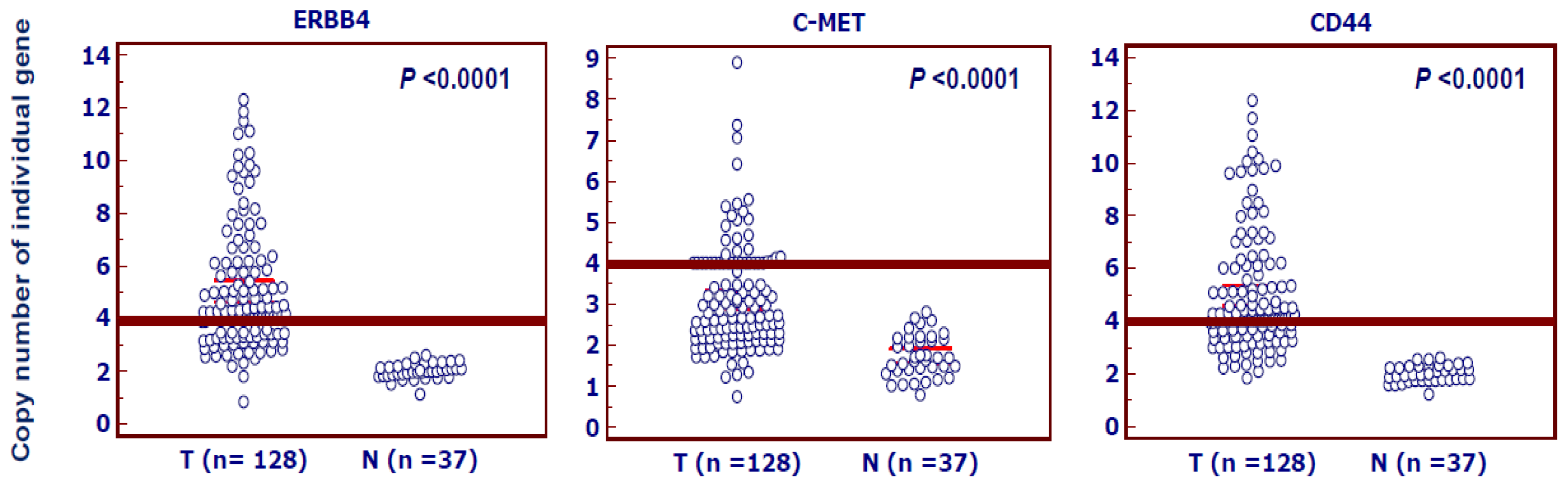
2.1. Highly Frequent Amplification of ERBB4, C-MET and CD44 in Gastric Cancer</title>
<p>Real-time quantitative PCR assay was performed to analyze the copy number of thirty candidate genes in 30 pairs of gastric cancer and normal gastric tissues. With a gene copy number of 4 or more defined as gene amplification, we found that <italic>ERBB4</italic>, <italic>C-MET</italic> and <italic>CD44</italic> genes were frequently amplified in gastric cancers, however, other genes were not or infrequently amplified in gastric cancers, ranging from 0 to 8% (data not shown). Subsequently, we used the same method to analyze the copy number of <italic>ERBB4</italic>, <italic>C-MET</italic> and <italic>CD44</italic> genes in the 128 gastric cancers and 37 normal controls. The data showed that the prevalence of amplification of <italic>ERBB4</italic>, <italic>C-MET</italic> and <italic>CD44</italic> was 67% (86/128), 30% (39/128) and 66% (84/128), respectively, but not in the normal gastric tissues. Copy number of each gene corresponding to each individual case of gastric cancers and normal gastric tissues was showed in <xref ref-type="fig" rid="f1-ijms-13-04714">Figure 1</xref>. Statistical analysis showed that the copy number of each gene in gastric cancers was significantly higher than normal gastric tissues (<xref ref-type="fig" rid="f1-ijms-13-04714">Figure 1</xref>).</p></sec>
<sec sec-type="intro">
<title>2.2. Association of Amplification of ERBB4, C-MET and CD44 with Clinicopathological Characteristics in Gastric Cancer</title>
<p>Because highly frequent amplification of <italic>ERBB4</italic>, <italic>C-MET</italic> and <italic>CD44</italic> was demonstrated in gastric cancer, their association with clinicopathological characteristics was analyzed in a cohort of clinically well-characterized gastric cancers. As shown in <xref ref-type="table" rid="t1-ijms-13-04714">Table 1</xref>, there was a positive association of amplification of <italic>ERBB4</italic> (OR = 2.62, 95% CI = 1.23–5.59, <italic>P</italic> &lt; 0.05) and <italic>CD44</italic> (OR = 2.28, 95% CI = 1.08–4.79, <italic>P</italic> &lt; 0.05) with tumor differentiation. <italic>C-MET</italic> amplification was found to be significantly positively associated with tumor invasion (OR = 2.00, 95% CI = 1.03–3.89, <italic>P</italic> &lt; 0.05). <italic>CD44</italic> amplification was significantly positively associated with lymph node metastasis (OR = 2.23, 95% CI = 1.05–4.73, <italic>P</italic> &lt; 0.05). Moreover, our data also showed that <italic>CD44</italic> amplification was significantly positively associated with the number of lymph node metastasis (OR = 1.70, 95% CI = 1.08–2.69, <italic>P</italic> &lt; 0.05). Notably, there was a significantly positive association of amplification of these genes with cancer-related death (<xref ref-type="table" rid="t1-ijms-13-04714">Table 1</xref>). In order to assess the independent association of gene amplification with age, tumor differentiation, tumor stage, lymph node metastasis and survival status, we conducted multiple multivariable logistic regressions (<xref ref-type="table" rid="t2-ijms-13-04714">Table 2</xref>). Similar to univariate analysis, after adjustment, amplification of <italic>ERBB4</italic> (OR = 2.95, 95% CI = 1.27–6.86, <italic>P</italic> &lt; 0.05) and <italic>CD44</italic> (OR = 2.49, 95% CI = 1.08–5.79, <italic>P</italic> &lt; 0.05) remained significantly associated with poor tumor differentiation (<xref ref-type="table" rid="t2-ijms-13-04714">Table 2</xref>). Similarly, amplification of these genes remained significantly positively associated with cancer-related death (<xref ref-type="table" rid="t2-ijms-13-04714">Table 2</xref>).</p></sec>
<sec>
<title>
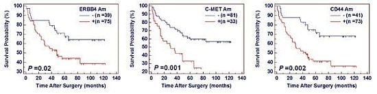
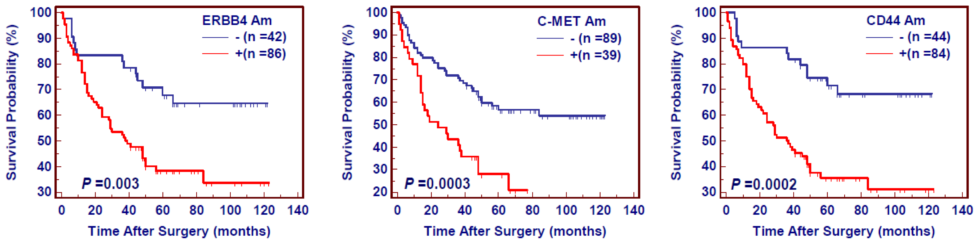
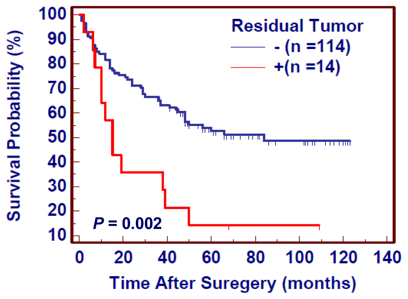
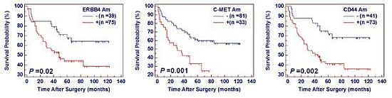
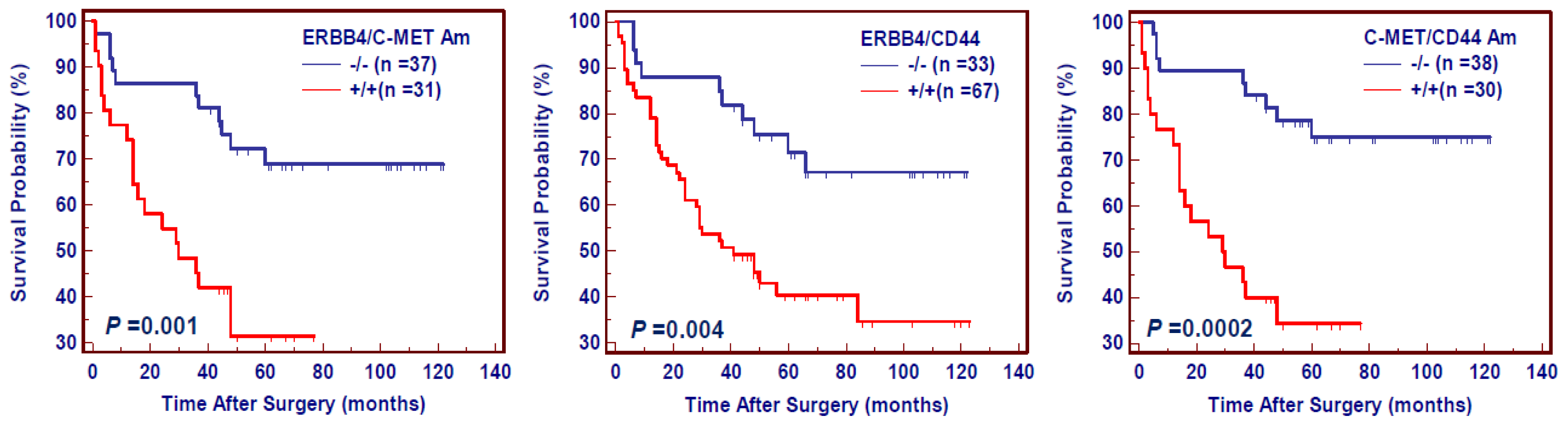
2.3. The Effect of Amplification of ERBB4, C-MET and CD44 on Poor Survival in Gastric Cancer</title>
<p>The Kaplan-Meier estimator of the survivorship function was used to evaluate the impact of amplification of these three genes on the survival of gastric cancer patients. The survival of gastric cancer patients with and without gene amplification was compared using the log-rank test. As shown in <xref ref-type="fig" rid="f2-ijms-13-04714">Figure 2</xref>, amplification of <italic>ERBB4</italic>, <italic>C-MET</italic> and <italic>CD44</italic> significantly affected the poor survival of gastric cancer patients.</p>
<p>Numerous evidences showed that residual tumor after surgery is an independent risk factor for gastric cancer patients. We thus attempted to evaluate the effect of residual tumor after surgery on the survival of gastric cancer patients. As shown in <xref ref-type="fig" rid="f3-ijms-13-04714">Figure 3</xref>, the patients with residual tumor after surgery had significantly shorter survival times than the patients without residual tumor (343.2 months <italic>vs.</italic> 601.2 months on average, <italic>P</italic> = 0.03). Given the impact of residual tumor after surgery on poor survival in gastric cancer, we excluded the patients with residual tumor to explore the effect of gene amplification on poor prognosis of gastric cancer patients. Similar to the findings in <xref ref-type="fig" rid="f2-ijms-13-04714">Figure 2</xref>, the patients with gene amplification had shorter survival times than the patients without gene amplification (<italic>ERBB4</italic>: 508.8 months <italic>vs.</italic> 777.6 on average, <italic>P</italic> = 0.002; <italic>C-MET</italic>: 382.8 months <italic>vs.</italic> 690.0 months on average, <italic>P</italic> = 0.0005; <italic>CD44</italic>: 490.8 months <italic>vs.</italic> 798.0 months on average, <italic>P</italic> = 0.0002) (<xref ref-type="fig" rid="f4-ijms-13-04714">Figure 4</xref>). Multivariate Cox regression analysis indicated that gene amplification may be served as a predictor of poor prognosis for gastric cancer patients (<italic>ERBB4</italic>: HR = 2.00, 95% CI = 1.02–3.86, <italic>P</italic> = 0.04; <italic>C-MET</italic>: HR = 2.10, 95% CI = 1.20–3.69, <italic>P</italic> = 0.01; <italic>CD44</italic>: HR = 2.59, 95% CI = 1.34–5.01, <italic>P</italic> = 0.005) as an independently variable with respect to gender, age, differentiation, lymph node metastasis, and tumor stage (<xref ref-type="table" rid="t3-ijms-13-04714">Table 3</xref>).</p>
<p>Gastric cancer is chronic proliferative disease characterized by genetic alterations of multiple genes, including gene amplification. In the present study, a high prevalence of concomitant amplification of 2 of 3 genes was found in gastric cancer, ranging from 28% (36/128) to 60% (77/128). As shown in <xref ref-type="fig" rid="f5-ijms-13-04714">Figure 5</xref>, concomitant amplification of 2 of 3 genes significantly shortened survival times (<italic>ERBB4/C-MET</italic>: 378.0 months <italic>vs.</italic> 795.6 on average, <italic>P</italic> = 0.001; <italic>ERBB4/CD44</italic>: 483.6 months <italic>vs.</italic> 817.2 months on average, <italic>P</italic> = 0.0004; <italic>C-MET/CD44</italic>: 372.0 months <italic>vs.</italic> 822.0 months on average, <italic>P</italic> &lt; 0.0001), and might be more prognostic of poor survival than amplification of individual gene in gastric cancer.</p></sec></sec>
<sec sec-type="discussion">
<title>3. Discussion</title>
<p>Although gastric cancer is a lethal disease around the world, the causes of gastric cancer are not completely understood. What is clear is that gastric cancer is a multistep process involving multiple genetic and epigenetic events, such as gene amplification. Gene amplification is frequent in solid tumors and represents one of the major molecular pathways through which the oncogenic potential of proto-oncogenes is overactivated during tumorigenesis [<xref ref-type="bibr" rid="b7-ijms-13-04714">7</xref>,<xref ref-type="bibr" rid="b8-ijms-13-04714">8</xref>]. Thus, gene amplification in general and specifically amplicons, have importance for both prognosis and targeted therapies in human cancers, including gastric cancer [<xref ref-type="bibr" rid="b9-ijms-13-04714">9</xref>–<xref ref-type="bibr" rid="b12-ijms-13-04714">12</xref>].</p>
<p>In the present study, we identified three frequently amplified genes in a cohort of well-characterized gastric cancer using real-time quantitative PCR method, including <italic>ERBB4</italic>, <italic>C-MET</italic> and <italic>CD44</italic>. ERBB4 (or HER4) is a member of the Tyr protein kinase family and the epidermal growth factor receptor subfamily, and plays an important role in normal cell growth and in neoplastic transformation [<xref ref-type="bibr" rid="b13-ijms-13-04714">13</xref>]. <italic>ERBB4</italic> amplification, resulting in its overexpression and ligand-independent activation has been found in a variety of human malignancies [<xref ref-type="bibr" rid="b6-ijms-13-04714">6</xref>,<xref ref-type="bibr" rid="b14-ijms-13-04714">14</xref>]. <italic>C-MET</italic> oncogene encodes the receptor tyrosine kinase (RTK) for hepatocyte growth factor (HGF) and regulates genetic programs leading to cell growth, invasion and protection from apoptosis during tumorigenesis [<xref ref-type="bibr" rid="b15-ijms-13-04714">15</xref>]. <italic>C-MET</italic> is frequently amplified in human cancers, including gastric cancer [<xref ref-type="bibr" rid="b16-ijms-13-04714">16</xref>,<xref ref-type="bibr" rid="b17-ijms-13-04714">17</xref>]. Its amplification is closely associated with poor prognosis of cancer patients and resistance to tyrosine kinase inhibitors [<xref ref-type="bibr" rid="b16-ijms-13-04714">16</xref>–<xref ref-type="bibr" rid="b20-ijms-13-04714">20</xref>]. CD44 is a receptor for hyaluronic acid (HA), which plays an important role in cell migration, tumor growth and progression through its affinity for HA, possibly also through its affinity for other ligands such as osteopontin, collagens, and matrix metalloproteinases (MMPs) [<xref ref-type="bibr" rid="b21-ijms-13-04714">21</xref>]. Accumulating evidences have shown that CD44 is abundantly expressed in cancer-initiating cells (CICs), and has thus been implicated as a CIC marker in several malignancies of haematopoietic and epithelial origin, including gastric cancer [<xref ref-type="bibr" rid="b21-ijms-13-04714">21</xref>,<xref ref-type="bibr" rid="b22-ijms-13-04714">22</xref>]. Moreover, <italic>CD44</italic> amplification is also found in gastric cancer [<xref ref-type="bibr" rid="b6-ijms-13-04714">6</xref>]. Notable, there was a higher amplification frequency of these genes in the present study than that of studies carried out in Europe and the US [<xref ref-type="bibr" rid="b23-ijms-13-04714">23</xref>]. One possibility is that the different dietary and environmental factors, such as nitrates, carbohydrates and salt, may potentially cause these distinct genetic alterations in gastric carcinogenesis [<xref ref-type="bibr" rid="b24-ijms-13-04714">24</xref>].</p>
<p>Given gene amplification plays a critical role in gastric tumorigenesis, we investigated their clinical significances and prognostic values in a cohort of gastric cancer patients who had known survival data. Our data showed that amplification of these genes was closely associated with poor tumor differentiation and cancer-related death, suggesting that these genes may contribute to oncologic outcomes of gastric cancer patients. More importantly, similar to observations in the previous studies [<xref ref-type="bibr" rid="b17-ijms-13-04714">17</xref>], <italic>C-MET</italic> amplification was correlated with poor survival of gastric cancer patients in the present study. Notably, our data showed that amplification of <italic>ERBB4</italic> and <italic>CD44</italic> was significantly associated with poor prognosis in gastric cancer, is new to the literature.</p>
<p>To date, inhibitors and antibodies targeting specific molecules are vigorously being developed, and some have been demonstrated to be effective in clinical settings, including gastric cancer [<xref ref-type="bibr" rid="b25-ijms-13-04714">25</xref>,<xref ref-type="bibr" rid="b26-ijms-13-04714">26</xref>]. In the present study, we identified three frequently amplified genes and demonstrated that they were significantly associated with clinical outcome in gastric cancer, suggesting that they may be served as potential therapeutic targets for gastric cancer. Moreover, the prognostic markers may have another role in predicting and guiding the clinical treatment of cancer patients by allowing the identification of patients suited to current therapies. For example, <italic>C-MET</italic> amplification may identify a subset of epithelial cancers with extreme sensitivity to the selective tyrosine kinase inhibitor and define a patient group that is appropriate for clinical trials of targeted therapy using MET inhibitors [<xref ref-type="bibr" rid="b27-ijms-13-04714">27</xref>].</p></sec>
<sec>
<title>4. Experimental Section</title>
<sec sec-type="subjects">
<title>4.1. Patients</title>
<p>With the approval of our institutional review board and human ethics committee, where required, we studied 128 patients with gastric cancer who underwent surgery at the First Affiliated Hospital of Xi’an Jiaotong University School of Medicine from 1999 to 2006. A total of 37 tissues from the patients with chronic gastritis were obtained from the First Affiliated Hospital of Xi’an Jiaotong University School of Medicine as normal controls. None of these patients received chemotherapy and radiotherapy before the surgery. Informed consent was obtained from each patient before the surgery. The histologic diagnosis of tumors was made and agreed upon by at least two senior pathologists at Department of Pathology of the Hospital based on World Health Organization (WHO) criteria. Relevant clinicopathologic characteristics were obtained from the patients’ files or by interview with the patients or their relatives, and the details were summarized in <xref ref-type="table" rid="t4-ijms-13-04714">Table 4</xref>.</p></sec>
<sec>
<title>4.2. Tissues and DNA Preparation</title>
<p>Serial sections from each tumor sample were cut. One section (5 μm) was stained by hematoxylin and eosin (H &amp; E), and was marked as a tumor representative tissue by an expert surgical pathologist for gastric cancer. The next section (8 μm) was deparaffinized and stained with hematoxylin. Tumor tissues were isolated by manual microdissection under an inverted microscope using the marked H &amp; E section for target tissue identification. Genomic DNA was extracted from isolated tissues as previously described [<xref ref-type="bibr" rid="b28-ijms-13-04714">28</xref>]. Briefly, the tissues dissected were first treated with xylene for 12 hours at room temperature to remove the paraffin. All tissues were subsequently subjected to digestion with 1% sodium dodecyl sulfate (SDS) and proteinase K at 48 °C for 48 to 72 hours with addition of several spiking aliquots of concentrated proteinase K to facilitate digestion. Genomic DNA was isolated from the digested tissues followed by standard phenol-chloroform extraction and ethanol precipitation protocol, and stored at −80 °C until use.</p></sec>
<sec sec-type="methods">
<title>4.3. Copy Number Analysis</title>
<p>We analyzed the copy number of candidate genes in gastric cancers and normal gastric tissues by real-time quantitative PCR technique on a CFX384 Thermal Cycler Dice<sup>TM</sup> real-time PCR system (Bio-Rad Laboratories, Inc., Hercules, CA, USA) as described previously [<xref ref-type="bibr" rid="b29-ijms-13-04714">29</xref>]. This method was well established and validated by florescence <italic>in situ</italic> hybridization (FISH) [<xref ref-type="bibr" rid="b29-ijms-13-04714">29</xref>,<xref ref-type="bibr" rid="b30-ijms-13-04714">30</xref>], which has been widely used in the various human cancers [<xref ref-type="bibr" rid="b28-ijms-13-04714">28</xref>–<xref ref-type="bibr" rid="b32-ijms-13-04714">32</xref>]. Specific primers and TaqMan probes were designed using Primer Express 3.0 (Applied Biosystems: Foster City, CA, USA, 2004) to amplify these genes and the internal reference gene <italic>β-actin</italic>. The primers and TaqMan probes for <italic>ERBB4</italic>, <italic>C-MET</italic>, <italic>CD44</italic> and <italic>β-actin</italic> genes in the present study were presented in <xref ref-type="table" rid="t5-ijms-13-04714">Table 5</xref>. The primers and TaqMan probes for other 27 genes need be requested, including <italic>SHFM1</italic>, <italic>CARD4</italic>, <italic>ELN</italic>, <italic>ARF5</italic>, <italic>SLC25A40</italic>, <italic>NRAS</italic>, <italic>CDK5</italic>, <italic>CREM</italic>, <italic>LMO2</italic>, <italic>DNMT1</italic>, <italic>FMR2</italic>, <italic>PSPHL</italic>, <italic>KRAS</italic>, <italic>PEG10</italic>, <italic>CDC2L5</italic>, <italic>HRAS</italic>, <italic>MGAM</italic>, <italic>ZP3</italic>, <italic>EPO</italic>, <italic>GUSB</italic>, <italic>ZPBP</italic>, <italic>TMEM60</italic>, <italic>PEPIN1</italic>, <italic>BRAF</italic>, <italic>AHR</italic>, <italic>DNMT3B</italic> and <italic>EZH2</italic>. All TaqMan probes were labeled with 5′-FAM (6-carboxyfluorescein, fluorescent reporter) and 3′-TAMRA (6-carboxy-tetramethylrhodamine, fluorescent quencher). Using a PCR protocol described previously [<xref ref-type="bibr" rid="b29-ijms-13-04714">29</xref>], PCR amplification were carried out in buffer containing 16.6 mM ammonium sulfate, 67 mM Tris base, 2.5 mM MgCl<sub>2</sub>, 10 mM 2-mercaptoethanol, 0.1% DMSO, 0.2 mM each of dATP, dCTP, dGTP and dTTP, 600 nM each of forward and reverse primers, 200 nM TaqMan probe, 0.6 unit Platinum <italic>Taq</italic> polymerase and 2% Rox reference dye. Each sample was run in triplicate, and <italic>β-actin</italic> was run in parallel to standardize the input DNA. Standard curves were established using serial dilutions of normal leukocyte DNA with a quantity range of 3.75 to 60 ng per 2 μL. The specificity of real-time quantitative PCR for these genes was confirmed by running thee PCR products on a 1.5% agarose gel to show single specific bands of the PCR products at the expected sizes (data not shown). The efficiency of real-time quantitative PCR assays for each target was shown in <xref ref-type="table" rid="t5-ijms-13-04714">Table 5</xref>. Gene amplification was defined by a copy number ≥4.</p></sec>
<sec sec-type="methods">
<title>4.4. Statistical Analysis</title>
<p>The Mann–Whitney <italic>U</italic> test was used to compare copy number of these genes between gastric cancer and normal gastric tissues. Association of gene amplification with clinicopathological characteristics was assessed univariately using the SPSS statistical package (version 11.5; IBM Corporation: Chicago, IL, USA, 2003). Multivariate models were then developed that adjusted for the most important covariates, including age, differentiation, tumor stage, lymph node metastasis and survival status. Survival length was determined from the day of primary tumor surgery to the day of death or last clinical follow-up. The Kaplan–Meier method was used for survival analysis grouping with gene amplification. Differences between curves were analyzed using the log-rank test. Multivariate Cox regression analysis was used to evaluate the effect of gene amplification on survival of independently of gender, age, differentiation, lymph node metastasis, and tumor stage. All statistical analyses were performed using the SPSS statistical package. <italic>P</italic> values &lt; 0.05 were considered significant.</p></sec></sec>
<sec sec-type="conclusions">
<title>5. Conclusions</title>
<p>In summary, we identified three genes that were frequently amplified from thirty candidate genes in cancerous tissues, but not normal gastric tissues, and demonstrated that amplification of these genes was closely associated with clinicopathological characteristics, particularly tumor differentiation and cancer-related death. Importantly, amplification of these genes was significantly associated with poor survival in gastric cancer. Thus, these aberrantly amplified genes may be used as useful markers in evaluating poor prognosis and potential therapeutic targets for gastric cancer.</p></sec></body>
<back>
<ack>
<title>Acknowledgments</title>
<p>This work was supported by the National Science Foundation of China (No. 30901459, 30973372 and 81171969) and the National Key Program for Developing Basic Research (No. 2010CB933903).</p></ack>
<fn-group><fn id="fn1-ijms-13-04714">
<p><bold>Conflicts of Interest</bold></p>
<p>The authors declare no conflict of interest.</p></fn></fn-group>
<ref-list>
<title>References</title>
<ref id="b1-ijms-13-04714"><label>1</label><citation citation-type="journal"><person-group person-group-type="author"><name><surname>Crew</surname><given-names>K.D.</given-names></name><name><surname>Neugut</surname><given-names>A.I.</given-names></name></person-group><article-title>Epidemiology of gastric cancer</article-title><source>World J. Gastroenterol</source><year>2006</year><volume>12</volume><fpage>354</fpage><lpage>362</lpage><pub-id pub-id-type="pmid">16489633</pub-id></citation></ref>
<ref id="b2-ijms-13-04714"><label>2</label><citation citation-type="journal"><person-group person-group-type="author"><name><surname>Jemal</surname><given-names>A.</given-names></name><name><surname>Bray</surname><given-names>F.</given-names></name><name><surname>Center</surname><given-names>M.M.</given-names></name><name><surname>Ferlay</surname><given-names>J.</given-names></name><name><surname>Ward</surname><given-names>E.</given-names></name><name><surname>Forman</surname><given-names>D.</given-names></name></person-group><article-title>Global cancer statistics</article-title><source>CA Cancer J. Clin</source><year>2011</year><volume>61</volume><fpage>69</fpage><lpage>90</lpage><pub-id pub-id-type="doi">10.3322/caac.20107</pub-id><pub-id pub-id-type="pmid">21296855</pub-id></citation></ref>
<ref id="b3-ijms-13-04714"><label>3</label><citation citation-type="journal"><person-group person-group-type="author"><name><surname>Kim</surname><given-names>H.J.</given-names></name><name><surname>Karpeh</surname><given-names>M.S.</given-names></name></person-group><article-title>Surgical approaches and outcomes in the treatment of gastric cancer</article-title><source>Semin. Radiat. Oncol</source><year>2002</year><volume>12</volume><fpage>162</fpage><lpage>169</lpage><pub-id pub-id-type="doi">10.1053/srao.2002.30818</pub-id><pub-id pub-id-type="pmid">11979417</pub-id></citation></ref>
<ref id="b4-ijms-13-04714"><label>4</label><citation citation-type="journal"><person-group person-group-type="author"><name><surname>Peng</surname><given-names>D.F.</given-names></name><name><surname>Sugihara</surname><given-names>H.</given-names></name><name><surname>Mukaisho</surname><given-names>K.</given-names></name><name><surname>Tsubosa</surname><given-names>Y.</given-names></name><name><surname>Hattori</surname><given-names>T.</given-names></name></person-group><article-title>Alterations of chromosomal copy number during progression of diffuse-type gastric carcinomas: Metaphase- and array-based comparative genomic hybridization analyses of multiple samples from individual tumours</article-title><source>J. Pathol</source><year>2003</year><volume>201</volume><fpage>439</fpage><lpage>450</lpage><pub-id pub-id-type="doi">10.1002/path.1459</pub-id><pub-id pub-id-type="pmid">14595756</pub-id></citation></ref>
<ref id="b5-ijms-13-04714"><label>5</label><citation citation-type="journal"><person-group person-group-type="author"><name><surname>Buffart</surname><given-names>T.E.</given-names></name><name><surname>van Grieken</surname><given-names>N.C.</given-names></name><name><surname>Tijssen</surname><given-names>M.</given-names></name><name><surname>Coffa</surname><given-names>J.</given-names></name><name><surname>Ylstra</surname><given-names>B.</given-names></name><name><surname>Grabsch</surname><given-names>H.I.</given-names></name><name><surname>van de Velde</surname><given-names>C.J.</given-names></name><name><surname>Carvalho</surname><given-names>B.</given-names></name><name><surname>Meijer</surname><given-names>G.A.</given-names></name></person-group><article-title>High resolution analysis of DNA copy-number aberrations of chromosomes 8, 13, and 20 in gastric cancers</article-title><source>Virchows Arch</source><year>2009</year><volume>455</volume><fpage>213</fpage><lpage>223</lpage><pub-id pub-id-type="doi">10.1007/s00428-009-0814-y</pub-id><pub-id pub-id-type="pmid">19697059</pub-id></citation></ref>
<ref id="b6-ijms-13-04714"><label>6</label><citation citation-type="journal"><person-group person-group-type="author"><name><surname>Zhang</surname><given-names>D.</given-names></name><name><surname>Wang</surname><given-names>Z.</given-names></name><name><surname>Luo</surname><given-names>Y.</given-names></name><name><surname>Xu</surname><given-names>Y.</given-names></name><name><surname>Liu</surname><given-names>Y.</given-names></name><name><surname>Yang</surname><given-names>W.</given-names></name><name><surname>Zhang</surname><given-names>X.</given-names></name></person-group><article-title>Analysis of DNA copy number aberrations by multiple ligation-dependent probe amplification on 50 intestinal type gastric cancers</article-title><source>J. Surg. Oncol</source><year>2011</year><volume>103</volume><fpage>124</fpage><lpage>132</lpage><pub-id pub-id-type="doi">10.1002/jso.21792</pub-id><pub-id pub-id-type="pmid">21259245</pub-id></citation></ref>
<ref id="b7-ijms-13-04714"><label>7</label><citation citation-type="journal"><person-group person-group-type="author"><name><surname>Schwab</surname><given-names>M.</given-names></name></person-group><article-title>Amplification of oncogenes in human cancer cells</article-title><source>Bioessays</source><year>1998</year><volume>20</volume><fpage>473</fpage><lpage>479</lpage><pub-id pub-id-type="doi">10.1002/(SICI)1521-1878(199806)20:6&lt;473::AID-BIES5&gt;3.0.CO;2-N</pub-id><pub-id pub-id-type="pmid">9699459</pub-id></citation></ref>
<ref id="b8-ijms-13-04714"><label>8</label><citation citation-type="journal"><person-group person-group-type="author"><name><surname>Albertson</surname><given-names>D.G.</given-names></name></person-group><article-title>Gene amplification in cancer</article-title><source>Trends Genet</source><year>2006</year><volume>22</volume><fpage>447</fpage><lpage>455</lpage><pub-id pub-id-type="doi">10.1016/j.tig.2006.06.007</pub-id><pub-id pub-id-type="pmid">16787682</pub-id></citation></ref>
<ref id="b9-ijms-13-04714"><label>9</label><citation citation-type="journal"><person-group person-group-type="author"><name><surname>Hirono</surname><given-names>Y.</given-names></name><name><surname>Tsugawa</surname><given-names>K.</given-names></name><name><surname>Fushida</surname><given-names>S.</given-names></name><name><surname>Ninomiya</surname><given-names>I.</given-names></name><name><surname>Yonemura</surname><given-names>Y.</given-names></name><name><surname>Miyazaki</surname><given-names>I.</given-names></name><name><surname>Endou</surname><given-names>Y.</given-names></name><name><surname>Tanaka</surname><given-names>M.</given-names></name><name><surname>Sasaki</surname><given-names>T.</given-names></name></person-group><article-title>Amplification of epidermal growth factor receptor gene and its relationship to survival in human gastric cancer</article-title><source>Oncology</source><year>1995</year><volume>52</volume><fpage>182</fpage><lpage>188</lpage><pub-id pub-id-type="doi">10.1159/000227455</pub-id><pub-id pub-id-type="pmid">7715901</pub-id></citation></ref>
<ref id="b10-ijms-13-04714"><label>10</label><citation citation-type="journal"><person-group person-group-type="author"><name><surname>Sakakura</surname><given-names>C.</given-names></name><name><surname>Hagiwara</surname><given-names>A.</given-names></name><name><surname>Yasuoka</surname><given-names>R.</given-names></name><name><surname>Fujita</surname><given-names>Y.</given-names></name><name><surname>Nakanishi</surname><given-names>M.</given-names></name><name><surname>Masuda</surname><given-names>K.</given-names></name><name><surname>Kimura</surname><given-names>A.</given-names></name><name><surname>Nakamura</surname><given-names>Y.</given-names></name><name><surname>Inazawa</surname><given-names>J.</given-names></name><name><surname>Abe</surname><given-names>T.</given-names></name><etal/></person-group><article-title>Amplification and over-expression of the AIB1 nuclear receptor co-activator gene in primary gastric cancers</article-title><source>Int. J. Cancer</source><year>2000</year><volume>89</volume><fpage>217</fpage><lpage>223</lpage><pub-id pub-id-type="doi">10.1002/1097-0215(20000520)89:3&lt;217::AID-IJC2&gt;3.0.CO;2-6</pub-id><pub-id pub-id-type="pmid">10861496</pub-id></citation></ref>
<ref id="b11-ijms-13-04714"><label>11</label><citation citation-type="journal"><person-group person-group-type="author"><name><surname>Park</surname><given-names>J.H.</given-names></name><name><surname>Lee</surname><given-names>B.L.</given-names></name><name><surname>Yoon</surname><given-names>J.</given-names></name><name><surname>Kim</surname><given-names>J.</given-names></name><name><surname>Kim</surname><given-names>M.A.</given-names></name><name><surname>Yang</surname><given-names>H.K.</given-names></name><name><surname>Kim</surname><given-names>W.H.</given-names></name></person-group><article-title>Focal adhesion kinase (FAK) gene amplification and its clinical implications in gastric cancer</article-title><source>Hum. Pathol</source><year>2010</year><volume>41</volume><fpage>1664</fpage><lpage>1673</lpage><pub-id pub-id-type="doi">10.1016/j.humpath.2010.06.004</pub-id><pub-id pub-id-type="pmid">20869748</pub-id></citation></ref>
<ref id="b12-ijms-13-04714"><label>12</label><citation citation-type="journal"><person-group person-group-type="author"><name><surname>Begnami</surname><given-names>M.D.</given-names></name><name><surname>Fukuda</surname><given-names>E.</given-names></name><name><surname>Fregnani</surname><given-names>J.H.</given-names></name><name><surname>Nonogaki</surname><given-names>S.</given-names></name><name><surname>Montagnini</surname><given-names>A.L.</given-names></name><name><surname>da Costa</surname><given-names>W.L.</given-names><suffix>Jr</suffix></name><name><surname>Soares</surname><given-names>F.A.</given-names></name></person-group><article-title>Prognostic implications of altered human epidermal growth factor receptors (HERs) in gastric carcinomas: HER2 and HER3 are predictors of poor outcome</article-title><source>J. Clin. Oncol</source><year>2011</year><volume>29</volume><fpage>3030</fpage><lpage>3036</lpage><pub-id pub-id-type="doi">10.1200/JCO.2010.33.6313</pub-id><pub-id pub-id-type="pmid">21709195</pub-id></citation></ref>
<ref id="b13-ijms-13-04714"><label>13</label><citation citation-type="journal"><person-group person-group-type="author"><name><surname>Muraoka-Cook</surname><given-names>R.S.</given-names></name><name><surname>Feng</surname><given-names>S.M.</given-names></name><name><surname>Strunk</surname><given-names>K.E.</given-names></name><name><surname>Earp</surname><given-names>H.S.</given-names><suffix>III</suffix></name></person-group><article-title>ErbB4/HER4: Role in mammary gland development, differentiation and growth inhibition</article-title><source>J. Mammary Gland Biol. Neoplasia</source><year>2008</year><volume>13</volume><fpage>235</fpage><lpage>246</lpage><pub-id pub-id-type="doi">10.1007/s10911-008-9080-x</pub-id><pub-id pub-id-type="pmid">18437540</pub-id></citation></ref>
<ref id="b14-ijms-13-04714"><label>14</label><citation citation-type="journal"><person-group person-group-type="author"><name><surname>Kamath</surname><given-names>S.</given-names></name><name><surname>Buolamwini</surname><given-names>J.K.</given-names></name></person-group><article-title>Targeting EGFR and HER-2 receptor tyrosine kinases for cancer drug discovery and development</article-title><source>Med. Res. Rev</source><year>2006</year><volume>26</volume><fpage>569</fpage><lpage>594</lpage><pub-id pub-id-type="doi">10.1002/med.20070</pub-id><pub-id pub-id-type="pmid">16788977</pub-id></citation></ref>
<ref id="b15-ijms-13-04714"><label>15</label><citation citation-type="journal"><person-group person-group-type="author"><name><surname>Liu</surname><given-names>X.</given-names></name><name><surname>Yao</surname><given-names>W.</given-names></name><name><surname>Newton</surname><given-names>R.C.</given-names></name><name><surname>Scherle</surname><given-names>P.A.</given-names></name></person-group><article-title>Targeting the c-MET signaling pathway for cancer therapy</article-title><source>Expert Opin. Investig. Drugs</source><year>2008</year><volume>17</volume><fpage>997</fpage><lpage>1011</lpage><pub-id pub-id-type="doi">10.1517/13543784.17.7.997</pub-id><pub-id pub-id-type="pmid">18549337</pub-id></citation></ref>
<ref id="b16-ijms-13-04714"><label>16</label><citation citation-type="journal"><person-group person-group-type="author"><name><surname>Sierra</surname><given-names>J.R.</given-names></name><name><surname>Tsao</surname><given-names>M.S.</given-names></name></person-group><article-title>c-MET as a potential therapeutic target and biomarker in cancer</article-title><source>Ther. Adv. Med. Oncol</source><year>2011</year><volume>3</volume><fpage>S21</fpage><lpage>S35</lpage><pub-id pub-id-type="doi">10.1177/1758834011422557</pub-id><pub-id pub-id-type="pmid">22128285</pub-id></citation></ref>
<ref id="b17-ijms-13-04714"><label>17</label><citation citation-type="journal"><person-group person-group-type="author"><name><surname>Lee</surname><given-names>J.</given-names></name><name><surname>Seo</surname><given-names>J.W.</given-names></name><name><surname>Jun</surname><given-names>H.J.</given-names></name><name><surname>Ki</surname><given-names>C.S.</given-names></name><name><surname>Park</surname><given-names>S.H.</given-names></name><name><surname>Park</surname><given-names>Y.S.</given-names></name><name><surname>Lim</surname><given-names>H.Y.</given-names></name><name><surname>Choi</surname><given-names>M.G.</given-names></name><name><surname>Bae</surname><given-names>J.M.</given-names></name><name><surname>Sohn</surname><given-names>T.S.</given-names></name><etal/></person-group><article-title>Impact of MET amplification on gastric cancer: Possible roles as a novel prognostic marker and a potential therapeutic target</article-title><source>Oncol. Rep</source><year>2011</year><volume>25</volume><fpage>1517</fpage><lpage>1524</lpage><pub-id pub-id-type="pmid">21424128</pub-id></citation></ref>
<ref id="b18-ijms-13-04714"><label>18</label><citation citation-type="journal"><person-group person-group-type="author"><name><surname>Toschi</surname><given-names>L.</given-names></name><name><surname>Cappuzzo</surname><given-names>F.</given-names></name></person-group><article-title>Clinical implications of MET gene copy number in lung cancer</article-title><source>Future Oncol</source><year>2010</year><volume>6</volume><fpage>239</fpage><lpage>247</lpage><pub-id pub-id-type="doi">10.2217/fon.09.164</pub-id><pub-id pub-id-type="pmid">20146583</pub-id></citation></ref>
<ref id="b19-ijms-13-04714"><label>19</label><citation citation-type="journal"><person-group person-group-type="author"><name><surname>Go</surname><given-names>H.</given-names></name><name><surname>Jeon</surname><given-names>Y.K.</given-names></name><name><surname>Park</surname><given-names>H.J.</given-names></name><name><surname>Sung</surname><given-names>S.W.</given-names></name><name><surname>Seo</surname><given-names>J.W.</given-names></name><name><surname>Chung</surname><given-names>D.H.</given-names></name></person-group><article-title>High MET gene copy number leads to shorter survival in patients with non-small cell lung cancer</article-title><source>J. Thorac. Oncol</source><year>2010</year><volume>5</volume><fpage>305</fpage><lpage>313</lpage><pub-id pub-id-type="doi">10.1097/JTO.0b013e3181ce3d1d</pub-id><pub-id pub-id-type="pmid">20107422</pub-id></citation></ref>
<ref id="b20-ijms-13-04714"><label>20</label><citation citation-type="journal"><person-group person-group-type="author"><name><surname>Park</surname><given-names>S.</given-names></name><name><surname>Choi</surname><given-names>Y.L.</given-names></name><name><surname>Sung</surname><given-names>C.O.</given-names></name><name><surname>An</surname><given-names>J.</given-names></name><name><surname>Seo</surname><given-names>J.</given-names></name><name><surname>Ahn</surname><given-names>M.J.</given-names></name><name><surname>Ahn</surname><given-names>J.S.</given-names></name><name><surname>Park</surname><given-names>K.</given-names></name><name><surname>Shin</surname><given-names>Y.K.</given-names></name><name><surname>Erkin</surname><given-names>O.C.</given-names></name><etal/></person-group><article-title>High MET copy number and MET overexpression: Poor outcome in non-small cell lung cancer patients</article-title><source>Histol. Histopathol</source><year>2012</year><volume>27</volume><fpage>197</fpage><lpage>207</lpage><pub-id pub-id-type="pmid">22207554</pub-id></citation></ref>
<ref id="b21-ijms-13-04714"><label>21</label><citation citation-type="journal"><person-group person-group-type="author"><name><surname>Zöller</surname><given-names>M.</given-names></name></person-group><article-title>CD44: Can a cancer-initiating cell profit from an abundantly expressed molecule?</article-title><source>Nat. Rev. Cancer</source><year>2011</year><volume>11</volume><fpage>254</fpage><lpage>267</lpage><pub-id pub-id-type="doi">10.1038/nrc3023</pub-id><pub-id pub-id-type="pmid">21390059</pub-id></citation></ref>
<ref id="b22-ijms-13-04714"><label>22</label><citation citation-type="journal"><person-group person-group-type="author"><name><surname>Takaishi</surname><given-names>S.</given-names></name><name><surname>Okumura</surname><given-names>T.</given-names></name><name><surname>Tu</surname><given-names>S.</given-names></name><name><surname>Wang</surname><given-names>S.S.</given-names></name><name><surname>Shibata</surname><given-names>W.</given-names></name><name><surname>Vigneshwaran</surname><given-names>R.</given-names></name><name><surname>Gordon</surname><given-names>S.A.</given-names></name><name><surname>Shimada</surname><given-names>Y.</given-names></name><name><surname>Wang</surname><given-names>T.C.</given-names></name></person-group><article-title>Identification of gastric cancer stem cells using the cell surface marker CD44</article-title><source>Stem Cells</source><year>2009</year><volume>27</volume><fpage>1006</fpage><lpage>1020</lpage><pub-id pub-id-type="doi">10.1002/stem.30</pub-id><pub-id pub-id-type="pmid">19415765</pub-id></citation></ref>
<ref id="b23-ijms-13-04714"><label>23</label><citation citation-type="journal"><person-group person-group-type="author"><name><surname>Rossi</surname><given-names>E.</given-names></name><name><surname>Klersy</surname><given-names>C.</given-names></name><name><surname>Manca</surname><given-names>R.</given-names></name><name><surname>Zuffardi</surname><given-names>O.</given-names></name><name><surname>Solcia</surname><given-names>E.</given-names></name></person-group><article-title>Correlation between genomic alterations assessed by array comparative genomic hybridization, prognostically informative histologic subtype, stage, and patient survival in gastric cancer</article-title><source>Hum. Pathol</source><year>2011</year><volume>42</volume><fpage>1937</fpage><lpage>1945</lpage><pub-id pub-id-type="doi">10.1016/j.humpath.2011.02.016</pub-id><pub-id pub-id-type="pmid">21676433</pub-id></citation></ref>
<ref id="b24-ijms-13-04714"><label>24</label><citation citation-type="journal"><person-group person-group-type="author"><name><surname>Ubukata</surname><given-names>H.</given-names></name><name><surname>Nagata</surname><given-names>H.</given-names></name><name><surname>Tabuchi</surname><given-names>T.</given-names></name><name><surname>Konishi</surname><given-names>S.</given-names></name><name><surname>Kasuga</surname><given-names>T.</given-names></name><name><surname>Tabuchi</surname><given-names>T.</given-names></name></person-group><article-title>Why is the coexistence of gastric cancer and duodenal ulcer rare? Examination of factors related to both gastric cancer and duodenal ulcer</article-title><source>Gastric Cancer</source><year>2011</year><volume>14</volume><fpage>4</fpage><lpage>12</lpage><pub-id pub-id-type="doi">10.1007/s10120-011-0005-9</pub-id><pub-id pub-id-type="pmid">21249411</pub-id></citation></ref>
<ref id="b25-ijms-13-04714"><label>25</label><citation citation-type="journal"><person-group person-group-type="author"><name><surname>Asaoka</surname><given-names>Y.</given-names></name><name><surname>Ikenoue</surname><given-names>T.</given-names></name><name><surname>Koike</surname><given-names>K.</given-names></name></person-group><article-title>New targeted therapies for gastric cancer</article-title><source>Expert Opin. Investig. Drugs</source><year>2011</year><volume>20</volume><fpage>595</fpage><lpage>604</lpage><pub-id pub-id-type="doi">10.1517/13543784.2011.566863</pub-id><pub-id pub-id-type="pmid">21406037</pub-id></citation></ref>
<ref id="b26-ijms-13-04714"><label>26</label><citation citation-type="journal"><person-group person-group-type="author"><name><surname>Yoong</surname><given-names>J.</given-names></name><name><surname>Michael</surname><given-names>M.</given-names></name><name><surname>Leong</surname><given-names>T.</given-names></name></person-group><article-title>Targeted therapies for gastric cancer: Current status</article-title><source>Drugs</source><year>2011</year><volume>71</volume><fpage>1367</fpage><lpage>1384</lpage><pub-id pub-id-type="doi">10.2165/11592530-000000000-00000</pub-id><pub-id pub-id-type="pmid">21812503</pub-id></citation></ref>
<ref id="b27-ijms-13-04714"><label>27</label><citation citation-type="journal"><person-group person-group-type="author"><name><surname>Smolen</surname><given-names>G.A.</given-names></name><name><surname>Sordella</surname><given-names>R.</given-names></name><name><surname>Muir</surname><given-names>B.</given-names></name><name><surname>Mohapatra</surname><given-names>G.</given-names></name><name><surname>Barmettler</surname><given-names>A.</given-names></name><name><surname>Archibald</surname><given-names>H.</given-names></name><name><surname>Kim</surname><given-names>W.J.</given-names></name><name><surname>Okimoto</surname><given-names>R.A.</given-names></name><name><surname>Bell</surname><given-names>D.W.</given-names></name><name><surname>Sgroi</surname><given-names>D.C.</given-names></name><etal/></person-group><article-title>Amplification of MET may identify a subset of cancers with extreme sensitivity to the selective tyrosine kinase inhibitor PHA-665752</article-title><source>Proc. Natl. Acad. Sci. USA</source><year>2006</year><volume>103</volume><fpage>2316</fpage><lpage>2321</lpage><pub-id pub-id-type="doi">10.1073/pnas.0508776103</pub-id><pub-id pub-id-type="pmid">16461907</pub-id></citation></ref>
<ref id="b28-ijms-13-04714"><label>28</label><citation citation-type="journal"><person-group person-group-type="author"><name><surname>Ji</surname><given-names>M.</given-names></name><name><surname>Guan</surname><given-names>H.</given-names></name><name><surname>Gao</surname><given-names>C.</given-names></name><name><surname>Shi</surname><given-names>B.</given-names></name><name><surname>Hou</surname><given-names>P</given-names></name></person-group><article-title>Highly frequent promoter methylation and PIK3CA amplification in non-small cell lung cancer (NSCLC)</article-title><source>BMC Cancer</source><year>2011</year><volume>11</volume><pub-id pub-id-type="doi">10.1186/1471-2407-11-147</pub-id></citation></ref>
<ref id="b29-ijms-13-04714"><label>29</label><citation citation-type="journal"><person-group person-group-type="author"><name><surname>Wu</surname><given-names>G.</given-names></name><name><surname>Mambo</surname><given-names>E.</given-names></name><name><surname>Guo</surname><given-names>Z.</given-names></name><name><surname>Hu</surname><given-names>S.</given-names></name><name><surname>Huang</surname><given-names>X.</given-names></name><name><surname>Gollin</surname><given-names>S.M.</given-names></name><name><surname>Trink</surname><given-names>B.</given-names></name><name><surname>Ladenson</surname><given-names>P.W.</given-names></name><name><surname>Sidransky</surname><given-names>D.</given-names></name><name><surname>Xing</surname><given-names>M.</given-names></name></person-group><article-title>Uncommon mutation but common amplifications of the <italic>PIK3CA</italic> gene in thyroid tumors</article-title><source>J. Clin. Endocrinol. Metab</source><year>2005</year><volume>90</volume><fpage>4688</fpage><lpage>4693</lpage><pub-id pub-id-type="doi">10.1210/jc.2004-2281</pub-id><pub-id pub-id-type="pmid">15928251</pub-id></citation></ref>
<ref id="b30-ijms-13-04714"><label>30</label><citation citation-type="journal"><person-group person-group-type="author"><name><surname>Engelman</surname><given-names>J.A.</given-names></name><name><surname>Zejnullahu</surname><given-names>K.</given-names></name><name><surname>Mitsudomi</surname><given-names>T.</given-names></name><name><surname>Song</surname><given-names>Y.</given-names></name><name><surname>Hyland</surname><given-names>C.</given-names></name><name><surname>Park</surname><given-names>J.O.</given-names></name><name><surname>Lindeman</surname><given-names>N.</given-names></name><name><surname>Gale</surname><given-names>C.M.</given-names></name><name><surname>Zhao</surname><given-names>X.</given-names></name><name><surname>Christensen</surname><given-names>J.</given-names></name><etal/></person-group><article-title><italic>MET</italic> amplification leads to gefitinib resistance in lung cancer by activating ERBB3 signaling</article-title><source>Science</source><year>2007</year><volume>316</volume><fpage>1039</fpage><lpage>1043</lpage><pub-id pub-id-type="doi">10.1126/science.1141478</pub-id><pub-id pub-id-type="pmid">17463250</pub-id></citation></ref>
<ref id="b31-ijms-13-04714"><label>31</label><citation citation-type="journal"><person-group person-group-type="author"><name><surname>Hou</surname><given-names>P.</given-names></name><name><surname>Liu</surname><given-names>D.</given-names></name><name><surname>Shan</surname><given-names>Y.</given-names></name><name><surname>Hu</surname><given-names>S.</given-names></name><name><surname>Studeman</surname><given-names>K.</given-names></name><name><surname>Condouris</surname><given-names>S.</given-names></name><name><surname>Wang</surname><given-names>Y.</given-names></name><name><surname>Trink</surname><given-names>A.</given-names></name><name><surname>El-Naggar</surname><given-names>A.K.</given-names></name><name><surname>Tallini</surname><given-names>G.</given-names></name><etal/></person-group><article-title>Genetic alterations and their relationship in the phosphatidylinositol 3-kinase/Akt pathway in thyroid cancer</article-title><source>Clin. Cancer Res</source><year>2007</year><volume>13</volume><fpage>1161</fpage><lpage>1170</lpage><pub-id pub-id-type="doi">10.1158/1078-0432.CCR-06-1125</pub-id><pub-id pub-id-type="pmid">17317825</pub-id></citation></ref>
<ref id="b32-ijms-13-04714"><label>32</label><citation citation-type="journal"><person-group person-group-type="author"><name><surname>Kawano</surname><given-names>O.</given-names></name><name><surname>Sasaki</surname><given-names>H.</given-names></name><name><surname>Okuda</surname><given-names>K.</given-names></name><name><surname>Yukiue</surname><given-names>H.</given-names></name><name><surname>Yokoyama</surname><given-names>T.</given-names></name><name><surname>Yano</surname><given-names>M.</given-names></name><name><surname>Fujii</surname><given-names>Y.</given-names></name></person-group><article-title><italic>PIK3CA</italic> gene amplification in Japanese non-small cell lung cancer</article-title><source>Lung Cancer</source><year>2007</year><volume>58</volume><fpage>159</fpage><lpage>160</lpage><pub-id pub-id-type="doi">10.1016/j.lungcan.2007.06.020</pub-id><pub-id pub-id-type="pmid">17681398</pub-id></citation></ref></ref-list>
<sec sec-type="display-objects">
<title>Figures and Tables</title>
<fig id="f1-ijms-13-04714" position="float">
<label>Figure 1</label>
<caption>
<p>The copy number of <italic>ERBB4</italic>, <italic>C-MET</italic> and <italic>CD44</italic> genes corresponding to each individual case of gastric cancers and normal gastric tissues (circle). Real-time quantitative PCR was performed to evaluate their copy numbers in a cohort of gastric cancers and normal gastric tissues. Details are as described in Methods. Horizontal lines indicate a 95% confidence interval for the sample mean. T: tumor tissues; N: normal gastric tissues.</p></caption>
<graphic xlink:href="ijms-13-04714f1.gif"/></fig>
<fig id="f2-ijms-13-04714" position="float">
<label>Figure 2</label>
<caption>
<p>The effect of amplification of <italic>ERBB4</italic>, <italic>C-MET</italic> and <italic>CD44</italic> on poor survival in gastric cancer. Kaplan-Meier survival curves were made according to the presence of gene amplification in a cohort of gastric cancers. The patients with gene amplification had significantly shorter survival times than the patients without gene amplification. Am, amplification; +, harboring gene amplification; −, the lack of gene amplification.</p></caption>
<graphic xlink:href="ijms-13-04714f2.gif"/></fig>
<fig id="f3-ijms-13-04714" position="float">
<label>Figure 3</label>
<caption>
<p>The effect of residual tumor after surgery on poor survival in gastric cancer. Survival was evaluated according to the presence of residual tumor after surgery in gastric cancers. Kaplan–Meier survival curves show that the patients with residual tumor had a significantly shorter survival time than the patients without residual tumor (<italic>P</italic> = 0.002). +, the patients with residual tumor; −, the patients without residual tumor.</p></caption>
<graphic xlink:href="ijms-13-04714f3.gif"/></fig>
<fig id="f4-ijms-13-04714" position="float">
<label>Figure 4</label>
<caption>
<p>The effect of amplification of <italic>ERBB4</italic>, <italic>C-MET</italic> and <italic>CD44</italic> on poor survival of gastric cancer patients without residual tumor after surgery. Kaplan–Meier analysis of survival was performed according to the status of gene amplification in a cohort of gastric cancers. The patients with gene amplification had poorer survival than the patients without gene amplification. Am, amplification; +, harboring gene amplification; −, the lack of gene amplification.</p></caption>
<graphic xlink:href="ijms-13-04714f4.gif"/></fig>
<fig id="f5-ijms-13-04714" position="float">
<label>Figure 5</label>
<caption>
<p>The effect of concomitant amplification of two of three genes on poor survival in gastric cancer. Survival was evaluated according to the presence of concomitant amplification of two of three genes in a number of gastric cancer patients without residual tumor after surgery. The patients with gene amplification had significantly shorter survival times than the patients without gene amplification. +/+, harboring concomitant amplification of two genes; −/−, the lack of gene amplification.</p></caption>
<graphic xlink:href="ijms-13-04714f5.gif"/></fig>
<table-wrap id="t1-ijms-13-04714" position="float">
<label>Table 1</label>
<caption>
<p>Amplification of individual genes in gastric cancer—univariate associations with clinicopathological characteristics (OR <xref ref-type="table-fn" rid="tfn1-ijms-13-04714">†</xref> and 95% CI).</p></caption>
<table frame="hsides" rules="groups">
<thead>
<tr>
<th align="center" valign="bottom">Genes</th>
<th align="center" valign="bottom">Male <italic>vs.</italic> Female</th>
<th align="center" valign="bottom">Age <xref ref-type="table-fn" rid="tfn2-ijms-13-04714">1</xref></th>
<th align="center" valign="bottom">Tumor Localization <xref ref-type="table-fn" rid="tfn3-ijms-13-04714">2</xref></th>
<th align="center" valign="bottom">Tumor Size <xref ref-type="table-fn" rid="tfn4-ijms-13-04714">3</xref></th>
<th align="center" valign="bottom">Differentiation <xref ref-type="table-fn" rid="tfn5-ijms-13-04714">4</xref></th></tr></thead>
<tbody>
<tr>
<td align="center" valign="top"><bold><italic>ERBB4</italic></bold></td>
<td align="center" valign="top">0.97 (0.39–2.39)</td>
<td align="center" valign="top">1.12 (0.79–1.54)</td>
<td align="center" valign="top">0.82 (0.53–1.29)</td>
<td align="center" valign="top">0.88 (0.55–1.40)</td>
<td align="center" valign="top">2.62 (1.23–5.59) <xref ref-type="table-fn" rid="tfn10-ijms-13-04714">*</xref></td></tr>
<tr>
<td align="center" valign="top"><bold><italic>C-MET</italic></bold></td>
<td align="center" valign="top">0.95 (0.38–2.41)</td>
<td align="center" valign="top">1.29 (0.90–1.85)</td>
<td align="center" valign="top">0.92 (0.59–1.44)</td>
<td align="center" valign="top">1.28 (0.80–2.05)</td>
<td align="center" valign="top">1.92 (0.87–4.27)</td></tr>
<tr>
<td align="center" valign="top"><bold><italic>CD44</italic></bold></td>
<td align="center" valign="top">1.06 (0.43–2.61)</td>
<td align="center" valign="top">1.15 (0.82–1.63)</td>
<td align="center" valign="top">0.80 (0.52–1.25)</td>
<td align="center" valign="top">0.88 (0.56–1.39)</td>
<td align="center" valign="top">2.28 (1.08–4.79) <xref ref-type="table-fn" rid="tfn10-ijms-13-04714">*</xref></td></tr>
<tr>
<td colspan="6" align="left" valign="top">
<hr/></td></tr>
<tr>
<td align="center" valign="top"><bold>Genes</bold></td>
<td align="center" valign="top"><bold>Tumor Invasion</bold> <xref ref-type="table-fn" rid="tfn6-ijms-13-04714">5</xref></td>
<td align="center" valign="top"><bold>Tumor Stage</bold> <xref ref-type="table-fn" rid="tfn7-ijms-13-04714">6</xref></td>
<td align="center" valign="top"><bold>Lymph Node Metastasis</bold></td>
<td align="center" valign="top"><bold>No. of LNM</bold> <xref ref-type="table-fn" rid="tfn8-ijms-13-04714">7</xref></td>
<td align="center" valign="top"><bold>Survival Status</bold> <xref ref-type="table-fn" rid="tfn9-ijms-13-04714">8</xref></td></tr>
<tr>
<td colspan="6" align="left" valign="top">
<hr/></td></tr>
<tr>
<td align="center" valign="top"><bold><italic>ERBB4</italic></bold></td>
<td align="center" valign="top">1.09 (0.65–1.85)</td>
<td align="center" valign="top">1.21 (0.81–1.83)</td>
<td align="center" valign="top">1.88 (0.89–4.01)</td>
<td align="center" valign="top">1.56 (0.99–2.46)</td>
<td align="center" valign="top">3.06 (1.41–6.63) <xref ref-type="table-fn" rid="tfn11-ijms-13-04714">**</xref></td></tr>
<tr>
<td align="center" valign="top"><bold><italic>C-MET</italic></bold></td>
<td align="center" valign="top">2.00 (1.03–3.89) <xref ref-type="table-fn" rid="tfn10-ijms-13-04714">*</xref></td>
<td align="center" valign="top">1.33 (0.86–2.08)</td>
<td align="center" valign="top">1.81 (0.80–4.09)</td>
<td align="center" valign="top">1.25 (0.82–1.92)</td>
<td align="center" valign="top">3.42 (1.51–7.71) <xref ref-type="table-fn" rid="tfn11-ijms-13-04714">**</xref></td></tr>
<tr>
<td align="center" valign="top"><bold><italic>CD44</italic></bold></td>
<td align="center" valign="top">1.28 (0.76–2.13)</td>
<td align="center" valign="top">1.37 (0.91–2.06)</td>
<td align="center" valign="top">2.23 (1.05–4.73) <xref ref-type="table-fn" rid="tfn10-ijms-13-04714">*</xref></td>
<td align="center" valign="top">1.70 (1.08–2.69) <xref ref-type="table-fn" rid="tfn10-ijms-13-04714">*</xref></td>
<td align="center" valign="top">4.08 (1.86–8.94) <xref ref-type="table-fn" rid="tfn11-ijms-13-04714">**</xref></td></tr></tbody></table>
<table-wrap-foot><fn id="tfn1-ijms-13-04714">
<label>†</label>
<p>OR: odds ratio with 95% confidence interval;</p></fn><fn id="tfn2-ijms-13-04714">
<label>1</label>
<p>Age (per 10 years);</p></fn><fn id="tfn3-ijms-13-04714">
<label>2</label>
<p>Tumor localization (gastric cardia; gastric body; gastric antrum);</p></fn><fn id="tfn4-ijms-13-04714">
<label>3</label>
<p>Tumor size (≤3 cm; &gt;3 cm and ≤5 cm; &gt;5 cm);</p></fn><fn id="tfn5-ijms-13-04714">
<label>4</label>
<p>Differentiation (well or moderate; poor or no differentiation);</p></fn><fn id="tfn6-ijms-13-04714">
<label>5</label>
<p>Tumor invasion (T1; T2; T3; T4);</p></fn><fn id="tfn7-ijms-13-04714">
<label>6</label>
<p>Tumor stage (I; II; III; IV);</p></fn><fn id="tfn8-ijms-13-04714">
<label>7</label>
<p>No. of LNM (lymph node metastasis) (0; 1–6; 7–15; &gt;16);</p></fn><fn id="tfn9-ijms-13-04714">
<label>8</label>
<p>Survival status (Alive <italic>vs.</italic> Dead);</p></fn><fn id="tfn10-ijms-13-04714">
<label>*</label>
<p><italic>P</italic> &lt; 0.05;</p></fn><fn id="tfn11-ijms-13-04714">
<label>**</label>
<p><italic>P</italic> &lt; 0.01.</p></fn></table-wrap-foot></table-wrap>
<table-wrap id="t2-ijms-13-04714" position="float">
<label>Table 2</label>
<caption>
<p>Amplification of individual genes in gastric cancer—multivariable models assessing age, differentiation, tumor stage, lymph node metastasis and survival status (OR <xref ref-type="table-fn" rid="tfn12-ijms-13-04714">†</xref> and 95% CI).</p></caption>
<table frame="hsides" rules="groups">
<thead>
<tr>
<th align="center" valign="middle">Genes</th>
<th align="center" valign="middle">Age <xref ref-type="table-fn" rid="tfn13-ijms-13-04714">1</xref></th>
<th align="center" valign="middle">Differentiation <xref ref-type="table-fn" rid="tfn14-ijms-13-04714">2</xref></th>
<th align="center" valign="middle">Tumor Stage <xref ref-type="table-fn" rid="tfn15-ijms-13-04714">3</xref></th>
<th align="center" valign="middle">Lymph Node Metastasis</th>
<th align="center" valign="middle">Survival Status <xref ref-type="table-fn" rid="tfn16-ijms-13-04714">4</xref></th></tr></thead>
<tbody>
<tr>
<td align="center" valign="top"><bold><italic>ERBB4</italic></bold></td>
<td align="center" valign="top">1.22 (0.81–1.83)</td>
<td align="center" valign="top">2.95 (1.27–6.86) <xref ref-type="table-fn" rid="tfn17-ijms-13-04714">*</xref></td>
<td align="center" valign="top">0.73 (0.39–1.39)</td>
<td align="center" valign="top">1.24 (0.38–3.98)</td>
<td align="center" valign="top">3.33 (1.28–8.67) <xref ref-type="table-fn" rid="tfn17-ijms-13-04714">*</xref></td></tr>
<tr>
<td align="center" valign="top"><bold><italic>C-MET</italic></bold></td>
<td align="center" valign="top">1.32 (0.88–1.97)</td>
<td align="center" valign="top">2.17 (0.92–5.14)</td>
<td align="center" valign="top">0.88 (0.45–1.70)</td>
<td align="center" valign="top">0.80 (0.23–2.84)</td>
<td align="center" valign="top">3.81 (1.35–10.8) <xref ref-type="table-fn" rid="tfn17-ijms-13-04714">*</xref></td></tr>
<tr>
<td align="center" valign="top"><bold><italic>CD44</italic></bold></td>
<td align="center" valign="top">1.19 (0.79–1.78)</td>
<td align="center" valign="top">2.49 (1.08–5.79) <xref ref-type="table-fn" rid="tfn17-ijms-13-04714">*</xref></td>
<td align="center" valign="top">0.80 (0.43–1.51)</td>
<td align="center" valign="top">1.20 (0.38–3.84)</td>
<td align="center" valign="top">4.23 (1.62–11.0) <xref ref-type="table-fn" rid="tfn18-ijms-13-04714">**</xref></td></tr></tbody></table>
<table-wrap-foot><fn id="tfn12-ijms-13-04714">
<label>†</label>
<p>OR: odds ratio with 95% confidence interval;</p></fn><fn id="tfn13-ijms-13-04714">
<label>1</label>
<p>Age (per 10 years);</p></fn><fn id="tfn14-ijms-13-04714">
<label>2</label>
<p>Differentiation (well/moderate; poor/no differentiation);</p></fn><fn id="tfn15-ijms-13-04714">
<label>3</label>
<p>Tumor stage (I; II; III; IV);</p></fn><fn id="tfn16-ijms-13-04714">
<label>4</label>
<p>Survival status (Alive <italic>vs.</italic> Dead);</p></fn><fn id="tfn17-ijms-13-04714">
<label>*</label>
<p><italic>P</italic> &lt; 0.05;</p></fn><fn id="tfn18-ijms-13-04714">
<label>**</label>
<p><italic>P</italic> &lt; 0.01.</p></fn></table-wrap-foot></table-wrap>
<table-wrap id="t3-ijms-13-04714" position="float">
<label>Table 3</label>
<caption>
<p>The effect of amplification of <italic>ERBB4</italic>, <italic>C-MET</italic> and <italic>CD44</italic> on overall survival in gastric cancer using multivariate Cox regression analysis.</p></caption>
<table frame="hsides" rules="groups">
<thead>
<tr>
<th align="center" valign="bottom">Covariate</th>
<th align="center" valign="bottom">Gene Amplification</th>
<th align="center" valign="bottom"><italic>P</italic> Value</th>
<th align="center" valign="bottom">HR <xref ref-type="table-fn" rid="tfn19-ijms-13-04714">*</xref></th>
<th align="center" valign="bottom">95% CI</th></tr></thead>
<tbody>
<tr>
<td align="left" valign="bottom">Gender</td>
<td align="center" valign="middle" rowspan="2"><italic>ERBB4</italic></td>
<td align="center" valign="middle" rowspan="2">0.04</td>
<td align="center" valign="middle" rowspan="2">2.00</td>
<td align="center" valign="middle" rowspan="2">1.02–3.86</td></tr>
<tr>
<td align="left" valign="bottom">Age</td></tr>
<tr>
<td align="left" valign="bottom">Differentiation</td>
<td align="center" valign="middle"><italic>C-MET</italic></td>
<td align="center" valign="middle">0.01</td>
<td align="center" valign="middle">2.10</td>
<td align="center" valign="middle">1.20–3.69</td></tr>
<tr>
<td align="left" valign="bottom">Lymph node metastasis</td>
<td align="center" valign="middle" rowspan="2"><italic>CD44</italic></td>
<td align="center" valign="middle" rowspan="2">0.005</td>
<td align="center" valign="middle" rowspan="2">2.59</td>
<td align="center" valign="middle" rowspan="2">1.34–5.01</td></tr>
<tr>
<td align="left" valign="bottom">Tumor stage</td></tr></tbody></table>
<table-wrap-foot><fn id="tfn19-ijms-13-04714">
<label>*</label>
<p>HR: Hazard ratio; CI: confidence interval; Significant at <italic>P</italic> &lt; 0.05.</p></fn></table-wrap-foot></table-wrap>
<table-wrap id="t4-ijms-13-04714" position="float">
<label>Table 4</label>
<caption>
<p>Characteristics of patients with gastric cancer.</p></caption>
<table frame="hsides" rules="groups">
<thead>
<tr>
<th align="center" valign="bottom">Characteristics</th>
<th align="center" valign="bottom">No. of Patients (%)</th></tr></thead>
<tbody>
<tr>
<td colspan="2" align="left" valign="top">Gender</td></tr>
<tr>
<td align="left" valign="top"> Male</td>
<td align="center" valign="top">101 (78.9)</td></tr>
<tr>
<td align="left" valign="top"> Female</td>
<td align="center" valign="top">27 (21.1)</td></tr>
<tr>
<td colspan="2" align="left" valign="top">Age, years</td></tr>
<tr>
<td align="left" valign="top"> Mean</td>
<td align="center" valign="top">59.42</td></tr>
<tr>
<td align="left" valign="top"> SD</td>
<td align="center" valign="top">13.062</td></tr>
<tr>
<td colspan="2" align="left" valign="top">Tumor localization</td></tr>
<tr>
<td align="left" valign="top"> gastric cardia</td>
<td align="center" valign="top">35 (27.3)</td></tr>
<tr>
<td align="left" valign="top"> gastric body</td>
<td align="center" valign="top">33 (25.8)</td></tr>
<tr>
<td align="left" valign="top"> gastric antrum</td>
<td align="center" valign="top">60 (46.9)</td></tr>
<tr>
<td colspan="2" align="left" valign="top">Tumor size (cm<sup>3</sup>)</td></tr>
<tr>
<td align="left" valign="top"> ≤3</td>
<td align="center" valign="top">43 (33.6)</td></tr>
<tr>
<td align="left" valign="top"> 3–5</td>
<td align="center" valign="top">46 (35.9)</td></tr>
<tr>
<td align="left" valign="top"> &gt;5</td>
<td align="center" valign="top">39 (30.5)</td></tr>
<tr>
<td colspan="2" align="left" valign="top">Differentiation</td></tr>
<tr>
<td align="left" valign="top"> well/moderate</td>
<td align="center" valign="top">53 (41.4)</td></tr>
<tr>
<td align="left" valign="top"> poor/undifferentiation</td>
<td align="center" valign="top">75 (58.6)</td></tr>
<tr>
<td colspan="2" align="left" valign="top">Tumor invasion</td></tr>
<tr>
<td align="left" valign="top"> T1</td>
<td align="center" valign="top">14 (10.9)</td></tr>
<tr>
<td align="left" valign="top"> T2</td>
<td align="center" valign="top">22 (17.2)</td></tr>
<tr>
<td align="left" valign="top"> T3</td>
<td align="center" valign="top">90 (70.3)</td></tr>
<tr>
<td align="left" valign="top"> T4</td>
<td align="center" valign="top">2 (1.6)</td></tr>
<tr>
<td colspan="2" align="left" valign="top">TNM stage</td></tr>
<tr>
<td align="left" valign="top"> I</td>
<td align="center" valign="top">29 (22.7)</td></tr>
<tr>
<td align="left" valign="top"> II</td>
<td align="center" valign="top">20 (15.6)</td></tr>
<tr>
<td align="left" valign="top"> III</td>
<td align="center" valign="top">73 (57.0)</td></tr>
<tr>
<td align="left" valign="top"> IV</td>
<td align="center" valign="top">6 (4.7)</td></tr>
<tr>
<td colspan="2" align="left" valign="top">Residual tumor</td></tr>
<tr>
<td align="left" valign="top"> Yes</td>
<td align="center" valign="top">14 (10.9)</td></tr>
<tr>
<td align="left" valign="top"> No</td>
<td align="center" valign="top">114 (89.1)</td></tr>
<tr>
<td colspan="2" align="left" valign="top">Lymph node metastasis (LNM)</td></tr>
<tr>
<td align="left" valign="top"> Yes</td>
<td align="center" valign="top">80 (62.5)</td></tr>
<tr>
<td align="left" valign="top"> No</td>
<td align="center" valign="top">48 (37.5)</td></tr>
<tr>
<td colspan="2" align="left" valign="top">No. of LNM</td></tr>
<tr>
<td align="left" valign="top"> N0</td>
<td align="center" valign="top">48(37.5)</td></tr>
<tr>
<td align="left" valign="top"> N1 (1–6)</td>
<td align="center" valign="top">47 (36.7)</td></tr>
<tr>
<td align="left" valign="top"> N2 (7–15)</td>
<td align="center" valign="top">27 (21.1)</td></tr>
<tr>
<td align="left" valign="top"> N3 (≥16)</td>
<td align="center" valign="top">6 (4.7)</td></tr>
<tr>
<td colspan="2" align="left" valign="top">Survival status</td></tr>
<tr>
<td align="left" valign="top"> Dead</td>
<td align="center" valign="top">66 (51.6)</td></tr>
<tr>
<td align="left" valign="top"> Alive</td>
<td align="center" valign="top">62 (48.4)</td></tr></tbody></table></table-wrap>
<table-wrap id="t5-ijms-13-04714" position="float">
<label>Table 5</label>
<caption>
<p>The primer and TaqMan probe sequences used in this study.</p></caption>
<table frame="hsides" rules="groups">
<thead>
<tr>
<th align="center" valign="top">Genes</th>
<th align="center" valign="top">Forward Primer Sequence (5′→3′)</th>
<th align="center" valign="top">Probe Sequence (5′→3′)</th>
<th align="center" valign="top">Reverse Primer Sequence (5′→3′)</th>
<th align="center" valign="top">Amplification Efficiency (%)</th></tr></thead>
<tbody>
<tr>
<td align="left" valign="top"><bold><italic>ERBB4</italic></bold></td>
<td align="left" valign="top">CCCTGAAGCCAGGCACTGT</td>
<td align="left" valign="top">6FAM-CTGCCGCCTCCACCTTACAGACACC-TAMRA</td>
<td align="left" valign="top">CCTAAAAAACCACAACTGAGCTTACA</td>
<td align="center" valign="top">84.2</td></tr>
<tr>
<td align="left" valign="top"><bold><italic>C-MET</italic></bold></td>
<td align="left" valign="top">ACCTGCCAGCGACATGTCTT</td>
<td align="left" valign="top">6FAM-CCACAATCATACTGCTGACA-TAMRA</td>
<td align="left" valign="top">GACACTGGCTGGGCTCTTCTATC</td>
<td align="center" valign="top">84.1</td></tr>
<tr>
<td align="left" valign="top"><bold><italic>CD44</italic></bold></td>
<td align="left" valign="top">GCTCTGAGCATCGGATTTGAG</td>
<td align="left" valign="top">6FAM-CCTGCAGGTAAGAGACCAGCACCCG-TAMRA</td>
<td align="left" valign="top">AGGCCGCCAGCTTTCC</td>
<td align="center" valign="top">85.0</td></tr>
<tr>
<td align="left" valign="top"><bold><italic>β-Actin</italic></bold></td>
<td align="left" valign="top">TCACCCACACTGTGCCCATCTACGA</td>
<td align="left" valign="top">6FAM-ATGCCCTCCCCCATGCCATCC-TAMRA</td>
<td align="left" valign="top">TCGGTGAGGATCTTCATGAGGTA</td>
<td align="center" valign="top">95.0</td></tr></tbody></table></table-wrap></sec></back></article>
