Microscopic Investigation of Reversible Nanoscale Surface Size Dependent Protein Conjugation


Abstract
:
1. Introduction
2. Results and Discussion
2.1. The Effect of Protein Conjugation to the Spectroscopic Feature of the Gold Colloid
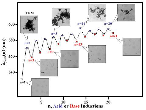
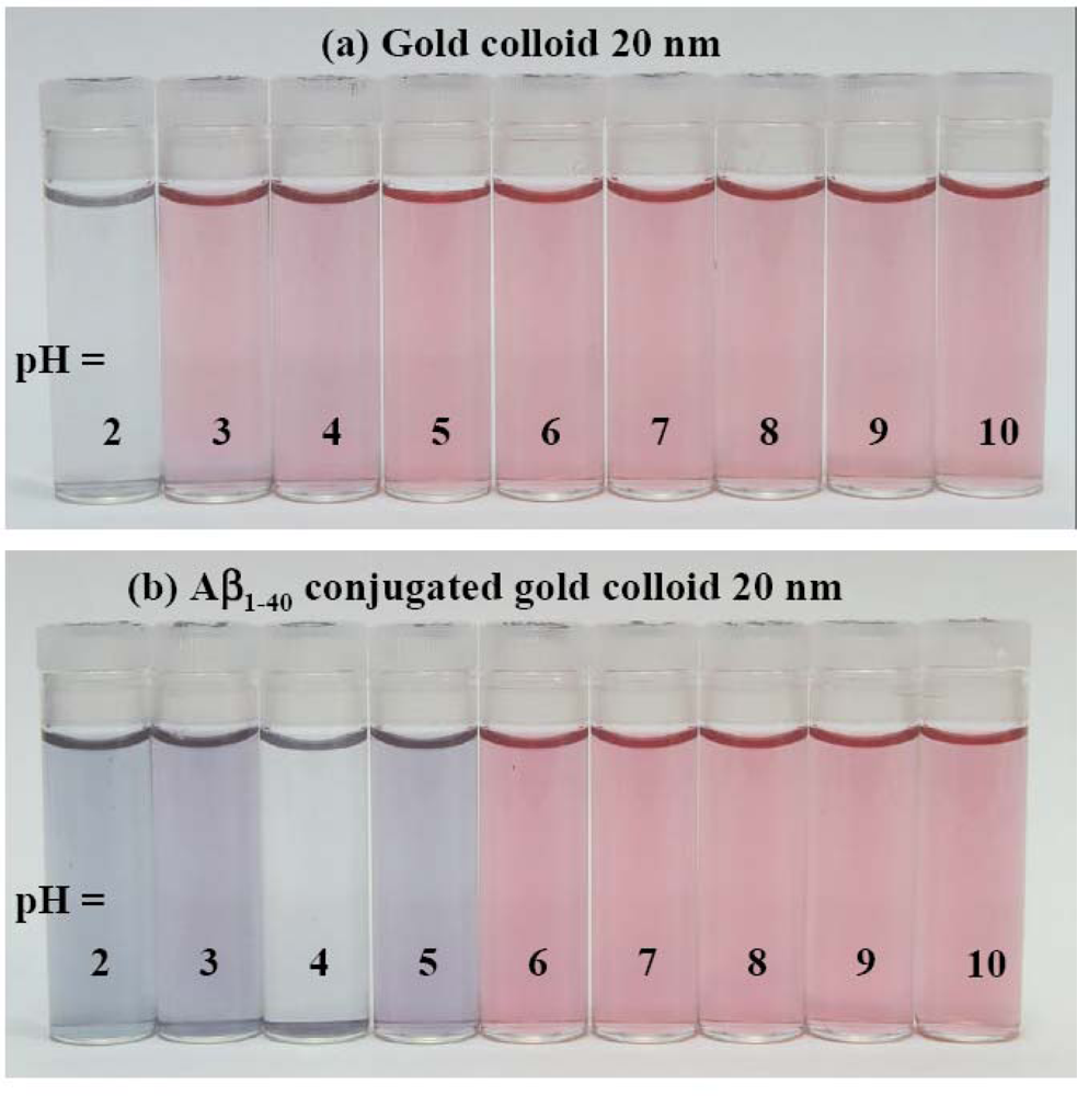
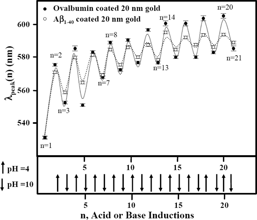
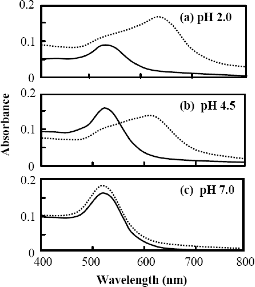
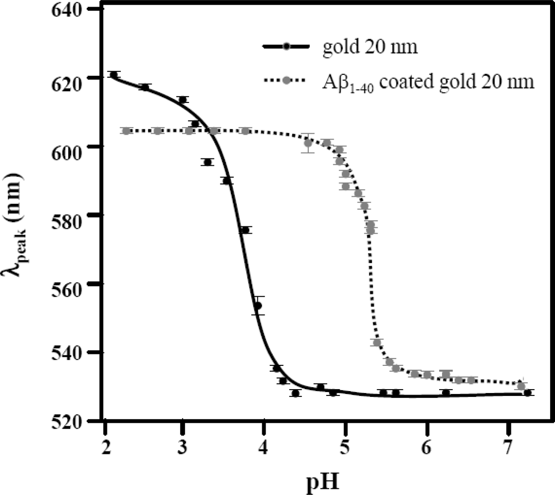
2.2. The pH Induced Reversible Color Change of Protein Coated Gold Colloid
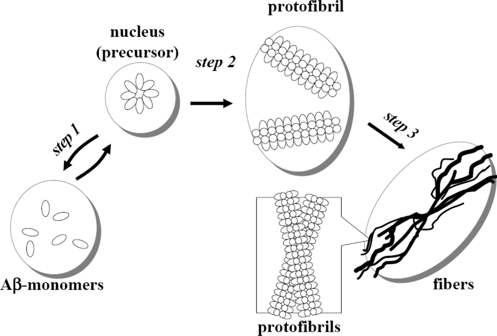
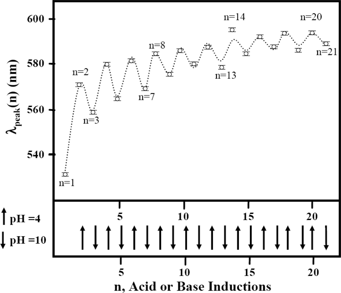
2.3. The Protein Dependent Reversible Process
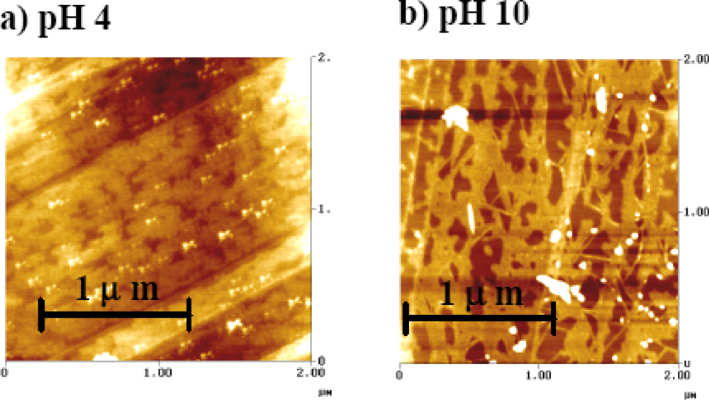
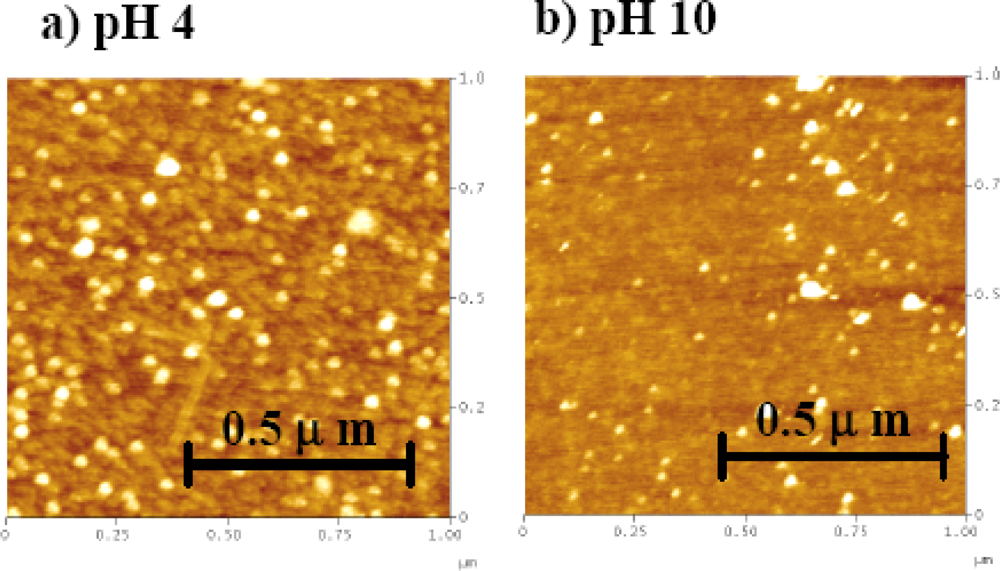
2.4. The AFM Study
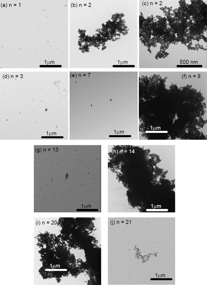
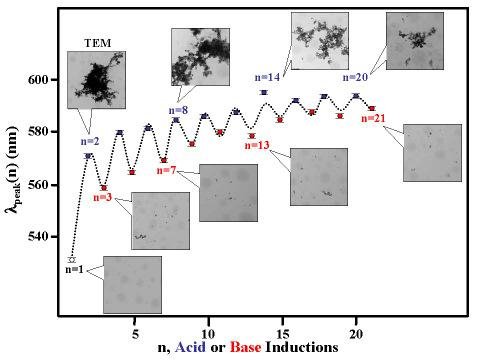
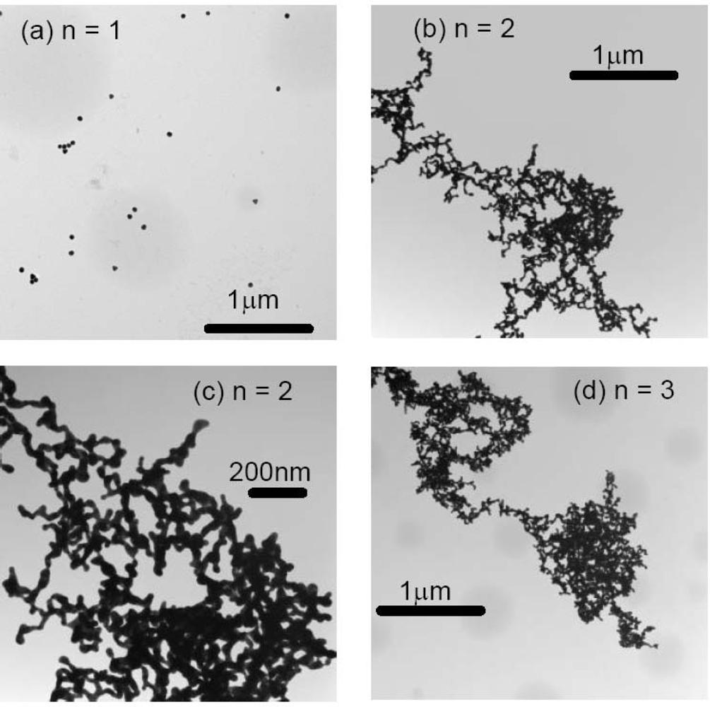
2.5. The TEM Study
3. Experimental Section
4. Conclusions
Acknowledgments
References and Notes
- Heller, W; Pugh, TL. Steric stabilization of colloidal solutions by adsorbtion of flexible macromolecules. J. Polymer Sci 1960, 48, 203–217. [Google Scholar]
- Roth, J. The colloidal gold marker systems for light and electron microscopic cytochemistry. In Techniques in Immunocytochemistry; Bullock, GR, Petrusz, P, Eds.; Academic Press: London, UK, 1983; pp. 217–284. [Google Scholar]
- Chow, MK; Zukoski, CF. Gold sols formation mechanisms: Role of colloidal stability. J. Colloid Interface Sci 1994, 165, 97–109. [Google Scholar]
- Mirkin, CA; Letsinger, RL; Mucic, RC; Storhoff, JJ. A DNA-based method for rationally assembling nanoparticles into macroscopic materials. Nature 1996, 382, 607–609. [Google Scholar]
- Nath, N; Chilkoti, A. Interfacial phase transition of an environmentally responsive elastin biopolymer adsorbed on functionalized gold nanoparticles studied by colloidal surface plasmon resonance. J. Am. Chem. Soc 2001, 123, 8197–8202. [Google Scholar]
- Maxwell, DJ; Taylor, JR; Nie, S. Self-assembled nanoparticle probes for recognition and detection of biomolecules. J. Am. Chem. Soc 2002, 124, 9606–9612. [Google Scholar]
- Katz, E; Shipway, AN; Willner, I. Biomaterial-nanoparticle hybrid systems: synthesis, properties, and applications. In Nanoparticles from Theory to Application; Schmid, G, Ed.; Wiley-VCH: Verlag, Germany, 2004; pp. 368–421. [Google Scholar]
- Daniel, MC; Astruc, D. Gold nanoparticles: Assembly, supramolecular chemistry, quantum-size-related properties, and applications toward biology, catalysis, and nanotechnology. Chem. Rev 2004, 104, 293–346. [Google Scholar]
- Verma, A; Simard, JM; Rotello, VM. Effect of ionic strength on the binding of a-chymotrypsin to nanoparticle receptors. Langmuir 2004, 20, 4178–4181. [Google Scholar]
- Goodrich, GP; Helfrich, MR; Overberg, JJ; Keating, CD. Effect of macromolecular crowding on DNA:Au nanoparticle bioconjugate assembly. Langmuir 2004, 20, 10246–10251. [Google Scholar]
- Lu, YA; Liu, J. Accelerated color change of gold nanoparticles assembled by DNAzymes for simple and fast colorimetric Pb2+ detection. J. Am. Chem. Soc 2004, 126, 12298–12305. [Google Scholar]
- Park, S; Brown, KA; Hamad-Schifferli, K. Changes in oligonucleotide conformation on nanoparticle surfaces by modification with mercaptohexanol. Nano Lett 2004, 4, 1925–1929. [Google Scholar]
- Glomm, WR. Functionalized gold nanoparticles for applications in bionanotechnology. J. Dispers. Sci. Technol 2005, 26, 389–414. [Google Scholar]
- Chah, S; Kumar, CV; Hammond, MR; Zare, RN. Denaturation and renaturation of self-assembled yeast iso-1-cytochrome c on Au. Anal. Chem 2004, 76, 2112–2117. [Google Scholar]
- Zhu, X; Yan, D; Fang, Y. In situ FTIR spectroscopic study of the conformational change of isotactic polypropylene during the crystallization process. J. Phys. Chem. B 2001, 105, 12461–12463. [Google Scholar]
- Harper, SM; Neil, LC; Gardner, KH. Structural basis of a phototropin light switch. Science 2003, 301, 1541–1544. [Google Scholar]
- Ohba, S; Hosomi, H; Ito, Y. In situ X-ray observation of pedal-like conformational change and dimerization of trans-cinnamamide in cocrystals with phthalic acid. J. Am. Chem. Soc 2001, 123, 6349–6352. [Google Scholar]
- Gupta, R; Ahmad, F. Protein stability: functional dependence of denaturational gibbs energy on urea concentration. Biochemistry 1999, 38, 2471–2479. [Google Scholar]
- Yokoyama, K; Welchons, DR. The conjugation of amyloid beta protein on the gold colloidal nanoparticles' surfaces. Nanotechnology 2007, 18, 105101–105107. [Google Scholar]
- Yokoyama, K; Briglio, NM; Sri Hartati, D; Tsang, SMW; MacCormac, JE; Welchons, DR. Nanoscale size dependence in the conjugation of amyloid beta and ovalbumin proteins on the surface of gold colloidal particles. Nanotechnology 2008, 19, 375101–375108. [Google Scholar]
- Aisenbrey, C; Borowik, T; Bystrom, R; Bokvist, M; Lindstrom, F; Misiak, H; Sani, M; Gröbner, G. How is protein aggregation in amyloidogenic diseases modulated by biological membranes? Eur. Biophys. J 2008, 37, 247–255. [Google Scholar]
- Selkoe, DJ. The molecular pathology of Alzheimer's disease. Nuron 1991, 6, 487–498. [Google Scholar]
- Terry, RD. Neuropathological changes in Alzheimer disease. Prog. Brain Res 1994, 101, 383–390. [Google Scholar]
- Glenner, GG; Wong, CW. Alzheimer's disease: initial report of the purification and characterization of a novel cerebrovascular amyloid protein. Biochem. Biophys. Res. Commun 1984, 120, 885–890. [Google Scholar]
- Masters, CL; Simms, G; Weinman, NA; Multhaup, G; McDonald, BL; Beyreuther, K. Amyloid Plaque core protein in Alzheimer disease and Down syndrome. Proc. Natl. Acad .Sci. USA 1985, 82, 4245–4249. [Google Scholar]
- Walsh, DM; Lomakin, A; Benedek, GB; Condron, MM; Teplow, DB. Amyloid β-protein fibrillogenesis. J. Biol. Chem 1997, 272, 22364–22372. [Google Scholar]
- Lomakin, A; Chung, DS; Benedek, GB; Kirshner, DA; Teplow, DB. On the nucleation and growth of amyloid -protein fibrils: detection of nuclei and quantitation of rate constants. Proc. Natl. Acad. Sci. USA 1996, 93, 1125–1129. [Google Scholar]
- Kirschner, DA; Inouye, H; Duffy, LK; Sinclair, A; Lind, M; Selkoe, DJ. Synthetic peptide homologous to β protein from Alzheimer disease forms amyloid-like fibrils in vitro. Proc. Natl. Acad .Sci. USA 1987, 84, 6953–6957. [Google Scholar]
- Lepère, M; Chevallard, C; Hernandez, JF; Mitraki, A; Guenoun, P. Multiscale surface self-assembly of an amyloid-like peptide. Langmuir 2007, 23, 8150–8155. [Google Scholar]
- Kusumoto, Y; Lomakin, A; Teplow, DB; Benedek, GB. Temperature dependence of amyloid beta-protein fibrillization. Proc. Natl. Acad. Sci. USA 1998, 95, 12277–12282. [Google Scholar]
- Coles, M; Bicknell, W; Watson, AA; Fairlie, DP; Craik, DJ. Solution structure of amyloid beta-peptide(1–40) in a water-micelle environment. Is the membranespanning domain where we think it is? Biochemistry 1998, 37, 11064–11077. [Google Scholar]
- Shao, HY; Jao, SC; Ma, K; Zagorski, MG. Solution structures of micelle-bound amyloid beta-(1–40) and beta-(1–42) peptides of Alzheimer's disease. J. Mol. Biol 1999, 285, 755–773. [Google Scholar]
- Giacomelli, CE; Norde, W. Conformational changes of the amyloid beta-peptide (1–40) adsorbed on solid surfaces. Macromol. Biosci 2005, 5, 401–407. [Google Scholar]
- Rocha, S; Krastev, R; Thunemann, AF; Pereira, MC; Mohwald, H; Brezesinski, G. Adsorption of amyloid beta-peptide at polymer surfaces: a neutron reflectivity study. Chem. Phys. Chem 2005, 6, 2527–2534. [Google Scholar]
- Kowalewski, T; Holtzman, DM. In situ atomic force microscopy study of Alzheimer's beta-amyloid peptide on different substrates: new insights into mechanism of beta-sheet formation. Proc. Natl. Acad. Sci. USA 1999, 96, 3688–3693. [Google Scholar]
- Schladitz, C; Vieira, EP; Hermel, H; Mohwald, H. Amyloid-beta-sheet formation at the air-water interface. Biophys. J 1999, 77, 3305–3310. [Google Scholar]
- Rocha, S; Thünemann, AF; Pereira, MDC; Coelho, M; Möhwald, H; Brezesinski, G. Influence of fluorinated and hydrogenated nanoparticles on the structure and fibrillogenesis of amyloid beta-peptide. Biophys Chem 2008, 137, 35–42. [Google Scholar]
- Kogan, MJ; Bastus, NG; Amigo, R; Grillo-Bosch, D; Araya, E; Turiel, A; Labarta, A; Giralt, E; Puntes, VF. Nanoparticle-mediated local and remote manipulation of protein aggregation. Nano Lett 2006, 6, 110–115. [Google Scholar]
- Fezoui, Y; Hartley, DM; Harper, JD; Khurana, R; Walsh, DM; Condron, MM; Selkoe, DJ; Lansbury, PT, Jr; Fink, AL; Teplow, DB. An improved method of preparing the amyloid β-protein for fibrillogenesis and neurotoxicity experiments. Amyloid: Int. J. Exp. Clin. Invest 2000, 7, 166–178. [Google Scholar]
- Oliver, C. Conjugation of colloidal gold to proteins. In Methods in Molecular Biology: Immunocytochemical Methods and Protocols, 2nd Ed; Javois, LC, Ed.; Humana Press Inc: Totowa, NJ, USA, 1999; pp. 331–334. [Google Scholar]
- Barrow, CJ; Yasuda, A; Kenny, PT; Zagorski, MG. Solution conformations and aggregational properties of synthetic amyloid beta-peptides of Alzheimer's disease. Analysis of circular dichroism spectra. J. Mol. Biol 1992, 225, 1075–1093. [Google Scholar]
- Wood, SJ; Maleeff, B; Hart, T; Wetzel, R. Physical, morphological and functional differences between pH 5.8 and 7.4 aggregates of the Alzheimer's amyloid peptide Abeta. J. Mol. Biol 1996, 256, 870–877. [Google Scholar]
- Hilbich, C; Kisters-Woike, B; Reed, J; Masters, CL; Beyreuther, K. Aggregation and secondary structure of synthetic amyloid beta A4 peptides of Alzheimer's disease. J. Mol. Biol 1991, 218, 149–163. [Google Scholar]
- Hu, HY; Du, HN. α-to-β structural transformation of ovalbumin: Heat and pH effects. J. Protein Chem 2000, 19, 177–183. [Google Scholar]
- Edelhoch, H. Spectroscopic determination of tryptophan and tyrosine in proteins. Biochemistry 1967, 6, 1948–1954. [Google Scholar]


| A (nm) | A – D (nm) | B (nm) | C | D (nm) | E | |
|---|---|---|---|---|---|---|
| Ovalbumin coated | 545(4) | 531(4) | 14(4) | 0.42(7) | 14(2) | 0.02(1) |
| Aβ1–40 coated | 542(2) | 532(2) | 21(2) | 0.29(3) | 10(1) | 0.07(2) |
| Gold 20 nm | Gold 20 nm coated with ovalbumin | Gold 20 nm coated with Aβ1–40 | ||||
|---|---|---|---|---|---|---|
| n | Occupancy (%) | Number of gold particles per aggregate | Occupancy (%) | Number of gold particles per aggregate | Occupancy (%) | Number of gold particles per aggregate |
| 1 | 0.37 ± 0.04 | - | 0.07 ± 0.03 | - | 0.06 ± 0.04 | - |
| 2 | 38 ± 4 | 350 | 55 ± 5 | 6,200 | 73 ± 1 | 24,000 |
| 3 | 27 ± 2 | 780 | 0.1 ± 0.04 | - | 0.6 ± 0.3 | - |
| 7 | 0.30 ± 0.02 | - | 0.6 ± 0.4 | - | ||
| 8 | 77 ± 3 | 19,000 | 31 ± 5 | 15,000 | ||
| 13 | 0.19 ± 0.01 | - | 0.4 ± 0.1 | - | ||
| 14 | 66 ± 5 | 22,000 | 13 ± 3 | 4,400 | ||
| 20 | 63 ± 4 | 25,000 | 7 ± 1 | 2,300 | ||
| 21 | 0.07 ± 0.03 | - | 0.5 ± 0.3 | - | ||
© 2009 by the authors; licensee Molecular Diversity Preservation International, Basel, Switzerland. This article is an open-access article distributed under the terms and conditions of the Creative Commons Attribution license (http://creativecommons.org/licenses/by/3.0/).
Share and Cite
Yokoyama, K.; Cho, H.; Cullen, S.P.; Kowalik, M.; Briglio, N.M.; Hoops, H.J.; Zhao, Z.; Carpenter, M.A. Microscopic Investigation of Reversible Nanoscale Surface Size Dependent Protein Conjugation. Int. J. Mol. Sci. 2009, 10, 2348-2366. https://doi.org/10.3390/ijms10052348
Yokoyama K, Cho H, Cullen SP, Kowalik M, Briglio NM, Hoops HJ, Zhao Z, Carpenter MA. Microscopic Investigation of Reversible Nanoscale Surface Size Dependent Protein Conjugation. International Journal of Molecular Sciences. 2009; 10(5):2348-2366. https://doi.org/10.3390/ijms10052348
Chicago/Turabian StyleYokoyama, Kazushige, Hyunah Cho, Sean P. Cullen, Matthew Kowalik, Nicole M. Briglio, Harold J. Hoops, Zhouying Zhao, and Michael A. Carpenter. 2009. "Microscopic Investigation of Reversible Nanoscale Surface Size Dependent Protein Conjugation" International Journal of Molecular Sciences 10, no. 5: 2348-2366. https://doi.org/10.3390/ijms10052348