Article

Four New Monoterpenoid Glycosides from the Flower Buds of Magnolia biondii

1
Collaborative Innovation Center for Respiratory Disease Diagnosis and Treatment & Chinese Medicine Development of Henan Province, Henan University of Chinese Medicine, Zhengzhou 450046, China
2
School of Pharmacy, Henan University of Chinese Medicine, Zhengzhou 450046, China
*
Author to whom correspondence should be addressed.
Molecules 2016, 21(6), 728; https://doi.org/10.3390/molecules21060728
Submission received: 19 April 2016 / Accepted: 30 April 2016 / Published: 3 June 2016
(This article belongs to the Section Natural Products Chemistry)

Abstract

:
Four new monoterpenoid glycosides 14, named magnoliaterpenoid A–D, were isolated from a 50% aqueous acetone extract of flower buds of Magnolia biondii, along with one known compound, (1′R,3′S,5′R,8′S,2Z,4E)-dihydrophaseic acid 3-O-β-d-glucopyranoside (5). Their structures and relative configuration were identified by extensive spectroscopic analysis (IR, UV, MS, 1D and 2D NMR). The aglycones of these four new compounds possess seven-membered rings systems, which are very rare. A plausible biosynthetic route for the four new compounds was proposed via the biogenetic isoprene rule. Compounds 1, 2, 3, and 4 showed no antimicrobial activity at the concentration range of 1.95–250 µg/mL.

Graphical Abstract

1. Introduction

The genus Magnolia (family Magnoliaceae) includes about 90 species worldwide, mainly distributed in tropical and temperate regions of Asia and North America to South America. In China, there are about 30 species, mostly distributed in the southern and northern provinces of China [1]. Flos Magnoliae, the flower buds of M. biondii, M. denudata or M. sprengeri, which is unique to China and mostly distributed in the Henan, Hubei, Zhejiang, Anhui, and Shanxi provinces, has been used as a traditional Chinese medicine for more than 1500 years to treat nasal congestion, headaches, sinusitis, and allergic rhinitis [2,3,4]. Its volatile oil has also been processed into all kinds of cosmetics which are widely used in China [4]. It has been reported to possess extensive anti-allergy [5,6], anti-inflammatory [7], anti-angiogenic [8], anti-platelet aggregation [9,10], antimicrobial [11] and antioxidant activities [12,13]. Several types of compounds have been isolated from the plant, including lignans [9], neolignans [9], sesquiterpenoids [14], alkaloids [15,16], and flavonoids [15,16]. In order to further investigate the active components of the herb, a systematic phytochemical study was carried out on the 50% aqueous acetone extract of flower buds of Magnolia biondii. As a result, four new monoterpenoid glycosides14, named magnoliaterpenoid A–D (Figure 1), were isolated and structurally identified, along with a known sesquiterpenoid glycoside, (1′R,3′S,5′R,8′S,2Z,4E)-dihydrophaseic acid 3-O-β-d-glucopyranoside (5). Herein, the isolation and structural elucidation of compounds 14 are reported, as well as their antimicrobial activity.

2. Results and Discussion

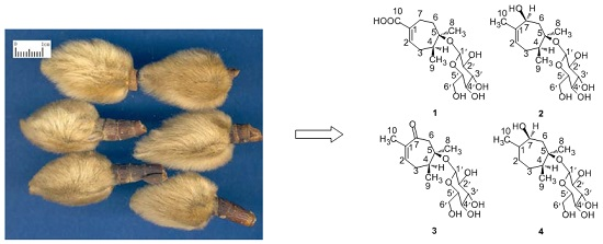
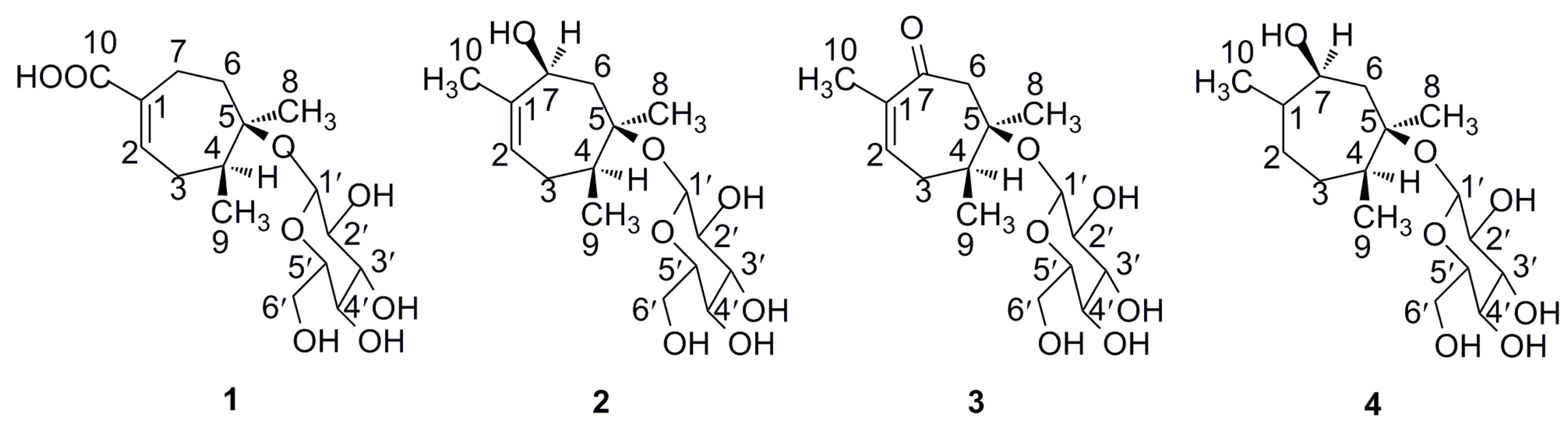
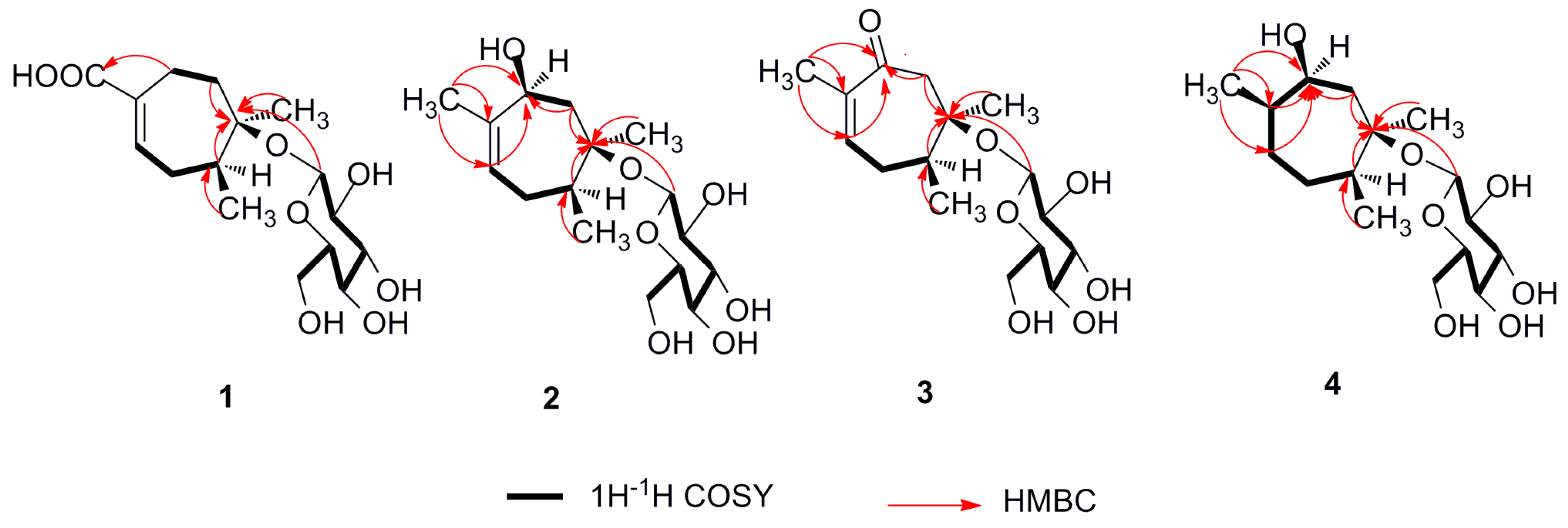
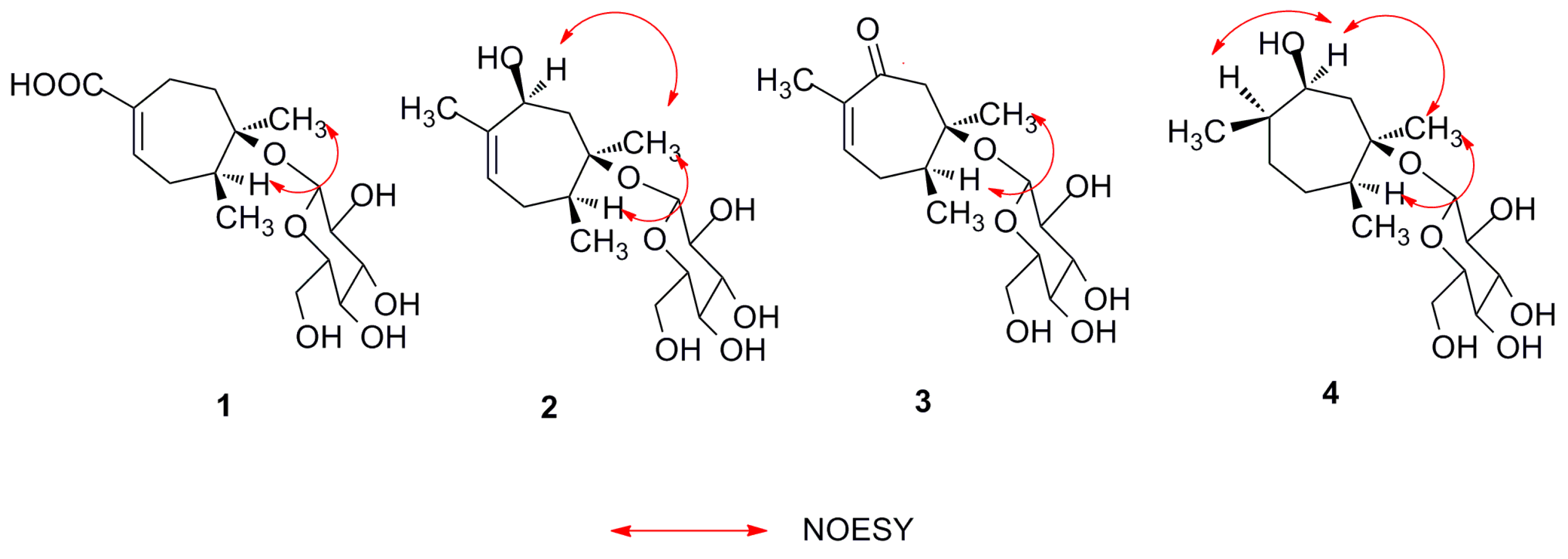
2.1. Structure Elucidation of Compounds 14

Compound 1, obtained as a colorless gum, was determined to have the molecular formula C16H26O8Cl by HRESIMS (m/z 381.1301 [M + Cl], calcd. 381.1310),with four degrees of unsaturation. Its UV spectrum showed an absorption maximum at 217 nm, indicating the presence of an α,β-unsaturated carbonyl moiety. The IR spectrum supported the presence of hydroxyl (3367 cm−1), carbonyl (1682 cm−1) and double bond (1652 cm−1) groups. The 1H-NMR spectroscopic data of 1 (Table 1) displayed the existence of two methyl singlets [δH 1.25 (3H, s, H-8), 1.23 (3H, br. s, H-9)] and one olefinic proton [δH 6.98 (1H, t, J = 2.2, 4.9Hz, H-2)] (See the Supplementary Materials). Figures S1–S36 showing the 1H-NMR, 13C-NMR , DEPT, HSQC, HMBC, 1H-1H COSY, NOESY, HR-ESI-MS, UV and IR spectra of compounds 14 can be found in the Supplementary Materials.
Closer examination of the 1H/13C/1H-1H COSY/HSQC/HMBC NMR data (Table 1) obtained for 1 revealed the presence of a glucose moiety [δ 4.46 (H-1′), 98.5 (C-1′); 3.11 (H-2′), 75.3 (C-2′); 3.21 (H-3′), 77.5 (C-3′); 3.26 (H-4′), 71.8 (C-4′); 3.35 (H-5′), 78.3 (C-5′); 3.80 (H-6′a), 3.63 (H-6′b), 62.9 (C-6′)], along with 10 carbon resonances, including two methyl [δC 24.8 (C-8), 23.3 (C-9)], three methylene [δC 28.6 (C-3), 24.5 (C-6), 26.3 (C-7)], two methine [δC 141.3 (C-2), 44.5 (C-4)], one carbonyl [δC 170.9 (C-10)], one oxygenated quaternary carbon [δC 80.5 (C-5)] and one olefinic quaternary carbon [δC 131.5 (C-1)]. Apart from one double bond, one carbonyl and one glucose moiety, the remaining unsaturation of 1 required it must contain a ring. Inspection of the 1H- and 13C-NMR spectra of 1 indicated a monoterpenoid glycoside with a structure similar to that of paeoveitol B [17]. The 7.8 Hz coupling constant for the anomeric proton H-1′ of the glucose at δH 4.46 confirmed its axial orientation. Acid hydrolysis and GC-MS analysis of the corresponding thiazolidine derivative substantiated the sugar unit as being β-d-glucose. In the 1H-1H COSY spectrum (Figure 2), the correlations of H-2/H-3/H-4 and H-6/H-7 displayed the key spin systems. In the HMBC spectrum (Figure 2), the correlation from H-8 to C-5 indicated that the 8-CH3 was located to C-5; the correlation from H-9 to C-4 indicated that the 9-CH3 was connected to C-4; the correlation from H-1′ to C-5 indicated that the β-D-glucose was linked to C-5; the correlations from H-2 and H-7 to C-10 indicated that the carbonyl (C-10) was linked to C-1; the oxygenated quaternary carbon (C-5) was located between C-4 and C-6 from the HMBC correlations from H-4 and H-6 to C-5. The relative configuration of compound 1 was established by the NOESY experiment (Figure 3) in which the correlation of H-4 and H-8 was observed. Thus, the structure of compound 1 was determined to be as shown in Figure 1, and it was assigned the trivial name magnoliaterpenoid A.
Compound 2 was isolated as a colorless gum and had a molecular formula C16H28O7Cl by HRESIMS (m/z 367.1519 [M + Cl], calcd. 367.1518), indicating three degrees of unsaturation. The IR spectrum showed the presence of hydroxyl (3345 cm−1) and double bond (1385 cm−1) groups. The 1H-NMR spectroscopic data of 2 (Table 1) revealed the existence of three methyl singlets [δH 1.21 (3H, s, H-8), 1.21 (3H, br. s, H-9), 1.70 (3H, s, H-10)] and one olefinic proton [δH 5.44 (1H, d, J = 4.2 Hz, H-2)] (See the Supplementary Materials). Comparison of its 13C-NMR and DEPT data with those of compound 1 indicated that the two compounds possessed a similar skeleton. One of the differences between these two compounds was that the methylene signal at C-7 (δC 26.3) in 1 was replaced by an oxygenated methine at C-7 (δC 71.9) in 2, which was established by the HMBC correlations (Figure 2) from H-2, H-6 and H-10 to C-7, along with the 1H-1H COSY correlations (Figure 2) of H-6/H-7. The other main difference was that the carbonyl at C-10 (δC 170.9) in 1 was replaced by a methyl at C-10 (δC 19.2) in 2, which was determined on the basis of the HMBC correlations (Figure 2) from H-10 to C-1, C-2 and C-7. The relative configuration of compound 2 was deduced from the NOESY experiment (Figure 3) in which the correlationof H-4/H-8 and H-7/H-8 was detected. Similarly, the sugar unit of compound 2 was confirmed as β-d-glucose using the same method as for compound 1. Therefore, the structure of 2 was assigned as shown in Figure 1, and it was given the trivial name magnoliaterpenoid B.
Compound 3 was isolated as a colorless gum and had a molecular formula C16H26O7Cl by HRESIMS (m/z 365.1357 [M + Cl], calcd. 365.1361), with four degrees of unsaturation. Its UV spectrum showed an absorption maximumat 240 nm, indicating the presence of an α,β-unsaturated carbonyl moiety. The IR spectrum supported the presence of hydroxyl (3364 cm−1), carbonyl (1655 cm−1) and double bond (1368 cm−1) groups. The 1H-NMR spectroscopic data of 3 (Table 1) revealed the existence of three methyl singlets [δH 1.28 (3H, s, H-8), 1.24 (3H, br. s, H-9), 1.72 (3H, s, H-10)] and one olefinic proton [δH 6.87 (1H, d, J = 5.4 Hz, H-2)] (See the Supplementary Materials). The 13C-NMR and DEPT spectroscopic data of 3 (Table 1) were highly similar to those of 2. The only difference between compound 2 and 3 was that the oxygenated methine at C-7 (δC 71.9) in 2 was replaced by a carbonyl at C-7 (δC 203.5) in 3, which was established by the HMBC correlations (Figure 2) from H-2, H-6 and H-10 to C-7. The relative configuration of compound 3 was assigned by the NOESY experiment (Figure 3) in which the correlation of H-4 and H-8 was observed. Similarly, the sugar unit of compound 3 was also confirmed as β-d-glucose using the same method as for compound 1. Therefore, the structure of 3 was established as shown in Figure 1, and the compound was trivially named magnoliaterpenoid C.
Compound 4 was isolated as a colorless gum. Its molecular formula was established as C16H30O7Na by HRESIMS (m/z 357.1878 [M + Na]+, calcd. 357.1883), with two degrees of unsaturation. The IR spectrum displayed the existence of hydroxyl (3362cm−1) groups. The 1H-NMR spectroscopic data of 4 (Table 1) supported the existence of three methyl singlets [δH 1.23 (3H, s, H-8), 1.14 (3H, br. s, H-9), 0.91 (3H, d, J = 6.2 Hz, H-10)] (See the Supplementary Materials). Critical analysis of 13C-NMR and DEPT spectroscopic data of 4 (Table 1) demonstrated that its structure was closely related to that of compound 2, except that the double bond at C-1 (δC 138.2) and C-2 (δC 124.6) in 2 was reduced to a methane at C-1 (δC 37.9) and a methylene at C-2 (δC 29.6) respectively in 4, which was established by the HMBC correlations (Figure 2) from H-10 to C-1 and C-2, along with the 1H-1H COSY correlations (Figure 2) of H-1 and H-2. The relative configuration of compound 4was similar to compound 2, which was deduced from the NOESY experiment (Figure 3) in which the correlation of H-4/H-8, H-7/H-8 and H-1/H-7 was detected. Similarly, the sugar unit of compound 4 was also confirmed as β-d-glucose using the same method as for compound 1. Thus, the structure of 4 was established as shown in Figure 1, and the compound was given the trivial name magnoliaterpenoid D.
The identity of compound 5 was determined by spectroscopic analysis and comparison with literature data [18].

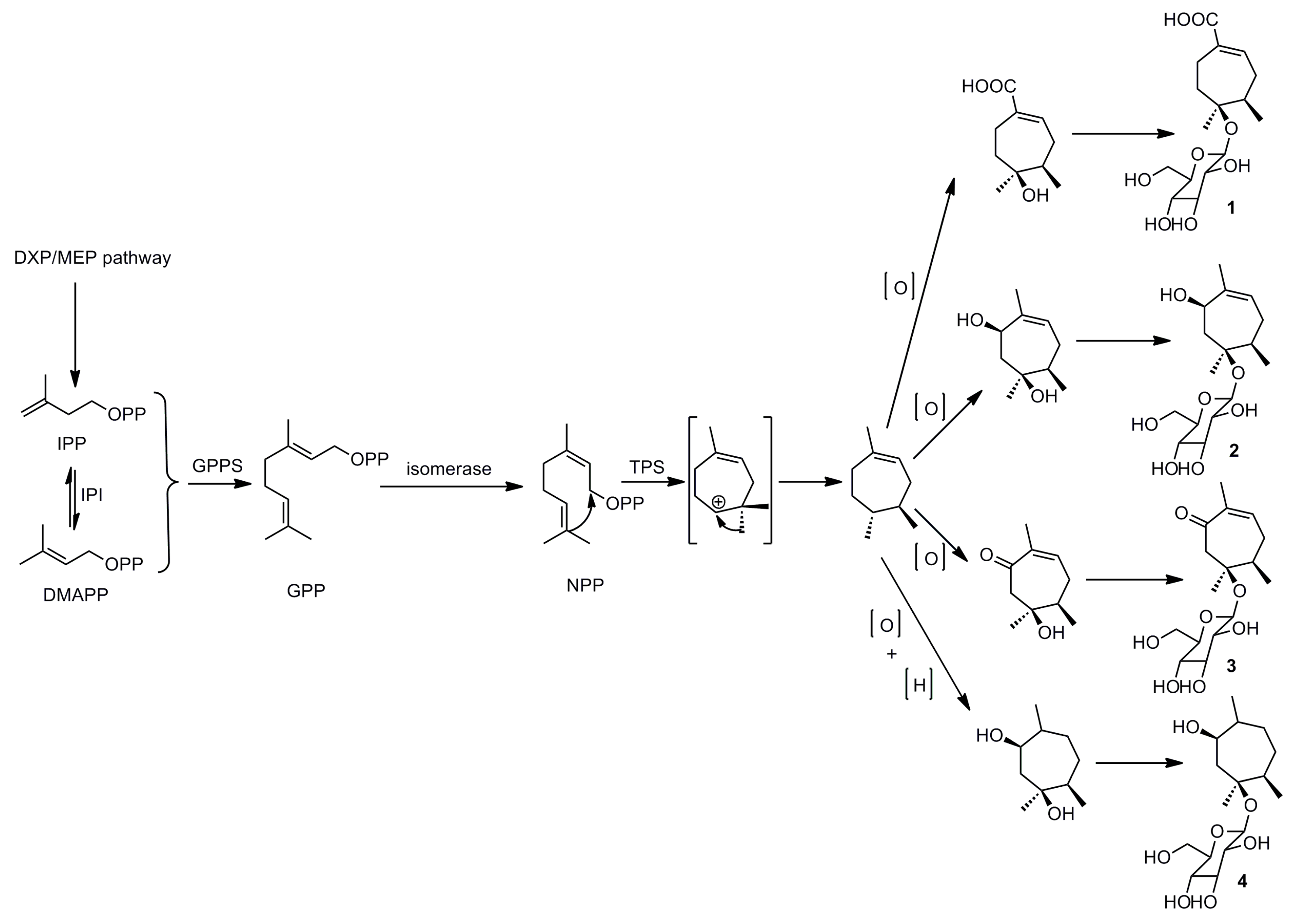
2.2. Plausible Biogenetic Pathway

Since the aglycones of these four new compounds contained 10 carbon atoms and belonged to the monoterpenoid class of compounds, we propose a plausible biosynthetic route via the biogenetic isoprene rule, as shown in Scheme 1.

2.3. Antimicrobial Activity

The four new compounds were tested for their antimicrobial activity against Gram-positive and negative bacteria and fungi at the concentration range of 1.95–250 μg/mL. None of them exhibited antimicrobial activity at the tested concentrations.

3. Experimental Section

3.1. General Procedures

Optical rotations were measured on an AP-IV polarimeter (Rudolph, NJ, USA). UV spectra were measured with a Thermo EVO 300 spectrometer (Thermo Fisher Scientific, Madison, WI, USA). IR spectra were recorded on a Thermo Nicolet IS 10 spectrometer (Thermo Fisher Scientific). NMR spectra were scanned on an Avance III spectrometer (500 MHz for 1H-NMR and 125 MHz for 13C-NMR, Bruker, Zug, Switzerland). HR-ESI-MS was recorded on a Bruker MicroTOF-Q II spectrometer (Bruker Daltonics, Bremen, Germany). GC analysis was carried out on a Shimadzu GC-MS-QP2010E instrument (Shimadzu Corporation, Tokyo, Japan) using a RXI-5 ms capillary column (30 m × 0.25 mm × 0.25 µm, Restek, Bellefonte, PA, USA). Preparative HPLC was performed on a LC-3000 instrument (Chuangxintongheng, Beijing, China) equipped with a UV-3000 detector using YMC HPLC columns (5 µm, 10.0 × 250 mm and 5 µm, 20.0 × 250 mm), with a flow rate of 2.0 mL/min and 8.0 mL/min, respectively. Column chromatography was performed using DiaionHP-20 (Mitsubishi Chemical Corporation, Tokyo, Japan), ODS-B (50 µm, Daiso Co., Ltd., Tokyo, Japan), MCI GEL CHP20p (75–150 µm, Mitsubishi Chemical Corporation), and Toyopearl HW-40 (TOSOH Corporation, Tokyo, Japan). Thin-layer chromatography was carried out on self-made silica gel G (Qingdao Marine Chemical Industry, Qingdao, China) plates. The chemical reagents were supplied by Beijing Chemical Plant (Beijing, China) and Tianjin No. 3 Reagent Plant (Tianjin, China).

3.2. Plant Material

The flower buds of Magnolia biondii were collected from Nanzhao, Henan Province, China, and identified by Prof. Cheng-Ming Dong of the Henan University of Traditional Chinese Medicine, China. A voucher specimen (No. 20140609) has been deposited in Department of Natural Medicinal Chemistry, School of Pharmacy, Henan University of Chinese Medicine.

3.3. Extraction and Isolation

The air-dried flower buds of Magnolia biondii (5.0 kg) were extracted with aqueous acetone (50% v/v, 3 × 20 L) at room temperature. The combined solutions were evaporated under vacuum to give a crude extract (463 g). The crude extract was suspended in H2O (2 L) and then successively extracted with petroleum ether, EtOAc and n-BuOH (2 L × 5), respectively. The n-BuOH fraction (60.0 g) was subjected to Diaion HP-20 column chromatography (10.0 × 60.0 cm) and eluted with EtOH–H2O (0:100, 20:80, 40:60, 80:20, 95:5, v/v, each 20 L) to afford five fractions (Fr. I–Fr. V). Fr. II (14.0 g) was then subjected to ODS column chromatography (4.0 × 30.0 cm) and eluted successively with a MeOH–H2O (0:100, 5:95, 10:90, 15:85, 20:80, 25:75, 35:65,45:55, 100:0, v/v, each 1.5 L) gradient to give nine fractions (Fr. 1–Fr. 9). Fr. 6 (1.3 g) was further separated by Toyopearl HW-40 column chromatography (2.0 × 50.0 cm) with MeOH–H2O (25:75) as the eluent to obtain eight subfractions (Fr. 6A–Fr. 6H). Fr. 6B (50.0 mg) was further purified by preparative HPLC on a YMC HPLC column (5 µm, 20.0 × 250 mm, flow rate 8 mL/min) with MeOH–H2O (25:75) to afford compound 1 (4.6 mg, tR = 77.5 min). Fr. 6C (110.0 mg) was further isolated by preparative HPLC on a YMC HPLC column (5 µm, 20.0 × 250 mm, flow rate 8 mL/min) with MeOH–H2O (30:70) to afford compound 2 (36.0 mg, tR = 59.5 min). Fr. 6E (120.0 mg) was also further purified by preparative HPLC on a YMC HPLC column (5 µm, 20.0 × 250 mm, flow rate 8 mL/min) with MeOH–H2O (35:65) to afford compound 3 (50.0 mg, tR = 26.0 min) and compound 4 (2.0 mg, tR = 15.0 min). Compound 5 (12.0 mg, tR = 60.0 min) was obtained from Fr. 6G by preparative HPLC on a YMC HPLC column (5 µm, 20.0 × 250 mm, flow rate 8 mL/min) with MeOH–H2O (15:85).

3.4. Compound characterization

Magnoliaterpenoid A (1): colorless gum; [ α ] D 20 –28.8 (MeOH, 0.10); UV λmax nm (log ε): 217 (0.8); IR (iTR): 3367, 2943, 2879, 1682, 1652, 1424, 1203, 1140 cm−1; 1H- and 13C-NMR, see Table 1; HRESIMS: m/z = 381.1301 [M + Cl], (C16H26O8Cl, calcd.381.1310).
Magnoliaterpenoid B (2): colorless gum; [ α ] D 20 –13.4 (MeOH,0.72); UV λmax nm (log ε): 203 (0.5); IR (iTR): 3345, 2969, 2917, 2884, 1385,1367, 1073, 918, 860cm−1; 1H- and 13C-NMR, see Table 1; HRESIMS: m/z = 367.1519 [M + Cl], (C16H28O7Cl, calcd.367.1518).
Magnoliaterpenoid C (3): colorless gum; [ α ] D 20 –15.5 (MeOH, 1.0); UV λmax nm (log ε): 240 (0.8); IR (iTR): 3364, 2973, 2923, 2886, 1655, 1368, 1073, 904 cm−1; 1H- and 13C-NMR, see Table 1; HRESIMS: m/z = 365.1357 [M + Cl], (C16H26O7Cl, calcd. 365.1361).
Magnoliaterpenoid D (4): colorless gum; [ α ] D 20 –6.6 (MeOH, 0.03); UV λmax nm (log ε): 202 (0.6); IR (iTR): 3362, 2926, 2871, 1100, 995 cm−1; 1H- and 13C-NMR, see Table 1; HRESIMS: m/z = 357.1878 [M + Na]+, (C16H30O7Na, calcd. 357.1883).

3.5. Acid Hydrolysis and Sugar Analysis

Compounds 14 (1.0 mg, respectively) were refluxed with 8% HCl (2 mL) for 3 h. After the reaction mixture was extracted with EtOAc (2 mL × 3), the aqueous layer was dried under vacuum. Then the residue was dissolved in pyridine (0.3 mL) containing l-cysteine methyl ester hydrochloride (1.5 mg) and heated at 60 °C for 1 h. A 0.3 mL solution of phenyl isothiocyanate (1.5 mg) in pyridine was added to the mixture, which was heated at 60 °C for 1 h. The reaction mixture was directly analyzed by gas chromatography (GC). The d-configuration of glucose was confirmed by comparing the retention time with a standard sample [tR (min): d-glucose (8.5)].

3.6. Antimicrobial Assay

The antimicrobial activity of compounds 14 was evaluated against Gram-positive bacteria (Staphylococcus aureus ATCC25923), Gram-negative bacteria (Escherichia coli ATCC35150 and Proteus vulgaris ATCC33420) and fungi (Aspergillusniger ATCC6257 and Candida albicans ATCC90029) by a microdilution titre technique [19]. Kanamycin and fluconazole was used as positive controls. All tests were performed in triplicate.

4. Conclusions

In conclusion, although these four new compounds did not show antimicrobial activity at the tested concentration range of 1.95–250 µg/mL, the aglycones of these four new compounds possess seven-membered rings system which are very rare and a plausible biogenetic pathway of 14 was proposed via the biogenetic isoprene rule. This kind of skeleton was also isolated from the Magnoliaceae for the first time.

Supplementary Materials

Supplementary materials can be accessed at: https://www.mdpi.com/1420-3049/21/6/728/s1.

Acknowledgments

This work was supported by the Key Project of Chinese Ministry of Education of China (2003078).

Author Contributions

W.-S.F. and Y.-H.H. conceived and designed the experiments; Y.-H.H. performed the experiments; X.-K.Z., J.-C.W., Y.-G.C. and Y.-L.Z. analyzed the data; K.S. contributed reagents/materials/analysis tools; W.-S.F. wrote the paper. All authors read and approved the final manuscript.

Conflicts of Interest

The authors declare that there are no conflicts of interest.

References

  1. Cai, H.M.; Fan, W.; Wang, X.D. Research progress on chemical constituents and pharmacological effects of Magnolia. China Pharm. 2013, 22, 3735–3737. [Google Scholar]
  2. Chinese Pharmacopoeia Commission. The Pharmacopoeia of the People’s Republic of China, 2015 ed.; China Medical Science Press: Beijing, China, 2015; Part I; p. 182. [Google Scholar]
  3. Shen, Y.; Pang, E.C.K.; Xue, C.C.L.; Zhao, Z.Z.; Lin, J.G.; Li, G.G. Inhibitions of mast cell-derived histamine release by different Flos Magnoliae species in rat peritoneal mast cells. Phytomedicine 2008, 15, 808–814. [Google Scholar] [CrossRef] [PubMed]
  4. Zhu, X.W.; Yang, J.K.; Hu, D.W. Summarize of study on the application in medicine function and the ingredient of Magnolia liliflora. Strait Pharm. J. 2002, 14, 5–7. [Google Scholar]
  5. Tsuruga, T.; Ebizuka, Y.; Nakajima, J.; Chun, Y.T.; Noguchi, H.; Irtaka, Y.; Sankawa, U. Biologically active constituents of Magnolia salicifolia: Inhibitiors of induced histamine release from Rat Mast Cells. Chem. Pharm. Bull. 1991, 39, 3265–3271. [Google Scholar] [CrossRef] [PubMed]
  6. Kim, G.C.; Lee, S.G.; Park, B.S.; Kim, J.Y.; Song, Y.S.; Kim, J.M.; Yoo, K.S.; Huh, G.Y.; Jeong, M.H.; Lim, Y.J.; et al. MagnoliaeFlos induces apoptosis of RBL-2H3 cells via mitochondria and caspase. Int. Arch. Allergy Immunol. 2003, 131, 101–110. [Google Scholar] [CrossRef] [PubMed]
  7. Kimura, M.; Suzuki, J.; Yamada, T.; Yoshizaki, M.; Kikuchi, T.; Shigetoshi, K.; Matsuda, S. Anti-inflammatory effect of neolignans newly isolated from the Crude Drug “Shin-i” (Flos Magnoliae). Planta Med. 1985, 291–293. [Google Scholar] [CrossRef] [PubMed]
  8. Kobayashi, S.; Kimura, I.; Kimura, M. Inhibitory effect of Magnosalin derived from Flos Magnoliae on tube formation of rat vascular endothelial cells during the angiogenic process. Biol. Pharm. Bull. 1996, 19, 1304–1306. [Google Scholar] [CrossRef] [PubMed]
  9. Kuroyanagi, M.; Yoshida, K.; Yamamoto, A.; Miwa, M. Bicyclo[3.2.1]octane and 6-Oxabicyclo[3.2.2]nonane type neolignans from Magnolia denudate. Chem. Pharm. Bull. 2000, 48, 832–837. [Google Scholar] [PubMed]
  10. Ma, Y.L.; Han, G.Q. Biologically active lignans from Magnolia biondii Pamp. China J. Chin. Mater. Med. 1995, 20, 102–104. [Google Scholar]
  11. Liang, Z.; Yang, E.Y. The therapeutic effects of irrigating therapy with magnolia injection on chronic maxillary sinusitis experimentally induced among rabbits. Chinese J. Otolaryngol. Integr. Tradit. West. Med. 2005, 13, 6–10. [Google Scholar]
  12. Lee, J.; Lee, D.; Jang, D.S.; Nam, J.W.; Kim, J.P.; Park, K.H.; Yang, M.S.; Seo, E.K. Two new stereoisomers of tetrahydrofuranoid lignans from the Flower Buds of Magnolia fargesii. Chem. Pharm. Bull. 2007, 55, 137–139. [Google Scholar] [CrossRef] [PubMed]
  13. Lee, J.; Seo, E.K.; Jang, D.S.; Ha, T.J.; Kim, J.P.; Nam, J.W.; Bae, G.; Lee, Y.M.; Yang, M.S.; Kim, J.S. Two new stereoisomers of neolignan and lignin from the Flower Buds of Magnolia fargesii. Chem. Pharm. Bull. 2009, 57, 298–301. [Google Scholar] [CrossRef] [PubMed]
  14. Schühly, W.; Skarbina, J.; Kunert, O.; Nandi, O.I.; Bauer, R. Chemical characterization of Magnolia biondii. Nat. Prod. Commun. 2009, 4, 231–234. [Google Scholar] [PubMed]
  15. Chen, Y.Y.; Wang, B.; Cao, C.Y.; Qiao, L.; Han, G.Q. Study on the hydrophilic components of Magnolia biondii Pamp. Acta Pharm. Sin. 1994, 29, 506–510. [Google Scholar]
  16. Lee, J.; Kim, N.H.; Nam, J.W.; Lee, Y.M.; Jang, D.S.; Kim, Y.S.; Nam, S.H.; Seo, E.K.; Yang, M.S.; Kim, J.S. Scopoletin from the Flower Buds of Magnolia fargesii inhibits protein glycation, aldose reductase, and cataractogenesis ex vivo. Arch. Pharm. Res. 2010, 33, 1317–1323. [Google Scholar] [CrossRef] [PubMed]
  17. Liang, W.J.; Ma, Y.B.; Geng, C.A.; Huang, X.Y.; Xu, H.B.; Zhang, X.M.; Chen, J.J. Paeoveitols A–E from Paeonia veitchii. Fitoterapia 2015, 106, 36–40. [Google Scholar] [CrossRef] [PubMed]
  18. Youn, U.J.; Lee, J.; Nam, J.W.; Lee, Y.J.; Seo, E.K. Identification of a new isomer of dihydrophaseic acid 3'-O-β-d-glucopyranoside from Nelumbo nucifera. Bull. Korean Chem. Soc. 2011, 32, 4083–4085. [Google Scholar] [CrossRef]
  19. Rahman, M.M.; Gray, A.I. A benzoisofuranone derivative and carbazole alkaloids from Murraya koenigii and their antimicrobial activity. Phytochemistry 2005, 66, 1601–1606. [Google Scholar] [CrossRef] [PubMed]
  • Sample Availability: Samples of the compounds 14 are available from the authors.
Figure 1. Structures of compounds 14.
Molecules 21 00728 g001
Figure 2. Key COSY and HMBC correlations of compounds 14.
Molecules 21 00728 g002
Figure 3. Key NOESY correlations of compounds 14.
Molecules 21 00728 g003
Scheme 1. Plausible biogenetic route of 14.
Molecules 21 00728 sch001
Table 1. 1H- and 13C-NMR data for compounds 14 (500/125 MHz, in CD3OD).
Position1 2 3 4
δHδCδHδCδHδCδHδC
1 131.5 (s) 138.2 (s) 135.8 (s)1.40 (1H, m)37.9 (d)
26.98 (1H, t, 2.2, 4.9)141.3 (d)5.44 (1H, d, 4.2)124.6 (d)6.87 (1H, d, 5.4)148.3 (d)1.40 (2H, m)29.6 (t)
32.02 (1H, m)28.6 (t)1.84 (1H, m)28.3 (t)2.28 (1H, m)28.6 (t)1.01 (1H, m)28.3 (t)
2.41 (1H, m) 2.05 (1H, m) 2.48 (1H, m) 1.76 (1H, m)
41.72 (1H, m)44.5 (d)1.76 (1H, m)44.6 (d)2.16 (1H, m)46.7 (d)1.93 (1H, m)41.3 (d)
5 80.5 (s) 80.3 (s) 79.3 (s) 81.1 (s)
62.15 (2H, m)24.5 (t)1.21 (1H, m) 35.1 (t)2.28 (1H, m) 40.4 (t)1.23 (1H, m) 35.1 (t)
2.27 (1H, dd, 5.7, 11.6) 2.66 (1H, m) 2.17 (1H, m)
72.45 (2H, m)26.3 (t)4.08 (1H, br. s) 71.9 (d) 203.5 (s)3.84 (1H, m)71.9 (d)
81.25 (3H, s)24.8 (q)1.21 (3H, s)24.1 (q)1.28 (3H, s)24.9 (q)1.23 (3H, s)24.1 (q)
91.23 (3H, s)23.3 (q)1.21 (3H, s)23.6 (q)1.24 (3H, s)23.5 (q)1.14 (3H, s)23.6 (q)
10 170.9 (s)1.70 (3H, s)19.2 (q)1.72 (3H, s)15.6 (q)0.91 (3H, d, 6.2)18.8 (q)
1′4.46 (1H, d, 7.8)98.5 (d)4.45 (1H, d, 7.8)98.4 (d)4.45 (1H, d, 7.8)98.5 (d)4.45 (1H, d, 7.7)98.6 (d)
2′3.11 (1H, m)75.3 (d)3.12 (1H, m)75.2 (d)3.12 (1H, m)75.2 (d)3.10 (1H, m)75.4 (d)
3′3.21 (1H, m)77.5 (d)3.20 (1H, m)77.5 (d)3.20 (1H, m)77.5 (d)3.23 (1H, m)77.8 (d)
4′3.26 (1H, m)71.8 (d)3.23 (1H, m)71.7 (d)3.26 (1H, m)71.6 (d)3.16 (1H, m)72.2 (d)
5′3.35 (1H, m)78.3 (d)3.34 (1H, m)78.3 (d)3.33 (1H, m)78.2 (d)3.33 (1H, m)78.2 (d)
6′3.63 (1H, m)62.9 (t)3.62 (1H, dd, 5.6, 11.9)62.8 (t)3.61 (1H, m)62.8 (t)3.56 (1H, m)63.2 (t)
3.80 (1H, m) 3.78 (1H, dd, 2.1, 11.9) 3.78 (1H, m) 3.81 (1H, m)

