Feature Papers in 'Tissues and Organs'
A topical collection in Cells (ISSN 2073-4409). This collection belongs to the section "Tissues and Organs".
Viewed by 1926
Share This Topical Collection
Editor
 Prof. Dr. Bruce A. Bunnell
Prof. Dr. Bruce A. Bunnell
E-Mail
Collection Editor
Health Science Center, University of North Texas, Fort Worth, TX, USA
Interests: mesenchymal stem cells; adipose; regeneration; SVF; exosomes; therapy; tissue engineering
Special Issues, Collections and Topics in MDPI journals
Topical Collection Information
Dear Colleagues,
This Topical Collection entitled, “Feature Papers on Tissues and Organs”, is interested in publishing high-quality research articles, communications, and review articles focused on cutting-edge research in the fields of tissues and organs. As the Topical Collection aims to illustrate, through selected works, research at the forefront of tissue engineering and organ creation, we encourage Editorial Board Members of the Tissues and Organs Section of Cells to contribute feature papers reflecting the latest progress in their research field, or to invite papers from relevant experts and colleagues. Unsolicited manuscripts presenting original research findings on these topics are also welcome for review and consideration for publication.
We welcome manuscripts that investigate any combination of cells, matrices, structures, cell biology, and assessments of physiologic function. We are also interested in systems modeling studies that are based on original datasets. Relevant research topics include, but are not limited to, the following:
- Stem cells of any origin;
- Organ-on-a-chip (microphysiological systems);
- Matrices and novel matrix generation;
- Tissue engineering;
- 3D printing of matrices for tissue engineering;
- Microenvironment and microenvironmental changes in tissue matrices;
- Solid organ generation;
- Histopathology;
- Regeneration;
- Genomics and genetics of these topics.
Prof. Dr. Bruce A. Bunnell
Collection Editor
Manuscript Submission Information
Manuscripts should be submitted online at www.mdpi.com by registering and logging in to this website. Once you are registered, click here to go to the submission form. Manuscripts can be submitted until the deadline. All submissions that pass pre-check are peer-reviewed. Accepted papers will be published continuously in the journal (as soon as accepted) and will be listed together on the collection website. Research articles, review articles as well as short communications are invited. For planned papers, a title and short abstract (about 100 words) can be sent to the Editorial Office for announcement on this website.
Submitted manuscripts should not have been published previously, nor be under consideration for publication elsewhere (except conference proceedings papers). All manuscripts are thoroughly refereed through a single-blind peer-review process. A guide for authors and other relevant information for submission of manuscripts is available on the Instructions for Authors page. Cells is an international peer-reviewed open access semimonthly journal published by MDPI.
Please visit the Instructions for Authors page before submitting a manuscript.
The Article Processing Charge (APC) for publication in this open access journal is 2700 CHF (Swiss Francs).
Submitted papers should be well formatted and use good English. Authors may use MDPI's
English editing service prior to publication or during author revisions.
Published Papers (2 papers)
Open AccessArticle
Hypoxia Increases the Efficiencies of Cellular Reprogramming and Oncogenic Transformation in Human Blood Cell Subpopulations In Vitro and In Vivo
by
Adrián Moratilla, Diana Martín, Marta Cadenas-Martín, Martha Stokking, Maria Angustias Quesada, Francisco Arnalich and Maria P. De Miguel
Cells 2024, 13(11), 971; https://doi.org/10.3390/cells13110971 (registering DOI) - 4 Jun 2024
Abstract
Patients with chronic hypoxia show a higher tumor incidence; however, no primary common cause has been recognized. Given the similarities between cellular reprogramming and oncogenic transformation, we directly compared these processes in human cells subjected to hypoxia. Mouse embryonic fibroblasts were employed as
[...] Read more.
Patients with chronic hypoxia show a higher tumor incidence; however, no primary common cause has been recognized. Given the similarities between cellular reprogramming and oncogenic transformation, we directly compared these processes in human cells subjected to hypoxia. Mouse embryonic fibroblasts were employed as controls to compare transfection and reprogramming efficiency; human adipose-derived mesenchymal stem cells were employed as controls in human cells. Easily obtainable human peripheral blood mononuclear cells (PBMCs) were chosen to establish a standard protocol to compare cell reprogramming (into induced pluripotent stem cells (iPSCs)) and oncogenic focus formation efficiency. Cell reprogramming was achieved for all three cell types, generating actual pluripotent cells capable for differentiating into the three germ layers. The efficiencies of the cell reprogramming and oncogenic transformation were similar. Hypoxia slightly increased the reprogramming efficiency in all the cell types but with no statistical significance for PBMCs. Various PBMC types can respond to hypoxia differently; lymphocytes and monocytes were, therefore, reprogrammed separately, finding a significant difference between normoxia and hypoxia in monocytes in vitro. These differences were then searched for in vivo. The iPSCs and oncogenic foci were generated from healthy volunteers and patients with chronic obstructive pulmonary disease (COPD). Although higher iPSC generation efficiency in the patients with COPD was found for lymphocytes, this increase was not statistically significant for oncogenic foci. Remarkably, a higher statistically significant efficiency in COPD monocytes was demonstrated for both processes, suggesting that physiological hypoxia exerts an effect on cell reprogramming and oncogenic transformation in vivo in at least some cell types.
Full article
►▼
Show Figures
Open AccessArticle
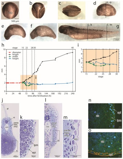
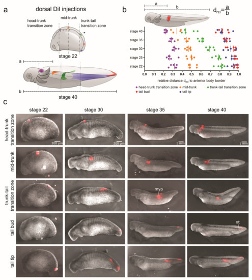
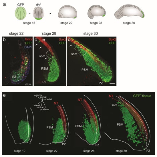
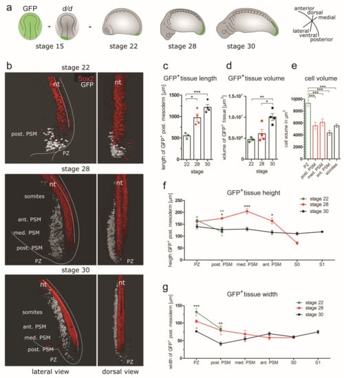
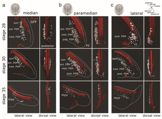
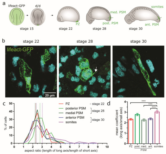
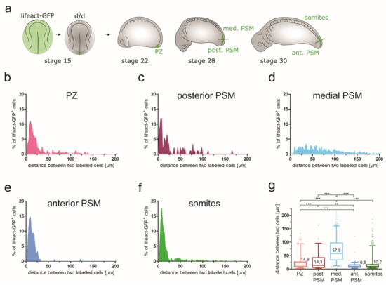
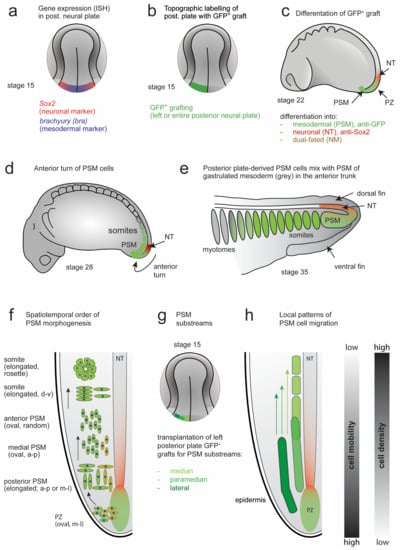
The Role of Posterior Neural Plate-Derived Presomitic Mesoderm (PSM) in Trunk and Tail Muscle Formation and Axis Elongation
by
Barbara K. Stepien, Verena Pawolski, Marc-Christoph Wagner, Thomas Kurth, Mirko H. H. Schmidt and Hans-Henning Epperlein
Cited by 1 | Viewed by 1587
Abstract
Elongation of the posterior body axis is distinct from that of the anterior trunk and head. Early drivers of posterior elongation are the neural plate/tube and notochord, later followed by the presomitic mesoderm (PSM), together with the neural tube and notochord. In axolotl,
[...] Read more.
Elongation of the posterior body axis is distinct from that of the anterior trunk and head. Early drivers of posterior elongation are the neural plate/tube and notochord, later followed by the presomitic mesoderm (PSM), together with the neural tube and notochord. In axolotl, posterior neural plate-derived PSM is pushed posteriorly by convergence and extension of the neural plate. The PSM does not go through the blastopore but turns anteriorly to join the gastrulated paraxial mesoderm. To gain a deeper understanding of the process of axial elongation, a detailed characterization of PSM morphogenesis, which precedes somite formation, and of other tissues (such as the epidermis, lateral plate mesoderm and endoderm) is needed. We investigated these issues with specific tissue labelling techniques (DiI injections and GFP
+ tissue grafting) in combination with optical tissue clearing and 3D reconstructions. We defined a spatiotemporal order of PSM morphogenesis that is characterized by changes in collective cell behaviour. The PSM forms a cohesive tissue strand and largely retains this cohesiveness even after epidermis removal. We show that during embryogenesis, the PSM, as well as the lateral plate and endoderm move anteriorly, while the net movement of the axis is posterior.
Full article
►▼
Show Figures


