31 pages, 1115 KiB  
Review
ER Stress-Sensor Proteins and ER-Mitochondrial Crosstalk—Signaling Beyond (ER) Stress Response
by Vaishali Kumar and Shuvadeep Maity
Biomolecules 2021, 11(2), 173; https://doi.org/10.3390/biom11020173 - 28 Jan 2021
Cited by 80 | Viewed by 10340
Abstract
Recent studies undoubtedly show the importance of inter organellar connections to maintain cellular homeostasis. In normal physiological conditions or in the presence of cellular and environmental stress, each organelle responds alone or in coordination to maintain cellular function. The Endoplasmic reticulum (ER) and [...] Read more.
Recent studies undoubtedly show the importance of inter organellar connections to maintain cellular homeostasis. In normal physiological conditions or in the presence of cellular and environmental stress, each organelle responds alone or in coordination to maintain cellular function. The Endoplasmic reticulum (ER) and mitochondria are two important organelles with very specialized structural and functional properties. These two organelles are physically connected through very specialized proteins in the region called the mitochondria-associated ER membrane (MAM). The molecular foundation of this relationship is complex and involves not only ion homeostasis through the shuttling of calcium but also many structural and apoptotic proteins. IRE1alpha and PERK are known for their canonical function as an ER stress sensor controlling unfolded protein response during ER stress. The presence of these transmembrane proteins at the MAM indicates its potential involvement in other biological functions beyond ER stress signaling. Many recent studies have now focused on the non-canonical function of these sensors. In this review, we will focus on ER mitochondrial interdependence with special emphasis on the non-canonical role of ER stress sensors beyond ER stress. Full article
(This article belongs to the Special Issue Endoplasmic Reticulum Stress in Diseases)
Show Figures

Figure 1

11 pages, 2948 KiB  
Article
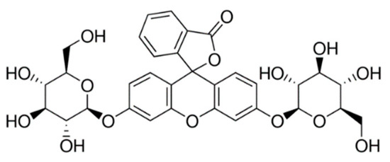
Human Saliva-Mediated Hydrolysis of Eugenyl-β-D-Glucoside and Fluorescein-di-β-D-Glucoside in In Vivo and In Vitro Models
by Mariusz Dziadas, Adam Junka and Henryk Jeleń
Biomolecules 2021, 11(2), 172; https://doi.org/10.3390/biom11020172 - 27 Jan 2021
Cited by 3 | Viewed by 3128
Abstract
Eugenyl-β-D-glucopyranoside, also referred to as Citrusin C, is a natural glucoside found among others in cloves, basil and cinnamon plants. Eugenol in a form of free aglycone is used in perfumeries, flavourings, essential oils and in medicinal products. Synthetic Citrusin C was incubated [...] Read more.
Eugenyl-β-D-glucopyranoside, also referred to as Citrusin C, is a natural glucoside found among others in cloves, basil and cinnamon plants. Eugenol in a form of free aglycone is used in perfumeries, flavourings, essential oils and in medicinal products. Synthetic Citrusin C was incubated with human saliva in several in vitro models together with substrate-specific enzyme and antibiotics (clindamycin, ciprofloxacin, amoxicillin trihydrate and potassium clavulanate). Citrusin C was detected using liquid chromatography with tandem mass spectrometry (LC-MS/MS). Citrusin C was completely degraded only when incubated with substrate-specific A. niger glucosidase E.C 3.2.1.21 (control sample) and when incubated with human saliva (tested sample). The addition of antibiotics to the above-described experimental setting, stopped Citrusin C degradation, indicating microbiologic origin of hydrolysis observed. Our results demonstrate that Citrusin C is subjected to complete degradation by salivary/oral cavity microorganisms. Extrapolation of our results allows to state that in the human oral cavity, virtually all β-D-glucosides would follow this type of hydrolysis. Additionally, a new method was developed for an in vivo rapid test of glucosidase activity in the human mouth on the tongue using fluorescein-di-β-D-glucoside as substrate. The results presented in this study serve as a proof of concept for the hypothesis that microbial hydrolysis path of β-D-glucosides begins immediately in the human mouth and releases the aglycone directly into the gastrointestinal tract. Full article
(This article belongs to the Collection Feature Papers in Biochemistry)
Show Figures

Figure 1

13 pages, 1322 KiB  
Article
The Effect of Acute Intense Exercise on Activity of Antioxidant Enzymes in Smokers and Non-Smokers
by Hadi Nobari, Hamzeh Abdi Nejad, Mehdi Kargarfard, Soghra Mohseni, Katsuhiko Suzuki, José Carmelo Adsuar and Jorge Pérez-Gómez
Biomolecules 2021, 11(2), 171; https://doi.org/10.3390/biom11020171 - 27 Jan 2021
Cited by 29 | Viewed by 4074
Abstract
Acute intense exercise causes significant oxidative stress and consequently an increase in total antioxidant capacity; however, the mechanisms and combined effects of intense exercise and smoking on oxidative stress among active and non-active smokers are not clear. The aim of this study was [...] Read more.
Acute intense exercise causes significant oxidative stress and consequently an increase in total antioxidant capacity; however, the mechanisms and combined effects of intense exercise and smoking on oxidative stress among active and non-active smokers are not clear. The aim of this study was to investigate the effect of acute intense exercise on antioxidant enzyme activity responses in active and non-active individuals exposed to cigarette smoke. The study included 40 subjects who were equally classified as: smokers that did exercise (SE), smokers that did not do exercise (SnE), non-smokers that did exercise (NSE), and non-smokers that did not do exercise (NSnE). The adjusted Astrand test was used to exhaust the subjects. Salivary enzymes of peroxidase (POX), catalase (CAT), and superoxide dismutase (SOD) were measured, by spectrophotometry methods, at 3 different time points: pre-test (TP1), post-test (TP2), and one hour after finishing the test (TP3). Significant (p < 0.05) group x time interactions were found for the three enzymes. Salivary POX, CAT and SOD increased in all groups from TP1 to TP2 and decreased from TP2 to TP3. Only the NSE showed a significant difference between TP1 to TP3 in POX and SOD by +0.011 ± 0.007 and +0.075 ± 0.020 (U/mL), respectively. The NSE showed significantly higher activity of POX, CAT and SOD in TP2 compared to the other groups. Furthermore, NSE and NSnE had higher activity of POX, CAT and SOD in TP1 and TP3 (p < 0.05) compared with SE and SnE. Only in the NSnE, were no differences observed in CAT compared with SE and SnE in TP3. These results showed that the antioxidant activity at rest and in the recovery time after the acute intense exercise was lower in SE and SnE compared with NSE and NSnE, suggesting that smoking habit may reduce the ameliorating effect of regular physical activity on acute exercise-induced oxidative stress. Full article
Show Figures

Figure 1

10 pages, 2614 KiB  
Article
Protective Effect of γ-mangostin Isolated from the Peel of Garcinia mangostana against Glutamate-Induced Cytotoxicity in HT22 Hippocampal Neuronal Cells
by Ji Yun Baek, Kiwon Jung, Young-Mi Kim, Hyun-Young Kim, Ki Sung Kang and Young-Won Chin
Biomolecules 2021, 11(2), 170; https://doi.org/10.3390/biom11020170 - 27 Jan 2021
Cited by 19 | Viewed by 3918
Abstract
The aim of the present study was to examine the protective effect of γ-mangostin, a component of the mangosteen shell, against oxidative damage to nerve cells induced by excessive glutamate, a known excitatory neurotransmitter. To investigate the effect of γ-mangostin on apoptosis, 5 [...] Read more.
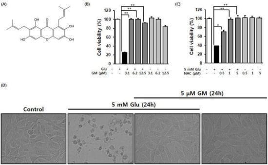
The aim of the present study was to examine the protective effect of γ-mangostin, a component of the mangosteen shell, against oxidative damage to nerve cells induced by excessive glutamate, a known excitatory neurotransmitter. To investigate the effect of γ-mangostin on apoptosis, 5 mM of glutamate was used to induce apoptotic cell death in mouse hippocampal HT22 cells. In this study, γ-mangostin was found to exert a stronger protection than N-acetyl cysteine against glutamate-induced cell damage. γ-Mangostin showed prevented glutamate-induced apoptosis in HT22 cells by reducing the production of reactive oxygen species and stimulating the expression of heme oxygenase-1 protein. In addition, glutamate significantly induced the accumulation of intracellular calcium ions, whereas treatment with γ-mangostin markedly reduced it. Hoechst 33342 staining showed an improvement in glutamate-induced nuclear condensation following γ-mangostin treatment. Furthermore, the number of annexin V-positive cells was significantly reduced following treatment with γ-mangostin. Western blot analysis showed the inhibition of glutamate-induced mitogen-activated protein kinase phosphorylation by γ-mangostin. γ-mangostin also inhibited the regulation of the intrinsic mitochondrial apoptotic pathway. Thus, the results of this study suggest that γ-mangostin is an active ingredient of mangosteen and exerts neuroprotective activities in HT22 cells. Full article
Show Figures

Figure 1

21 pages, 2990 KiB  
Article
Molecular Modeling Studies on the Multistep Reactivation Process of Organophosphate-Inhibited Acetylcholinesterase and Butyrylcholinesterase
by Jakub Jończyk, Jędrzej Kukułowicz, Kamil Łątka, Barbara Malawska, Young-Sik Jung, Kamil Musilek and Marek Bajda
Biomolecules 2021, 11(2), 169; https://doi.org/10.3390/biom11020169 - 27 Jan 2021
Cited by 7 | Viewed by 3479
Abstract
Poisoning with organophosphorus compounds used as pesticides or misused as chemical weapons remains a serious threat to human health and life. Their toxic effects result from irreversible blockade of the enzymes acetylcholinesterase and butyrylcholinesterase, which causes overstimulation of the cholinergic system and often [...] Read more.
Poisoning with organophosphorus compounds used as pesticides or misused as chemical weapons remains a serious threat to human health and life. Their toxic effects result from irreversible blockade of the enzymes acetylcholinesterase and butyrylcholinesterase, which causes overstimulation of the cholinergic system and often leads to serious injury or death. Treatment of organophosphorus poisoning involves, among other strategies, the administration of oxime compounds. Oximes reactivate cholinesterases by breaking the covalent bond between the serine residue from the enzyme active site and the phosphorus atom of the organophosphorus compound. Although the general mechanism of reactivation has been known for years, the exact molecular aspects determining the efficiency and selectivity of individual oximes are still not clear. This hinders the development of new active compounds. In our research, using relatively simple and widely available molecular docking methods, we investigated the reactivation of acetyl- and butyrylcholinesterase blocked by sarin and tabun. For the selected oximes, their binding modes at each step of the reactivation process were identified. Amino acids essential for effective reactivation and those responsible for the selectivity of individual oximes against inhibited acetyl- and butyrylcholinesterase were identified. This research broadens the knowledge about cholinesterase reactivation and demonstrates the usefulness of molecular docking in the study of this process. The presented observations and methods can be used in the future to support the search for new effective reactivators. Full article
(This article belongs to the Special Issue Cholinesterase Research)
Show Figures

Figure 1

15 pages, 658 KiB  
Review
TOR Signaling Pathway in Cardiac Aging and Heart Failure
by Nastaran Daneshgar, Peter S. Rabinovitch and Dao-Fu Dai
Biomolecules 2021, 11(2), 168; https://doi.org/10.3390/biom11020168 - 27 Jan 2021
Cited by 35 | Viewed by 5960
Abstract
Mechanistic Target of Rapamycin (mTOR) signaling is a key regulator of cellular metabolism, integrating nutrient sensing with cell growth. Over the past two decades, studies on the mTOR pathway have revealed that mTOR complex 1 controls life span, health span, and aging by [...] Read more.
Mechanistic Target of Rapamycin (mTOR) signaling is a key regulator of cellular metabolism, integrating nutrient sensing with cell growth. Over the past two decades, studies on the mTOR pathway have revealed that mTOR complex 1 controls life span, health span, and aging by modulating key cellular processes such as protein synthesis, autophagy, and mitochondrial function, mainly through its downstream substrates. Thus, the mTOR pathway regulates both physiological and pathological processes in the heart from embryonic cardiovascular development to maintenance of cardiac homeostasis in postnatal life. In this regard, the dysregulation of mTOR signaling has been linked to many age-related pathologies, including heart failure and age-related cardiac dysfunction. In this review, we highlight recent advances of the impact of mTOR complex 1 pathway and its regulators on aging and, more specifically, cardiac aging and heart failure. Full article
(This article belongs to the Collection TOR Signaling Pathway)
Show Figures

Figure 1

13 pages, 6967 KiB  
Article
Nanobody-Based Indirect Competitive ELISA for Sensitive Detection of 19-Nortestosterone in Animal Urine
by Yuan-yuan Yang, Yu Wang, Yi-feng Zhang, Feng Wang, Yi-fan Liang, Jin-yi Yang, Zhen-lin Xu, Yu-dong Shen and Hong Wang
Biomolecules 2021, 11(2), 167; https://doi.org/10.3390/biom11020167 - 27 Jan 2021
Cited by 11 | Viewed by 3860
Abstract
Nanobody (Nb), a new type of biorecognition element generally from Camelidae, has the characteristics of small molecular weight, high stability, great solubility and high expression level in E. coli. In this study, with 19-nortestosterone (19-NT), an anabolic androgenic steroid as target [...] Read more.
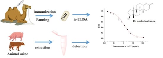
Nanobody (Nb), a new type of biorecognition element generally from Camelidae, has the characteristics of small molecular weight, high stability, great solubility and high expression level in E. coli. In this study, with 19-nortestosterone (19-NT), an anabolic androgenic steroid as target drug, three specific Nbs against 19-NT were selected from camel immune library by phage display technology. The obtained Nbs showed excellent thermostability and organic solvent tolerance. The nanobody Nb2F7 with the best performance was used to develop a sensitive indirect competitive enzyme-linked immunosorbent assay (ic-ELISA) for 19-NT detection. Under optimized conditions, the standard curve of ic-ELISA was fitted with a half-maximal inhibitory concentration (IC50) of 1.03 ng/mL and a detection limit (LOD) of 0.10 ng/mL for 19-NT. Meanwhile, the developed assay had low cross- reactivity with analogs and the recoveries of 19-NT ranged from 82.61% to 99.24% in spiked samples. The correlation coefficient between ic-ELISA and the ultra-performance liquid chromatography/mass spectrometry (UPLC-MS/MS) method was 0.9975, which indicated that the nanobody-based ic-ELISA could be a useful tool for a rapid analysis of 19-NT in animal urine samples. Full article
(This article belongs to the Special Issue The Therapeutic and Diagnostic Potential of Nanobodies)
Show Figures

Graphical abstract

10 pages, 758 KiB  
Review
Galectin-3 and sST2 as Prognosticators for Heart Failure Requiring Extracorporeal Life Support: Jack n’ Jill
by Jianli Bi, Vidu Garg and Andrew R. Yates
Biomolecules 2021, 11(2), 166; https://doi.org/10.3390/biom11020166 - 27 Jan 2021
Cited by 9 | Viewed by 3630
Abstract
Extracorporeal life support provides perfusion for patients with heart failure to allow time for recovery, function as a bridge for patients to heart transplantation, or serve as destination therapy for long term mechanical device support. Several biomarkers have been employed in attempt to [...] Read more.
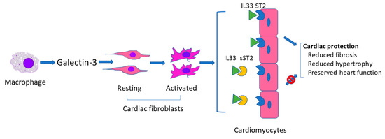
Extracorporeal life support provides perfusion for patients with heart failure to allow time for recovery, function as a bridge for patients to heart transplantation, or serve as destination therapy for long term mechanical device support. Several biomarkers have been employed in attempt to predict these outcomes, but it remains to be determined which are suitable to guide clinical practice relevant to extracorporeal life support. Galectin-3 and soluble suppression of tumorigenicity-2 (sST2) are two of the more promising candidates with the greatest supporting evidence. In this review, we address the similarities and differences between galectin-3 and sST2 for prognostic prediction in adults and children with heart failure requiring extracorporeal life support and highlight the significant lack of progress in pediatric biomarker discovery and utilization. Full article
(This article belongs to the Special Issue Molecular Biomarkers In Cardiology)
Show Figures

Figure 1

11 pages, 4035 KiB  
Article
Thymoquinone Protects Neurons in the Cerebellum of Rats through Mitigating Oxidative Stress and Inflammation Following High-Fat Diet Supplementation
by Aziza Alrafiah
Biomolecules 2021, 11(2), 165; https://doi.org/10.3390/biom11020165 - 27 Jan 2021
Cited by 13 | Viewed by 2756
Abstract
High-fat diet (HFD) is a major problem causing neuronal damage. Thymoquinone (TQ) could regulate oxidative stress and the inflammatory process. Hence, the present study elucidated the significant role of TQ on oxidative stress, inflammation, as well as morphological changes in the cerebellum of [...] Read more.
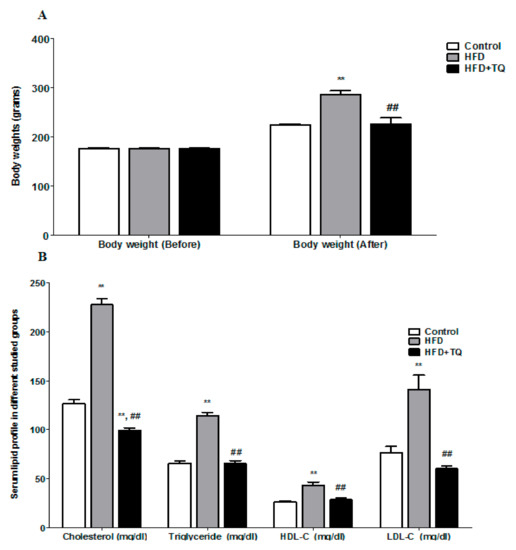
High-fat diet (HFD) is a major problem causing neuronal damage. Thymoquinone (TQ) could regulate oxidative stress and the inflammatory process. Hence, the present study elucidated the significant role of TQ on oxidative stress, inflammation, as well as morphological changes in the cerebellum of rats with HFD. Rats were divided into three groups as (1) control, (2) saturated HFD for eight weeks and (3) HFD supplementation (four weeks) followed by TQ 300 mg/kg/day treated (four weeks). After treatment, blood samples were collected to measure oxidative stress markers glutathione (GSH), malondialdehyde (MDA), superoxide dismutase (SOD), and inflammatory cytokines. Furthermore, neuronal morphological changes were also observed in the cerebellum of the rats. HFD rats show higher body weight (286.5 ± 7.4 g) as compared with the control group (224.67 ± 1.78 g). TQ treatment significantly (p < 0.05) lowered the body weight (225.83 ± 13.15 g). TQ produced a significant (p < 0.05) reduction in cholesterol, triglycerides, high-density lipoprotein (HDL), and low-density lipoprotein (LDL). The antioxidative enzymes significantly reduced in HFD rats (GSH, 1.46 ± 0.36 mol/L and SOD, 99.13 ± 5.41 µmol/mL) as compared with the control group (GSH, 6.25 ± 0.36 mol/L and SOD, 159.67 ± 10.67 µmol/mL). MDA was increased significantly in HFD rats (2.05 ± 0.25 nmol/L) compared to the control group (0.695 ± 0.11 nmol/L). Surprisingly, treatment with TQ could improve the level of GSH, MDA, and SOD. TQ treatment significantly (p < 0.05) reduced the inflammatory markers as compared with HFD alone. TQ treatment minimizes neuronal damage as well as reduces inflammation and improves antioxidant enzymes. TQ can be considered as a promising agent in preventing the neuronal morphological changes in the cerebellum of obese populations. Full article
Show Figures

Figure 1

19 pages, 5042 KiB  
Article
LPS Tolerance Inhibits Cellular Respiration and Induces Global Changes in the Macrophage Secretome
by Joseph Gillen, Thunnicha Ondee, Devikala Gurusamy, Jiraphorn Issara-Amphorn, Nathan P. Manes, Sung Hwan Yoon, Asada Leelahavanichkul and Aleksandra Nita-Lazar
Biomolecules 2021, 11(2), 164; https://doi.org/10.3390/biom11020164 - 27 Jan 2021
Cited by 39 | Viewed by 5187
Abstract
Inflammatory response plays an essential role in the resolution of infections. However, inflammation can be detrimental to an organism and cause irreparable damage. For example, during sepsis, a cytokine storm can lead to multiple organ failures and often results in death. One of [...] Read more.
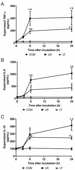
Inflammatory response plays an essential role in the resolution of infections. However, inflammation can be detrimental to an organism and cause irreparable damage. For example, during sepsis, a cytokine storm can lead to multiple organ failures and often results in death. One of the strongest triggers of the inflammatory response is bacterial lipopolysaccharides (LPS), acting mostly through Toll-like receptor 4 (TLR4). Paradoxically, while exposure to LPS triggers a robust inflammatory response, repeated or prolonged exposure to LPS can induce a state of endotoxin tolerance, a phenomenon where macrophages and monocytes do not respond to new endotoxin challenges, and it is often associated with secondary infections and negative outcomes. The cellular mechanisms regulating this phenomenon remain elusive. We used metabolic measurements to confirm differences in the cellular metabolism of naïve macrophages and that of macrophages responding to LPS stimulation or those in the LPS-tolerant state. In parallel, we performed an unbiased secretome survey using quantitative mass spectrometry during the induction of LPS tolerance, creating the first comprehensive secretome profile of endotoxin-tolerant cells. The secretome changes confirmed that LPS-tolerant macrophages have significantly decreased cellular metabolism and that the proteins secreted by LPS-tolerant macrophages have a strong association with cell survival, protein metabolism, and the metabolism of reactive oxygen species. Full article
(This article belongs to the Special Issue Integrative Multi-Omics in Biomedical Research)
Show Figures

Figure 1

13 pages, 2048 KiB  
Article
Effect of Humanizing Mutations on the Stability of the Llama Single-Domain Variable Region
by Miguel A. Soler, Barbara Medagli, Jiewen Wang, Sandra Oloketuyi, Gregor Bajc, He Huang, Sara Fortuna and Ario de Marco
Biomolecules 2021, 11(2), 163; https://doi.org/10.3390/biom11020163 - 26 Jan 2021
Cited by 19 | Viewed by 5983
Abstract
In vivo clinical applications of nanobodies (VHHs) require molecules that induce minimal immunoresponse and therefore possess sequences as similar as possible to the human VH domain. Although the relative sequence variability in llama nanobodies has been used to identify scaffolds with partially humanized [...] Read more.
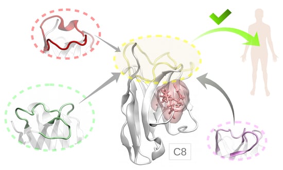
In vivo clinical applications of nanobodies (VHHs) require molecules that induce minimal immunoresponse and therefore possess sequences as similar as possible to the human VH domain. Although the relative sequence variability in llama nanobodies has been used to identify scaffolds with partially humanized signature, the transformation of the Camelidae hallmarks in the framework2 still represents a major problem. We assessed a set of mutants in silico and experimentally to elucidate what is the contribution of single residues to the VHH stability and how their combinations affect the mutant nanobody stability. We described at molecular level how the interaction among residues belonging to different structural elements enabled a model llama nanobody (C8WT, isolated from a naïve library) to be functional and maintain its stability, despite the analysis of its primary sequence would classify it as aggregation-prone. Five chimeras formed by grafting CDRs isolated from different nanobodies into C8WT scaffold were successfully expressed as soluble proteins and both tested clones preserved their antigen binding specificity. We identified a nanobody with human hallmarks that seems suitable for humanizing selected camelid VHHs by grafting heterologous CDRs in its scaffold and could serve for the preparation of a synthetic library of human-like single domains. Full article
(This article belongs to the Special Issue The Therapeutic and Diagnostic Potential of Nanobodies)
Show Figures

Graphical abstract

17 pages, 2638 KiB  
Article
Anti-Inflammatory Effects of Rosmarinic Acid-Loaded Nanovesicles in Acute Colitis through Modulation of NLRP3 Inflammasome
by Sonia Marinho, Matilde Illanes, Javier Ávila-Román, Virginia Motilva and Elena Talero
Biomolecules 2021, 11(2), 162; https://doi.org/10.3390/biom11020162 - 26 Jan 2021
Cited by 70 | Viewed by 4670
Abstract
Ulcerative colitis (UC), one of the two main types of inflammatory bowel disease, has no effective treatment. Rosmarinic acid (RA) is a polyphenol that, when administered orally, is metabolised in the small intestine, compromising its beneficial effects. We used chitosan/nutriose-coated niosomes loaded with [...] Read more.
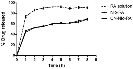
Ulcerative colitis (UC), one of the two main types of inflammatory bowel disease, has no effective treatment. Rosmarinic acid (RA) is a polyphenol that, when administered orally, is metabolised in the small intestine, compromising its beneficial effects. We used chitosan/nutriose-coated niosomes loaded with RA to protect RA from gastric degradation and target the colon and evaluated their effect on acute colitis induced by 4% dextran sodium sulphate (DSS) for seven days in mice. RA-loaded nanovesicles (5, 10 and 20 mg/kg) or free RA (20 mg/kg) were orally administered from three days prior to colitis induction and during days 1, 3, 5 and 7 of DSS administration. RA-loaded nanovesicles improved body weight loss and disease activity index as well as increased mucus production and decreased myeloperoxidase activity and TNF-α production. Moreover, RA-loaded nanovesicles downregulated protein expression of inflammasome components such as NLR family pyrin domain-containing 3 (NLRP3), adaptor protein (ASC) and caspase-1, and the consequent reduction of IL-1β levels. Furthermore, nuclear factor erythroid 2-related factor 2 (Nrf2) and heme oxygenase-1 (HO-1) protein expression increased after the RA-loaded nanovesicles treatment However, these mechanistic changes were not detected with the RA-free treatment. Our findings suggest that the use of chitosan/nutriose-coated niosomes to increase RA local bioavailability could be a promising nutraceutical strategy for oral colon-targeted UC therapy. Full article
Show Figures

Figure 1

3 pages, 156 KiB  
Editorial
Cell-Free Approaches and Therapeutic Biomolecules for Cardiac Regeneration
by Mariann Gyöngyösi
Biomolecules 2021, 11(2), 161; https://doi.org/10.3390/biom11020161 - 26 Jan 2021
Cited by 1 | Viewed by 2060
Abstract
In contrast with some adult human organs, such as liver or skin, the adult human heart shows very limited self-regeneration capacity, attributed to the negligible presence of resident cardiac stem cells or cardiac progenitors [...] Full article
19 pages, 2049 KiB  
Article
Computational Insights into the Potential of Withaferin-A, Withanone and Caffeic Acid Phenethyl Ester for Treatment of Aberrant-EGFR Driven Lung Cancers
by Vidhi Malik, Vipul Kumar, Sunil C. Kaul, Renu Wadhwa and Durai Sundar
Biomolecules 2021, 11(2), 160; https://doi.org/10.3390/biom11020160 - 26 Jan 2021
Cited by 15 | Viewed by 4947
Abstract
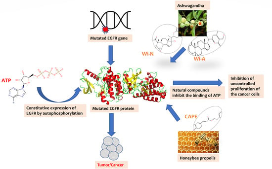
The anticancer activities of Withaferin-A (Wi-A) and Withanone (Wi-N) from Ashwagandha and Caffeic Acid Phenethyl Ester (CAPE) from honeybee propolis have been well documented. Here, we examined the binding potential of these natural compounds to inhibit the constitutive phosphorylation of epidermal growth factor [...] Read more.
The anticancer activities of Withaferin-A (Wi-A) and Withanone (Wi-N) from Ashwagandha and Caffeic Acid Phenethyl Ester (CAPE) from honeybee propolis have been well documented. Here, we examined the binding potential of these natural compounds to inhibit the constitutive phosphorylation of epidermal growth factor receptors (EGFRs). Exon 20 insertion mutants of EGFR, which show resistance to various FDA approved drugs and are linked to poor prognosis of lung cancer patients, were the primary focus of this study. Apart from exon 20 insertion mutants, the potential of natural compounds to serve as ATP competitive inhibitors of wildtype protein and other common mutants of EGFR, namely L858R and exon19del, were also examined. The potential of natural compounds was compared to the positive controls such as erlotinib, TAS6417 and poziotinib. Similar to known inhibitors, Wi-A and Wi-N could displace and binds at the ATP orthosteric site of exon19del, L858R and exon20, while CAPE was limited to wildtype EGFR and exon 20 insertion mutants only. Moreover, the binding free energy of the natural drugs against EGFRs was also comparable to the positive controls. This computational study suggests that Wi-A and Wi-N have potential against multiple mutated EGFRs, warranting further in vitro and in vivo experiments. Full article
(This article belongs to the Section Bioinformatics and Systems Biology)
Show Figures

Graphical abstract

22 pages, 3745 KiB  
Article
Transcriptome Analysis Reveals Altered Inflammatory Pathway in an Inducible Glial Cell Model of Myotonic Dystrophy Type 1
by Cuauhtli N. Azotla-Vilchis, Daniel Sanchez-Celis, Luis E. Agonizantes-Juárez, Rocío Suárez-Sánchez, J. Manuel Hernández-Hernández, Jorge Peña, Karla Vázquez-Santillán, Norberto Leyva-García, Arturo Ortega, Vilma Maldonado, Claudia Rangel, Jonathan J. Magaña, Bulmaro Cisneros and Oscar Hernández-Hernández
Biomolecules 2021, 11(2), 159; https://doi.org/10.3390/biom11020159 - 26 Jan 2021
Cited by 15 | Viewed by 4666
Abstract
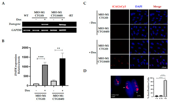
Myotonic dystrophy type 1 (DM1), the most frequent inherited muscular dystrophy in adults, is caused by the CTG repeat expansion in the 3′UTR of the DMPK gene. Mutant DMPK RNA accumulates in nuclear foci altering diverse cellular functions including alternative splicing regulation. DM1 [...] Read more.
Myotonic dystrophy type 1 (DM1), the most frequent inherited muscular dystrophy in adults, is caused by the CTG repeat expansion in the 3′UTR of the DMPK gene. Mutant DMPK RNA accumulates in nuclear foci altering diverse cellular functions including alternative splicing regulation. DM1 is a multisystemic condition, with debilitating central nervous system alterations. Although a defective neuroglia communication has been described as a contributor of the brain pathology in DM1, the specific cellular and molecular events potentially affected in glia cells have not been totally recognized. Thus, to study the effects of DM1 mutation on glial physiology, in this work, we have established an inducible DM1 model derived from the MIO-M1 cell line expressing 648 CUG repeats. This new model recreated the molecular hallmarks of DM1 elicited by a toxic RNA gain-of-function mechanism: accumulation of RNA foci colocalized with MBNL proteins and dysregulation of alternative splicing. By applying a microarray whole-transcriptome approach, we identified several gene changes associated with DM1 mutation in MIO-M1 cells, including the immune mediators CXCL10, CCL5, CXCL8, TNFAIP3, and TNFRSF9, as well as the microRNAs miR-222, miR-448, among others, as potential regulators. A gene ontology enrichment analyses revealed that inflammation and immune response emerged as major cellular deregulated processes in the MIO-M1 DM1 cells. Our findings indicate the involvement of an altered immune response in glia cells, opening new windows for the study of glia as potential contributor of the CNS symptoms in DM1. Full article
(This article belongs to the Special Issue Molecular Basis of Neuromuscular Diseases)
Show Figures

Figure 1