Diagnostics 2020, 10(8), 516; https://doi.org/10.3390/diagnostics10080516 - 25 Jul 2020
Cited by 4 | Viewed by 6602
Abstract
Skin staining due to iron leakage into the subcutaneous tissue can sometimes occur during intravenous iron infusion. We describe a case of lateral antebrachial cutaneous nerve (LACN) entrapment due to extravasated iron after an intravenous iron infusion. A 41-year-old woman received an intravenous
[...] Read more.
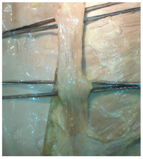
Skin staining due to iron leakage into the subcutaneous tissue can sometimes occur during intravenous iron infusion. We describe a case of lateral antebrachial cutaneous nerve (LACN) entrapment due to extravasated iron after an intravenous iron infusion. A 41-year-old woman received an intravenous ferric carboxymaltose infusion for iron deficiency anemia. However, during the infusion, extravasation of iron occurred and brown pigmentation developed on the lateral side of the cubital fossa. Sixteen months later, the patient still had some staining in her anterolateral elbow and proximal forearm. In addition, she complained of tingling pain over her left forearm. Ultrasonography (US) revealed a lateral antebrachial cutaneous nerve (LACN) under the stained area. When we swept the stained area with the US transducer, she reported a tingling pain on her left lateral forearm, the region innervated by the left LACN. Therefore, we considered that the pain resulted from the compression of the left LACN by the leaked iron during the intravenous infusion. Leaked iron can compress the cutaneous nerve and result in neuropathic pain and cosmetic problems. When patients with skin staining after iron infusion have neuropathic pain, clinicians should consider the possibility of entrapment of the cutaneous nerves.
Full article
(This article belongs to the Special Issue The Diagnostic Value of Nerve and Muscle Ultrasound in Neuromuscular Disorders)
►
Show Figures

Figure 1


