Cells 2022, 11(19), 2940; https://doi.org/10.3390/cells11192940 - 20 Sep 2022
Cited by 10 | Viewed by 4072
Abstract
AKT serine-threonine kinase (AKT) and its effectors are essential for maintaining cell proliferation, apoptosis, autophagy, endoplasmic reticulum (ER) stress, mitochondrial morphogenesis (fission/fusion), ferroptosis, necroptosis, DNA damage response (damage and repair), senescence, and migration of cancer cells. Several lncRNAs and circRNAs also regulate the
[...] Read more.
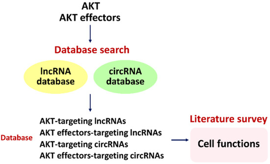
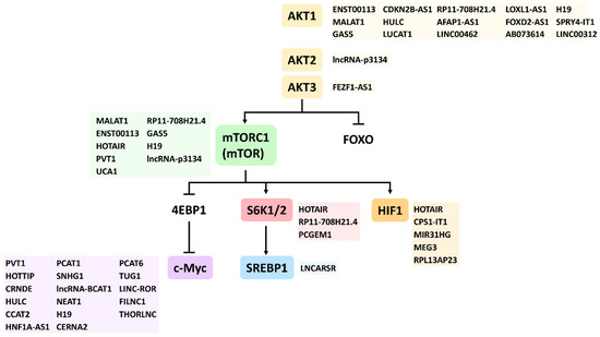
AKT serine-threonine kinase (AKT) and its effectors are essential for maintaining cell proliferation, apoptosis, autophagy, endoplasmic reticulum (ER) stress, mitochondrial morphogenesis (fission/fusion), ferroptosis, necroptosis, DNA damage response (damage and repair), senescence, and migration of cancer cells. Several lncRNAs and circRNAs also regulate the expression of these functions by numerous pathways. However, the impact on cell functions by lncRNAs and circRNAs regulating AKT and its effectors is poorly understood. This review provides comprehensive information about the relationship of lncRNAs and circRNAs with AKT on the cell functions of cancer cells. the roles of several lncRNAs and circRNAs acting on AKT effectors, such as FOXO, mTORC1/2, S6K1/2, 4EBP1, SREBP, and HIF are explored. To further validate the relationship between AKT, AKT effectors, lncRNAs, and circRNAs, more predicted AKT- and AKT effector-targeting lncRNAs and circRNAs were retrieved from the LncTarD and circBase databases. Consistently, using an in-depth literature survey, these AKT- and AKT effector-targeting database lncRNAs and circRNAs were related to cell functions. Therefore, some lncRNAs and circRNAs can regulate several cell functions through modulating AKT and AKT effectors. This review provides insights into a comprehensive network of AKT and AKT effectors connecting to lncRNAs and circRNAs in the regulation of cancer cell functions.
Full article
(This article belongs to the Special Issue Regulatory Roles of Non-coding RNAs in Cancer)
►
Show Figures
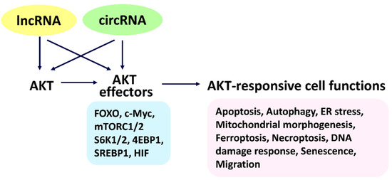
Figure 1


