Cancers 2022, 14(9), 2242; https://doi.org/10.3390/cancers14092242 - 29 Apr 2022
Cited by 12 | Viewed by 3814
Abstract
Endometriosis, a painful gynecological condition accompanied by inflammation in women of reproductive age, is associated with an increased risk of ovarian cancer. We evaluated the role of peritoneal heme accumulated during menstrual cycling, as well as peritoneal and lesional macrophage phenotype, in promoting
[...] Read more.
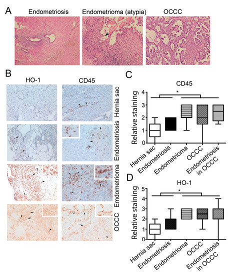
Endometriosis, a painful gynecological condition accompanied by inflammation in women of reproductive age, is associated with an increased risk of ovarian cancer. We evaluated the role of peritoneal heme accumulated during menstrual cycling, as well as peritoneal and lesional macrophage phenotype, in promoting an oncogenic microenvironment. We quantified the heme-degrading enzyme, heme oxygenase-1 (HO-1, encoded by Hmox1) in normal peritoneum, endometriotic lesions and endometriosis-associated ovarian cancer (EAOC) of clear cell type (OCCC). HO-1 was expressed primarily in macrophages and increased in endometrioma and OCCC tissues relative to endometriosis and controls. Further, we compared cytokine expression profiles in peritoneal macrophages (PM) and peripheral blood mononuclear cells (PBMC) in women with endometriosis versus controls as a measure of a tumor-promoting environment in the peritoneum. We found elevated levels of HO-1 along with IL-10 and the pro-inflammatory cytokines (IL-1β, IL-16, IFNγ) in PM but not in PBMC from endometriosis patients. Using LysM-Cre:Hmox1flfl conditional knockout mice, we show that a deficiency of HO-1 in macrophages led to the suppression of growth of ID8 ovarian tumors implanted into the peritoneum. The restriction of ID8 ovarian tumor growth was associated with an increased number of Mac3+ macrophage and B cells in LysM-Cre:Hmox1flfl mice compared to controls. Functional experiments in ovarian cancer cell lines show that HO-1 is induced by heme. Low levels of exogenous heme promoted ovarian cancer colony growth in soft agar. Higher doses of heme led to slower cancer cell colony growth in soft agar and the induction of HO-1. These data suggest that perturbation of heme metabolism within the endometriotic niche and in cancer cells themselves may be an important factor that influences tumor initiation and growth.
Full article
(This article belongs to the Special Issue Cancer-Mediated Immunomodulation—Pathogenetic, Diagnostic and Therapeutic Aspects)
►
Show Figures

Figure 1


