Strongyloides stercoralis Hyperinfection in an HIV-Infected Patient Successfully Treated with Subcutaneous Ivermectin

 and
and
Abstract
:
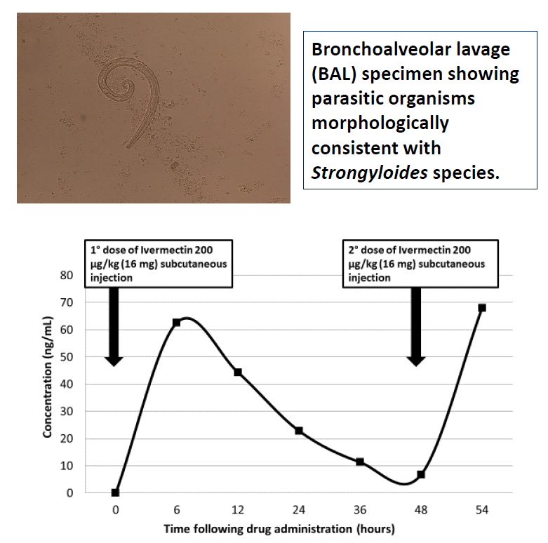
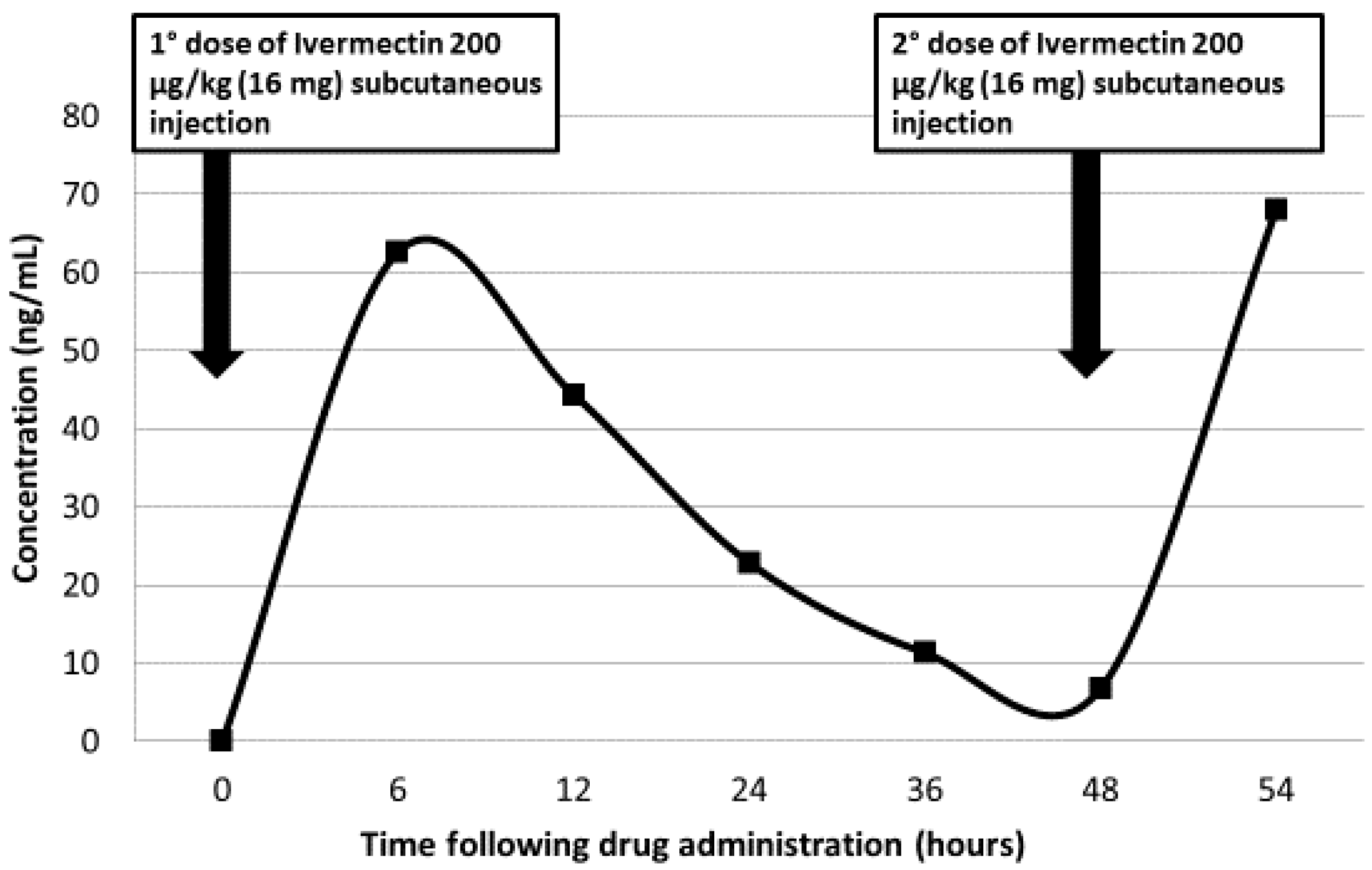
1. Case Report
2. Discussion
Author Contributions
Funding
Conflicts of Interest
References
- Kitzman, D.; Wei, S.Y.; Fleckenstein, L. Liquid chromatographic assay of ivermectin in human plasma for application to clinical pharmacokinetic studies. J. Pharm. Biomed. Anal. 2006, 40, 1013–1020. [Google Scholar] [CrossRef] [PubMed]
- Schär, F.; Trostdorf, U.; Giardina, F.; Khieu, V.; Muth, S.; Marti, H.; Vounatsou, P.; Odermatt, P. Strongyloides stercoralis: Global distribution and risk factors. PLoS Negl. Trop. Dis. 2013, 7, e2288. [Google Scholar] [CrossRef] [PubMed] [Green Version]
- Keiser, P.B.; Nutman, T.B. Strongyloides stercoralis in the immunocompromised population. Clin. Microbiol. Rev. 2004, 17, 208–217. [Google Scholar] [CrossRef] [PubMed]
- Mejia, R.; Nutman, T.B. Screening, prevention, and treatment for hyperinfection syndrome and disseminated infections caused by Strongyloides stercoralis. Curr. Opin. Infect. Dis. 2012, 25, 458–463. [Google Scholar] [CrossRef] [PubMed]
- Cooper, A.J.R.; Dholakia, S.; Holland, C.V.; Friend, P.J. Helminths in organ transplantation. Lancet Infect. Dis. 2017, 17, e166–e176. [Google Scholar] [CrossRef]
- Buonfrate, D.; Requena-Mendez, A.; Angheben, A.; Muñoz, J.; Gobbi, F.; Van Den Ende, J.; Bisoffi, Z. Severe strongyloidiasis: A systematic review of case reports. BMC Infect. Dis. 2013, 13, 78. [Google Scholar] [CrossRef] [PubMed]
- Schwartz, B.; Mawhorter, S. Parasitic infections in solid organ transplantation. Am. J. Transplant. 2013, 13, 280–303. [Google Scholar] [CrossRef] [PubMed]
- Greaves, D.; Coggle, S.; Pollard, C.; Aliyu, S.H.; Moore, E.M. Strongyloides stercoralis infection. BMJ 2013, 347, f4610. [Google Scholar] [CrossRef] [PubMed]
- Barrett, J.; Broderick, C.; Soulsby, H.; Wade, P.; Newsholme, W. Subcutaneous ivermectin use in the treatment of severe Strongyloides stercoralis infection: Two case reports and a discussion of the literature. J. Antimicrob. Chemother. 2016, 71, 220–225. [Google Scholar] [CrossRef] [PubMed]
- Chiodini, P.L.; Reid, A.J.; Wiselka, M.J.; Firmin, R.; Foweraker, J. Parenteral ivermectin in Strongyloides hyperinfection. Lancet 2000, 355, 43–44. [Google Scholar] [CrossRef]
- Marty, F.M.; Lowry, C.M.; Rodriguez, M.; Milner, D.A.; Pieciak, W.S.; Sinha, A.; Fleckenstein, L.; Baden, L. Treatment of human disseminated strongyloidiasis with a parenteral veterinary formulation of ivermectin. Clin. Infect. Dis. 2005, 41, e5–e8. [Google Scholar] [CrossRef] [PubMed]
- Turner, S.A.; Maclean, J.D.; Fleckenstein, L.; Greenaway, C. Parental administration of ivermectin in a patient with disseminated strongyloidiasis. Am. J. Trop. Med. Hyg. 2005, 73, 911–914. [Google Scholar] [PubMed]
- Fusco, D.N.; Downs, J.A.; Satlin, M.J.; Pahuja, M.; Ramos, L.; Barie, P.S.; Fleckenstein, L.; Murray, H.W. Non-oral treatment with ivermectin for disseminated strongyloidiasis. Am. J. Trop. Med. Hyg. 2010, 83, 879–883. [Google Scholar] [CrossRef] [PubMed]
- Grein, J.D.; Mathisen, G.E.; Donovan, S.; Fleckenstein, L. Serum ivermectin levels after enteral and subcutaneous administration for Strongyloides hyperinfection: A case report. Scand. J. Infect. Dis. 2010, 42, 234–236. [Google Scholar] [CrossRef] [PubMed]
- Donadello, K.; Cristallini, S.; Taccone, F.S.; Lorent, S.; Vincent, J.L.; de Backer, D.; Jacobs, F. Strongyloides disseminated infection successfully treated with parenteral ivermectin: Case report with drug concentration measurements and review of the literature. Int. J. Antimicrob. Agents 2013, 42, 580–583. [Google Scholar] [CrossRef] [PubMed]
- Van Westerloo, D.J.; Landman, G.W.; Prichard, R.; Lespine, A.; Visser, L.G. Persistent coma in Strongyloides hyperinfection syndrome associated with persistently increased ivermectin levels. Clin. Infect. Dis. 2014, 58, 143–144. [Google Scholar] [CrossRef] [PubMed]
- Knopp, S.; Mgeni, A.F.; Khamis, I.S.; Steinmann, P.; Stothard, J.R.; Rollinson, D.; Marti, H.; Utzinger, J. Diagnosis of soil-transmitted helminths in the era of preventive chemotherapy: Effect of multiple stool sampling and use of different diagnostic techniques. PLoS Negl. Trop. Dis. 2008, 2, e331. [Google Scholar] [CrossRef] [PubMed]
- Verweij, J.J.; Canales, M.; Polman, K.; Ziem, J.; Brienen, E.A.; Polderman, A.M.; van Lieshout, L. Molecular diagnosis of Strongyloides stercoralis in faecal samples using real-time PCR. Trans. R. Soc. Trop. Med. Hyg. 2009, 103, 342–346. [Google Scholar] [CrossRef] [PubMed]
- Requena-Méndez, A.; Chiodini, P.; Bisoffi, Z.; Buonfrate, D.; Gotuzzo, E.; Muñoz, J. The laboratory diagnosis and follow up of strongyloidiasis: A systematic review. PLoS Negl. Trop. Dis. 2013, 7, e2002. [Google Scholar] [CrossRef] [PubMed]
- Knopp, S.; Salim, N.; Schindler, T.; Karagiannis Voules, D.A.; Rothen, J.; Lweno, O.; Mohammed, A.S.; Singo, R.; Benninghoff, M.; Nsojo, A.A.; et al. Diagnostic accuracy of Kato-Katz, FLOTAC, Baermann, and PCR methods for the detection of light-intensity hookworm and Strongyloides stercoralis infections in Tanzania. Am. J. Trop. Med. Hyg. 2014, 90, 535–545. [Google Scholar] [CrossRef] [PubMed]
- Buonfrate, D.; Requena-Mendez, A.; Angheben, A.; Cinquini, M.; Cruciani, M.; Fittipaldo, A.; Giorli, G.; Gobbi, F.; Piubelli, C.; Bisoffi, Z. Accuracy of molecular biology techniques for the diagnosis of Strongyloides stercoralis infection—A systematic review and metaanalysis. PLoS Negl. Trop. Dis. 2018, 9, e0006229. [Google Scholar] [CrossRef]
- Requena-Méndez, A.; Buonfrate, D.; Gomez-Junyent, J.; Zammarchi, L.; Bisoffi, Z.; Muñoz, J. Evidence-based guidelines for screening and management of strongyloidiasis in non-endemic countries. Am. J. Trop. Med. Hyg. 2017, 97, 645–652. [Google Scholar] [CrossRef] [PubMed]
© 2018 by the authors. Licensee MDPI, Basel, Switzerland. This article is an open access article distributed under the terms and conditions of the Creative Commons Attribution (CC BY) license (http://creativecommons.org/licenses/by/4.0/).
Share and Cite
Grossi, P.A.; Lombardi, D.; Petrolo, A.; Rovelli, C.; Di Rosa, Z.; Perriccioli, G.; Rossi, A.; Minoja, G.; Scaglione, F.; Dalla Gasperina, D. Strongyloides stercoralis Hyperinfection in an HIV-Infected Patient Successfully Treated with Subcutaneous Ivermectin. Trop. Med. Infect. Dis. 2018, 3, 46. https://doi.org/10.3390/tropicalmed3020046
Grossi PA, Lombardi D, Petrolo A, Rovelli C, Di Rosa Z, Perriccioli G, Rossi A, Minoja G, Scaglione F, Dalla Gasperina D. Strongyloides stercoralis Hyperinfection in an HIV-Infected Patient Successfully Treated with Subcutaneous Ivermectin. Tropical Medicine and Infectious Disease. 2018; 3(2):46. https://doi.org/10.3390/tropicalmed3020046
Chicago/Turabian StyleGrossi, Paolo Antonio, Domenico Lombardi, Alessia Petrolo, Cristina Rovelli, Zaira Di Rosa, Giorgio Perriccioli, Agostino Rossi, Giulio Minoja, Francesco Scaglione, and Daniela Dalla Gasperina. 2018. "Strongyloides stercoralis Hyperinfection in an HIV-Infected Patient Successfully Treated with Subcutaneous Ivermectin" Tropical Medicine and Infectious Disease 3, no. 2: 46. https://doi.org/10.3390/tropicalmed3020046