Review

Glucose, Fructose, and Urate Transporters in the Choroid Plexus Epithelium

1
Department of Pathology and Host Defense, Faculty of Medicine, Kagawa University, 1750-1 Ikenobe, Miki-cho, Kita-gun, Kagawa 761-0793, Japan
2
Department of Supportive and Promotive Medicine of the Municipal Hospital, Faculty of Medicine, Kagawa University, 1750-1 Ikenobe, Miki-cho, Kita-gun, Kagawa 761-0793, Japan
3
Department of Gastroenterology and Neurology, Faculty of Medicine, Kagawa University, 1750-1 Ikenobe, Miki-cho, Kita-gun, Kagawa 761-0793, Japan
4
Department of Anesthesiology, Faculty of Medicine, Kagawa University, 1750-1 Ikenobe, Miki-cho, Kita-gun, Kagawa 761-0793, Japan
5
Department of Neurological Intractable Disease Research, Faculty of Medicine, Kagawa University, 1750-1 Ikenobe, Miki-cho, Kita-gun, Kagawa 761-0793, Japan
*
Author to whom correspondence should be addressed.
Int. J. Mol. Sci. 2020, 21(19), 7230; https://doi.org/10.3390/ijms21197230
Submission received: 26 August 2020 / Revised: 25 September 2020 / Accepted: 28 September 2020 / Published: 30 September 2020
(This article belongs to the Special Issue Choroid Plexus: Novel Functions for an Old Structure)

Abstract

:
The choroid plexus plays a central role in the regulation of the microenvironment of the central nervous system by secreting the majority of the cerebrospinal fluid and controlling its composition, despite that it only represents approximately 1% of the total brain weight. In addition to a variety of transporter and channel proteins for solutes and water, the choroid plexus epithelial cells are equipped with glucose, fructose, and urate transporters that are used as energy sources or antioxidative neuroprotective substrates. This review focuses on the recent advances in the understanding of the transporters of the SLC2A and SLC5A families (GLUT1, SGLT2, GLUT5, GLUT8, and GLUT9), as well as on the urate-transporting URAT1 and BCRP/ABCG2, which are expressed in choroid plexus epithelial cells. The glucose, fructose, and urate transporters repertoire in the choroid plexus epithelium share similar features with the renal proximal tubular epithelium, although some of these transporters exhibit inversely polarized submembrane localization. Since choroid plexus epithelial cells have high energy demands for proper functioning, a decline in the expression and function of these transporters can contribute to the process of age-associated brain impairment and pathophysiology of neurodegenerative diseases.

1. Introduction

The composition of the interstitial fluid (ISF) of the central nervous system (CNS) is tightly controlled by the blood-brain barrier (BBB) and blood-cerebrospinal fluid barrier (BCSFB) in order to ensure the appropriate microenvironments for neuronal signaling [1,2]. The BBB is composed of endothelial cells interconnected by tight junctions, the basement membrane, pericytes, and the end feet of astrocytes, with high transendothelial electrical resistance ranging between 1500 and 2000 Ω∙cm2 [1,3,4]. Endothelial cells of capillaries in the choroid plexus (CP) parenchyma are fenestrated, with the pores measuring 30 to 50 nm in diameter, allowing a facilitated diffusion of molecules with molecular weights up to ~800 kDa [3,5,6,7]. In contrast, the entry of macromolecules into the cerebrospinal fluid (CSF) is restricted by the BCSFB, which is composed of a monolayer of CP epithelial cells joined by tight junctions. The electrical resistance of the CP epithelial cell layer is in the intermediate range (~150 Ω∙cm2), indicating its permeable nature similar to some kidney and gut segments [1,4,8,9]. These two structures function as the interface between blood and brain fluid (i.e., brain ISF and CSF), preventing plasma proteins such as albumin and circulating blood cells (erythrocytes and leukocytes) from entering freely into the brain parenchyma. Moreover, they protect the brain from physiological fluctuations in plasma components and from blood-borne neurotoxic substances [1,2,3]. In contrast to the BBB or BCSFB, the ependymal cells that line the ventricular wall to separate the CSF from the brain ISF are interconnected with gap junctions, making them permeable to most substances, including macromolecules [10].
In addition to their barrier function, the BBB and BCSFB are also responsible for maintaining the brain microenvironment by regulating the supply of essential nutrients such as glucose and amino acids, the exchange of electrolytes between the brain ISF and the circulating blood, and the removal of metabolic wastes and neurotoxic substances such as drug metabolites and amyloid β (Aβ) [1,2,3,11,12]. To fulfill such needs, brain capillary endothelial cells and CP epithelial cells are equipped with several specific transporter proteins [1,2,3]. However, these two cells show significant dissimilarities in the expression profiles of transporters and their localization, which ultimately leads to their functional differences [1,3,4]. One of the primary functions of the CP is to produce and secrete a major fraction of the CSF, about 500 mL a day in human adults [8,13]. To meet the requirements for such a high yield demand, the CP epithelial cells have a spectrum of ion and organic solute transporters that are distinguishable from those present in the BBB endothelial cells [1,10]. These transporting functions require high energy expenditure; therefore, the CP receives its blood supply at a high rate (4 mL/min per gram), which is 5–10 times higher than the blood supply rate to the brain parenchyma [10,13]. Moreover, the CP epithelial cells contain a large number of mitochondria, which comprise 12–15% of the adult cell volume [14].
This review focuses on the recent knowledge advances on the transporter proteins expressed in CP epithelial cells—in particular, those belonging to the facilitated diffusion glucose transporter (SLC2A/GLUT) and sodium/glucose cotransporter (SLC5A/SGLT) families, which are specific to hexose (glucose and fructose) and urate transport. These molecules are used by brain cells as either energy sources [1,15,16] or neuroprotective substances [17,18]. Herein is summarized the localization of these transporters in CP epithelial cells, discussing their physiological function, as well as the pathophysiological relevance of their expression at BCSFB in neurological diseases. It also describes the similarity of CP epithelial cells to renal proximal tubular epithelial cells, in terms of their morphological properties and the expression repertoire of transporter proteins.

2. Glucose Transporters in Choroid Plexus Epithelial Cells

Since glucose is a large polar molecule, it cannot cross the lipid bilayer of the plasma membrane by simple diffusion [19]. Therefore, cells are equipped with glucose transporters to enable glucose entry, which belong to either the SLC2A/GLUT [20] or SLC5A/SGLT [21] families. Except for GLUT1, which is ubiquitously expressed, each glucose transporter has a distinct cell- and tissue-specific expression pattern [16].

2.1. SLC2A Family

Proteins of the SLC2A family, encoded by SLC2A genes, are integral membrane proteins comprised of ~500 amino acid residues organized in 12 membrane-spanning domains with intracellularly located amino and carboxyl terminals and a single site of N-linked glycosylation [19,20]. The 14 members of the SLC2A/GLUT family are categorized into three classes based on sequence similarity: Class 1 (GLUTs 1–4 and 14); Class 2 (GLUTs 5, 7, 9, and 11); and Class 3 (GLUTs 6, 8, 10, 12, and HMIT) [20].

2.1.1. GLUT1 (SLC2A1)

The brain is strictly dependent on glucose as its energetic source. Since the brain has almost no glucose storage, this macromolecule needs to be continuously supplied from the blood to brain cells to ensure normal brain functions [1,15,16]. GLUT1 plays a critical role on glucose transport from circulating blood to the brain parenchyma through the BBB [1,15,16,20]. Although GLUT1 is localized in both the luminal and abluminal membranes of the microvascular endothelial cells, it is asymmetrically distributed with four-fold higher expression on the abluminal membrane [16,22].
Similar to the BBB-forming endothelial cells, CP epithelial cells also use GLUT1 as the main glucose transporter [1,23]. Quantitative targeted absolute proteomics of human and rat CP revealed that GLUT1/Glut1 is the hexose transporter protein most abundantly expressed in CP [24]. A transcriptome study revealed that Slc2a1, which encodes Glut1 protein, is expressed in the rat CP at a three-fold higher level in E15 embryos than in adults [25]. Immunohistochemical studies further revealed that GLUT1/Glut1 is mainly present in the basolateral side of CP epithelial cell membranes (Figure 1a) [26,27]. In contrast to brain microvascular endothelial cells, the capillary endothelial cells in the CP parenchyma were negative for GLUT1/Glut1 (Figure 1a) [26].

2.1.2. GLUT12 (SLC2A12)

Among other Slc2a family members that predominantly transport glucose, mRNAs for Slc2a3, a4, a8, a12, a13, and a15 were identified in the rat CP; however, only Slc2a12 is expressed at a functionally significant level in adult rats [25,29]. GLUT12/Glut12 is a member of the class 3 GLUT protein and is expressed in human and mouse brains at very low levels and in the mouse CP, as well as the heart, prostate, skeletal muscle, placenta, small intestine, and adipose tissue [20,30,31,32]. The role of GLUT12 in glucose homeostasis under physiological or pathological conditions remains to be clarified [20]. Nevertheless, some GLUT12 unique functional features were reported, including its capacity to increase glucose transport in the presence of extracellular sodium, as well as its potential to transport α-methyl-D-glucose, which is a substrate of the SGLT family [33]. Moreover, intriguingly, GLUT12/Glut12 expression was found to be upregulated in the frontal cortex of patients with Alzheimer’s disease (AD) [30] and in the brain of AD mouse models, aged mice, and in mice upon receiving intracerebroventricular injection of Aβ oligomers [34]. Although Slc2a12 transcripts have been identified as being highly expressed in adult mice [35] and rats [25,29], the localization of the GLUT12 protein in the CP has not been confirmed by immunohistochemistry (Figure 1b) [28].

2.1.3. GLUT3 (SLC2A3)

GLUT3 is the primary mediator of glucose uptake into neurons [15,20]. It is sometimes grouped together with GLUT14, which was identified as a duplicon of GLUT3 and exclusively expressed in human testes, due to their 94.5% identity in an amino acid sequence [36]. Transcriptome studies using rat CP suggested that Slc2a3 was more expressed at the E15 developmental stage than in adults [25,29]. Immunohistochemical and immunoblot analyses did not confirm the expression of Glut3 protein in the CP [37], although a quantitative proteomic analysis of adult human CP revealed the expression of GLUT3 and 14 [24].

2.1.4. GLUT4 (SLC2A4)

GLUT4 is mainly expressed in adipocytes, skeletal muscle, and cardiomyocytes, where it functions as an insulin-responsive glucose transporter [20,38]. In the brain, GLUT4 is present at the hippocampal nerve terminals and is mobilized by neuronal activity to support the energetic demands of firing synapses [39]. Several lines of evidence suggest that GLUT4 plays a pivotal role in hippocampal memory processing [40]. Transcriptomic and in-situ hybridization studies have revealed that Slc2a4 is expressed only in embryonic rat and mouse CP [25,29,41].

2.2. SLC5A Family

Human SLC5A encodes 12 glycosylated membrane proteins of 60–80 kDa that have 14 membrane-spanning domains [21,42,43], four of which—SGLT1 (SLC5A1), SGLT2 (SLC5A2), SGLT4 (SLC5A9), and SGLT5 (SLC5A10))—are sodium/glucose cotransporters. SGLT3 (SLC5A4) is not a transporter but a glucosensor expressed in the enteric nervous system and muscle [44]. The other SLC5A family members cotransport sodium and myo-inositol (SMIT1 (SLC5A3) and SGLT6 (SMIT2 and SLC5A11)), iodide (NIS (SLC5A5)), multivitamin (SMVT (SLC5A6)), choline (CHT1 (SLC5A7)), and monocarboxylate (SMCT1 (SLC5A8) and SMCT2 (SLC5A12)) [43].

2.2.1. SGLT1 (SLC5A1)

SGLT1 is a high-affinity and low-capacity glucose transporter that transports two sodium ions along with each glucose molecule [21,42,43]. SGLT1 is expressed in the intestinal epithelial cells [19,43,45,46] and in the apical membrane of the S3 segment of the renal proximal tubule [19,21,46,47,48]. Immunohistochemical studies revealed the presence of Sglt1 in neurons of the cerebral cortices, hippocampus, amygdala, hypothalamus, and cerebellar Purkinje cells in the rat brain [49,50]. It is still controversial whether SGLT1 is expressed in the BBB. The expression of Sglt1 was reported in rat and pig brain capillaries and in primary capillary endothelial cells from porcine or bovine brains [51,52], which was upregulated after transient ischemia [51] or oxygen-glucose deprivation [52]. In contrast, several studies failed to confirm a SGLT1/Sglt1 presence in immortalized or induced pluripotent stem cell-derived human brain microvascular endothelial cells and in the rat BBB by immunocytochemistry or immunohistochemistry [45,49,50,53]. Our immunohistochemical results also suggested the absence of SGLT1 in the capillaries of human brains (Figure 2a). In addition, the absence of transport across the BBB of α-methyl-4-deoxy-4-[18F]fluoro-D-glucopyranoside (Me-4-FDG), a SGLT-specific molecular imaging probe, suggests that SGLTs do not significantly contribute to the glucose transport into the brain [49,50,54].
Balen et al. reported the expression of Sglt1 in CP epithelial and ependymal cells in the rat brain, with CP epithelial cells showing granular immunoreactivity, suggesting localization in intracellular organelles [45]. Transcriptome studies of lateral ventricle CP of embryonic (E15) and adult (10-week-old) rats further reported that the expression of the Slc5a1 transcript was 2.38-times higher in adults than in the E15 embryo [29,56]. However, reports on SGLT1 expression in the CP are conflicting [1], and we could not confirm the expression of SGLT1 in CP epithelial cells (Figure 2b) [55].

2.2.2. SGLT2 (SLC5A2)

SGLT2 is a low-affinity and high-capacity glucose transporter with a stoichiometry of 1:1 for sodium and glucose [19,42,47,57]. It is predominantly localized at the brush border membrane of the proximal tubules (S1 and S2 segments) [21,42,47,58,59], where it is responsible for the uptake of 90% of the glucose from the glomerular filtrate [58]. SGLT2 inhibitors have been successfully used for the treatment of type 2 diabetes mellitus (T2DM) [46,60,61]. Although SGLT2 is mainly expressed in renal proximal tubular epithelial cells, studies have suggested its presence in other organs, including the cerebellum, hippocampus, isolated brain microvessels, heart, salivary gland, liver, thyroid gland, and pancreatic alpha cells [21,49,62,63]. Functional assays using rat midbrain slices indicated that Sglt2 is accountable for 20% of the total Me-4-FDG uptake [49]. In turn, Wright et al. suggested that SGLT2 (and SGLT1) expressed in the heart and brain may behave as glucose receptors [42,64], and the possibility that SGLT inhibitors may act in brain regions governing appetite and satiety to induce SGLT inhibitor-induced hyperphagia has not been conclusively ruled out [46]. SGLT2 in the brain may also affect the pathophysiology of epilepsy; however, the effects of SGLT2 inhibitors on murine epilepsy models are conflicting [65,66]. Brain SGLT2 and SGLT1 expressions are increased upon traumatic brain injury [67]. The expression of SGLT2 was also observed in tumor cells and proliferating microvasculature of high-grade astrocytomas; thus, Me-4-FDG could serve as a highly sensitive positron emission tomography (PET) probe for this malignant brain tumor [68]. In contrast to the tumor microvasculature, normal brain microvascular endothelial cells lack SGLT2/Sglt2 immunoreactivity (Figure 2c) [49,55].
We have recently reported the expression of SGLT2/Sglt2 in CP epithelial and ependymal cells in human and mouse brains [55]. Immunohistochemical staining revealed intracellular granular immunoreactivity in these CSF-facing cells with an anti-SGLT2 antibody raised against amino acid residues 220–266 of human SGLT2 (NBP1-92384, Novus Biologicals) (Figure 2d) [55], whereas immunohistochemistry using an antibody targeting the 250–350 residues of human SGLT2 (ab85626, Abcam) showed immunoreactivity on the basolateral membrane (Figure 2e, unpublished data). Furthermore, the expression of Slc5a2 in mouse CP was confirmed by genetic analysis [55]. The functional significance of SGLT2 in CP epithelial cells remains to be elucidated.

2.3. Physiological and Pathophysiological Considerations of Glucose Transporters in Choroid Plexus Epithelial Cells

Due to the rapid consumption of glucose by brain cells, a downhill gradient of glucose from blood towards the brain ISF is maintained. Basal, steady-state brain ISF glucose levels in normoglycemic subjects (plasma glucose concentration of 6 mM) is estimated to be 1.4 mM [16], which matches well with the measured value in the rat brain ISF [69]. According to this concentration gradient, GLUT1 favors the blood-to-brain transport of circulating glucose.
The glucose level in the CSF is 50–80 mg/100 mL (2.78–4.44 mM) in normoglycemic individuals, or approximately 50–60% of the plasma glucose levels, which creates the downhill gradient from the blood to CSF, then to the brain ISF. The net glucose transfer from the blood to the CSF was confirmed using in-situ perfusion of sheep CP, and the process of glucose transport across CP was shown to be independent of the sodium ions [70]. The net glucose flux across the BCSFB is estimated at approximately 1.4 mmol/day, which is much less than the net flux of glucose across the BBB (about 600 mmol/day) [23]. The basolateral-predominant expression of glucose transporters (GLUT1 (Figure 1a), and maybe SGLT2 (Figure 2e)), in CP epithelial cells suggest that they may be important contributors for supplying glucose to CP epithelial cells in order to meet their high metabolic demands or for increasing the water permeability of the basolateral membrane [1,23]. The transport of water along with the solute has been established in SGLT1, as well as in several other symporter-type cotransporters [13,71,72]. In contrast to the abundant expression of glucose transporters (mainly GLUT1) in the basolateral membrane, information is scarce on the glucose transporters expressed on the apical membrane of CP epithelial cells. The expression of GLUT1 in the apical membrane of CP epithelial cells has been reported only in the shark brain [73]. The transporter(s) that are responsible for glucose secretion to the CSF by CP epithelial cells in mammals remains to be clarified.
Several studies demonstrated that the CP undergoes age-associated morphological, and functional alterations. CP epithelial cells exhibit reduced height, total volume, and length of apical villi, resulting in a more flattened appearance [74,75,76,77]. The accumulation of lipofuscin and thickening of the basement membrane, CP stroma, and vessel walls were also reported [74,75,76]. Reduced CSF production in the elderly has also been observed in humans, rats, and sheep [74,75,76,78]. The metabolic activity of the CP epithelial cells declines with aging, with CP epithelial cells isolated from old (24 months of age) rats showing decreased intracellular dehydrogenase activity as compared with younger (three to four months of age) rats [79]. Moreover, CP epithelial cells lacking cytochrome C oxidase then accumulate with age [80], whereas the expression of lactate dehydrogenase (necessary for anaerobic respiration) and succinate dehydrogenase (necessary for oxidative respiration) decrease [81,82], overall resulting in reduced energetic output in senescent CP epithelial cells [74,82]. These age-associated CP alterations are more prominent in AD, which are believed to be linked to the increasing Aβ burden in the CP [74,77,78,83,84]. By dynamic 18F-fluorodeoxyglucose (FDG) PET scans, AD subjects were found to have reduced 18F-FDG metabolism in the CP compared with subjects with amnestic mild cognitive impairment or healthy subjects [85]. This glucose metabolism imbalance in patients with AD may be attributed to a reduced glucose transport by GLUT1 [85]. CP epithelial cells need high energy to exert their homeostatic and secretory functions; therefore, they are equipped with a large number of mitochondria to meet their high energy demands [14,74]. A failure in glucose transport and subsequent metabolic derangement should affect various functions of CP epithelial cells, including CSF production, transport across the BCSFB, and the secretion of growth factors into the CSF [74]. Thus, impairments in glucose metabolism in the CP epithelial cells may contribute to age-related brain function decline, as well as to the pathophysiology of neurodegenerative diseases.
The presence of SGLT2 in CP epithelial cells raises the possibility that SGLT2 inhibitors, a new class of antidiabetic drugs, may have additional effects on the CNS [55]. SGLT2 inhibitors may modulate CSF production, the composition of glucose and/or sodium in the CSF, and/or glucose metabolism in the CP epithelial cells. Although no significant CNS side effects have been described for SGLT2 inhibitors, the impact of such compounds on the CP epithelial cells merits further investigation to ensure the better care and safety of T2DM patients. Studies using Sglt2 knockout and mutant mice (SAMP10-ΔSglt2) [86,87]—in particular, models of targeted knockdown in the CP epithelial and ependymal cells [88], could pave the way for a better understanding of the Sglt2 functions in these cells.

3. Fructose Transporters in Choroid Plexus Epithelial Cells

Fructose is an isomer of glucose, meaning that it shares the same molecular formula (C6H12O6), whereas it has a different stereochemical structure. Fructose most frequently holds an active open-chain structure and interacts more rapidly with proteins than glucose to form reactive adducts (Maillard reaction) [89,90,91]. Due to its enhanced reactivity, an excessive fructose intake can promote the formation of advanced glycation end products, resulting in diabetic complications and neurodegeneration [89,90,92]. Fructose consumption in Western diets has increased, mostly due to the intake of refined or processed sugars, such as high-fructose corn syrup [89,90,93]. Increased fructose ingestion correlates with the development of metabolic diseases such as obesity, insulin resistance, T2DM, nonalcoholic fatty liver disease, hypertension, hyperuricemia, and dyslipidemia [90,93,94]. Furthermore, results from animal studies suggest that high fructose intake may be linked to cognitive decline derived from insulin resistance, reduced neurogenesis, mitochondrial dysfunction, and a reduction of hippocampal plasticity [89,90,93,95,96,97,98]. Epidemiological studies have also suggested an association between the consumption of a Western diet (high in saturated fats and added sugars) with cognitive dysfunction in humans [99,100]. However, the short- or long-term effects of fructose intake on cognitive function in humans remain to be investigated [89,101].
Although researchers have been trying to better understand the fructose transport process from the blood to the brain, the collected data are still inconclusive. To date, GLUT5, which is the only fructose-specific facilitative glucose transporter [102], has been identified in the human BBB [103]. However, radiolabeled fructose injection into the rat common carotid artery resulted in no measurable uptake of fructose in the brain in the short period, suggesting the insignificant transport of fructose across the BBB [104].
There are seven SLC2A and two SLC5A family proteins that can transport fructose—specifically, GLUT2, GLUT5, GLUT7, GLUT8, GLUT9a/b, GLUT11, GLUT12, SGLT4, and SGLT5. Among these, five proteins/mRNAs have been identified in CP epithelial cells (GLUT5, GLUT8, GLUT9a/b, GLUT12, and SGLT5). In this section, we discuss the role of GLUT5, GLUT8, and SGLT5. GLUT9 will be discussed in the following section, as it has been recently recognized as a urate transporter. GLUT12 was already mentioned in the previous section.

3.1. GLUT5 (SLC2A5)

GLUT5 is a class 2 GLUT protein that was first identified in and initially cloned from the human small intestine [20,105]. Among nine members of the SLC2A and SLC5A family proteins able to transport fructose, GLUT5 is the sole fructose transporter, being unable to transport other sugars such as glucose, galactose, or mannose [102]. SLC2A5 mRNA and/or protein was identified in the small intestine, kidneys, fat, skeletal muscle, brain, and sperm [20,102].
In the brain, GLUT5/Glut5 is expressed in microglia [106], cerebellar Purkinje cells [107,108], the human BBB [103], rat hippocampus [95,109], rat cerebral cortex [107,108], rat olfactory bulb [108], rat optic tract and its terminal fields [107], and in rat vestibular and cochlear nuclei [107]. GLUT5 is the sole hexose transporter found in microglia [110]. An in-situ hybridization study further revealed that neurons expressing Glut5 (cerebellar Purkinje cells; cerebral cortical neurons; and neurons in the granule cell layer, mitral cell layer, and periglomerular cells in the olfactory bulb) also expressed Khk and Aldoc, which encode enzymes that can catabolize fructose via the fructose-1-phosphate pathway [108], indicating the possibility that these neurons may use fructose, in addition to glucose, for energy production. However, it is still unclear whether high-fructose diets can induce Glut5 upregulation in the brain [95,102,109].
GLUT5/Glut5 has also been identified in ependymal cells [107,111] and CP epithelial cells [24,25,29,111]. A transcriptomic analysis of embryonic and adult rat CP revealed that Slc2a5 was more expressed in adult CP [25,29]. Although a proteomic analysis revealed GLUT5/Glut5 expression in humans but not in rat CPs [24], our study revealed the presence of GLUT5/Glut5 protein in humans as well as in the rat CP and confirmed the expression of Slc2a5 and the Glut5 protein in the mouse brain [111]. Moreover, immunohistochemical data demonstrated that GLUT5 is present in the apical side of the CP epithelial and ependymal cells (Figure 3a) [111].

3.2. GLUT8 (SLC2A8)

GLUT8 belongs to the class 3 GLUT proteins and is predominantly expressed in the testes and brain [20,113]. It has a high affinity for glucose, and fructose and galactose compete for its transport activity [20]. A recent study identified GLUT8 as a mammalian trehalose transporter [114,115]. GLUT8 can be found in intracellular compartments such as endosomes, lysosomes, and the endoplasmic reticulum, and no conventional signals, including stimulation by insulin, membrane depolarization, or activation of protein kinase A or C, can induce its translocation to the plasma membrane [113]. Thus, GLUT8 seems to be responsible for transporting sugars into or out of intracellular organelles [113].
GLUT8 has been detected in several brain regions, including the cerebral cortex, cerebellum, certain hypothalamic nuclei, the amygdala, dentate gyrus of the hippocampus, and subventricular areas [112,113,115]. GLUT8-expressing cells are mainly neurons [116], although astrocytic and/or microglial expression was also suggested [112,115]. Slc2a8 knockout mice showed hyperactive behavior and increased arousal [117], as well as increased cell proliferation in the dentate gyrus without memory disturbances [118]. In addition to neuronal and glial cells, GLUT8/Glut8 was also found to be expressed by CP epithelial and ependymal cells in human and mouse brains [28]. The subcellular localization of GLUT8 in these cells was also cytoplasmic (Figure 3b). A transcriptomic analysis detected a higher expression of Slc2a8 in the adult rat CP than in the embryonic CP [29].

3.3. SGLT5 (SLC5A10)

SGLT5 is expressed almost exclusively in the kidney cortex and has a relatively high affinity and capacity for mannose and fructose relative to glucose and galactose [42,43,119]. SGLT5, along with SGLT4, is present in the luminal membrane of the proximal tubule and contributes to the reabsorption of fructose [120]. Transcriptome studies on the rat CP showed that the Slc5a10 transcript was present at low levels only in the embryonic stage [25,29]. The physiological significance of SGLT5 in the CP remains unknown.

3.4. Physiological and Pathophysiological Considerations of Fructose Transporters in Choroid Plexus Epithelial Cells

Estimates of blood fructose concentrations can vary significantly [102], with serum fructose concentrations in healthy, diabetic, and nondiabetic subjects being reported of 8.1 ± 1.0 μM, 12.0 ± 3.8 μM, and 7.7 ± 1.6 μM, respectively [121]. Moreover, serum fructose levels in heathy humans consuming high-fructose or high-sucrose diets can reach 200–500 μM, which is still an order of magnitude lower than the normal blood glucose level (5.5 mM) [102]. These low blood fructose levels can be attributed to the lower intestinal absorption rate compared with glucose and to the efficient clearance of fructose, mainly by the liver (50–70%) and, to a lesser extent, by the kidneys (20%) [102]. Interestingly, fructose concentrations in the CSF are on the order of 100 μM, exceeding the plasma fructose level [122,123]. In addition, a close correlation between glucose and fructose concentrations in CSF was reported [123]. These observations may be explained by the recent report that fructose is produced in the human brain via the polyol pathway [124].
The physiological functions of fructose in the brain are not fully understood. In Drosophila, nutritional carbohydrate ingestion results in increased hemolymph fructose levels, which, in turn, act on a nutrient sensor [125]. Whether fructose plays a similar role in nutrient sensing in mammalian brains warrants investigation. However, the intraventricular injection of fructose showed a stimulation of the food intake in rats, whereas glucose delivered directly to the brain had an opposite effect [97,101,126]. These studies suggest a direct action of fructose on the hypothalamic feeding center. Furthermore, fructose may represent an alternative energy source by neuronal or glial cells [102,108,112,127] and may alter the neuronal and glial interactions [124].
GLUT5 is expressed in the apical side of the CP epithelial and ependymal cells (Figure 3a) [111], whereas no fructose transporters were described in the basolateral membrane of the CP epithelial cells. Considering the higher fructose concentration in CSF than in serum, it can be speculated that GLUT5 in the CP epithelial cells can import fructose from the CSF to CP epithelial cells rather than transport it from the blood to the CSF. Adult rat and human CP transcriptomic analyses revealed low expressions of Khk and Aldoc [29,128], which are genes involved in fructose catabolism via the fructose-1-phosphate pathway [108], suggesting that CP epithelial cells may use fructose for energy production.
Mice and rats exposed to high-fructose feeding exhibited reduced hippocampal neurogenesis and synaptic plasticity, concomitantly with increased circulating tumor necrosis factor-α levels, and oxidative damage in the hippocampus [96,97]. Due to their close proximity to the CP and direct exposure to the CSF, the subventricular and hippocampal subgranular zones, the two major sites where adult neurogenesis occurs, are under the influence of the CSF contents [6,75]. Given its high reactivity, CSF fructose may cause macromolecular modifications in neural progenitor cells residing in these adult neurogenic niches [89,90,91]. Provided that fructose is transported from the CSF to CP epithelial cells, a decrease in GLUT5 expression in CP epithelial cells could cause an increase in the CSF fructose concentration, which, in turn, could result in the impairment of periventricular structures, including the hippocampus. Therefore, changes in the GLUT5 expression during aging and in neurodegenerative diseases, as well as the direction of fructose transport across the BCSFB, deserve further investigation.

4. Urate Transporters in Choroid Plexus Epithelial Cells

Uric acid (C5H4N4O3) is the final product of purine (adenosine and guanine) metabolism in humans and some higher primates [129,130]. Uric acid is endogenously derived from nucleic acids and can be exogenously obtained from animal proteins and fructose catabolism [108,129,131,132]. Not only is hyperuricemia the main determinant of gout, it is also implicated in the pathogenesis of metabolic syndrome, hypertension, atherosclerosis, coronary artery disease, heart failure, and atrial fibrillation [129,131]. Hyperuricemia is currently considered to contribute to the cardiovascular risk in patients with hypertension [133], despite some evidence suggesting that urate per se is not a risk factor but only a marker of cardiovascular damage [129]. Regardless of its pathogenic potential, 90% of the urate filtered in the kidney glomeruli is reabsorbed, suggesting that urate may also have a physiological role [131,134]. Urate is the most abundant aqueous antioxidant, accounting for up to 60% of the plasma antioxidative capacity [131,134] and has gained interest due to its neuroprotective properties [17]. Urate levels are inversely associated with the risk of Parkinson’s disease (PD) and AD, and with the progression of PD, amyotrophic lateral sclerosis (ALS), Huntington’s disease, and multiple system atrophy [17,135,136]. A recent meta-analysis revealed low serum urate levels in patients with multiple sclerosis and neuromyelitis optica [137]. Owing to its neuroprotective potential, therapeutic applications of urate have been investigated for PD, ALS, and acute ischemic stroke [17,18,138]. In contrast, some studies suggested that hyperuricemia may be an independent risk factor for white matter atrophy and memory deficits [139,140] by acting as a potent inflammatory stimulant, thereby resulting in neuroinflammation and hippocampal gliosis [141].
Urate needs to pass through the BBB and/or BCSFB by specific transporters to enter the CSF and brain ISF. Among urate transporters, GLUT9/SLC2A9, urate transporter 1 (URAT1/SLC22A12), and breast cancer resistance protein (BCRP/ABCG2) were reported to play important roles in the regulation of serum uric acid [130,134] and are all expressed in the BBB and/or BCSFB [142,143,144].

4.1. GLUT9 (SLC2A9)

GLUT9 is a class 2 transporter that was initially considered a glucose and fructose transporter [20]; however, its substrate is now known to be urate instead [20,134,145,146]. Two distinct N-terminal isoforms, GLUT9a (540 amino acids) and GLUT9b (511 amino acids), are generated by alternative splicing of the 5′ ends of SLC2A9. These two variants are differently expressed, with GLUT9a being ubiquitously expressed and present in the basolateral membrane of the proximal tubular epithelium of the human kidneys, whereas GLUT9b is expressed only in the liver and kidneys, specifically in the apical membrane of the collecting duct [20,134,147]. Genome-wide association studies to find gene loci associated with urate levels identified SLC2A9 as the major locus that was significantly associated with gout [20,131,134,145,148]. Inactivating mutations in SLC2A9 cause hypouricemia with a massive renal excretion of urate in humans [20,146], suggesting that GLUT9 is essential for urate reabsorption.
An in-situ hybridization study of adult mouse brains revealed that Slc2a9 is expressed by Purkinje cells, neurons in the dentate gyrus, hippocampal pyramidal neurons, cells in the cerebral cortices, and neurons in the olfactory bulb [108]. Glut9 was also detected by Western blotting of membrane fractions of tissue samples where Slc2a9 was detected [108]. Glut9 was also found to be present in the mouse substantia nigra and MES23.5 cell line, a hybrid of rat embryonic mesencephalon cells with mouse neuroblastoma cells [149].
An immunohistochemical analysis of mouse brain frozen sections revealed the expression pattern of Glut9 in ependymal cells, tanycytes lining the floor of the ventral third ventricle, and NeuN-positive cells in the brain parenchyma, although submembrane localization (apical or basolateral) was inconclusive [143]. Furthermore, immunohistochemistry using a methacarn-fixed mouse brain revealed Glut9 expression on the luminal membrane of the brain capillary endothelium [143]. Although these approaches failed to show Glut9 expression in the CP, the presence of Slc2a9 was identified not only in ependymal cells but, also, in CP epithelial cells by highly sensitive in-situ hybridization [143]. We have recently reported the expression of GLUT9 in CP epithelial and ependymal cells of autopsied human brains; however, immunoreactivity signals in the CP were weak and heterogeneous among subjects [142]. In our data, GLUT9 was mainly localized on the apical side of CP epithelial and ependymal cells (Figure 4a) [113].
Recent studies suggest a uricosuric effect of SGLT2 inhibitors [130,150,151], although SGLT2 does not appear to have the ability to transport urate [152]. It is believed that a high glucose concentration in the lumen of proximal tubules may suppress the activity of GLUT9b [151,152]. Nevertheless, previous studies indicated that excessive glucose or fructose would not compete against urate for GLUT9 transport [20,146].

4.2. URAT1 (SLC22A12)

URAT1 is a member of the SLC22A transporter family, which has a pivotal role in the movement of small molecules (endogenous metabolites, drugs, and toxins) between tissues and interfacing fluids in the renal proximal tubules, hepatocytes, and CP [153]. URAT1 mediates urate uptake in exchange for intracellular organic anions such as lactate and nicotinate and is a main mediator of urate reabsorption in the kidneys [154]. URAT1 is located in the apical membrane of the proximal tubular epithelial cells and is a drug target for hyperuricemia [154]. Moreover, mutations in SLC22A12 are causative of idiopathic renal hypouricemia [154]. A human transcriptomic analysis revealed that SLC22A12 is mainly expressed in the kidneys and is present in low levels in adipose tissue, the colon, liver, stomach, brain, and testes [155].
In the mouse brain, Urat1 expression was mainly detected in the apical surfaces, including the cilia of ependymal cells lining the ventricles and aqueduct, except for tanycytes in the ventral third ventricle [144]. The weak immunoreactivity in the CP and brain parenchyma was considered nonspecific, as the detected signal was similarly observed in Slc22a12 knockout mice [144]. Although mouse Urat1 was demonstrated to be present in the brain capillaries and CP by immunoblotting [156], immunohistochemistry data failed to confirm Urat1 expression in these structures [144]. In contrast, in-situ hybridization revealed Slc22a12 signals in the CP and brain parenchyma, as well as in ependymal cells [143]. Our immunohistochemical study using autopsied human brains further revealed that URAT1 is located in the basolateral side of CP epithelial cells (Figure 4b) [142].

4.3. BCRP (ABCG2)

BCRP/ABCG2 is a member of a large ATP-binding cassette (ABC) family, which are categorized as active transporters that use ATP to transport substrates across the membrane [4]. BCRP/ABCG2 is expressed in the placenta, liver, intestines, kidneys, heart, and brain and is involved in the cellular efflux of a large number of chemically and structurally diverse compounds, including sulfoconjugated organic anions, hydrophobic and amphiphilic drugs, and Aβ peptides [4,130,157]. Furthermore, BCRP/ABCG2 plays an important role in the renal and gastrointestinal excretion of urate, whereas the dysfunction of BCRP/ABCG2 has proven to be a major cause of hyperuricemia and gout [130,134].
In the brain, BCRP/ABCG2 was identified at both the BBB and BCSFB [4]. It is expressed on the luminal membrane of endothelial cells in humans, monkeys, mice, and rats [4,11,143,158,159,160]. Quantitative proteomic analyses revealed that the levels of BCRP/Bcrp at the BBB are highest in monkeys (14–16 fmol/μg protein) [158,161], followed by humans (3.8–10.9 fmol/μg protein) [162] and rats and mice (4–5 fmol/μg protein) [158,161]. An age-dependent increase in Bcrp expression in the brain capillaries was observed in cynomolgus monkeys [161]. BCRP/Bcrp was found in the CP of humans, nonhuman primates, mice, and rats. Transcriptomic analyses of mouse and rat CP revealed that Abcg2 was significantly upregulated in the embryonic CP as compared with the adult CP [29,56,163]. An in-situ hybridization study confirmed the expression of Abcg2 in the mouse CP but not in ependymal cells [143]. A quantitative proteomic analysis demonstrated that BCRP/Bcrp is present in the human and rat CP, with two-fold higher expression in humans (0.7 fmol/μg protein) than in rats (0.3 fmol/μg protein) [24]. BCRP levels in the CP, however, were significantly lower than those in the BBB [4,163]. Immunohistochemical studies indicated that BCRP/Bcrp is located in the CSF side membrane in human and mouse CP epithelial cells (Figure 4c) [143,159,163,164]. The apical localization of ABCG2 implies that it would transport its substrates, including urate and Aβ peptides, into the CSF [143,163].

4.4. Physiological and Pathophysiological Considerations of Urate Transporters in Choroid Plexus Epithelial Cells

The normal serum reference levels of uric acid are 1.5–6.0 mg/dL (89–357 μM) in women and 2.5–7.0 mg/dL (149–417 μM) in men [134], whereas the CSF urate concentration is 10- to 20-fold lower than in plasma; however, a positive correlation between plasma and CSF urate levels was reported [165,166]. Urate concentrations in rat CSF (6.13 ± 0.68 μM) [167] were found to be higher than in the rat brain ISF (approximately 1 μM) [168]. Moreover, rats fed a high uric acid diet exhibited an increase in urate levels in the hippocampus as compared to those fed a control diet [141], suggesting the presence of a transporting system of dietary uric acid into the brain. Taking into consideration the herein described localization of urate transporters at the BBB and BCSFB, it is plausible that brain ISF urate may be mainly derived from the CSF [142,143,144]. BCRP/ABCG2 is the main urate transporter at the BBB, which is expressed on the luminal membrane of brain capillary endothelial cells and likely acts to excrete urate to the blood, rather than to transport it into the brain parenchyma [4,143,158]. In contrast, immunohistochemical and in-situ hybridization studies of human and mouse brains revealed that URAT1/Urat1, GLUT9/Glut9, and BCRP/Bcrp are all expressed in CP epithelial and/or ependymal cells [142,143,144]. Although there are some differences in the protein expression pattern between humans and mice, urate seems to be transported from the blood to the CSF via CP epithelial cells (see Figure 5) and then from the CSF to the brain parenchyma via ependymal cells [142,143,144]. Considering the inverse association between urate levels and the risk and progression of neurodegenerative diseases, including AD and PD, and the antioxidative and neuroprotective properties of urate, it is intriguing to investigate whether urate transporters in these cells exhibit expression changes with aging or in patients with age-associated neurodegenerative disorders.

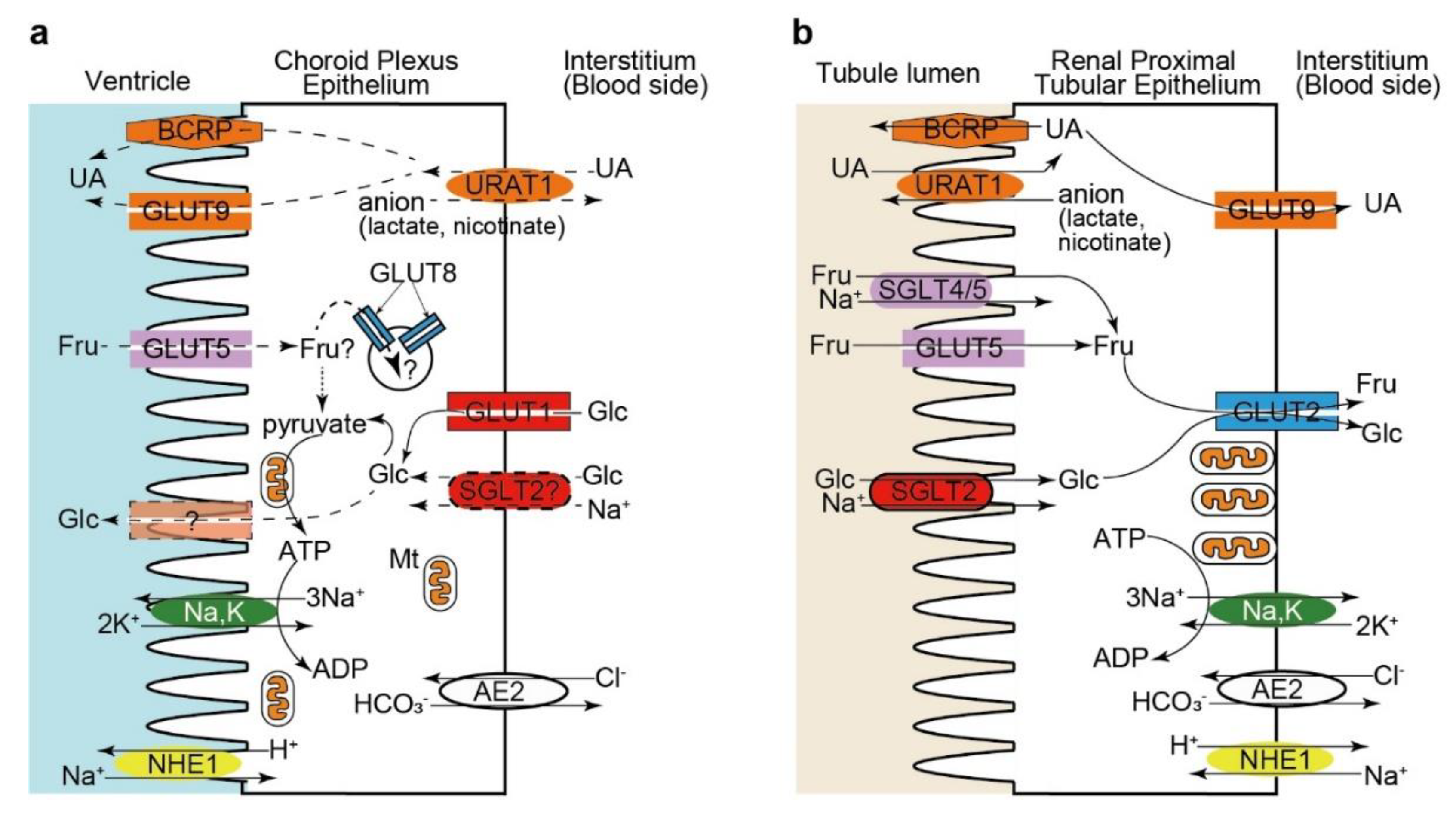
5. Hexose and Urate Transporters in the Choroid Plexus Epithelium: Comparison with the Renal Proximal Tubular Epithelium

Due to their morphological and functional similarities, CP is impressively described as the “kidney” of the brain [10]. CP epithelial cells share morphological features with the epithelia involved in extensive transcellular transport, such as the proximal tubular epithelial cells, including the presence of brush borders or microvilli on the apical membrane, basolateral infoldings (basal labyrinth), the formation of tight junctions between neighboring cells, and the abundance in mitochondria [1,5,6,10,169,170]. They both form a leaky barrier with transepithelial electrical resistance in the intermediate range due to the expression of claudin 2, the only pore-forming claudin [1,5,6], and they are responsible for purifying the extracellular fluid (plasma for the proximal tubular epithelium and CSF for the CP epithelium), resulting in the transfer of a relatively large volume of fluid. One of the prominent features of CP epithelial cells is their apparent inverse polarization: some of the “basolateral transporters”, such as Na+,K+-ATPase and NHE1, are expressed in the apical membrane of CP epithelial cells (Figure 5) [10,13]. However, not all “basolateral transporters” show inverted localization, which is the case of AE2, KCC3, and NBCn1, which occupy the basolateral membrane domain in both the CP and proximal tubular epithelium (Figure 5) [13]. A recent study investigating the subcellular distribution of an array of proteins known to be apical and basolateral determinants further revealed that CP epithelial cells are normally polarized according with the localization of the major polarity protein complexes and the membrane phospholipids phosphatidylinositol 4,5-bisphosphate and phosphatidylinositol 3,4,5-triphosphate [5,171]. However, several deviations from other polarized epithelia, including (1) the expression of P- and N-cadherins instead of E-cadherin, (2) the apical localization of Lgl2, one of the proteins that establish and maintain the basolateral membrane domain, (3) the lack of AP-1B expression, which is necessary for basolateral membrane recycling, and (4) the apical localization of syntaxin-4, a SNARE protein providing specificity to the fusion of transport vesicles to the basolateral membrane, were identified in the CP epithelial cells [5,171]. Further studies are warranted to clarify which molecular deviations are determinant for the atypical submembrane localization of certain transporters in CP epithelial cells.
The expression pattern of hexose and urate transporters in CP epithelial cells resembles that in proximal tubular epithelial cells, although some transporters have different submembrane localizations. Our immunohistochemical study showed that GLUT9 and URAT1 exhibited an inverted localization between CP epithelial cells and proximal tubular epithelial cells (Figure 5) [142]. SGLT2 may also be present in the basolateral membrane in CP epithelial cells (Figure 2), being another example of inverted polarity of the CP epithelial cells (Figure 5). In contrast, GLUT5 [102] and BCRP [172] were localized on the apical side in both cell types (Figure 5). These partial deviations in the submembrane localization of urate and glucose transporters in CP epithelial cells may originate from the expression differences of cell polarity-determinant proteins, as described above [5,171].

6. Conclusions and Future Perspectives

In summary, CP epithelial cells express (1) GLUT1 and SGLT2 as glucose transporters, mainly on the basolateral membrane, (2) fructose transporter GLUT5 on the apical membrane, (3) fructose and glucose transporter GLUT8 in the cytoplasmic membrane compartment, and (4) urate transporters GLUT9 and BCRP/ABCG2 on the apical membrane and URAT1 on the basolateral membrane (Figure 5). The transporters and ion channels expressed in CP epithelial cells that are involved in the secretion of CSF have been extensively studied [5,10,13,23]. Recent knowledge on the advances on glucose, fructose, and urate transporters expressed in CP epithelial cells highlight their potential relevance for the physiology and pathophysiology of the CNS. Despite of some differences in submembrane localization, the expression of common transporters for hexose and urate in CP epithelial cells and renal proximal tubular epithelial cells provides additional evidence for the similarity of these epithelia. However, the physiological and pathophysiological aspects of these transporters expressed in CP epithelial cells are still largely unknown and await further investigation. How and to which direction CP epithelial cells transport glucose, fructose, and urate across the BCFSB through these transporters should be clarified in future studies. Although it is difficult to investigate the transporter function in CP epithelial cells in humans, the analyses of the CSF composition and glucose metabolism in subjects who have mutations in these transporter genes or those who are treated with specific inhibitors (for example, those having T2DM and treated with SGLT2 inhibitors) may clarify some functional roles of these transporters. The functional decline in these transporters in CP epithelial cells could impair the homeostasis of the brain microenvironment and affect the periventricular structures, including the hippocampus, thereby contributing to the pathogenesis of cognitive impairment and dementia. Although CP may only have an indirect influence on the pathogenesis of these conditions, a better understanding of how the functional impairment of the CP epithelium can modulate pathological processes in the brain could reveal new potential therapeutic targets in the CP-CSF system for treating and preventing dementia and other age-associated neurodegenerative disorders [13,74,75,76,78].

Author Contributions

Conceptualization, Y.C. and M.U.; investigation, Y.C., R.M., K.M., K.W., W.N., N.U., and K.Y.; writing—original draft preparation, Y.C. and M.U.; writing—review and editing, Y.C., M.K., and M.U.; supervision, M.K. and M.U.; and funding acquisition, M.U., Y.C., R.M., and N.U. All authors have read and agreed to the published version of the manuscript.

Funding

This research was funded by JSPS KAKENHI grant numbers 17K08788 (Masaki Ueno), 17K15896 (Naoya Uemura), 19K07508 (Yoichi Chiba), and 20K16193 (Ryuta Murakami) and the research grant from the Hyakujushi Bank Academic Culture Promotion Foundation (Yoichi Chiba).

Acknowledgments

The authors thank K. Yasutomi for her editorial assistance, M. Kawauchi for her technical assistance.

Conflicts of Interest

The authors declare no conflict of interest.

Abbreviations

ISFInterstitial fluid
CNSCentral nervous system
BBBBlood-brain barrier
BCSFBBlood-cerebrospinal fluid barrier
CPChoroid plexus
CSFCerebrospinal fluid
Amyloid β
GLUTGlucose transporter
SGLTSodium/glucose cotransporter
ADAlzheimer disease
Me-4-FDGα-methyl-4-deoxy-4-[18F]fluoro-D-glucopyranoside
T2DMType 2 diabetes mellitus
PETPositron emission tomography
PDParkinson’s disease
ALSAmyotrophic lateral sclerosis
URAT1Urate transporter 1
BCRPBreast cancer resistance protein
ABCATP-binding cassette
FDG18F-fluorodeoxyglucose

References

  1. Redzic, Z. Molecular biology of the blood-brain and the blood-cerebrospinal fluid barriers: Similarities and differences. Fluids Barriers CNS 2011, 8, 3. [Google Scholar] [CrossRef] [PubMed] [Green Version]
  2. Sweeney, M.D.; Zhao, Z.; Montagne, A.; Nelson, A.R.; Zlokovic, B.V. Blood-Brain Barrier: From Physiology to Disease and Back. Physiol. Rev. 2019, 99, 21–78. [Google Scholar] [CrossRef] [PubMed]
  3. Ueno, M.; Chiba, Y.; Murakami, R.; Matsumoto, K.; Fujihara, R.; Uemura, N.; Yanase, K.; Kamada, M. Disturbance of Intracerebral Fluid Clearance and Blood-Brain Barrier in Vascular Cognitive Impairment. Int. J. Mol. Sci. 2019, 20, 2600. [Google Scholar] [CrossRef] [Green Version]
  4. Morris, M.E.; Rodriguez-Cruz, V.; Felmlee, M.A. SLC and ABC Transporters: Expression, Localization, and Species Differences at the Blood-Brain and the Blood-Cerebrospinal Fluid Barriers. AAPS J. 2017, 19, 1317–1331. [Google Scholar] [CrossRef] [PubMed]
  5. Damkier, H.; Praetorius, J. Structure of the Mammalian Chorod Plexus. In Role of the Choroid Plexus in Health and Disease; Praetorius, J., Blazer-Yost, B., Damkier, H., Eds.; Springer: New York, NY, USA, 2020; pp. 1–33. [Google Scholar] [CrossRef]
  6. Ghersi-Egea, J.F.; Strazielle, N.; Catala, M.; Silva-Vargas, V.; Doetsch, F.; Engelhardt, B. Molecular anatomy and functions of the choroidal blood-cerebrospinal fluid barrier in health and disease. Acta Neuropathol. 2018, 135, 337–361. [Google Scholar] [CrossRef] [Green Version]
  7. Strazielle, N.; Ghersi-Egea, J.F. Physiology of blood-brain interfaces in relation to brain disposition of small compounds and macromolecules. Mol. Pharm. 2013, 10, 1473–1491. [Google Scholar] [CrossRef]
  8. Praetorius, J.; Damkier, H.H. Transport across the choroid plexus epithelium. Am. J. Physiol. Cell Physiol. 2017, 312, C673–C686. [Google Scholar] [CrossRef]
  9. Pardridge, W.M. CSF, blood-brain barrier, and brain drug delivery. Expert Opin. Drug Deliv. 2016, 13, 963–975. [Google Scholar] [CrossRef]
  10. Johanson, C.E.; Keep, R.F. Blending Established and New Perspectives on Choroid Plexus-CSF Dynamics. In Role of the Choroid Plexus in Health and Disease; Praetorius, J., Blazer-Yost, B., Damkier, H., Eds.; Springer: New York, NY, USA, 2020; pp. 35–81. [Google Scholar] [CrossRef]
  11. Matsumoto, K.; Chiba, Y.; Fujihara, R.; Kubo, H.; Sakamoto, H.; Ueno, M. Immunohistochemical analysis of transporters related to clearance of amyloid-beta peptides through blood-cerebrospinal fluid barrier in human brain. Histochem. Cell Biol. 2015, 144, 597–611. [Google Scholar] [CrossRef]
  12. Ueno, M.; Chiba, Y.; Murakami, R.; Matsumoto, K.; Kawauchi, M.; Fujihara, R. Blood-brain barrier and blood-cerebrospinal fluid barrier in normal and pathological conditions. Brain Tumor Pathol. 2016, 33, 89–96. [Google Scholar] [CrossRef]
  13. Damkier, H.H.; Brown, P.D.; Praetorius, J. Cerebrospinal fluid secretion by the choroid plexus. Physiol. Rev. 2013, 93, 1847–1892. [Google Scholar] [CrossRef] [PubMed] [Green Version]
  14. Spector, R.; Keep, R.F.; Robert Snodgrass, S.; Smith, Q.R.; Johanson, C.E. A balanced view of choroid plexus structure and function: Focus on adult humans. Exp. Neurol. 2015, 267, 78–86. [Google Scholar] [CrossRef] [PubMed] [Green Version]
  15. Takahashi, S. Metabolic compartmentalization between astroglia and neurons in physiological and pathophysiological conditions of the neurovascular unit. Neuropathology 2020, 40, 121–137. [Google Scholar] [CrossRef] [PubMed] [Green Version]
  16. Simpson, I.A.; Carruthers, A.; Vannucci, S.J. Supply and demand in cerebral energy metabolism: The role of nutrient transporters. J. Cereb. Blood Flow Metab. 2007, 27, 1766–1791. [Google Scholar] [CrossRef]
  17. Paganoni, S.; Schwarzschild, M.A. Urate as a Marker of Risk and Progression of Neurodegenerative Disease. Neurotherapeutics 2017, 14, 148–153. [Google Scholar] [CrossRef]
  18. Amaro, S.; Jimenez-Altayo, F.; Chamorro, A. Uric acid therapy for vasculoprotection in acute ischemic stroke. Brain Circ. 2019, 5, 55–61. [Google Scholar] [CrossRef]
  19. Navale, A.M.; Paranjape, A.N. Glucose transporters: Physiological and pathological roles. Biophys. Rev. 2016, 8, 5–9. [Google Scholar] [CrossRef] [Green Version]
  20. Mueckler, M.; Thorens, B. The SLC2 (GLUT) family of membrane transporters. Mol. Asp. Med. 2013, 34, 121–138. [Google Scholar] [CrossRef] [Green Version]
  21. Wright, E.M.; Loo, D.D.; Hirayama, B.A. Biology of human sodium glucose transporters. Physiol. Rev. 2011, 91, 733–794. [Google Scholar] [CrossRef] [Green Version]
  22. Farrell, C.L.; Pardridge, W.M. Blood-brain barrier glucose transporter is asymmetrically distributed on brain capillary endothelial lumenal and ablumenal membranes: An electron microscopic immunogold study. Proc. Natl. Acad. Sci. USA 1991, 88, 5779–5783. [Google Scholar] [CrossRef] [Green Version]
  23. Hladky, S.B.; Barrand, M.A. Fluid and ion transfer across the blood-brain and blood-cerebrospinal fluid barriers; a comparative account of mechanisms and roles. Fluids Barriers CNS 2016, 13, 19. [Google Scholar] [CrossRef] [PubMed] [Green Version]
  24. Uchida, Y.; Zhang, Z.; Tachikawa, M.; Terasaki, T. Quantitative targeted absolute proteomics of rat blood-cerebrospinal fluid barrier transporters: Comparison with a human specimen. J. Neurochem. 2015, 134, 1104–1115. [Google Scholar] [CrossRef] [PubMed] [Green Version]
  25. Saunders, N.R.; Dziegielewska, K.M.; Mollgard, K.; Habgood, M.D.; Wakefield, M.J.; Lindsay, H.; Stratzielle, N.; Ghersi-Egea, J.F.; Liddelow, S.A. Influx mechanisms in the embryonic and adult rat choroid plexus: A transcriptome study. Front. Neurosci. 2015, 9, 123. [Google Scholar] [CrossRef] [PubMed] [Green Version]
  26. Harik, S.I.; Kalaria, R.N.; Andersson, L.; Lundahl, P.; Perry, G. Immunocytochemical localization of the erythroid glucose transporter: Abundance in tissues with barrier functions. J. Neurosci. 1990, 10, 3862–3872. [Google Scholar] [CrossRef] [Green Version]
  27. Cornford, E.M.; Hyman, S.; Cornford, M.E.; Damian, R.T. Glut1 glucose transporter in the primate choroid plexus endothelium. J. Neuropathol. Exp. Neurol. 1998, 57, 404–414. [Google Scholar] [CrossRef] [Green Version]
  28. Murakami, R.; Chiba, Y.; Tsuboi, K.; Matsumoto, K.; Kawauchi, M.; Fujihara, R.; Mashima, M.; Kanenishi, K.; Yamamoto, T.; Ueno, M. Immunoreactivity of glucose transporter 8 is localized in the epithelial cells of the choroid plexus and in ependymal cells. Histochem. Cell Biol. 2016, 146, 231–236. [Google Scholar] [CrossRef]
  29. Liddelow, S.A.; Dziegielewska, K.M.; Ek, C.J.; Habgood, M.D.; Bauer, H.; Bauer, H.C.; Lindsay, H.; Wakefield, M.J.; Strazielle, N.; Kratzer, I.; et al. Mechanisms that determine the internal environment of the developing brain: A transcriptomic, functional and ultrastructural approach. PLoS ONE 2013, 8, e65629. [Google Scholar] [CrossRef] [Green Version]
  30. Pujol-Gimenez, J.; Martisova, E.; Perez-Mediavilla, A.; Lostao, M.P.; Ramirez, M.J. Expression of the glucose transporter GLUT12 in Alzheimer’s disease patients. J. Alzheimers Dis. 2014, 42, 97–101. [Google Scholar] [CrossRef]
  31. Gil-Iturbe, E.; Castilla-Madrigal, R.; Barrenetxe, J.; Villaro, A.C.; Lostao, M.P. GLUT12 expression and regulation in murine small intestine and human Caco-2 cells. J. Cell Physiol. 2019, 234, 4396–4408. [Google Scholar] [CrossRef]
  32. Gil-Iturbe, E.; Arbones-Mainar, J.M.; Moreno-Aliaga, M.J.; Lostao, M.P. GLUT12 and adipose tissue: Expression, regulation and its relation with obesity in mice. Acta Physiol. 2019, 226, e13283. [Google Scholar] [CrossRef]
  33. Pujol-Gimenez, J.; Perez, A.; Reyes, A.M.; Loo, D.D.; Lostao, M.P. Functional characterization of the human facilitative glucose transporter 12 (GLUT12) by electrophysiological methods. Am. J. Physiol. Cell Physiol. 2015, 308, C1008–C1022. [Google Scholar] [CrossRef] [Green Version]
  34. Gil-Iturbe, E.; Solas, M.; Cuadrado-Tejedo, M.; Garcia-Osta, A.; Escote, X.; Ramirez, M.J.; Lostao, M.P. GLUT12 Expression in Brain of Mouse Models of Alzheimer’s Disease. Mol. Neurobiol. 2020, 57, 798–805. [Google Scholar] [CrossRef] [PubMed]
  35. Ho, H.T.; Dahlin, A.; Wang, J. Expression Profiling of Solute Carrier Gene Families at the Blood-CSF Barrier. Front. Pharmacol. 2012, 3, 154. [Google Scholar] [CrossRef] [PubMed] [Green Version]
  36. Wu, X.; Freeze, H.H. GLUT14, a duplicon of GLUT3, is specifically expressed in testis as alternative splice forms. Genomics 2002, 80, 553–557. [Google Scholar] [CrossRef] [PubMed]
  37. Vannucci, S.J. Developmental expression of GLUT1 and GLUT3 glucose transporters in rat brain. J. Neurochem. 1994, 62, 240–246. [Google Scholar] [CrossRef]
  38. Huang, S.; Czech, M.P. The GLUT4 glucose transporter. Cell Metab. 2007, 5, 237–252. [Google Scholar] [CrossRef] [PubMed] [Green Version]
  39. Ashrafi, G.; Wu, Z.; Farrell, R.J.; Ryan, T.A. GLUT4 Mobilization Supports Energetic Demands of Active Synapses. Neuron 2017, 93, 606–615. [Google Scholar] [CrossRef]
  40. McNay, E.C.; Pearson-Leary, J. GluT4: A central player in hippocampal memory and brain insulin resistance. Exp. Neurol. 2020, 323, 113076. [Google Scholar] [CrossRef]
  41. Vannucci, S.J.; Rutherford, T.; Wilkie, M.B.; Simpson, I.A.; Lauder, J.M. Prenatal expression of the GLUT4 glucose transporter in the mouse. Dev. Neurosci. 2000, 22, 274–282. [Google Scholar] [CrossRef]
  42. Szablewski, L. Distribution of glucose transporters in renal diseases. J. Biomed. Sci. 2017, 24, 64. [Google Scholar] [CrossRef] [Green Version]
  43. Wright, E.M. Glucose transport families SLC5 and SLC50. Mol. Asp. Med. 2013, 34, 183–196. [Google Scholar] [CrossRef] [PubMed]
  44. Diez-Sampedro, A.; Hirayama, B.A.; Osswald, C.; Gorboulev, V.; Baumgarten, K.; Volk, C.; Wright, E.M.; Koepsell, H. A glucose sensor hiding in a family of transporters. Proc. Natl. Acad. Sci. USA 2003, 100, 11753–11758. [Google Scholar] [CrossRef] [Green Version]
  45. Balen, D.; Ljubojevic, M.; Breljak, D.; Brzica, H.; Zlender, V.; Koepsell, H.; Sabolic, I. Revised immunolocalization of the Na+-D-glucose cotransporter SGLT1 in rat organs with an improved antibody. Am. J. Physiol. Cell Physiol. 2008, 295, C475–C489. [Google Scholar] [CrossRef] [PubMed] [Green Version]
  46. Ferrannini, E. Sodium-Glucose Co-transporters and Their Inhibition: Clinical Physiology. Cell Metab. 2017, 26, 27–38. [Google Scholar] [CrossRef] [PubMed] [Green Version]
  47. Ghezzi, C.; Loo, D.D.F.; Wright, E.M. Physiology of renal glucose handling via SGLT1, SGLT2 and GLUT2. Diabetologia 2018, 61, 2087–2097. [Google Scholar] [CrossRef] [Green Version]
  48. Sabolic, I.; Vrhovac, I.; Eror, D.B.; Gerasimova, M.; Rose, M.; Breljak, D.; Ljubojevic, M.; Brzica, H.; Sebastiani, A.; Thal, S.C.; et al. Expression of Na+-D-glucose cotransporter SGLT2 in rodents is kidney-specific and exhibits sex and species differences. Am. J. Physiol. Cell Physiol. 2012, 302, C1174–C1188. [Google Scholar] [CrossRef] [Green Version]
  49. Yu, A.S.; Hirayama, B.A.; Timbol, G.; Liu, J.; Basarah, E.; Kepe, V.; Satyamurthy, N.; Huang, S.C.; Wright, E.M.; Barrio, J.R. Functional expression of SGLTs in rat brain. Am. J. Physiol. Cell Physiol. 2010, 299, C1277–C1284. [Google Scholar] [CrossRef] [Green Version]
  50. Yu, A.S.; Hirayama, B.A.; Timbol, G.; Liu, J.; Diez-Sampedro, A.; Kepe, V.; Satyamurthy, N.; Huang, S.C.; Wright, E.M.; Barrio, J.R. Regional distribution of SGLT activity in rat brain in vivo. Am. J. Physiol. Cell Physiol. 2013, 304, C240–C247. [Google Scholar] [CrossRef]
  51. Elfeber, K.; Kohler, A.; Lutzenburg, M.; Osswald, C.; Galla, H.J.; Witte, O.W.; Koepsell, H. Localization of the Na+-D-glucose cotransporter SGLT1 in the blood-brain barrier. Histochem. Cell Biol. 2004, 121, 201–207. [Google Scholar] [CrossRef]
  52. Vemula, S.; Roder, K.E.; Yang, T.; Bhat, G.J.; Thekkumkara, T.J.; Abbruscato, T.J. A functional role for sodium-dependent glucose transport across the blood-brain barrier during oxygen glucose deprivation. J. Pharmacol. Exp. Ther. 2009, 328, 487–495. [Google Scholar] [CrossRef]
  53. Al-Ahmad, A.J. Comparative study of expression and activity of glucose transporters between stem cell-derived brain microvascular endothelial cells and hCMEC/D3 cells. Am. J. Physiol. Cell Physiol. 2017, 313, C421–C429. [Google Scholar] [CrossRef] [PubMed]
  54. Sala-Rabanal, M.; Hirayama, B.A.; Ghezzi, C.; Liu, J.; Huang, S.C.; Kepe, V.; Koepsell, H.; Yu, A.; Powell, D.R.; Thorens, B.; et al. Revisiting the physiological roles of SGLTs and GLUTs using positron emission tomography in mice. J. Physiol. 2016, 594, 4425–4438. [Google Scholar] [CrossRef] [PubMed]
  55. Chiba, Y.; Sugiyama, Y.; Nishi, N.; Nonaka, W.; Murakami, R.; Ueno, M. Sodium/glucose cotransporter 2 is expressed in choroid plexus epithelial cells and ependymal cells in human and mouse brains. Neuropathology 2020. [Google Scholar] [CrossRef] [PubMed]
  56. Liddelow, S.A.; Temple, S.; Mollgard, K.; Gehwolf, R.; Wagner, A.; Bauer, H.; Bauer, H.C.; Phoenix, T.N.; Dziegielewska, K.M.; Saunders, N.R. Molecular characterisation of transport mechanisms at the developing mouse blood-CSF interface: A transcriptome approach. PLoS ONE 2012, 7, e33554. [Google Scholar] [CrossRef]
  57. Kanai, Y.; Lee, W.S.; You, G.; Brown, D.; Hediger, M.A. The human kidney low affinity Na+/glucose cotransporter SGLT2. Delineation of the major renal reabsorptive mechanism for D-glucose. J. Clin. Investig. 1994, 93, 397–404. [Google Scholar] [CrossRef]
  58. Vallon, V.; Platt, K.A.; Cunard, R.; Schroth, J.; Whaley, J.; Thomson, S.C.; Koepsell, H.; Rieg, T. SGLT2 mediates glucose reabsorption in the early proximal tubule. J. Am. Soc. Nephrol. 2011, 22, 104–112. [Google Scholar] [CrossRef] [Green Version]
  59. Vrhovac, I.; Balen Eror, D.; Klessen, D.; Burger, C.; Breljak, D.; Kraus, O.; Radovic, N.; Jadrijevic, S.; Aleksic, I.; Walles, T.; et al. Localizations of Na(+)-D-glucose cotransporters SGLT1 and SGLT2 in human kidney and of SGLT1 in human small intestine, liver, lung, and heart. Pflugers Arch. 2015, 467, 1881–1898. [Google Scholar] [CrossRef]
  60. American Diabetes Association. 9. Pharmacologic Approaches to Glycemic Treatment: Standards of Medical Care in Diabetes—2019. Diabetes Care 2019, 42, S90–S102. [Google Scholar] [CrossRef] [Green Version]
  61. Dandona, P.; Chaudhuri, A. Sodium-glucose co-transporter 2 inhibitors for type 2 diabetes mellitus: An overview for the primary care physician. Int. J. Clin. Pract. 2017, 71, e12937. [Google Scholar] [CrossRef] [Green Version]
  62. Bonner, C.; Kerr-Conte, J.; Gmyr, V.; Queniat, G.; Moerman, E.; Thevenet, J.; Beaucamps, C.; Delalleau, N.; Popescu, I.; Malaisse, W.J.; et al. Inhibition of the glucose transporter SGLT2 with dapagliflozin in pancreatic alpha cells triggers glucagon secretion. Nat. Med. 2015, 21, 512–517. [Google Scholar] [CrossRef]
  63. Enerson, B.E.; Drewes, L.R. The rat blood-brain barrier transcriptome. J. Cereb. Blood Flow Metab. 2006, 26, 959–973. [Google Scholar] [CrossRef] [PubMed] [Green Version]
  64. Wright, E.M.; Loo, D.D.; Hirayama, B.A.; Turk, E. Surprising versatility of Na+-glucose cotransporters: SLC5. Physiology 2004, 19, 370–376. [Google Scholar] [CrossRef] [PubMed]
  65. Erdogan, M.A.; Yusuf, D.; Christy, J.; Solmaz, V.; Erdogan, A.; Taskiran, E.; Erbas, O. Highly selective SGLT2 inhibitor dapagliflozin reduces seizure activity in pentylenetetrazol-induced murine model of epilepsy. BMC Neurol. 2018, 18, 81. [Google Scholar] [CrossRef] [PubMed] [Green Version]
  66. Melo, I.S.; Santos, Y.M.O.; Costa, M.A.; Pacheco, A.L.D.; Silva, N.; Cardoso-Sousa, L.; Pereira, U.P.; Goulart, L.R.; Garcia-Cairasco, N.; Duzzioni, M.; et al. Inhibition of sodium glucose cotransporters following status epilepticus induced by intrahippocampal pilocarpine affects neurodegeneration process in hippocampus. Epilepsy Behav. 2016, 61, 258–268. [Google Scholar] [CrossRef]
  67. Oerter, S.; Forster, C.; Bohnert, M. Validation of sodium/glucose cotransporter proteins in human brain as a potential marker for temporal narrowing of the trauma formation. Int. J. Legal Med. 2019, 133, 1107–1114. [Google Scholar] [CrossRef]
  68. Kepe, V.; Scafoglio, C.; Liu, J.; Yong, W.H.; Bergsneider, M.; Huang, S.C.; Barrio, J.R.; Wright, E.M. Positron emission tomography of sodium glucose cotransport activity in high grade astrocytomas. J. Neurooncol. 2018, 138, 557–569. [Google Scholar] [CrossRef] [Green Version]
  69. Chatard, C.; Sabac, A.; Moreno-Velasquez, L.; Meiller, A.; Marinesco, S. Minimally Invasive Microelectrode Biosensors Based on Platinized Carbon Fibers for in Vivo Brain Monitoring. ACS Cent. Sci. 2018, 4, 1751–1760. [Google Scholar] [CrossRef]
  70. Deane, R.; Segal, M.B. The transport of sugars across the perfused choroid plexus of the sheep. J. Physiol. 1985, 362, 245–260. [Google Scholar] [CrossRef]
  71. Steffensen, A.B.; Zeuthen, T. Cotransport of Water in the Choroid Plexus Epithelium: From Amphibians to Mammals. In Role of the Choroid Plexus in Health and Disease; Praetorius, J., Blazer-Yost, B., Damkier, H., Eds.; Springer: New York, NY, USA, 2020; pp. 99–124. [Google Scholar] [CrossRef]
  72. Sasseville, L.J.; Cuervo, J.E.; Lapointe, J.Y.; Noskov, S.Y. The structural pathway for water permeation through sodium-glucose cotransporters. Biophys. J. 2011, 101, 1887–1895. [Google Scholar] [CrossRef] [Green Version]
  73. Balmaceda-Aguilera, C.; Cortes-Campos, C.; Cifuentes, M.; Peruzzo, B.; Mack, L.; Tapia, J.C.; Oyarce, K.; Garcia, M.A.; Nualart, F. Glucose transporter 1 and monocarboxylate transporters 1, 2, and 4 localization within the glial cells of shark blood-brain-barriers. PLoS ONE 2012, 7, e32409. [Google Scholar] [CrossRef] [Green Version]
  74. Van Cauwenberghe, C.; Gorle, N.; Vandenbroucke, R.E. Roles of the Choroid Plexus in Aging. In Role of the Choroid Plexus in Health and Disease; Praetorius, J., Blazer-Yost, B., Damkier, H., Eds.; Springer: New York, NY, USA, 2020; pp. 209–232. [Google Scholar] [CrossRef]
  75. Marques, F.; Sousa, J.C.; Brito, M.A.; Pahnke, J.; Santos, C.; Correia-Neves, M.; Palha, J.A. The choroid plexus in health and in disease: Dialogues into and out of the brain. Neurobiol. Dis. 2017, 107, 32–40. [Google Scholar] [CrossRef] [PubMed] [Green Version]
  76. Kaur, C.; Rathnasamy, G.; Ling, E.A. The Choroid Plexus in Healthy and Diseased Brain. J. Neuropathol. Exp. Neurol. 2016, 75, 198–213. [Google Scholar] [CrossRef] [PubMed]
  77. Serot, J.M.; Bene, M.C.; Faure, G.C. Choroid plexus, aging of the brain, and Alzheimer’s disease. Front. Biosci. 2003, 8, s515–s521. [Google Scholar] [CrossRef] [PubMed] [Green Version]
  78. Balusu, S.; Brkic, M.; Libert, C.; Vandenbroucke, R.E. The choroid plexus-cerebrospinal fluid interface in Alzheimer’s disease: More than just a barrier. Neural Regen. Res. 2016, 11, 534–537. [Google Scholar] [CrossRef] [PubMed]
  79. Emerich, D.F.; Schneider, P.; Bintz, B.; Hudak, J.; Thanos, C.G. Aging reduces the neuroprotective capacity, VEGF secretion, and metabolic activity of rat choroid plexus epithelial cells. Cell Transplant. 2007, 16, 697–705. [Google Scholar] [CrossRef]
  80. Cottrell, D.A.; Blakely, E.L.; Johnson, M.A.; Ince, P.G.; Borthwick, G.M.; Turnbull, D.M. Cytochrome c oxidase deficient cells accumulate in the hippocampus and choroid plexus with age. Neurobiol. Aging 2001, 22, 265–272. [Google Scholar] [CrossRef]
  81. Gorle, N.; Van Cauwenberghe, C.; Libert, C.; Vandenbroucke, R.E. The effect of aging on brain barriers and the consequences for Alzheimer’s disease development. Mamm. Genome 2016, 27, 407–420. [Google Scholar] [CrossRef]
  82. Emerich, D.F.; Skinner, S.J.; Borlongan, C.V.; Vasconcellos, A.V.; Thanos, C.G. The choroid plexus in the rise, fall and repair of the brain. Bioessays 2005, 27, 262–274. [Google Scholar] [CrossRef]
  83. Gonzalez-Marrero, I.; Gimenez-Llort, L.; Johanson, C.E.; Carmona-Calero, E.M.; Castaneyra-Ruiz, L.; Brito-Armas, J.M.; Castaneyra-Perdomo, A.; Castro-Fuentes, R. Choroid plexus dysfunction impairs beta-amyloid clearance in a triple transgenic mouse model of Alzheimer’s disease. Front. Cell Neurosci. 2015, 9, 17. [Google Scholar] [CrossRef] [Green Version]
  84. Brkic, M.; Balusu, S.; Van Wonterghem, E.; Gorle, N.; Benilova, I.; Kremer, A.; Van Hove, I.; Moons, L.; De Strooper, B.; Kanazir, S.; et al. Amyloid beta Oligomers Disrupt Blood-CSF Barrier Integrity by Activating Matrix Metalloproteinases. J. Neurosci. 2015, 35, 12766–12778. [Google Scholar] [CrossRef] [Green Version]
  85. Daouk, J.; Bouzerar, R.; Chaarani, B.; Zmudka, J.; Meyer, M.E.; Baledent, O. Use of dynamic (18)F-fluorodeoxyglucose positron emission tomography to investigate choroid plexus function in Alzheimer’s disease. Exp. Gerontol. 2016, 77, 62–68. [Google Scholar] [CrossRef] [PubMed]
  86. Unno, K.; Yamamoto, H.; Toda, M.; Hagiwara, S.; Iguchi, K.; Hoshino, M.; Takabayashi, F.; Hasegawa-Ishii, S.; Shimada, A.; Hosokawa, M.; et al. Novel frame-shift mutation in Slc5a2 encoding SGLT2 in a strain of senescence-accelerated mouse SAMP10. Biochem. Biophys. Res. Commun. 2014, 454, 89–94. [Google Scholar] [CrossRef] [PubMed]
  87. Unno, K.; Takagi, Y.; Konishi, T.; Suzuki, M.; Miyake, A.; Kurotaki, T.; Hase, T.; Meguro, S.; Shimada, A.; Hasegawa-Ishii, S.; et al. Mutation in Sodium-Glucose Cotransporter 2 Results in Down-Regulation of Amyloid Beta (A4) Precursor-Like Protein 1 in Young Age, Which May Lead to Poor Memory Retention in Old Age. Int. J. Mol. Sci. 2020, 21, 5579. [Google Scholar] [CrossRef]
  88. Myung, J.; Schmal, C.; Hong, S.; Tsukizawa, Y.; Rose, P.; Zhang, Y.; Holtzman, M.J.; De Schutter, E.; Herzel, H.; Bordyugov, G.; et al. The choroid plexus is an important circadian clock component. Nat. Commun. 2018, 9, 1062. [Google Scholar] [CrossRef] [PubMed]
  89. Stephan, B.C.; Wells, J.C.; Brayne, C.; Albanese, E.; Siervo, M. Increased fructose intake as a risk factor for dementia. J. Gerontol. Ser. A Biol. Sci. Med. Sci. 2010, 65, 809–814. [Google Scholar] [CrossRef] [PubMed] [Green Version]
  90. Lowette, K.; Roosen, L.; Tack, J.; Vanden Berghe, P. Effects of high-fructose diets on central appetite signaling and cognitive function. Front. Nutr. 2015, 2, 5. [Google Scholar] [CrossRef] [Green Version]
  91. Dills, W.L., Jr. Protein fructosylation: Fructose and the Maillard reaction. Am. J. Clin. Nutr. 1993, 58, 779S–787S. [Google Scholar] [CrossRef]
  92. Semchyshyn, H.M.; Miedzobrodzki, J.; Bayliak, M.M.; Lozinska, L.M.; Homza, B.V. Fructose compared with glucose is more a potent glycoxidation agent in vitro, but not under carbohydrate-induced stress in vivo: Potential role of antioxidant and antiglycation enzymes. Carbohydr. Res. 2014, 384, 61–69. [Google Scholar] [CrossRef]
  93. Lakhan, S.E.; Kirchgessner, A. The emerging role of dietary fructose in obesity and cognitive decline. Nutr. J. 2013, 12, 114. [Google Scholar] [CrossRef] [Green Version]
  94. Basaranoglu, M.; Basaranoglu, G.; Bugianesi, E. Carbohydrate intake and nonalcoholic fatty liver disease: Fructose as a weapon of mass destruction. Hepatobiliary Surg. Nutr. 2015, 4, 109–116. [Google Scholar] [CrossRef]
  95. Jimenez-Maldonado, A.; Ying, Z.; Byun, H.R.; Gomez-Pinilla, F. Short-term fructose ingestion affects the brain independently from establishment of metabolic syndrome. Biochim. Biophys. Acta Mol. Basis Dis. 2018, 1864, 24–33. [Google Scholar] [CrossRef] [PubMed]
  96. van der Borght, K.; Kohnke, R.; Goransson, N.; Deierborg, T.; Brundin, P.; Erlanson-Albertsson, C.; Lindqvist, A. Reduced neurogenesis in the rat hippocampus following high fructose consumption. Regul. Pept. 2011, 167, 26–30. [Google Scholar] [CrossRef] [PubMed]
  97. Cisternas, P.; Salazar, P.; Serrano, F.G.; Montecinos-Oliva, C.; Arredondo, S.B.; Varela-Nallar, L.; Barja, S.; Vio, C.P.; Gomez-Pinilla, F.; Inestrosa, N.C. Fructose consumption reduces hippocampal synaptic plasticity underlying cognitive performance. Biochim. Biophys. Acta 2015, 1852, 2379–2390. [Google Scholar] [CrossRef] [Green Version]
  98. Agrawal, R.; Noble, E.; Vergnes, L.; Ying, Z.; Reue, K.; Gomez-Pinilla, F. Dietary fructose aggravates the pathobiology of traumatic brain injury by influencing energy homeostasis and plasticity. J. Cereb. Blood Flow Metab. 2016, 36, 941–953. [Google Scholar] [CrossRef]
  99. Francis, H.; Stevenson, R. The longer-term impacts of Western diet on human cognition and the brain. Appetite 2013, 63, 119–128. [Google Scholar] [CrossRef]
  100. Ye, X.; Gao, X.; Scott, T.; Tucker, K.L. Habitual sugar intake and cognitive function among middle-aged and older Puerto Ricans without diabetes. Br. J. Nutr. 2011, 106, 1423–1432. [Google Scholar] [CrossRef] [Green Version]
  101. Reichelt, A.C.; Stoeckel, L.E.; Reagan, L.P.; Winstanley, C.A.; Page, K.A. Dietary influences on cognition. Physiol. Behav. 2018, 192, 118–126. [Google Scholar] [CrossRef]
  102. Douard, V.; Ferraris, R.P. Regulation of the fructose transporter GLUT5 in health and disease. Am. J. Physiol. Endocrinol. Metab. 2008, 295, E227–E237. [Google Scholar] [CrossRef] [PubMed] [Green Version]
  103. Mantych, G.J.; James, D.E.; Devaskar, S.U. Jejunal/kidney glucose transporter isoform (Glut-5) is expressed in the human blood-brain barrier. Endocrinology 1993, 132, 35–40. [Google Scholar] [CrossRef]
  104. Oldendorf, W.H. Brain uptake of radiolabeled amino acids, amines, and hexoses after arterial injection. Am. J. Physiol. 1971, 221, 1629–1639. [Google Scholar] [CrossRef] [Green Version]
  105. Kayano, T.; Burant, C.F.; Fukumoto, H.; Gould, G.W.; Fan, Y.S.; Eddy, R.L.; Byers, M.G.; Shows, T.B.; Seino, S.; Bell, G.I. Human facilitative glucose transporters. Isolation, functional characterization, and gene localization of cDNAs encoding an isoform (GLUT5) expressed in small intestine, kidney, muscle, and adipose tissue and an unusual glucose transporter pseudogene-like sequence (GLUT6). J. Biol. Chem. 1990, 265, 13276–13282. [Google Scholar] [PubMed]
  106. Horikoshi, Y.; Sasaki, A.; Taguchi, N.; Maeda, M.; Tsukagoshi, H.; Sato, K.; Yamaguchi, H. Human GLUT5 immunolabeling is useful for evaluating microglial status in neuropathological study using paraffin sections. Acta Neuropathol. 2003, 105, 157–162. [Google Scholar] [CrossRef] [PubMed]
  107. Kojo, A.; Yamada, K.; Yamamoto, T. Glucose transporter 5 (GLUT5)-like immunoreactivity is localized in subsets of neurons and glia in the rat brain. J. Chem. Neuroanat. 2016, 74, 55–70. [Google Scholar] [CrossRef] [PubMed]
  108. Oppelt, S.A.; Zhang, W.; Tolan, D.R. Specific regions of the brain are capable of fructose metabolism. Brain Res. 2017, 1657, 312–322. [Google Scholar] [CrossRef] [Green Version]
  109. Shu, H.J.; Isenberg, K.; Cormier, R.J.; Benz, A.; Zorumski, C.F. Expression of fructose sensitive glucose transporter in the brains of fructose-fed rats. Neuroscience 2006, 140, 889–895. [Google Scholar] [CrossRef]
  110. Shah, K.; Desilva, S.; Abbruscato, T. The role of glucose transporters in brain disease: Diabetes and Alzheimer’s Disease. Int. J. Mol. Sci. 2012, 13, 12629–12655. [Google Scholar] [CrossRef]
  111. Ueno, M.; Nishi, N.; Nakagawa, T.; Chiba, Y.; Tsukamoto, I.; Kusaka, T.; Miki, T.; Sakamoto, H.; Yamaguchi, F.; Tokuda, M. Immunoreactivity of glucose transporter 5 is located in epithelial cells of the choroid plexus and ependymal cells. Neuroscience 2014, 260, 149–157. [Google Scholar] [CrossRef]
  112. Mashima, M.; Chiba, Y.; Murakami, R.; Uemura, N.; Matsumoto, K.; Kawauchi, M.; Kanenishi, K.; Hata, T.; Ueno, M. Glucose transporter 8 immunoreactivity in astrocytic and microglial cells in subependymal areas of human brains. Neurosci. Lett. 2017, 636, 90–94. [Google Scholar] [CrossRef]
  113. Schmidt, S.; Joost, H.G.; Schurmann, A. GLUT8, the enigmatic intracellular hexose transporter. Am. J. Physiol. Endocrinol. Metab. 2009, 296, E614–E618. [Google Scholar] [CrossRef]
  114. Mayer, A.L.; Higgins, C.B.; Heitmeier, M.R.; Kraft, T.E.; Qian, X.; Crowley, J.R.; Hyrc, K.L.; Beatty, W.L.; Yarasheski, K.E.; Hruz, P.W.; et al. SLC2A8 (GLUT8) is a mammalian trehalose transporter required for trehalose-induced autophagy. Sci. Rep. 2016, 6, 38586. [Google Scholar] [CrossRef] [PubMed]
  115. Narita, H.; Tanji, K.; Miki, Y.; Mori, F.; Wakabayashi, K. Trehalose intake and exercise upregulate a glucose transporter, GLUT8, in the brain. Biochem. Biophys. Res. Commun. 2019, 514, 672–677. [Google Scholar] [CrossRef] [PubMed]
  116. Reagan, L.P.; Rosell, D.R.; Alves, S.E.; Hoskin, E.K.; McCall, A.L.; Charron, M.J.; McEwen, B.S. GLUT8 glucose transporter is localized to excitatory and inhibitory neurons in the rat hippocampus. Brain Res. 2002, 932, 129–134. [Google Scholar] [CrossRef]
  117. Schmidt, S.; Gawlik, V.; Holter, S.M.; Augustin, R.; Scheepers, A.; Behrens, M.; Wurst, W.; Gailus-Durner, V.; Fuchs, H.; Hrabe de Angelis, M.; et al. Deletion of glucose transporter GLUT8 in mice increases locomotor activity. Behav. Genet. 2008, 38, 396–406. [Google Scholar] [CrossRef] [PubMed] [Green Version]
  118. Membrez, M.; Hummler, E.; Beermann, F.; Haefliger, J.A.; Savioz, R.; Pedrazzini, T.; Thorens, B. GLUT8 is dispensable for embryonic development but influences hippocampal neurogenesis and heart function. Mol. Cell Biol. 2006, 26, 4268–4276. [Google Scholar] [CrossRef] [PubMed] [Green Version]
  119. Grempler, R.; Augustin, R.; Froehner, S.; Hildebrandt, T.; Simon, E.; Mark, M.; Eickelmann, P. Functional characterisation of human SGLT-5 as a novel kidney-specific sodium-dependent sugar transporter. FEBS Lett. 2012, 586, 248–253. [Google Scholar] [CrossRef] [Green Version]
  120. Gonzalez-Vicente, A.; Cabral, P.D.; Hong, N.J.; Asirwatham, J.; Saez, F.; Garvin, J.L. Fructose reabsorption by rat proximal tubules: Role of Na(+)-linked cotransporters and the effect of dietary fructose. Am. J. Physiol. Renal Physiol. 2019, 316, F473–F480. [Google Scholar] [CrossRef]
  121. Kawasaki, T.; Akanuma, H.; Yamanouchi, T. Increased fructose concentrations in blood and urine in patients with diabetes. Diabetes Care 2002, 25, 353–357. [Google Scholar] [CrossRef] [Green Version]
  122. Lutz, N.W.; Viola, A.; Malikova, I.; Confort-Gouny, S.; Audoin, B.; Ranjeva, J.P.; Pelletier, J.; Cozzone, P.J. Inflammatory multiple-sclerosis plaques generate characteristic metabolic profiles in cerebrospinal fluid. PLoS ONE 2007, 2, e595. [Google Scholar] [CrossRef] [Green Version]
  123. Hwang, J.J.; Johnson, A.; Cline, G.; Belfort-DeAguiar, R.; Snegovskikh, D.; Khokhar, B.; Han, C.S.; Sherwin, R.S. Fructose levels are markedly elevated in cerebrospinal fluid compared to plasma in pregnant women. PLoS ONE 2015, 10, e0128582. [Google Scholar] [CrossRef] [Green Version]
  124. Hwang, J.J.; Jiang, L.; Hamza, M.; Dai, F.; Belfort-DeAguiar, R.; Cline, G.; Rothman, D.L.; Mason, G.; Sherwin, R.S. The human brain produces fructose from glucose. JCI Insight 2017, 2, e90508. [Google Scholar] [CrossRef]
  125. Miyamoto, T.; Slone, J.; Song, X.; Amrein, H. A fructose receptor functions as a nutrient sensor in the Drosophila brain. Cell 2012, 151, 1113–1125. [Google Scholar] [CrossRef] [PubMed] [Green Version]
  126. Cha, S.H.; Wolfgang, M.; Tokutake, Y.; Chohnan, S.; Lane, M.D. Differential effects of central fructose and glucose on hypothalamic malonyl-CoA and food intake. Proc. Natl. Acad. Sci. USA 2008, 105, 16871–16875. [Google Scholar] [CrossRef] [Green Version]
  127. Funari, V.A.; Crandall, J.E.; Tolan, D.R. Fructose metabolism in the cerebellum. Cerebellum 2007, 6, 130–140. [Google Scholar] [CrossRef]
  128. Rouillard, A.D.; Gundersen, G.W.; Fernandez, N.F.; Wang, Z.; Monteiro, C.D.; McDermott, M.G.; Ma’ayan, A. The harmonizome: A collection of processed datasets gathered to serve and mine knowledge about genes and proteins. Database 2016, 2016, baw100. [Google Scholar] [CrossRef] [PubMed]
  129. Cortese, F.; Giordano, P.; Scicchitano, P.; Faienza, M.F.; De Pergola, G.; Calculli, G.; Meliota, G.; Ciccone, M.M. Uric acid: From a biological advantage to a potential danger. A focus on cardiovascular effects. Vascul. Pharmacol. 2019, 120, 106565. [Google Scholar] [CrossRef] [PubMed]
  130. Keenan, R.T. The biology of urate. Semin. Arthritis Rheum. 2020, 50, S2–S10. [Google Scholar] [CrossRef]
  131. El Ridi, R.; Tallima, H. Physiological functions and pathogenic potential of uric acid: A review. J. Adv. Res. 2017, 8, 487–493. [Google Scholar] [CrossRef]
  132. Bjornstad, P.; Lanaspa, M.A.; Ishimoto, T.; Kosugi, T.; Kume, S.; Jalal, D.; Maahs, D.M.; Snell-Bergeon, J.K.; Johnson, R.J.; Nakagawa, T. Fructose and uric acid in diabetic nephropathy. Diabetologia 2015, 58, 1993–2002. [Google Scholar] [CrossRef] [Green Version]
  133. Williams, B.; Mancia, G.; Spiering, W.; Agabiti Rosei, E.; Azizi, M.; Burnier, M.; Clement, D.L.; Coca, A.; de Simone, G.; Dominiczak, A.; et al. 2018 ESC/ESH Guidelines for the management of arterial hypertension. Eur. Heart J. 2018, 39, 3021–3104. [Google Scholar] [CrossRef]
  134. Maiuolo, J.; Oppedisano, F.; Gratteri, S.; Muscoli, C.; Mollace, V. Regulation of uric acid metabolism and excretion. Int. J. Cardiol. 2016, 213, 8–14. [Google Scholar] [CrossRef] [Green Version]
  135. Engel, B.; Gomm, W.; Broich, K.; Maier, W.; Weckbecker, K.; Haenisch, B. Hyperuricemia and dementia—A case-control study. BMC Neurol. 2018, 18, 131. [Google Scholar] [CrossRef] [PubMed] [Green Version]
  136. Liu, H.; Wei, X.; Yang, W.; Reynolds, G.P. Agricultural work and reduced circulating uric acid are both associated with initial hospital admission for Parkinson’s disease. J. Neural Transm. 2020, 127, 779–783. [Google Scholar] [CrossRef] [PubMed] [Green Version]
  137. Wang, L.; Hu, W.; Wang, J.; Qian, W.; Xiao, H. Low serum uric acid levels in patients with multiple sclerosis and neuromyelitis optica: An updated meta-analysis. Mult. Scler. Relat. Disord. 2016, 9, 17–22. [Google Scholar] [CrossRef] [PubMed]
  138. Bakshi, R.; Xu, Y.; Mueller, K.A.; Chen, X.; Granucci, E.; Paganoni, S.; Sadri-Vakili, G.; Schwarzschild, M.A. Urate mitigates oxidative stress and motor neuron toxicity of astrocytes derived from ALS-linked SOD1(G93A) mutant mice. Mol. Cell Neurosci. 2018, 92, 12–16. [Google Scholar] [CrossRef]
  139. Afsar, B.; Elsurer, R.; Covic, A.; Johnson, R.J.; Kanbay, M. Relationship between uric acid and subtle cognitive dysfunction in chronic kidney disease. Am. J. Nephrol. 2011, 34, 49–54. [Google Scholar] [CrossRef] [Green Version]
  140. Verhaaren, B.F.; Vernooij, M.W.; Dehghan, A.; Vrooman, H.A.; de Boer, R.; Hofman, A.; Witteman, J.C.; Niessen, W.J.; Breteler, M.M.; van der Lugt, A.; et al. The relation of uric acid to brain atrophy and cognition: The Rotterdam Scan Study. Neuroepidemiology 2013, 41, 29–34. [Google Scholar] [CrossRef]
  141. Shao, X.; Lu, W.; Gao, F.; Li, D.; Hu, J.; Li, Y.; Zuo, Z.; Jie, H.; Zhao, Y.; Cen, X. Uric Acid Induces Cognitive Dysfunction through Hippocampal Inflammation in Rodents and Humans. J. Neurosci. 2016, 36, 10990–11005. [Google Scholar] [CrossRef] [Green Version]
  142. Uemura, N.; Murakami, R.; Chiba, Y.; Yanase, K.; Fujihara, R.; Mashima, M.; Matsumoto, K.; Kawauchi, M.; Shirakami, G.; Ueno, M. Immunoreactivity of urate transporters, GLUT9 and URAT1, is located in epithelial cells of the choroid plexus of human brains. Neurosci. Lett. 2017, 659, 99–103. [Google Scholar] [CrossRef]
  143. Tomioka, N.H.; Tamura, Y.; Takada, T.; Shibata, S.; Suzuki, H.; Uchida, S.; Hosoyamada, M. Immunohistochemical and in situ hybridization study of urate transporters GLUT9/URATv1, ABCG2, and URAT1 in the murine brain. Fluids Barriers CNS 2016, 13, 22. [Google Scholar] [CrossRef] [Green Version]
  144. Tomioka, N.H.; Nakamura, M.; Doshi, M.; Deguchi, Y.; Ichida, K.; Morisaki, T.; Hosoyamada, M. Ependymal cells of the mouse brain express urate transporter 1 (URAT1). Fluids Barriers CNS 2013, 10, 31. [Google Scholar] [CrossRef] [Green Version]
  145. Vitart, V.; Rudan, I.; Hayward, C.; Gray, N.K.; Floyd, J.; Palmer, C.N.; Knott, S.A.; Kolcic, I.; Polasek, O.; Graessler, J.; et al. SLC2A9 is a newly identified urate transporter influencing serum urate concentration, urate excretion and gout. Nat. Genet. 2008, 40, 437–442. [Google Scholar] [CrossRef] [PubMed]
  146. Anzai, N.; Ichida, K.; Jutabha, P.; Kimura, T.; Babu, E.; Jin, C.J.; Srivastava, S.; Kitamura, K.; Hisatome, I.; Endou, H.; et al. Plasma urate level is directly regulated by a voltage-driven urate efflux transporter URATv1 (SLC2A9) in humans. J. Biol. Chem. 2008, 283, 26834–26838. [Google Scholar] [CrossRef] [PubMed] [Green Version]
  147. Mandal, A.K.; Mount, D.B. The molecular physiology of uric acid homeostasis. Annu. Rev. Physiol. 2015, 77, 323–345. [Google Scholar] [CrossRef] [PubMed]
  148. Döring, A.; Gieger, C.; Mehta, D.; Gohlke, H.; Prokisch, H.; Coassin, S.; Fischer, G.; Henke, K.; Klopp, N.; Kronenberg, F.; et al. SLC2A9 influences uric acid concentrations with pronounced sex-specific effects. Nat. Genet. 2008, 40, 430–436. [Google Scholar] [CrossRef]
  149. Bi, M.; Jiao, Q.; Du, X.; Jiang, H. Glut9-mediated Urate Uptake Is Responsible for Its Protective Effects on Dopaminergic Neurons in Parkinson’s Disease Models. Front. Mol. Neurosci. 2018, 11, 21. [Google Scholar] [CrossRef] [Green Version]
  150. Li, J.; Badve, S.V.; Zhou, Z.; Rodgers, A.; Day, R.; Oh, R.; Lee, M.; Perkovic, V.; de Zeeuw, D.; Mahaffey, K.W.; et al. The effects of canagliflozin on gout in type 2 diabetes: A post-hoc analysis of the CANVAS Program. Lancet Rheumatol. 2019, 1, e220–e228. [Google Scholar] [CrossRef]
  151. Bailey, C.J. Uric acid and the cardio-renal effects of SGLT2 inhibitors. Diabetes Obes. Metab. 2019, 21, 1291–1298. [Google Scholar] [CrossRef] [Green Version]
  152. Chino, Y.; Samukawa, Y.; Sakai, S.; Nakai, Y.; Yamaguchi, J.; Nakanishi, T.; Tamai, I. SGLT2 inhibitor lowers serum uric acid through alteration of uric acid transport activity in renal tubule by increased glycosuria. Biopharm. Drug Dispos. 2014, 35, 391–404. [Google Scholar] [CrossRef] [Green Version]
  153. Nigam, S.K. The SLC22 Transporter Family: A Paradigm for the Impact of Drug Transporters on Metabolic Pathways, Signaling, and Disease. Annu. Rev. Pharmacol. Toxicol. 2018, 58, 663–687. [Google Scholar] [CrossRef]
  154. Enomoto, A.; Kimura, H.; Chairoungdua, A.; Shigeta, Y.; Jutabha, P.; Cha, S.H.; Hosoyamada, M.; Takeda, M.; Sekine, T.; Igarashi, T.; et al. Molecular identification of a renal urate anion exchanger that regulates blood urate levels. Nature 2002, 417, 447–452. [Google Scholar] [CrossRef]
  155. Fagerberg, L.; Hallstrom, B.M.; Oksvold, P.; Kampf, C.; Djureinovic, D.; Odeberg, J.; Habuka, M.; Tahmasebpoor, S.; Danielsson, A.; Edlund, K.; et al. Analysis of the human tissue-specific expression by genome-wide integration of transcriptomics and antibody-based proteomics. Mol. Cell Proteom. 2014, 13, 397–406. [Google Scholar] [CrossRef] [PubMed] [Green Version]
  156. Imaoka, T.; Kusuhara, H.; Adachi-Akahane, S.; Hasegawa, M.; Morita, N.; Endou, H.; Sugiyama, Y. The renal-specific transporter mediates facilitative transport of organic anions at the brush border membrane of mouse renal tubules. J. Am. Soc. Nephrol. 2004, 15, 2012–2022. [Google Scholar] [CrossRef] [PubMed] [Green Version]
  157. Do, T.M.; Noel-Hudson, M.S.; Ribes, S.; Besengez, C.; Smirnova, M.; Cisternino, S.; Buyse, M.; Calon, F.; Chimini, G.; Chacun, H.; et al. ABCG2- and ABCG4-mediated efflux of amyloid-beta peptide 1-40 at the mouse blood-brain barrier. J. Alzheimers Dis. 2012, 30, 155–166. [Google Scholar] [CrossRef] [PubMed]
  158. Hoshi, Y.; Uchida, Y.; Tachikawa, M.; Inoue, T.; Ohtsuki, S.; Terasaki, T. Quantitative atlas of blood-brain barrier transporters, receptors, and tight junction proteins in rats and common marmoset. J. Pharm. Sci. 2013, 102, 3343–3355. [Google Scholar] [CrossRef]
  159. Tachikawa, M.; Watanabe, M.; Hori, S.; Fukaya, M.; Ohtsuki, S.; Asashima, T.; Terasaki, T. Distinct spatio-temporal expression of ABCA and ABCG transporters in the developing and adult mouse brain. J. Neurochem. 2005, 95, 294–304. [Google Scholar] [CrossRef]
  160. Cooray, H.C.; Blackmore, C.G.; Maskell, L.; Barrand, M.A. Localisation of breast cancer resistance protein in microvessel endothelium of human brain. Neuroreport 2002, 13, 2059–2063. [Google Scholar] [CrossRef]
  161. Ito, K.; Uchida, Y.; Ohtsuki, S.; Aizawa, S.; Kawakami, H.; Katsukura, Y.; Kamiie, J.; Terasaki, T. Quantitative membrane protein expression at the blood-brain barrier of adult and younger cynomolgus monkeys. J. Pharm. Sci. 2011, 100, 3939–3950. [Google Scholar] [CrossRef]
  162. Uchida, Y.; Ohtsuki, S.; Katsukura, Y.; Ikeda, C.; Suzuki, T.; Kamiie, J.; Terasaki, T. Quantitative targeted absolute proteomics of human blood-brain barrier transporters and receptors. J. Neurochem. 2011, 117, 333–345. [Google Scholar] [CrossRef]
  163. Ghersi-Egea, J.F.; Vasiljevic, A.; Blondel, S.; Strazielle, N. Neuroprotective Mechanisms at the Blood-CSF Barrier of the Developing and Adult Brain. In Role of the Choroid Plexus in Health and Disease; Praetorius, J., Blazer-Yost, B., Damkier, H., Eds.; Springer: New York, NY, USA, 2020; pp. 193–207. [Google Scholar] [CrossRef]
  164. Mollgard, K.; Dziegielewska, K.M.; Holst, C.B.; Habgood, M.D.; Saunders, N.R. Brain barriers and functional interfaces with sequential appearance of ABC efflux transporters during human development. Sci. Rep. 2017, 7, 11603. [Google Scholar] [CrossRef] [Green Version]
  165. Bowman, G.L.; Shannon, J.; Frei, B.; Kaye, J.A.; Quinn, J.F. Uric acid as a CNS antioxidant. J. Alzheimers Dis. 2010, 19, 1331–1336. [Google Scholar] [CrossRef] [Green Version]
  166. Shu, Y.; Li, H.; Zhang, L.; Wang, Y.; Long, Y.; Li, R.; Qiu, W.; Lu, Z.; Hu, X.; Peng, F. Elevated cerebrospinal fluid uric acid during relapse of neuromyelitis optica spectrum disorders. Brain Behav. 2017, 7, e00584. [Google Scholar] [CrossRef] [PubMed]
  167. Walter, G.A.; Phillis, J.W.; O’Reagan, M.H. Determination of rat cerebrospinal fluid concentrations of adenosine, inosine, hypoxanthine, xanthine and uric acid by high performance liquid chromatography. J. Pharm. Pharmacol. 1988, 40, 140–142. [Google Scholar] [CrossRef] [PubMed]
  168. De Luca, M.A.; Cauli, O.; Morelli, M.; Simola, N. Elevation of striatal urate in experimental models of Parkinson’s disease: A compensatory mechanism triggered by dopaminergic nigrostriatal degeneration? J. Neurochem. 2014, 131, 284–289. [Google Scholar] [CrossRef] [PubMed] [Green Version]
  169. Pavelka, M.; Roth, J. Basal Labyrinth. In Functional Ultrastructure; Springer: Vienna, Austria, 2010; pp. 178–179. [Google Scholar] [CrossRef]
  170. Spector, R.; Johanson, C. Micronutrient and urate transport in choroid plexus and kidney: Implications for drug therapy. Pharm. Res. 2006, 23, 2515–2524. [Google Scholar] [CrossRef]
  171. Christensen, I.B.; Mogensen, E.N.; Damkier, H.H.; Praetorius, J. Choroid plexus epithelial cells express the adhesion protein P-cadherin at cell-cell contacts and syntaxin-4 in the luminal membrane domain. Am. J. Physiol. Cell Physiol. 2018, 314, C519–C533. [Google Scholar] [CrossRef]
  172. Huls, M.; Brown, C.D.; Windass, A.S.; Sayer, R.; van den Heuvel, J.J.; Heemskerk, S.; Russel, F.G.; Masereeuw, R. The breast cancer resistance protein transporter ABCG2 is expressed in the human kidney proximal tubule apical membrane. Kidney Int. 2008, 73, 220–225. [Google Scholar] [CrossRef] [Green Version]
Figure 1. Representative microphotographs of immunohistochemical staining for (a) GLUT1 (clone SP168, ab150299, Abcam, Cambridge, UK, 1:200, antigen retrieval with citrate buffer (pH 6)) and (b) GLUT12 (bs-2540R, Bioss, Boston, MA, USA) [28] in human choroid plexus (CP). (a) GLUT1 is mainly located in the basolateral membrane of CP epithelial cells, whereas no GLUT1 is detected in capillary endothelial cells of the CP parenchyma (arrows). Erythrocytes in the capillary show high GLUT1 expression (arrowhead). (b) No apparent GLUT12 immunoreactivity is observed in CP epithelial cells. Scale bars: 50 μm.
Ijms 21 07230 g001
Figure 2. Representative microphotographs of immunohistochemical staining for (a,b) SGLT1 (ab14685, Abcam) [55], (c,d) SGLT2 (NBP1-92384, Novus Biologicals, Centennial, CO, USA) [55], and (e) SGLT2 (ab85626, Abcam, 5 μg/mL, antigen retrieval with citrate buffer (pH 6)) in autopsied human brains. Endothelial cells of brain microcapillaries express neither SGLT1 (a) nor SGLT2 (c). CP epithelial cells are devoid of SGLT1 immunoreactivity (b). In contrast, SGLT2 is expressed in CP epithelial cells, with an intracytoplasmic granular pattern (d), or along the plasma membrane of the basal side (e, arrows), depending on the antibodies used. Scale bars: 50 μm.
Ijms 21 07230 g002
Figure 3. Representative microphotographs of immunohistochemical staining for (a) GLUT5 (18905, IBL, Takasaki, Japan) [111] and (b) GLUT8 (bs-4241R, Bioss) [28] in human CP. (a) GLUT5 is mainly located in the apical side of CP epithelial cells. (b) Intracytoplasmic granular immunoreactivity for GLUT8 is observed in CP epithelial cells, as well as in ependymal cells (arrowheads) and subependymal glial cells (arrow) [112]. Scale bars: 50 μm.
Ijms 21 07230 g003
Figure 4. Representative microphotographs of immunohistochemical staining for (a) GLUT9 (ab104623, Abcam) [142], (b) URAT1 (BMP064, MBL, Nagoya, Japan) [142], and (c) BCRP/ABCG2 (clone BXP-21, ab3380, Abcam, 1:100, antigen retrieval with citrate buffer (pH 6)) in the human CP. (a) GLUT9 is mainly located in the apical side of CP epithelial cells. (b) Immunoreactivity for URAT1 is observed in the basolateral side of CP epithelial cells. (c) Along with endothelial cells in the CP parenchyma (arrowheads), BCRP/ABCG2 is expressed on the apical surface of CP epithelial cells (arrows). Scale bars: 50 μm.
Ijms 21 07230 g004
Figure 5. Polarized distribution of glucose (Glc), fructose (Fru), and urate (UA) transporters in choroid plexus (CP) epithelial cells (a) and renal proximal tubular epithelial cells (b). Putative directions of Glc, Fru, and UA transport through CP epithelial cells are indicated by dashed arrows. The transporter for Glc in the apical side of CP epithelial cells in mammals is still uncertain (indicated by a semitransparent rectangle labeled with a question mark). GLUT8 is expressed in the intracellular membranes, such as lysosomes, endoplasmic reticulum, and endosomes. As examples for the partially inverted distribution of transporters between CP epithelial cells and renal proximal tubular epithelial cells, the localization of Na+,K+-ATPase (Na and K) and NHE1 is indicated. AE2 is located in the basolateral membrane in both cells. Mt, mitochondria.
Ijms 21 07230 g005

