


Dent. J. 2024, 12(5), 138; https://doi.org/10.3390/dj12050138 - 9 May 2024
Abstract
►
Show Figures
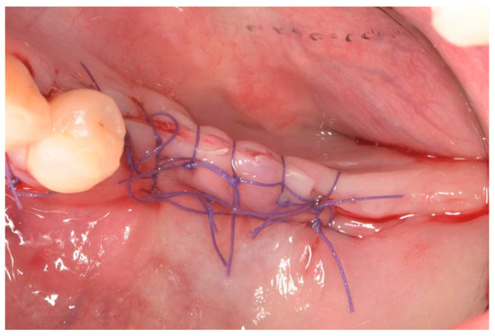
There is no current consensus on the parameters that determine the difficulty of mandibular third molar extraction in terms of the time required, which is essential to prevent complications and optimize the time of the intervention. This study aims to obtain, using the
[...] Read more.
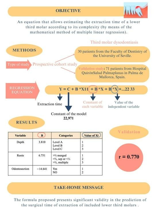
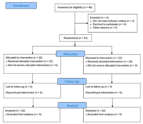
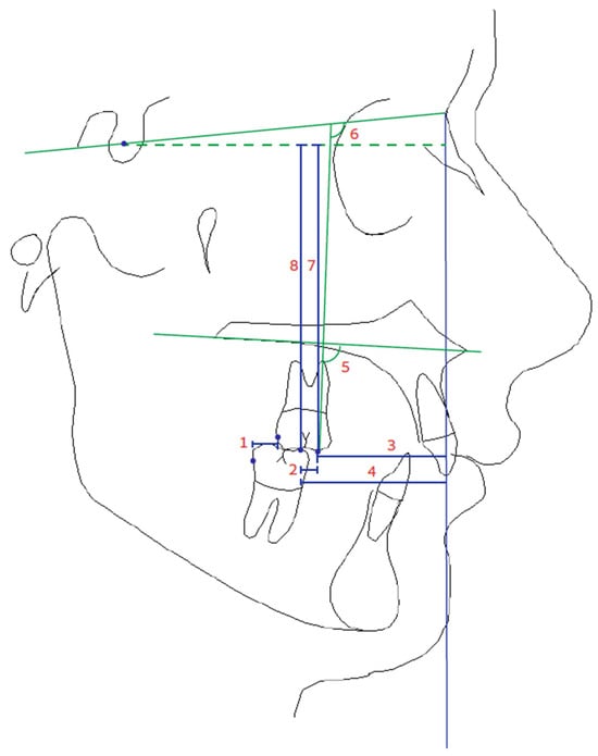
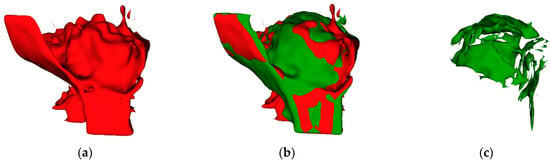
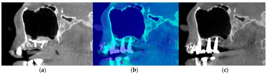
There is no current consensus on the parameters that determine the difficulty of mandibular third molar extraction in terms of the time required, which is essential to prevent complications and optimize the time of the intervention. This study aims to obtain, using the mathematical method of multiple linear regression, an equation that allows estimating the extraction time of a lower third molar according to its complexity, as well as to validate this equation in a sample of external wisdom teeth. Methods: A prospective cohort study on a sample of patients of the Master of Oral Surgery of the University of Seville in which multiple linear regression coefficients were calculated with a subsequent validation study of the results in the sample of patients operated in the Hospital Palmaplanas of Mallorca. Results: The regression line obtained after applying the statistical methodology to the cohort of patients from the University of Seville obtained significant dependent variables such as depth, roots, and odontosection. Once applied to the cohort of patients from the Palmaplanas Hospital in Mallorca, a regression coefficient was obtained between the data received and the estimated 0.770. Conclusions: The formula proposed in this article presents significant validity in the prediction of the surgical time of extraction of the lower third molars included.
Full article
Graphical abstract


