Article

A Glimpse into the Role and Effectiveness of Splenectomy for Isolated Metachronous Spleen Metastasis of Colorectal Cancer Origin: Long-Term Survivals Can Be Achieved

by
Beatrice Mihaela Tivadar
,
Traian Dumitrascu
* and
Catalin Vasilescu
Department of General Surgery, Fundeni Clinical Institute, Carol Davila University of Medicine and Pharmacy, Fundeni Street No. 258, 022328 Bucharest, Romania
*
Author to whom correspondence should be addressed.
J. Clin. Med. 2024, 13(8), 2362; https://doi.org/10.3390/jcm13082362
Submission received: 6 March 2024 / Revised: 15 April 2024 / Accepted: 15 April 2024 / Published: 18 April 2024
(This article belongs to the Special Issue Recent Advances in the Management of Colorectal Cancer)

Abstract

:
Background: Many papers exploring the role of resectioning metastases in colorectal cancer (CRC) have focused mainly on liver and lung sites, showing improved survival compared with non-resectional therapies. However, data about exceptional metastatic sites such as splenic metastases (SMs) are scarce. This paper aims to assess the role and effectiveness of splenectomy in the case of isolated metachronous SM of CRC origin. Methods: The patients’ data were extracted after a comprehensive literature search through public databases for articles reporting patients with splenectomies for isolated metachronous SM of CRC origin. Potential predictors of survival were explored, along with demographic, diagnostic, pathology, and treatment data for each patient. Results: A total of 83 patients with splenectomies for isolated metachronous SM of CRC origin were identified. The primary CRC was at an advanced stage (Duke’s C—70.3%) and on the left colon (45.5%) for most patients, while the median interval between CRC resection and SM was 24 months. The median overall survival after splenectomy was 84 months, and patients younger than 62 years presented statistically significantly worse overall survival rates than those ≥62 years old (p = 0.011). There was no significant impact on the long-term outcomes for factors including primary tumor location or adjuvant chemotherapy (p values ≥ 0.070, ns). Laparoscopic splenectomy was increasingly used in the last 20 years from 2002 (33.3% vs. 0%, p < 0.001). Conclusions: Splenectomy is the optimal treatment for patients with isolated metachronous SM of CRC, with the laparoscopic approach being increasingly used and having the potential to become a standard of care. Encouraging long-term survival rates were reported in the context of a multidisciplinary approach. Younger ages are associated with worse survival. Perioperative chemotherapy in the context of a patient diagnosed with SM of CRC origin appears to be a reasonable option, although the present study failed to show any significant impact on long-term survival.

1. Introduction

Colorectal cancer (CRC) is a common cancer worldwide, being estimated in 2024 to be the third most frequent type of cancer, both in men and women, in the United States. Although over time, the CRC death rate has decreased in both men and women, CRC is still estimated to represent the third cause of death by cancer in men and the fourth cause of death by cancer in women [1]. Due to population growth and demographic aging, the number of new CRC patients is expected to increase in many countries, while the treatment costs with CRC patients are substantial [2].
Approximately 22% of patients with CRC have metastatic disease at diagnosis, while 38% of patients have regional disease, and 32% of patients have localized disease. The stage of the disease at the time of diagnosis strongly influences the prognosis of a patient with CRC. Thus, the five-year survival of CRC patients with distant metastasis is only 14%, while for the regional and localized diseases, the five-year survival is 73% and 91%, respectively [1]. Geographical disparities regarding the survival rates were observed for patients diagnosed with CRC. Thus, better survival rates were observed in Western European countries compared with Central and South American countries, a situation explained by better screening programs and treatment options in Western Europe [2].
In the United States, in the last few years, there has been a trend toward increasing incidence of CRC in individuals younger than 50 years. The prevalence of advanced CRC is higher in younger versus older patients, a situation explained by the lack of screening in asymptomatic younger patients and higher percentages of misdiagnosis in younger symptomatic patients compared with the older ones [2,3].
Nowadays, managing a patient with CRC is multimodal, and each patient’s situation should be discussed in a multidisciplinary tumor board to maximize the patient’s chances of survival. Higher survival rates can be obtained even for advanced CRC with developments in surgery, chemotherapy, and radiotherapy according to cancer location, stage, and patient performance status.
Distant metastases in CRC can be present at the time of diagnosis (synchronous metastases) or occur at an interval after resection of the primary tumor (metachronous metastases). A few studies identified synchronous metastases at diagnosis of CRC in 17.7% to 22% of patients, while metachronous metastases occurred in 18% to 70% of patients after resection of the primary tumor [4,5,6,7,8,9].
For metastatic CRC, there is an improvement in survival for patients with a multimodal approach, where surgery (i.e., resection) plays a crucial role [6,9,10]. Furthermore, the indication for resection is extended in metastatic CRC, particularly for those patients with single-site metastasis [8]. Thus, significant improvements in survival have been observed, particularly for liver and lung metastases of CRC, with the advances in surgical methods, cancer-directed therapy, chemo-radiotherapy, and optimization of targeted therapies [1,6,11,12].
The most frequent pattern of metastatic disease in CRC is represented by the liver and/or lung (up to 91.% of patients with synchronous metastases and up to 81.5% of patients with metachronous metastases) [5,7,8,9,13]. A Dutch study including 5671 CRC patients with curative intent surgery has identified as the most common sites of metachronous metastatic disease the liver (60%), lungs (39%), extra-regional lymph nodes (22%), and peritoneum (19%), other less frequent sites being the brain and the bones [4]. Another Dutch study, including 160,278 patients diagnosed with CRC, identified synchronous metastases in 21% of the patients, with liver, lung, and peritoneum being the most frequent sites [6]. Other isolated sites of metastatic diseases in CRC are rare. Resection of metastases of CRC origin has been associated with significant improvement in survival compared with that of patients who did not undergo resection, particularly for liver, lung, and even limited peritoneal metastases [6,8,14,15].
Metastases in CRC have multiple sites in many patients for synchronous and metachronous settings [4,6,9,13]. Isolated metachronous metastases of CRC are rare, the most frequent sites being the liver (29%), the lung (10%), and the peritoneum (8%); isolated single-site metastases to other organs represent less than 5% of patients [4].
Many papers exploring the role and effectiveness of resection of metastases in CRC have focused mainly on liver and lung sites or limited peritoneal disease [8]. Only a few papers have examined the potential role of resection for rare sites of CRC metastases, such as splenic metastases (SMs) [16,17,18,19,20,21,22]. Thus, SM from CRC generally occurs in the context of advanced metastatic disease, usually after cancer has spread to sites such as the liver, lungs, peritoneum, and other sites [4,7,23]. Diagnosing isolated SM of CRC without further organ involvement is exceptional, representing less than 0.2% of resected metastases of CRC origin [18,24].
While the reason metastases of CRC rarely occur in the spleen is not fully understood, the question remains: can these patients with isolated SM of CRC benefit from surgical resection or not? Currently, the diagnosis and long-term benefit of resection for isolated SM of CRC origin are poorly addressed in case report papers and a few literature reviews, including a limited number of patients. The present study aims to assess the role and effectiveness of resection in isolated metachronous SM of CRC origin based on data provided by the current literature.

2. Patients and Methods

2.1. Patients

The patients’ data were extracted after a comprehensive literature search of the PubMed-Medline, Google Scholar, and Elsevier-Scopus databases for articles reporting data of patients with metachronous SM of CRC origin. The search strategy combined keywords such as “spleen metastasis”, “isolated spleen metastasis”, “colon cancer”, “rectal cancer”, “colorectal cancer”, and “splenectomy”. Up to January 2024, several publications were retrieved, and the search was refined, including only cases where there were no other metastatic sites at the time of diagnosis of SM and only the cases where there was a single-site metachronous SM of CRC origin, all treated by resection (i.e., splenectomy). Furthermore, the articles’ references were reviewed to identify potential other patients to be included in the analyses. No language restrictions were made. BT and TD independently screened the literature and extracted data; any disagreement was discussed and managed under CV’s supervision.
A few studies were excluded from the analysis if clear parameters of interest could not be extracted from the text or if essential results were not reported. From each study, we pulled data regarding the number, age, and gender of patients, the site of the primary tumor, the stage of the disease at the time of primary tumor resection, the interval between the primary tumor resection and the resection of SM, clinical signs and symptoms, imaging methods and carcinoembryonic antigen (CEA) serum level at the time of SM diagnosis, the number and size of metastases in the spleen, the type of resection/approach performed for the SM, data about oncological medical treatment, and lastly, the overall survival status of these patients.
All included articles described metachronous SM of CRC origin (adenocarcinoma-only patients) confirmed by pathology of the resected specimens, presenting as isolated metastases without other involvement at the time of SM resection. While a few cases were reported where a resection for another metastatic site was performed before or after the splenectomy for SM, these cases were not excluded.

2.2. Statistical Analysis
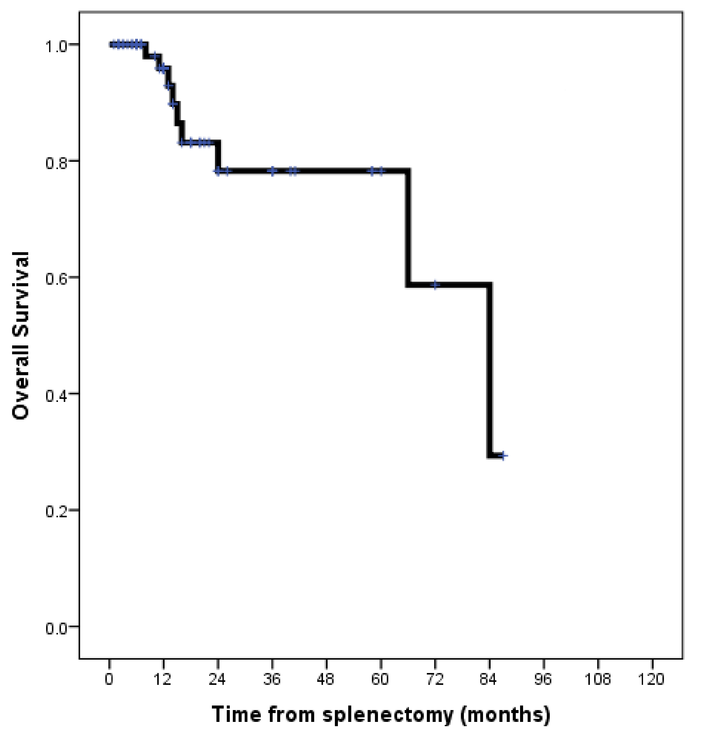
Numeric data are presented as median (range), while categorical variables are expressed as numbers (percentages). However, the survival is presented as median (range) and mean (±SD) because the median was not reached in several survival analyses due to the high number of censored patients. Fisher’s exact test (two-tailed) was used to compare the categorical variables between the groups. The survival curves were estimated using the Kaplan–Meier method and were compared using the long-rank test. The median follow-up time was assessed using the reversed Kaplan–Meier curves. A p-value < 0.05 was considered statistically significant.

3. Results

The search in the abovementioned databases identified 83 patients with splenectomies for metachronous isolated SM of CRC origin (no other sites of metastases at the time of splenectomy for SM), extracted from papers published between 1965 and 2024 (Supplementary Table S1, references [16,17,18,20,25,26,27,28,29,30,31,32,33,34,35,36,37,38,39,40,41,42,43,44,45,46,47,48,49,50,51,52,53,54,55,56,57,58,59,60,61,62,63,64,65,66,67,68,69,70,71,72,73,74,75,76,77] are cited in Supplementary Materials). The median age of patients at the time of splenectomy was 62 years (range, 22–84 years), with a slightly male gender predominance (50.6%).
The primary CRC origin was the left colon (including the sigmoid) in 38 patients (45.8%), the right colon in 24 patients (28.9%), and the rectum in 10 patients (12%). Two patients (2.4%) had the transverse colon as the primary site of the tumor, and one patient (1.2%) had a double primary tumor location (left and right colon). In eight patients (9.6%), the primary tumor location was the colon, but the exact location was not specified. Thus, overall, 73 patients (88%) had the colon as the primary tumor location.
Data about T, N, and Duke’s stages of the primary CRC were available for 44 patients (53%), 45 patients (54.2%), and 65 patients (78.3%), respectively. Thus, 28 patients (out of 44 patients—63.6%) have had T3 stages, 12 patients (27.3%) have had T4 stages, and only 4 patients (9.1%) have had a T2 stage. No patient was in a T1 stage. Loco-regional positive lymph nodes of the resected primary CRC were observed in 27 patients (out of 45 patients—60%). Regarding the Duke’s stage of the primary CRC, the most significant part of the patients were Duke C—45 patients (out of 64 patients—70.3%), followed by Duke’s B stage—13 patients (20.3%); Duke’s A and D stages were observed in three patients in each group (13.7%). Negative resection margins were obtained in all patients with available data after primary CRC resection (available data in 45 patients—54.2%).
An adjuvant chemotherapy after resection of the primary CRC was performed in 40 patients (out of the 52 patients with available data)—76.9%. The most frequently used chemotherapy regimens included 5-fluorouracil (10 patients out of 40 patients with chemotherapy—25%), FOLFOX (8 patients—20%), and capecitabine (3 patients—7.5%); in the remaining patients with adjuvant chemotherapy (18 patients—47.5%), the used regimen was not specified.
The median interval between primary CRC resection and the splenectomy for SM was 24 months (range, 3–180 months) (data available for 79 patients—95.2%).
Data about clinical signs and symptoms, imaging, and CEA serum level at the time of splenectomy for SM were available for 79 patients (95.2%), 74 patients (89.2%), and 63 patients (75.9%), respectively.
The most significant portion of patients were asymptomatic at the time of splenectomy for SM—66 patients (out of 79 patients with available data—83.5%). Only 13 patients were symptomatic (16.5%). The main clinical signs and symptoms are shown in Table 1.
The imaging methods for detecting metachronous isolated SM of CRC are shown in Table 2. Thus, a contrast-enhanced computed tomography (CT) was performed in the most significant portion of patients—64 patients (out of 74 patients—86.5%), followed by a positron emission computed tomography (PET-CT) (22 patients—29.7%).
An elevated CEA serum level was observed at the time of splenectomy for SM of CRC origin in 52 patients (out of 63 patients with available data—82.5%). The median CEA serum level in the present cohort of patients was 26.2 ng/mL (range, 2.6–363 ng/mL) (data available for 51 patients—61.4%).
The most significant portion of the patients in the present cohort underwent an open splenectomy (68 patients—81.9%), while the remaining 15 patients (18.1%) were laparoscopically approached. In the open approach group of patients, two patients (2.4%) have had associated procedures (right oophorectomy for a serous cyst in one patient and partial gastrectomy in the other patient). However, in the last 20 years (2003–2024), the rate of laparoscopic splenectomies for SM of CRC origin was significantly higher than the rate reported between 1965 and 2002 (33.3% vs. 0%, p < 0.001).
Data about the number and size of SM of CRC were available in 75 patients (90.4%) and 63 patients (75.9%), respectively. Thus, a solitary metastasis was observed in 69 patients (out of 75 patients with available data—92%), two metastases in 4 patients (5.3%), and multiple spleen metastases in 2 patients (2.7%). The median diameter of the SM was 4.5 cm (range, 1–18 cm).
Data about perioperative chemotherapy at the time of splenectomy for SM of CRC origin were available in 47 patients (56.6%). Thus, chemotherapy was performed in 22 patients (out of 47 patients with available data—46.8%). The chemotherapy regimens are shown in Table 3. Targeted therapy was used in only six patients (27.3%). It is worth mentioning that in the last 20 years (2003–2024), the rate of perioperative chemotherapy at the time of splenectomy for SM of CRC origin was significantly higher than the rate reported between 1965 and 2002 (60% vs. 8.3%, p = 0.002).
Data about the overall survival and status were available in 70 patients (84.3%). Thus, 61 patients (out of 70 patients with available data—87.1%) were alive at the time of the last follow-up (53 patients without recurrence—75.7% and 8 patients with disease recurrence—11.4%). Only nine patients (12.6%) died of the disease recurrence during the follow-up after splenectomy for SM of CRC. The estimated median overall survival time after splenectomy was 84 months (range, 1–87 months; mean 66.5 ± 5.9 months). However, the median follow-up time after splenectomy was only 12 months (range, 1–87 months; mean 22.2 ± 2.8 months).
The estimated one-year and five-year overall survivals in the present cohort were 96% and 78%, respectively (Figure 1).
Furthermore, several comparative survival analyses were performed, based on the available data in each group, to assess potential predictors of long-term survival in patients with splenectomies for metachronous isolated SM of CRC origin (Table 4). Thus, no significant impact on the long-term outcomes was observed for factors such as gender, primary tumor location (colon vs. rectum, left vs. right colon), Duke’s stage of the primary tumor, presence or absence of positive lymph nodes after primary tumor resection, presence or absence of adjuvant chemotherapy after primary tumor resection or after SM resection, interval time from primary tumor resection to splenectomy for SM, presence or absence of symptoms or signs, the value of CEA serum level, type of approach for splenectomy (laparoscopic vs. open), number and size of SM, and period of time (1965–2002 vs. 2003–2024) (p values ≥ 0.070, ns). Interestingly, patients younger than 62 presented statistically significantly worse overall survival rates than those ≥62 years old (p = 0.011), as shown in Table 4 and Figure 2.

4. Discussion

Intraparenchimatous SM from solid tumors is widely considered exceptional and usually occurs in the context of multi-visceral disseminated cancer (more than 50% of the cases) and rarely as a solitary lesion. Autopsy, ultrasonography, and splenectomy series have reported a broad spectrum of prevalence for SM in solid cancers, ranging between 0.15% and 9.8% [24,33,37,51,78,79,80,81,82]. In the context of multi-visceral metastatic disease, the most common sources of SM are melanoma (34%), breast cancer (12%), and ovarian cancer (12%), with only 10% arising from CRC [71]. Other studies identified lung cancer (18.6–30.5%), melanoma (8.4–15.8%), and breast cancers (10.2–12.3%) as the most frequent sources of SM [24,79,83]. A review published in 2001 identified 50 patients with splenectomies for SM, gynecological cancers being the most frequent source (60%), while CRC origin was observed in 11% of the patients [84]. Another study, including 93 patients with solitary SM, identified CRC and ovarian cancer as the most frequent sources [80]. Nevertheless, a few extensive studies analyzing the data of 84 to 115 patients with splenectomies for SM identified as the most frequent primary sources of SM, ovarian cancer (31.3–46.4%), melanoma (16.7–27.8%), and CRC (10.7–13.9%) [56,85].
An autopsy study published in 1974 reported a 4.4% incidence of SM from CRC origin (none as isolated SM) [86], while a few clinical studies identified an incidence of 0.2% to 0.5% for isolated SM from CRC origin [24,37,60].
Several studies reviewing the patients with splenectomies for SM of CRC origin reached conflicting results about the number of published cases [16,17,18]. In a study published in 2007, 42 patients with isolated SM of CRC origin were described in the literature [19], while another study published in 2016 identified 48 patients with SM of CRC origin [20]. Interestingly, in a study published in 2022, only 39 patients with isolated SM of CRC were retrieved from the English literature [22]. Nevertheless, a Japanese-language paper analyzed the data of 75 Japanese patients with synchronous and metachronous SM of CRC origin [21]. To the best of our knowledge, the present study is the largest to date, including only patients with splenectomies for metachronous isolated SM of CRC origin.
The presumed factors that may restrict metastasis to the spleen include the constant high blood flow through the spleen with rhythmic contraction by the sinusoidal splenic architecture (not allowing potential tumor embolus to soil), the sharp angle of the splenic artery with the celiac axis, the absence of afferent lymphatics to the spleen, the scarcity of lymphatic vessels extending into the intrasplenic parenchyma, the splenic capsule (acting as a shield that prevents intraparenchymal metastasis), and the good immune surveillance in the spleen that inhibits tumor cell proliferation [16,44,51,87]. Old experimental studies have shown that the growth rate of inoculated adenocarcinoma cells into the spleen is significantly slower compared with the liver [25]. Conversely, the propensity of CRC to spread using the lymphatic and vascular systems may explain, at least in part, the high incidence of liver, lung, and lymph node metastases [4]. A hypothesis suggested that the rarity of SM in epithelial cancers originates from cancer cells undergoing pooling within the spleen, being exposed to pro-apoptotic signals, and consequently failing to survive [88]. Nevertheless, the rarity of metastases of solid cancers into the spleen remains unexplained mainly, even though it is the most vascular organ.
Several reviews reporting patients with splenectomies for SM of CRC origin, identified the colon (88–92.3%), particularly the left colon, as the most frequent site for the primary tumor (61.5–88%), presenting vastly advanced stages of the primary tumor (Duke’s stages C in 63–67.9% of the patients), with a median interval time from primary tumor resection to SM diagnosis/resection of 18 months (range, 1–144 months), the most significant portion of patients being asymptomatic at the time of SM diagnosis (78.6%). An elevated CEA serum level was observed in 55% to 83.9% of the patients at the time of SM diagnosis, while the imaging diagnosis included CT scan in 87.1% of the patients and PET-CT in 12.9% of the patients. A metachronous pattern for SM was reported in 83.9–92.9% of the patients, the most significant portion of SM being solitary (92–99%) and treated with splenectomy (94–96.4%) [17,18,19,20,22,65,87].
In the present cohort, the colon was the most frequent site for the primary tumor (88%), with the left colon as the first source (45.8%). Advanced disease of the primary tumor (Duke’s C) was observed in 70.3% of the patients, with a median interval from primary tumor resection to splenectomy for SM of CRC origin of 24 months (range, 3–180 months). A solitary SM was observed in 92% of the patients. The characteristics of the patients in our study appear consistent with those reported in previous studies addressing the same topic [17,18,19,20,22,65,87]. Interestingly, the study of Japanese patients by Kurumiya and co-workers in 2019 identified the right colon as the leading site for primary tumors (38.7%) [21].
Except for the study of Kurumiya and co-workers [21], all other studies (including the present one) identified the left colon as the primary source for SM of CRC origin, as highlighted above. One might explain this feature by the possibility of a retrograde spread from the inferior mesenteric vein to the spleen via the splenic vein [34], particularly in portal hypertension [31]. The prevalence of the right colon as a primary source of SM of CRC origin in the Japanese study of Kurumiya and co-workers [21] appears to be at odds with the theory of Indudhara and co-workers for SM occurrence in CRC [34]. Different metastatic patterns were previously described between the colon and rectal cancers [5,7]. A previous study has shown that the prevalence of liver, lung, and bone metastases is higher in the left vs. right colon cancer [5]. Rectal cancers spread significantly more frequently to the thorax, the bone, and the nervous system than colon cancers [7].
It is widely accepted that patients with metastatic disease after curative intent therapy for CRC usually will develop the metastases within the first three years after surgery (86%), with a median time to diagnosis of first metastasis of 17 months (range, 10–29 months). The median time to diagnose liver metastases is 15 months, while for lung and peritoneum metastases, the median time to diagnosis is 22 months and 16 months, respectively [4,7]. In the present cohort, the median time from primary CRC resection to SM resection was 24 months (range, 3–180 months). Furthermore, in a study of 93 patients with solitary SM of different types of cancer origin, the median interval time from the primary tumor resection to the SM diagnosis was 28 months (range, 0–264 months); for SM of CRC origin, the median interval time from the primary tumor to the SM diagnosis was 27 months (range, 0–132 months) [80]. Thus, one might speculate that metachronous SM occurs later after CRC resection, compared with the liver, lung, or peritoneal metastases. The slow progression by the splenic function of immune surveillance might explain the long interval between resection of the primary CRC and SM occurrence [16].
Most patients in the present cohort were asymptomatic at the time of SM diagnosis (83.5%), consistent with previously reported series of SM of CRC origin [17,18,19,20,22,65,87]. In a limited number of patients reported in the literature, SM may be complicated by splenic and portal vein thrombosis [89], spleen abscess [19], or spleen rupture [36,90].
An elevated CEA serum level was present in a high proportion of patients in the present cohort (82.5%), leading to further imaging investigations to detect SM. In almost all cases of the present study, radiologic diagnosis was established by CT (performed in 86.5% of the patients), followed by PET-CT (performed in 29.7%). It is worth mentioning the case of two patients in whom SM was first diagnosed on the operative specimen of splenectomy, and further investigations led to a colon carcinoma diagnosis [90,91].
Whenever a splenic mass is detected by imaging (particularly in the context of a patient with previous malignancies, including CRC), it raises the suspicion of metastatic disease based on clinical history, CEA serum levels, and appearance on imaging studies, and it is also necessary to exclude the presence of other metastatic lesions by a PET-CT. A previous history of cancer has been identified as the single independent predictor of malignancy in a splenic lesion [85].
SM should be differentiated from other primary lesions in the spleen, particularly in the context of liberal use and development of modern imaging techniques. Of note, the most common tumors of the spleen are benign; isolated metastatic lesions to the spleen remain an exceptional appearance. Other lesions, such as hemangioma, infarction, and spleen infectious or inflammatory conditions, should also be ruled out. Thus, a new lesion in the spleen in the imaging follow-up of a patient who underwent resection for CRC may pose some challenging problems of diagnosis and treatment. An accurate imaging differentiation of benign/malignant spleen lesions is critical for the proper management, particularly in the context of a patient with previous malignancies; commonly, an SM is hypoechoic on ultrasonography, hypodense on contrast-enhanced CT in the venous phase, hypointense on T1-weighted MRI sequences images following contrast administration, and intense hypermetabolic on PET-CT [58,92,93,94,95,96]. It is worth mentioning the case of a patient diagnosed with concurrent rectosigmoid carcinoma and primary splenic malignant lymphoma mimicking an SM of CRC origin [97]. CT scans reveal the most significant part of SM. However, a PET-CT is recommended not necessarily to identify the SM but to rule out other metastatic sites. Mestner and co-workers showed a 100% sensitivity and specificity of PET-CT in diagnosing SM [94].
Although in the past, the presence of SM in solid cancers was considered an advanced disease with no indication for surgery [98], nowadays, for isolated SM, total splenectomy is the treatment of choice. This situation is associated with encouraging survival [24,87]. While an open technique in the setting of previously operated patients seems the first choice [87], other studies suggest that a laparoscopic approach can be successfully performed for isolated SM of solid cancers, with all the advantages of minimally invasive surgery [60,69,99,100,101]. The oncological safety of a minimally invasive approach for spleen malignancies (including SM) was recently demonstrated [102]. A minimally invasive splenectomy is associated with low morbidity rates and almost nil mortality rates for both benign and malignant pathologies [102,103], although a malignant pathology is a risk factor for postoperative morbidity [104].
In the present cohort, SM was predominately resected by open splenectomy (81.9%). However, in the last 20 years (2003–2024), the rate of laparoscopic splenectomies for SM of CRC origin was significantly higher than the rate reported between 1965 and 2002 (33.3% vs. 0%, p < 0.001). One might conclude that the laparoscopic approach will be increasingly used as a standard of care for the few patients diagnosed with SM of solid cancers, including CRC.
While not the particular subject of this paper, there are reports of partial splenectomy being performed for SM [105,106,107,108], and thus, this may be a theme for future research. Partial splenectomy, including the laparoscopic approach, has been demonstrated as safe in experienced surgeon hands, with low morbidity rates, being proposed as an alternative to total splenectomy for a specific pathology to overcome potential complications of asplenia; a malignant pathology is, however, a very rare indication for partial splenectomy (1.5%) [106,107,109,110]. Indeed, total splenectomy may induce several significant complications: infections (including severe ones and mortality related to infections, which is 2–3-fold greater than that of the general population), vascular complications (such as venous thromboembolism, stroke, myocardial infarction, and pulmonary hypertension), and a high risk of developing cancer [111,112]. A total splenectomy may induce immunodeficiency, including a decrease in pro-inflammatory cytokines; this change is hindered with a partial splenectomy, particularly with a minimally invasive approach [113]. A few studies have associated total splenectomy with worse survival rates in CRC [114,115], while other studies did not find any significant impact [116]. Furthermore, one experimental study has shown that the growth rate of hematogenous lung metastases decreases after splenectomy [117], while another study has associated splenectomy with increased liver metastases in CRC [118].
For the exceptional cases of synchronous SM of CRC origin, resection of both sites in the same surgical session is recommended; a sequential approach including first resection of the primary tumor and splenectomy at a later time might be a safe option for patients with high surgical risks [119].
The liver is the most common site of metastasis from CRC (up to 50% of the patients), followed by the lungs (10–15% of the patients) [120]. The median overall survival of patients resected for metastatic CRC is reported to be around 38–59 months for liver-only metastases and 45–64 months for lung-only metastases [6,11,121,122,123]. Resection for peritoneal metastases of CRC origin is associated with a median overall survival time of 47.7 months [15]. Resection for isolated lung metastasis of CRC origin is associated with five- and ten-year survival rates of 25–58% and 17–28%, respectively. Resection for isolated liver metastasis of CRC origin is associated with five- and ten-year survival rates of 25–58% and 17–30%, respectively [120,121,122,123,124,125].
A study has shown that SM is associated with a worse prognosis than other metastatic sites in solid cancers [79], a situation that appears to be at odds with the results of the present study. In the present cohort, the median overall survival after splenectomy for isolated SM of CRC origin was 84 months, with 96% one-year and 78% five-year survival rates. Similar long-term outcomes were previously reported [17,18,19,20,22,65,87]. Thus, one might speculate that splenectomies for isolated SM from CRC origin are associated with encouraging long-term survival, apparently better than those reported after resectioning other isolated metastatic sites such as the liver or lung.
The present study did not find any significant impact on the long-term outcomes for factors such as gender, primary tumor location (colon vs. rectum, left vs. right colon), Duke’s stage of the primary tumor, presence or absence of positive lymph nodes after primary tumor resection, presence or absence of adjuvant chemotherapy after primary tumor resection or after SM resection, interval time from primary tumor resection to splenectomy for SM, presence or absence of symptoms or signs, the value of CEA serum level, type of approach for splenectomy (laparoscopic vs. open), number and size of SM, and period of time (1965–2002 vs. 2003–2024) (p values ≥ 0.070, ns).
Only one study in the English literature assesses potential prognostic factors for long-term survival in patients with splenectomies for SM of CRC origin showing no impact on survival for the age, gender, primary tumor location, Duke’s stage, SM size, synchronous/metachronous pattern, or CEA serum level in this study [17]. Another study reflecting the data of 75 Japanese patients with splenectomies for synchronous and metachronous SM of CRC origin (1989–2018) did not identify any impact on the long-term survival for the primary tumor site (right vs. left colon) or synchronous vs. metachronous pattern [21].
A study of 115 patients with splenectomies for SM of different solid cancer origin did not find any influence of age at time of splenectomy, gender, and the primary cancer origin on the long-term survival [56]; however, it appears that metachronous SM has statistically significant better long-term outcomes compared with synchronous SM [24,56].
Similar results (as in the studies mentioned above) were obtained in the present study, except for age. Thus, in the present study, patients younger than 62 years presented statistically significantly worse overall survival rates after splenectomies for SM of CRC origin than those ≥62 years (p = 0.011), as shown in Table 4 and Figure 2. Younger ages were previously associated with worse survival also after hepatectomies for liver metastases of CRC origin in a study [126]. Conversely, another study associated age < 65 years with better overall survival after resectioning liver and lung metastases of CRC origin [127].
A few studies have shown that the primary tumor site (left vs. right colon) appears to influence long-term outcomes after resection of liver and lung metastases of CRC origin but reached conflicting results of which location has the worst survival [127,128,129]. Other studies did not identify any prognostic value of the primary tumor site on the long-term outcomes after resection for liver or lung metastases of CRC origin [121,130], as was the case in the present study for SM of CRC origin.
No influence of the preoperative CEA serum level on the long-term outcomes after splenectomies for SM of CRC origin was reported in the present study. This situation is at odds with previous studies showing a prognostic role for preoperative CEA serum level after resection for liver and lung metastases of CRC origin [124,131].
In two studies, the overall survival was significantly better for patients resected for isolated SM of CRC origin compared with those with associated other metastatic sites at the time of splenectomy for SM [17,21]. A study on 26,170 patients with stage IV CRC identified the distant metastasis site and the number of metastasis sites as independent prognostic survival factors, highlighting the diverse treatment strategies for patients with different metastatic patterns [9].
Kurumiya and co-workers have shown that the five-year survival rates were significantly better for patients in the interval 2003–2018 (introduction of oxaliplatin in the adjuvant chemotherapy after splenectomy for SM) vs. 1989–2002 (80.8% vs. 50.1%, p = 0.031) [21]. Our study failed to show any significant improvement in survival in the last 20 years (Table 4). This is particularly interesting in the context of the fact that in the last 20 years (2003–2024), the rate of perioperative chemotherapy at the time of splenectomy for SM of CRC origin was significantly higher than the rate reported between 1965 and 2002 (60% vs. 8.3%, p = 0.002) in the present study. However, our analyses did not associate adjuvant chemotherapy with improved survival (Table 4). Another explanation for the different survival analyses in the two studies might be related to the differences regarding the rate of perioperative chemotherapy at the time of SM resection (87.5% in the Japanese study [21] and only 46.8% in our study). Furthermore, in our study, there was considerable heterogeneity regarding the chemotherapy protocols, as shown in Table 4.
The present study has a few significant limitations: the number of analyzed patients is low because SM of CRC origin is an exceptional pathology; the heterogeneity of the literature data may impact the results of the analyses performed in the present study, particularly regarding those related to oncological medical therapy and long-term survival; the short follow-up time after splenectomy for SM of CRC represents another limitation of the study, along with the high number of censored patients. Thus, the results of the present study should be regarded with caution for clinical decision-making, considering that excellent survival rates are probably overestimated.

5. Conclusions

Our literature search shows that the spleen remains an exceptional site for metastatic disease in CRC. SM appears to develop later after primary CRC resection, compared with other metastatic sites such as the liver, lungs, and peritoneum. An accurate imaging differentiation of benign/malignant spleen lesions is critical for proper management, particularly in a patient with previous malignancies. Splenectomy is the optimal treatment for patients with isolated metachronous SM of CRC origin fit for surgery, with the laparoscopic approach being increasingly used and having the potential to become a standard of care for the few patients diagnosed with SM of solid cancers, including CRC. Encouraging long-term survival rates were reported after splenectomies for isolated metachronous SM of CRC in the context of a multidisciplinary approach. Perioperative chemotherapy in the context of a patient diagnosed with SM of CRC origin appears to be a reasonable option, although the present study failed to show any significant impact on long-term survival. Younger ages are associated with worse survival. Further studies should focus on the potential benefit of partial splenectomies and modern chemotherapy regimens for SM of solid organ cancers, including CRC.

Supplementary Materials

The following supporting information can be downloaded at: https://www.mdpi.com/article/10.3390/jcm13082362/s1, Table S1. Characteristics of 83 patients with splenectomies for metachronous isolated SM of CRC origin.

Author Contributions

Conceptualization, B.M.T. and T.D.; Methodology, B.M.T. and T.D.; Validation, B.M.T., T.D. and C.V.; Formal Analysis, B.M.T. and T.D.; Investigation, B.M.T. and T.D.; Resources, B.M.T. and T.D.; Data Curation, B.M.T. and T.D.; Writing—Original Draft Preparation, B.M.T. and T.D.; Writing—Review and Editing, B.M.T., T.D. and C.V.; Visualization, B.M.T., T.D. and C.V.; Supervision, C.V. All authors have read and agreed to the published version of the manuscript.

Funding

This research received no external funding.

Institutional Review Board Statement

Not applicable.

Informed Consent Statement

Not applicable.

Data Availability Statement

Data are provided in Supplementary Table S1.

Conflicts of Interest

The authors declare no conflicts of interest.

References

  1. Siegel, R.L.; Giaquinto, A.N.; Jemal, A. Cancer Statistics, 2024. CA Cancer J. Clin. 2024, 74, 12–49. [Google Scholar] [CrossRef] [PubMed]
  2. Klimeck, L.; Heisser, T.; Hoffmeister, M.; Brenner, H. Colorectal Cancer: A Health and Economic Problem. Best Pract. Res. Clin. Gastroenterol. 2023, 66, 101839. [Google Scholar] [CrossRef] [PubMed]
  3. Siegel, R.L.; Wagle, N.S.; Cercek, A.; Smith, R.A.; Jemal, A. Colorectal Cancer Statistics, 2023. CA Cancer J. Clin. 2023, 73, 233–254. [Google Scholar] [CrossRef] [PubMed]
  4. Van Gestel, Y.R.B.M.; De Hingh, I.H.J.T.; Van Herk-Sukel, M.P.P.; Van Erning, F.N.; Beerepoot, L.V.; Wijsman, J.H.; Slooter, G.D.; Rutten, H.J.T.; Creemers, G.-J.M.; Lemmens, V.E.P.P. Patterns of Metachronous Metastases after Curative Treatment of Colorectal Cancer. Cancer Epidemiol. 2014, 38, 448–454. [Google Scholar] [CrossRef] [PubMed]
  5. Qiu, M.; Hu, J.; Yang, D.; Cosgrove, D.P.; Xu, R. Pattern of Distant Metastases in Colorectal Cancer: A SEER Based Study. Oncotarget 2015, 6, 38658–38666. [Google Scholar] [CrossRef] [PubMed]
  6. Van Der Geest, L.G.M.; Lam-Boer, J.; Koopman, M.; Verhoef, C.; Elferink, M.A.G.; De Wilt, J.H.W. Nationwide Trends in Incidence, Treatment and Survival of Colorectal Cancer Patients with Synchronous Metastases. Clin. Exp. Metastasis 2015, 32, 457–465. [Google Scholar] [CrossRef] [PubMed]
  7. Riihimäki, M.; Hemminki, A.; Sundquist, J.; Hemminki, K. Patterns of Metastasis in Colon and Rectal Cancer. Sci. Rep. 2016, 6, 29765. [Google Scholar] [CrossRef] [PubMed]
  8. Väyrynen, V.; Wirta, E.-V.; Seppälä, T.; Sihvo, E.; Mecklin, J.-P.; Vasala, K.; Kellokumpu, I. Incidence and Management of Patients with Colorectal Cancer and Synchronous and Metachronous Colorectal Metastases: A Population-Based Study. BJS Open 2020, 4, 685–692. [Google Scholar] [CrossRef] [PubMed]
  9. Wang, J.; Li, S.; Liu, Y.; Zhang, C.; Li, H.; Lai, B. Metastatic Patterns and Survival Outcomes in Patients with Stage IV Colon Cancer: A Population-based Analysis. Cancer Med. 2020, 9, 361–373. [Google Scholar] [CrossRef] [PubMed]
  10. Morris, V.K.; Kennedy, E.B.; Baxter, N.N.; Benson, A.B.; Cercek, A.; Cho, M.; Ciombor, K.K.; Cremolini, C.; Davis, A.; Deming, D.A.; et al. Treatment of Metastatic Colorectal Cancer: ASCO Guideline. J. Clin. Oncol. 2023, 41, 678–700. [Google Scholar] [CrossRef]
  11. Oweira, H.; Mehrabi, A.; Reissfelder, C.; Abdel-Rahman, O. A Real-World, Population-Based Analysis of the Outcomes of Colorectal Cancer Patients with Isolated Synchronous Liver or Lung Metastases Treated with Metastasectomy. World J. Surg. 2020, 44, 1604–1611. [Google Scholar] [CrossRef] [PubMed]
  12. Itenberg, E.R.; Lozano, A.M. Surgical and Interventional Management of Liver Metastasis. Clin. Colon Rectal Surg. 2024, 37, 80–84. [Google Scholar] [CrossRef]
  13. Holch, J.W.; Demmer, M.; Lamersdorf, C.; Michl, M.; Schulz, C.; Von Einem, J.C.; Modest, D.P.; Heinemann, V. Pattern and Dynamics of Distant Metastases in Metastatic Colorectal Cancer. Visc. Med. 2017, 33, 70–75. [Google Scholar] [CrossRef] [PubMed]
  14. Angelsen, J.-H.; Horn, A.; Sorbye, H.; Eide, G.E.; Løes, I.M.; Viste, A. Population-Based Study on Resection Rates and Survival in Patients with Colorectal Liver Metastasis in Norway. Br. J. Surg. 2017, 104, 580–589. [Google Scholar] [CrossRef] [PubMed]
  15. Bang, K.; Kim, J.E.; Kim, T.W.; Kim, S.Y.; Lim, S.; Park, I.J.; Kim, C.W.; Yoon, Y.S.; Hong, Y.S. Clinical Outcomes of Curative Surgical Resection of Peritoneal Metastasis in Patients with Colorectal Cancer: A Long-term Follow-up Study. Cancer Med. 2023, 12, 2861–2868. [Google Scholar] [CrossRef]
  16. Kim, J.C.; Jeong, C.S.; Kim, H.C.; Yu, C.S.; Kang, G.H.; Lee, M.G. Isolated Splenic Metastasis from Colorectal Carcinoma: A Case Report. J. Korean Med. Sci. 2000, 15, 355. [Google Scholar] [CrossRef]
  17. Fujita, N.; Shirai, Y.; Shimoda, S.; Yamai, K.; Hatakeyama, K. Clinical Significance of Splenectomy for Colorectal Cancer Metastases to the Spleen. Int. J. Clin. Oncol. 2000, 5, 121–125. [Google Scholar] [CrossRef]
  18. Okuyama, T. Isolated Splenic Metastasis of Sigmoid Colon Cancer: A Case Report. Jpn. J. Clin. Oncol. 2001, 31, 341–345. [Google Scholar] [CrossRef]
  19. Pisanu, A. Synchronous Isolated Splenic Metastasis from Colon Carcinoma and Concomitant Splenic Abscess: A Case Report and Review of the Literature. World J. Gastroenterol. 2007, 13, 5516. [Google Scholar] [CrossRef]
  20. Altaf, K.; Mckernan, G.; Skaife, P.; Slawik, S. Splenic Metastasis in Colorectal Cancer. Tech. Coloproctol. 2016, 20, 795–796. [Google Scholar] [CrossRef]
  21. Kurumiya, Y.; Kobayashi, S.; Sugawara, G. A Case of Long-Term Survival after Splenectomy for Metachronous Solitary Splenic Metastasis of Cecal Carcinoma:A Study of 75 Cases of Splenic Metastasis of Colon Cancer in Japan. J. Jpn. Soc. Coloproctol. 2019, 72, 215–219. [Google Scholar] [CrossRef]
  22. Zhao, L.; Sui, M.; Li, J.; Zhang, K. Case Report of Isolated Synchronous Multiple Splenic Metastases from Rectal Cancer: A Case Report and Brief Review of the Literature. Medicine 2022, 101, e29613. [Google Scholar] [CrossRef]
  23. Inaba, S.; Tanaka, T.; Yamagishi, H.; Iguchi, K.; Kurioka, H.; Oka, T. A Case of Colon Cancer Metastasizing to the Spleen. Jpn. J. Clin. Oncol. 1984, 14, 425–430. [Google Scholar] [PubMed]
  24. Sauer, J.; Sobolewski, K.; Dommisch, K. Splenic Metastases—Not a Frequent Problem, but an Underestimate Location of Metastases: Epidemiology and Course. J. Cancer Res. Clin. Oncol. 2009, 135, 667–671. [Google Scholar] [CrossRef] [PubMed]
  25. Miller, J.N.; Milton, G.W. An Experimental Comparison between Tumour Growth in the Spleen and Liver. J. Pathol. 1965, 90, 515–521. [Google Scholar] [CrossRef]
  26. Dunbar, W.H.; Beahrs, O.H.; Morlock, C.G. Solitary Splenic Metastasis Incidental to Rectal Carcinoma: Report of a Case. Mayo Clin. Proc. 1969, 44, 40–45. [Google Scholar]
  27. Waller, R.M.; Fajman, W.A. An Unusual Cause of an Isolated, Focal Splenic Defect Demonstrated By Liver-Spleen Scintigraphy. Clin. Nucl. Med. 1982, 7, 5–7. [Google Scholar] [CrossRef]
  28. Slavin, J.D.; Mathews, J.; Spencer, R.P. Splenectomy for Splenic Metastasis from Carcinoma of Colon. Clin. Nucl. Med. 1986, 11, 491–492. [Google Scholar] [CrossRef]
  29. Capizzi, P.J.; Allen, K.B.; Amerson, J.R.; Skandalakis, J.E. Isolated Splenic Metastasis From Rectal Carcinoma. South. Med. J. 1992, 85, 1003–1005. [Google Scholar] [CrossRef]
  30. Thomas, S.M.; Fitzgerald, J.B.; Pollock, R.E.; Evans, D.B. Isolated Splenic Metastases from Colon Carcinoma. Eur. J. Surg. Oncol. 1993, 19, 485–490. [Google Scholar]
  31. Pedrazzoli, P.; Catona, A.; Pavesi, L.; Gossemberg, M.; Cuna, G.R.D. Splenic Metastases in Patients with Portal Hypertension. Eur. J. Cancer 1995, 31, 1885–1886. [Google Scholar] [CrossRef]
  32. Mainprize, K.S.; Berry, A.R. Solitary Splenic Metastasis from Colorectal Carcinoma. Br. J. Surg. 1997, 84, 70. [Google Scholar]
  33. Ishida, H.; Konno, K.; Ishida, J.; Shirayama, K.; Naganuma, H.; Komatsuda, T.; Hamashima, Y.; Masamune, O. Isolated Splenic Metastases. J. Ultrasound Med. 1997, 16, 743–749. [Google Scholar] [CrossRef]
  34. Indudhara, R.; Vogt, D.; Levin, H.S.; Church, J. Isolated Splenic Metastases From Colon Cancer. South. Med. J. 1997, 90, 633–636. [Google Scholar] [CrossRef]
  35. Weathers, K.B.; Modesto, V.L.; Gordon, D. Isolated Splenic Metastasis from Colorectal Carcinoma: Report of a Case and Review of the Literature. Dis. Colon Rectum 1999, 42, 1345–1348. [Google Scholar] [CrossRef]
  36. Achuthan, R.; Joseph, A.; Haray, P.N. Splenic Metastasis from a Rectal Tumour: An Unusual Presentation. Ann. R. Coll. Surg. Engl. 1999, 81, 139. [Google Scholar]
  37. Lee, S.S.; Morgenstern, L.; Phillips, E.H.; Hiatt, J.R.; Margulies, D.R. Splenectomy for Splenic Metastases: A Changing Clinical Spectrum. Am. Surg. 2000, 66, 837–840. [Google Scholar]
  38. Place, R.J. Isolated Colon Cancer Metastasis to the Spleen. Am. Surg. 2001, 67, 454–457. [Google Scholar]
  39. Genna, M.; Leopardi, F.; Valloncini, E.; Molfetta, M.; De Manzoni, G.; Castelli, A. Metachronus splenic metastasis of colon cancer. A case report. Minerva Chir. 2003, 58, 811–814. [Google Scholar]
  40. Hashemzadeh, S.; Safari, M. Solitary Splenic Metastasis of Colon Cancer: A Case Report. Acta Med. Iran. 2004, 42, 467–470. [Google Scholar]
  41. Pizzirusso, F.; Gillet, J.-P.; Fobe, D. Isolated Spleen Metastatic Involvement From a Colorectal Adenocarcinoma Complicated With a Gastrosplenic Fistula: A Case Report and Literature Review. Acta Chir. Belg. 2004, 104, 214–216. [Google Scholar] [CrossRef]
  42. Cavallaro, A.; Modugno, P.; Specchia, M.; Pontenza, A.E.; Loschiavo, V.; Colli, R.; Lauriola, L.; Barone, C. Isolated Splenic Metastasis from Colon Cancer. J. Exp. Clin. Cancer Res. 2004, 23, 143–146. [Google Scholar] [PubMed]
  43. Lobato, L.; Pérez-Lara, J.; Moreno, F.J.; Oliva, H. Metástasis Esplénica Metacrónica de Cáncer de Colon: Presentación de Un Caso Infrecuente. Rev. Española Enfermedades Dig. 2006, 98, 629–631. [Google Scholar] [CrossRef]
  44. Avninder, S.; Bhatnagar, A.; Agrawal, U.; Saxena, S. Isolated Splenic Metastasis from Colorectal Mucinous Carcinoma. Int. J. Gastrointest. Cancer 2006, 37, 98–101. [Google Scholar] [CrossRef] [PubMed]
  45. Gencosmanoglu, R.; Aker, F.; Kir, G.; Tozun, N. Isolated Metachronous Splenic Metastasis from Synchronous Colon Cancer. World J. Surg. Oncol. 2006, 4, 42. [Google Scholar] [CrossRef] [PubMed]
  46. Popovic, M.; Barisic, G.; Krivokapic, Z. Isolated Splenic Metastases of Colorectal Carcinoma—Case Report and Review of Literature. Acta Chir. Iugosl. 2008, 55, 73–76. [Google Scholar] [CrossRef]
  47. Bigot, P.; Goodman, C.; Hamy, A.; Teyssedou, C.; Arnaud, J.P. Isolated Splenic Metastasis from Colorectal Cancer: Report of a Case. J. Gastrointest. Surg. 2008, 12, 981–982. [Google Scholar] [CrossRef] [PubMed]
  48. Gasent Blesa, J.M.; De La Morena, E.; Canales, J.B.L.; Martínez, D.V.; Vázquez, C. Clinical Case Report and Literature Review: Metachronous Colorectal Splenic Metastases. Clin. Transl. Oncol. 2008, 10, 445–447. [Google Scholar] [CrossRef] [PubMed]
  49. Montemurro, S.; Maselli, E.; Ruggieri, E.; Caliandro, C.; Rucci, A.; Zito, A.F.; Sciscio, V. Isolated Splenic Metastasis from Colon Cancer. Report of a Case. Tumori 2008, 94, 422–425. [Google Scholar] [CrossRef]
  50. Sileri, P.; D’Ugo, S.; Benavoli, D.; Stolfi, V.M.; Palmieri, G.; Mele, A.; Gaspari, A.L. Metachronous Splenic Metastasis from Colonic Carcinoma Five Years After Surgery: A Case Report and Literature Review. South. Med. J. 2009, 102, 733–735. [Google Scholar] [CrossRef]
  51. Busić, Z.; Cupurdija, K.; Kolovrat, M.; Cavka, V.; Cavka, M.; Patrlj, L.; Servis, D.; Kvesić, A. Isolated Splenic Metastasis from Colon Cancer—Case Report and Literature Review. Coll. Antropol. 2010, 34 (Suppl. S1), 287–290. [Google Scholar] [PubMed]
  52. Genc, V.; Akbari, M.; Karaca, A.S.; Cakmak, A.; Ekinci, C.; Gurel, M. Why Is Isolated Spleen Metastasis a Rare Entity? Turk. J. Gastroenterol. 2010, 21, 452–453. [Google Scholar] [CrossRef] [PubMed]
  53. Dogan, M.; Ozal, G.; Ekinci, C.; Utkan, G.; Urun, Y.; Yalcin, B.; Icli, F. Two Cases with Atypical Metastasis in Colorectal Cancer: Splenic and Renal Metastasis. Exp. Oncol. 2010, 32, 277–279. [Google Scholar] [PubMed]
  54. El M’rabet, F.Z.; Brahmi, S.A.; Rachidi, S.; Tizniti, S.; Amaarti, A.; Ait Taleb, K.; El Mesbahi, O. Splenic metastasis from colonic adenocarcinoma—About a case and literature review. Pan Afr. Med. J. 2011, 10, 44. [Google Scholar] [PubMed]
  55. Jain, S.; Munjal, S.; Yantiss, R.K.; Sonoda, T.; Fahey, T.J.; Ruggiero, J.T.; Anand, A.; Gersten, A.; Goldsmith, S.J.; Ocean, A.J. Isolated Splenic Metastasis from Rectal Carcinoma: A Rare Occurrence. Case Rep. Oncol. 2011, 4, 499–504. [Google Scholar] [CrossRef]
  56. Gatenby, P.A.C.; Mudan, S.S.; Wotherspoon, A.C. Splenectomy for Non-Haematological Metastatic Malignant Disease. Langenbecks Arch. Surg. 2011, 396, 625–638. [Google Scholar] [CrossRef] [PubMed]
  57. Pavlović, M.; Separović, R.; Vukelić-Marković, M.; Patrlj, L.; Kolovrat, M.; Kopljar, M.; Babić, N.; Kosuta, D.; Babić, Z. Isolated Splenic Metastasis from Colorectal Carcinoma in a High-Risk Patient: A Case Report. Coll. Antropol. 2011, 35, 1307–1310. [Google Scholar] [PubMed]
  58. Chekrine, T.; Tawfiq, N.; Benissa, N.; El Attar, H.; Bouchbika, Z.; Benchakroun, N.; Jouhadi, H.; Badre, L.; Sahraoui, S.; Benider, A. Métastase splénique isolée métachrone d’un adénocarcinome colique. Méd. Nucléaire 2012, 36, 329–331. [Google Scholar] [CrossRef]
  59. Takeuchi, T.; Desaki, R.; Ohkura, Y.; Noda, N.; Yuasa, H.; Ito, F. A Case of a Metachronous Solitary Splenic Metastasis from a Cecal Cancer. Nihon Rinsho Geka Gakkai Zasshi (J. Jpn. Surg. Assoc.) 2013, 74, 1071–1074. [Google Scholar] [CrossRef]
  60. Lopez Monclova, J.; Targarona Soler, E.; Peraza Solis, Y.; Vidal Gonzalez, P.; Balague Ponz, C.; Rodriguez Luppi, C.; Trias Folch, M. Laparoscopic Approach for Isolated Splenic Metastasis: Comprehensive Literature Review and Report of 6 Cases. Surg. Laparosc. Endosc. Percutaneous Tech. 2013, 23, 21–24. [Google Scholar] [CrossRef]
  61. Toyoshima, Y.; Nakano, S.; Akabane, H.; Inagaki, M.; Yanagida, N.; Shomura, H.; Sakurai, K. A Case of Colon Cancer Developed Metachronous Solitary Splenic Metastasis. Nihon Rinsho Geka Gakkai Zasshi (J. Jpn. Surg. Assoc.) 2014, 75, 134–139. [Google Scholar] [CrossRef]
  62. Badak, B. Isolated Splenic Metastasis of Colorectal Carcinoma: A Case Report. Turk. J. Colorectal Dis. 2016, 26, 101–103. [Google Scholar] [CrossRef]
  63. Efared, B.; Mazti, A.; Atsame-Ebang, G.; Tahiri, L.; El Bouhaddouti, H.; Hammas, N.; El Fatemi, H.; Chbani, L. An Unusual Site of Metastasis: Splenic Metastastasis from a Colon Cancer. J. Surg. Case Rep. 2016, 2016, rjw175. [Google Scholar] [CrossRef] [PubMed]
  64. Tartaro, M.L.; Cardona, M.A.; Serrano, M.S.; Cantin, M.; Ottone, N.E. Solitary Splenic Metastasis from a Mucinous Adenocarcinoma of the Cecum. A Case Report. Indian. J. Surg. 2016, 78, 490–492. [Google Scholar] [CrossRef]
  65. Abdou, J. Isolated Splenic Metastasis from Colon Cancer: Case Report. World J. Gastroenterol. 2016, 22, 4610. [Google Scholar] [CrossRef] [PubMed]
  66. Lucke-Wold, B.; Bonasso, P.C.; Turner, R.; Cassim, R. Adenocarcinoma of the Cecum with Rare Splenic Metastasis. West Va. Med. J. 2017, 113, 32–34. [Google Scholar]
  67. Gilardi, L.; Vadrucci, M. Isolated Metachronous Splenic Metastasis From Colon Cancer Found by 18F-FDG PET/CT. Clin. Nucl. Med. 2017, 42, 79–80. [Google Scholar] [CrossRef] [PubMed]
  68. Rizzo, F.; Calamia, S.; Mingoia, G.; Fulfaro, F.; Grassi, N.; Cipolla, C. Isolated Metachronous Splenic Metastasis from Colon Cancer: Possible Explanations for This Rare Entity. J. Gastrointest. Cancer 2019, 50, 143–146. [Google Scholar] [CrossRef] [PubMed]
  69. Dimitrov Dimitrov, D. Laparoscopic Splenectomy for Solitary Splenic Metastasis in Patients with Previous Open Surgery—Case Series. Int. J. Surg. Case Rep. 2019, 65, 83–86. [Google Scholar] [CrossRef]
  70. Miller-Ocuin, J.; Ashburn, J.; Zhou, Y.; Water, G. Rectal Cancer Recurrence as Isolated Splenic Metastasis Treated with Laparoscopic Splenectomy. ACS Case Rev. Surg. 2021, 3, 10–13. [Google Scholar]
  71. Hu, L.; Zhu, J.-Y.; Fang, L.; Yu, X.-C.; Yan, Z.-L. Isolated Metachronous Splenic Multiple Metastases after Colon Cancer Surgery: A Case Report and Literature Review. World J. Clin. Cases 2020, 8, 3320–3328. [Google Scholar] [CrossRef] [PubMed]
  72. Ognerubov, N.A.; Antipova, T.S.; Ognerubova, M.A. Isolated Splenic Metastases from Colon Cancer: Clinical Observations. J. Mod. Oncol. 2021, 23, 162–166. [Google Scholar] [CrossRef]
  73. Totikov, V.Z.; Totikov, Z.V.; Remizov, O.V.; Epkhiev, A.A. Isolated Splenic Metastasis from Colorectal Cancer. Pirogov Russ. J. Surg. 2022, 4, 91–95. [Google Scholar] [CrossRef]
  74. Xu, J.; Cai, Y.; Chen, Q.; Huang, Y.; He, Y. Isolated Splenic Metastases from Rectal Carcinoma Five Years after Surgery: Case Report. Am. J. Surg. Clin. Case Rep. 2022, 5, e32493. [Google Scholar]
  75. Chaudhary, K.; Basukala, S.; Neupane, M.; Bhulan, B.; Malla, K.; Bhugai, N.; Shrestha, K.; Shah, K. Splenic Metastasis Secondary to Sigmoid Colon Carcinoma: A Case Report and Literature Review. Authorea 2023. [Google Scholar] [CrossRef]
  76. Ramos, C.; Santos, V.; Lopes, A.; Ferreira, C.; Viana, P.; Quirino, F.; Miranda, L. Isolated Splenic Metastasis from Colon Cancer: Case Report. Clin. Surg. 2023, 8, 3618. [Google Scholar]
  77. Hong, Y.; Li, H.; Teng, F.; Chen, Z. Isolated Splenic Metastasis from Rectal Cancer 12 Years after the Primary Surgery: A Rare Case Report. Asian J. Surg. 2024, 47, 710–711. [Google Scholar] [CrossRef] [PubMed]
  78. Lam, K.Y.; Tang, V. Metastatic Tumors to the Spleen. Arch. Pathol. Lab. Med. 2000, 124, 526–530. [Google Scholar] [CrossRef]
  79. Schön, C.A.; Görg, C.; Ramaswamy, A.; Barth, P.J. Splenic Metastases in a Large Unselected Autopsy Series. Pathol.—Res. Pract. 2006, 202, 351–356. [Google Scholar] [CrossRef]
  80. Compérat, E.; Bardier-Dupas, A.; Camparo, P.; Capron, F.; Charlotte, F. Splenic Metastases: Clinicopathologic Presentation, Differential Diagnosis, and Pathogenesis. Arch. Pathol. Lab. Med. 2007, 131, 965–969. [Google Scholar] [CrossRef]
  81. Fernández-Aceñero, M.J.; Muela, M.A.; Portela, S.C.; Vorwald, P.W. Metastasis to the Pancreas and the Spleen: An Increasing Diagnostic and Therapeutic Challenge. Clin. Pract. 2011, 1, e44. [Google Scholar] [CrossRef] [PubMed]
  82. Delaunoit, T.; Peny, M.O.; Mignon, M.; Dili, A. Splenic Metastasis from Gastrointestinal Neoplasms: A Review. Acta Gastroenterol. Belg. 2012, 75, 3–4. [Google Scholar]
  83. Görg, C.; Hoffmann, A. Milzmetastasen bei 59 Patienten mit Karzinomen: Eine klinisch sonografische Studie über einen Zeitraum von 14 Jahren. Ultraschall Med. 2007, 29, 173–178. [Google Scholar] [CrossRef]
  84. Agha-Mohammadi, S.; Calne, R.Y. Solitary Splenic Metastasis: Case Report and Review of the Literature. Am. J. Clin. Oncol. 2001, 24, 306–310. [Google Scholar] [CrossRef]
  85. Pugalenthi, A.; Bradley, C.; Gonen, M.; Do, K.G.; Strong, V.; Jarnagin, W.; Coit, D.; Kingham, T.P. Splenectomy to Treat Splenic Lesions: An Analysis of 148 Cases at a Cancer Center. J. Surg. Oncol. 2013, 108, 521–525. [Google Scholar] [CrossRef]
  86. Berge, T. Splenic Metastases. Frequencies and Patterns. Acta Pathol. Microbiol. Scand. A 1974, 82, 499–506. [Google Scholar]
  87. Abi Saad, G.S.; Hussein, M.; El-Saghir, N.S.; Termos, S.; Sharara, A.I.; Shamseddine, A. Isolated Splenic Metastasis from Colorectal Cancer. Int. J. Clin. Oncol. 2011, 16, 306–313. [Google Scholar] [CrossRef] [PubMed]
  88. Peters, A.M. Why the Spleen Is a Very Rare Site for Metastases from Epithelial Cancers. Med. Hypotheses 2012, 78, 26–28. [Google Scholar] [CrossRef]
  89. Hiraiwa, K. Isolated Splenic Vein Thrombosis Secondary to Splenic Metastasis: A Case Report. World J. Gastroenterol. 2006, 12, 6561. [Google Scholar] [CrossRef]
  90. Verger, P.; Weinfeld, C.; Henni, Z.; Henni, T.; Blais, J.; Garnier, P.P. Hémopéritoine révélant une métastase splénique isolée secondairement attribuée à un adénocarcinome du cæcum. Rev. Méd. Interne 2000, 21, 463. [Google Scholar] [CrossRef]
  91. Aijaz, M.; Hasan, M.; Alam, F. Isolated Splenic Metastasis: An Unusual Presentation of Colonic Adenocarcinoma. Arch. Clin. Gastroenterol. 2019, 5, 27–30. [Google Scholar] [CrossRef]
  92. Giovagnoni, A. Tumours of the Spleen. Cancer Imaging 2005, 5, 73–77. [Google Scholar] [CrossRef] [PubMed]
  93. Kamaya, A.; Weinstein, S.; Desser, T.S. Multiple Lesions of the Spleen: Differential Diagnosis of Cystic and Solid Lesions. Semin. Ultrasound CT MRI 2006, 27, 389–403. [Google Scholar] [CrossRef]
  94. Metser, U.; Miller, E.; Kessler, A.; Lerman, H.; Lievshitz, G.; Oren, R.; Even-Sapir, E. Solid Splenic Masses: Evaluation with 18F-FDG PET/CT. J. Nucl. Med. 2005, 46, 52–59. [Google Scholar] [PubMed]
  95. Kaza, R.K.; Azar, S.; Al-Hawary, M.M.; Francis, I.R. Review: Primary and Secondary Neoplasms of the Spleen. Cancer Imaging 2010, 10, 173–182. [Google Scholar] [CrossRef] [PubMed]
  96. Huettl, F.; Lang, H. Chirurgie bei primären Milztumoren und Metastasen der Milz. Chirurgie 2023, 94, 994–999. [Google Scholar] [CrossRef] [PubMed]
  97. Wakasugi, M.; Yasuhara, Y.; Nakahara, Y.; Matsumoto, T.; Takemoto, H.; Takachi, K.; Nishioka, K.; Yoshida, K.; Oshima, S. Primary Splenic Malignant Lymphoma Mimicking Metastasis of Rectosigmoid Cancer: A Case Report. Int. J. Surg. Case Rep. 2018, 44, 11–15. [Google Scholar] [CrossRef] [PubMed]
  98. Pieslor, P.C.; White, C.A.; Varney, R.R. Splenic Metastasis From Colon Carcinoma Imaged Using Ln-111 OncoScint. Clin. Nucl. Med. 1995, 20, 553–554. [Google Scholar] [CrossRef] [PubMed]
  99. Gallotta, V.; Nero, C.; Lodoli, C.; Chiantera, V.; Pacelli, F.; Fagotti, A.; Costantini, B.; Scambia, G. Laparoscopic Splenectomy for Secondary Cytoreduction in Ovarian Cancer Patients With Localized Spleen Recurrence: Feasibility and Technique. J. Minim. Invasive Gynecol. 2016, 23, 425–428. [Google Scholar] [CrossRef]
  100. Macciò, A.; Sanna, E.; Lavra, F.; Chiappe, G.; Petrillo, M.; Madeddu, C. Laparoscopic Splenectomy Both for Primary Cytoreductive Surgery for Advanced Ovarian Cancer and for Secondary Surgery for Isolated Spleen Recurrence: Feasibility and Technique. BMC Surg. 2021, 21, 380. [Google Scholar] [CrossRef]
  101. Kang, D.; Zhao, D.; Jiang, X.; Li, D. Isolated Splenic Metastasis from Primary Fallopian Tube Carcinoma and the Application of Laparoscopic Splenectomy: A Case Report and Literature Review. Front. Oncol. 2023, 13, 1079044. [Google Scholar] [CrossRef] [PubMed]
  102. Burch, M.; Misra, M.; Phillips, E.H. Splenic Malignancy: A Minimally Invasive Approach. Cancer J. 2005, 11, 36–42. [Google Scholar] [CrossRef] [PubMed]
  103. Fu, X.; Yang, Z.; Tu, S.; Xin, W.; Chen, H.; Li, X.; Li, Y.; Xiao, W. Short- and Long-Term Outcomes of 486 Consecutive Laparoscopic Splenectomy in a Single Institution. Medicine 2021, 100, e25308. [Google Scholar] [CrossRef] [PubMed]
  104. Peltrini, R.; Di Nuzzo, M.M.; De Capua, M.; Andreuccetti, J.; D’Alessio, R.; Baldoni, D.; Bracale, U.; Pignata, G.; Corcione, F. Impact of Underlying Disease and Preoperative Factors on Postoperative Outcomes After Laparoscopic Splenectomy: A Bicentric Retrospective Analysis. Surg. Laparosc. Endosc. Percutaneous Tech. 2022, 32, 472–475. [Google Scholar] [CrossRef]
  105. Mudan, S.; Kumar, J.; Mafalda, N.C.; Kusano, T.; Reccia, I.; Zanallato, A.; Dalgleish, A.; Habib, N. Case Report on the Role of Radiofrequency-Assisted Spleen-Preserving Surgery for Splenic Metastasis in the Era of Check-Point Inhibitors. Medicine 2017, 96, e9106. [Google Scholar] [CrossRef] [PubMed]
  106. Costi, R.; Castro Ruiz, C.; Romboli, A.; Wind, P.; Violi, V.; Zarzavadjian Le Bian, A. Partial Splenectomy: Who, When and How. A Systematic Review of the 2130 Published Cases. J. Pediatr. Surg. 2019, 54, 1527–1538. [Google Scholar] [CrossRef] [PubMed]
  107. Romboli, A.; Annicchiarico, A.; Morini, A.; Castro Ruiz, C.; Pagliai, L.; Montali, F.; Costi, R. Laparoscopic Partial Splenectomy: A Critical Appraisal of an Emerging Technique. A Review of the First 457 Published Cases. J. Laparoendosc. Adv. Surg. Tech. 2021, 31, 1130–1142. [Google Scholar] [CrossRef]
  108. Hauge, T.; Dorenberg, E.; Goscinski, M. Partial Splenectomy after Preoperative Embolization in a Patient with Metastatic Melanoma—A Case Report. Int. J. Surg. Case Rep. 2022, 92, 106837. [Google Scholar] [CrossRef] [PubMed]
  109. Vasilescu, C.; Stanciulea, O.; Tudor, S.; Stanescu, D.; Colita, A.; Stoia, R.; Coriu, D.; Colita, A.; Arion, C. Laparoscopic Subtotal Splenectomy in Hereditary Spherocytosis: To Preserve the Upper or the Lower Pole of the Spleen? Surg. Endosc. 2006, 20, 748–752. [Google Scholar] [CrossRef]
  110. Liu, G.; Fan, Y. Feasibility and Safety of Laparoscopic Partial Splenectomy: A Systematic Review. World J. Surg. 2019, 43, 1505–1518. [Google Scholar] [CrossRef]
  111. Dragomir, M.; Petrescu, G.E.D.; Manga, G.E.; Călin, G.A.; Vasilescu, C. Patients After Splenectomy: Old Risks and New Perspectives. Chirurgia 2016, 111, 393–399. [Google Scholar] [CrossRef] [PubMed]
  112. Long, B.; Koyfman, A.; Gottlieb, M. Complications in the Adult Asplenic Patient: A Review for the Emergency Clinician. Am. J. Emerg. Med. 2021, 44, 452–457. [Google Scholar] [CrossRef] [PubMed]
  113. Dragomir, M.P.; Tudor, S.; Lacatus, M.; Stanciulea, O.; Trandafir, B.; Diaconu, A.; Coriu, D.; Colita, A.; Droc, G.; Purnichescu-Purtan, R.; et al. TNF-Alpha Releasing Capacity of the Whole Blood Drops after Open Total Splenectomy, but Increases after Partial/Subtotal or Minimally Invasive Splenectomy. Acta Chir. Belg. 2022, 122, 346–356. [Google Scholar] [CrossRef] [PubMed]
  114. McGory, M.L. The Significance of Inadvertent Splenectomy During Colorectal Cancer Resection. Arch. Surg. 2007, 142, 668. [Google Scholar] [CrossRef] [PubMed]
  115. Wakeman, C.J.; Dobbs, B.R.; Frizelle, F.A.; Bissett, I.P.; Dennett, E.R.; Hill, A.G.; Thompson-Fawcett, M.W. The Impact of Splenectomy on Outcome After Resection for Colorectal Cancer: A Multicenter, Nested, Paired Cohort Study. Dis. Colon Rectum 2008, 51, 213–217. [Google Scholar] [CrossRef] [PubMed]
  116. Lolle, I.; Pommergaard, H.-C.; Schefte, D.F.; Bulut, O.; Krarup, P.-M.; Rosenstock, S.J. Inadvertent Splenectomy During Resection for Colorectal Cancer Does Not Increase Long-Term Mortality in a Propensity Score Model: A Nationwide Cohort Study. Dis. Colon Rectum 2016, 59, 1150–1159. [Google Scholar] [CrossRef] [PubMed]
  117. Sonoda, K.; Izumi, K.; Matsui, Y.; Inomata, M.; Shiraishi, N.; Kitano, S. Decreased Growth Rate of Lung Metastatic Lesions after Splenectomy in Mice. Eur. Surg. Res. 2006, 38, 469–475. [Google Scholar] [CrossRef] [PubMed]
  118. Shiratori, Y.; Kawase, T.; Nakata, R.; Tanaka, M.; Hikiba, Y.; Okano, K.I.; Matsumura, M.; Niwa, Y.; Komatsu, Y.; Shiina, S.; et al. Effect of Splenectomy on Hepatic Metastasis of Colon Carcinoma and Natural Killer Activity in the Liver. Digest Dis. Sci. 1995, 40, 2398–2406. [Google Scholar] [CrossRef] [PubMed]
  119. Yokokawa, H.; Imaizumi, R.; Ito, Y.; Kono, T.; Koike, T.; Miyano, Y.; Oyama, K.; Shiozawa, S.; Yoshimatsu, K. An Octogenarian Case of Sequential Laparoscopic Surgery for Synchronous Isolated Splenic Metastasis From Cancer of the Cecum. Int. Surg. 2021, 105, 619–622. [Google Scholar] [CrossRef]
  120. Vatandoust, S. Colorectal Cancer: Metastases to a Single Organ. World J. Gastroenterol. 2015, 21, 11767. [Google Scholar] [CrossRef]
  121. Zabaleta, J.; Iida, T.; Falcoz, P.E.; Salah, S.; Jarabo, J.R.; Correa, A.M.; Zampino, M.G.; Matsui, T.; Cho, S.; Ardissone, F.; et al. Individual Data Meta-Analysis for the Study of Survival after Pulmonary Metastasectomy in Colorectal Cancer Patients: A History of Resected Liver Metastases Worsens the Prognosis. Eur. J. Surg. Oncol. 2018, 44, 1006–1012. [Google Scholar] [CrossRef]
  122. Tsilimigras, D.I.; Hyer, M.J.; Bagante, F.; Guglielmi, A.; Ruzzenente, A.; Alexandrescu, S.; Poultsides, G.; Sasaki, K.; Aucejo, F.; Pawlik, T.M. Resection of Colorectal Liver Metastasis: Prognostic Impact of Tumor Burden vs KRAS Mutational Status. J. Am. Coll. Surg. 2021, 232, 590–598. [Google Scholar] [CrossRef]
  123. Buisman, F.E.; Giardiello, D.; Kemeny, N.E.; Steyerberg, E.W.; Höppener, D.J.; Galjart, B.; Nierop, P.M.H.; Balachandran, V.P.; Cercek, A.; Drebin, J.A.; et al. Predicting 10-Year Survival after Resection of Colorectal Liver Metastases; an International Study Including Biomarkers and Perioperative Treatment. Eur. J. Cancer 2022, 168, 25–33. [Google Scholar] [CrossRef]
  124. Rena, O. Pulmonary Resection for Metastases from Colorectal Cancer: Factors Influencing Prognosis. Twenty-Year Experience. Eur. J. Cardio-Thorac. Surg. 2002, 21, 906–912. [Google Scholar] [CrossRef]
  125. Gkikas, A.; Kakos, C.; Lampridis, S.; Godolphin, P.J.; Patrini, D. Preoperative Prognostic Factors for 5-Year Survival Following Pulmonary Metastasectomy from Colorectal Cancer: A Systematic Review and Meta-Analysis. Eur. J. Cardio-Thorac. Surg. 2023, 63, ezad059. [Google Scholar] [CrossRef]
  126. Wang, H.-W.; Wang, L.-J.; Jin, K.-M.; Bao, Q.; Li, J.; Ge, S.-K.; Wang, K.; Xing, B.-C. Impact of Age of Onset on Survival after Hepatectomy for Patients with Colorectal Cancer Liver Metastasis: A Real-World Single-Center Experience. Curr. Oncol. 2022, 29, 8456–8467. [Google Scholar] [CrossRef]
  127. Peng, P.; Luan, Y.; Sun, P.; Wang, L.; Zeng, X.; Wang, Y.; Cai, X.; Ren, P.; Yu, Y.; Liu, Q.; et al. Prognostic Factors in Stage IV Colorectal Cancer Patients With Resection of Liver and/or Pulmonary Metastases: A Population-Based Cohort Study. Front. Oncol. 2022, 12, 850937. [Google Scholar] [CrossRef]
  128. Bingham, G.; Shetye, A.; Suresh, R.; Mirnezami, R. Impact of Primary Tumour Location on Colorectal Liver Metastases: A Systematic Review. World J. Clin. Oncol. 2020, 11, 294–307. [Google Scholar] [CrossRef]
  129. Yin, H.; Li, H.; Xu, J.; Wu, J. Primary Tumor Location Impacts Survival in Colorectal Cancer Patients after Primary Resection: A Population-based Propensity Score Matching Cohort Study. J. Gastrointest. Oncol. 2023, 14, 886–899. [Google Scholar] [CrossRef] [PubMed]
  130. Mahamid, A.; Abu-Zaydeh, O.; Kazlow, E.; Froylich, D.; Sawaied, M.; Goldberg, N.; Berger, Y.; Khoury, W.; Sadot, E.; Haddad, R. The Effects of Primary Tumor Location on Survival after Liver Resection for Colorectal Liver Metastasis in the Mediterranean Population. J. Clin. Med. 2023, 12, 5242. [Google Scholar] [CrossRef] [PubMed]
  131. Tang, F.; Huang, C.-W.; Tang, Z.-H.; Lu, S.-L.; Bai, T.; Huang, Q.; Li, X.-Z.; Zhang, B.; Wu, F.-X. Prognostic Role of Serum Carcinoembryonic Antigen in Patients Receiving Liver Resection for Colorectal Cancer Liver Metastasis: A Meta-Analysis. World J. Gastrointest. Surg. 2023, 15, 2890–2906. [Google Scholar] [CrossRef] [PubMed]
Figure 1. Kaplan–Meier survival curve estimating the overall survival in 70 patients with splenectomies for splenic metastases of colorectal cancer origin.
Jcm 13 02362 g001
Figure 2. Kaplan–Meier comparative survival curves for the overall survival in the group of patients < 62 years old vs. ≥ 62 years old with splenectomies for splenic metastases of colorectal cancer origin.
Jcm 13 02362 g002
Table 1. Clinical signs and symptoms in 79 patients with splenectomies for metachronous isolated SM of CRC origin.
Clinical Signs and SymptomsNo of Patients (%)
Asymptomatic66 patients (83.5%)
Symptomatic:13 patients (16.5%)
Abdominal pain7 patients (8.9%)
Fatigue1 patient (1.3%)
Fever1 patient (1.3%)
Hematuria1 patient (1.3%)
Malaise1 patient (1.3%)
Weight loss and abdominal pain1 patient (1.3%)
Hemoperitoneum due to rupture of the spleen1 patient (1.3%)
SM—splenic metastasis; CRC—colorectal cancer.
Table 2. Imaging methods for diagnosis in 74 patients with splenectomies for metachronous isolated SM of CRC origin.
Imaging MethodNo of Patients (%)
CT scan only45 patients (60.8%)
CT and PET-CT15 patients (20.3%)
PET-CT only6 patients (8.1%)
CT and MRI2 patients (2.7%)
Ultrasonography only2 patients (2.7%)
Radionuclide-only liver-spleen scintigraphy1 patient (1.4%)
CT and radionuclide liver-spleen scintigraphy1 patient (1.4%)
CT, PET-CT, and PET-MRI1 patient (1.4%)
CT and radionuclide liver-spleen scintigraphy1 patient (1.4%)
Abdominal-only roentgenography 1 patient (1.4%)
SM—splenic metastasis; CRC—colorectal cancer; CT—computed tomography; PET-CT—positron emission tomography; MRI—magnetic resonance imaging; PET-MRI—positron emission magnetic resonance imaging.
Table 3. Chemotherapy regimens used in 22 patients at the time of splenectomies for metachronous isolated SM of CRC origin.
Chemotherapy RegimenNo of Patients (%)
XELOX3 patients (13.6%)
FOLFIRI and targeted therapy3 patients (13.6%)
5-fluorouracil2 patients (9.1%)
FOLFOX2 patient (9.1%)
FOLFOX and targeted therapy1 patient (4.5%)
5-fluorouracil and targeted therapy1 patient (4.5%)
Capecitabine1 patient (4.5%)
Targeted-only therapy1 patient (4.5%)
Not specified8 patients (36.6%)
SM—splenic metastasis; CRC—colorectal cancer.
Table 4. Exploring potential predictors for the overall survival in patients with splenectomies for metachronous isolated SM of CRC origin.
ParameterMedian OS, MonthsMean OS, Months1-Year OS, %5-Year OS, %p Value
Gender 0.880, ns
M84 (1–84)69.8 ± 995%78%
FNR (2–87)64.7 ± 8.195%77%
Age 0.011
<62 yearsNR (2–58)38 ± 6.296%52%
≥62 years84 (1–87)78 ± 4.996%96%
Primary tumor 0.571, ns
Colon66 (1–87)63.4 ± 7.195%74%
Rectum84 (3–84)84100%100%
Primary tumor 0.689, ns
Right colonNR (2–87)65.7 ± 9.194%71%
Left colon (including sigmoid)66 (1–66)55.6 ± 9.196%76%
Duke’s stage 0.616, ns
A-B66 (2–66)61.4 ± 6.291%91%
C-D84 (1–87)74.7 ± 5.996%85%
N stage 0.157, ns
Negative66 (2–66)66100%100%
Positive84 (1–87)85.5 ± 1.1100%100%
Adjuvant chemotherapy after primary CRC resection 0.473, ns
YesNR (1–87)80 ± 6.6100%84%
No66 (12–84)75.7 ± 9100%100%
Interval from primary tumor resection 0.070, ns
<24 monthsNR (2–58)39.3 ± 6.695%54%
≥24 months84 (1–87)72.4 ± 6.695%90%
Signs or symptoms 0.356, ns
Yes84 (4–84)65.4 ± 1475%75%
No66 (1–87)65.3 ± 7.9100%81%
CEA serum level 0.122, ns
<26.2 ng/mLNR (6–60)55.8 ± 4100%91%
≥26.2 ng/mL66 (2–87)58.7 ± 10.483%73%
Splenectomy approach 0.414, ns
OpenNR (2–87)NA95%76%
Laparoscopic NR (1–36)NA100%100%
Number of SM 0.597, ns
1NR (1–87)NA95%81%
≥2NR (5–12)NA100%100%
Diameter of SM 0.689, ns
<4.5 cm66 (3–87)66.1 ± 8.395%82%
≥4.5 cm84 (1–84)71.3 ± 10.594%82%
Adjuvant chemotherapy after SM resection 0.141, ns
YesNR (1–58)48.7 ± 5.8100%75%
No84 (5–87)79 ± 5.4100%100%
Period 0.105, ns
1965–200284 (2–84)62.2 ± 8.795%75%
2003–2024NR (1–87)77.6 ± 6.2100%87%
SM—splenic metastasis; CRC—colorectal cancer; OS—overall survival; CEA—carcinoembryonic antigen; NR—not reached due to the increased number of censored patients; NA—not available; ns—not significant.
Disclaimer/Publisher’s Note: The statements, opinions and data contained in all publications are solely those of the individual author(s) and contributor(s) and not of MDPI and/or the editor(s). MDPI and/or the editor(s) disclaim responsibility for any injury to people or property resulting from any ideas, methods, instructions or products referred to in the content.

