Article

Screening of Antioxidative and Antiproliferative Activities of Crude Polysaccharides Extracted from Six Different Plants

by
Omowumi Oyeronke Adewale
1,2,*,
Patrycja Wińska
1,*,
Hanna Krawczyk
3,
Eryk Grzechnik
4 and
Joanna Cieśla
1
1
Drug and Cosmetics Biotechnology, Faculty of Chemistry, Warsaw University of Technology, 00-664 Warsaw, Poland
2
Department of Biochemistry, Osun State University, Osogbo P.M.B. 4494, Osun State, Nigeria
3
Organic Chemistry, Faculty of Chemistry, Warsaw University of Technology, 00-664 Warsaw, Poland
4
Polymer Chemistry and Technology, Faculty of Chemistry, Warsaw University of Technology, Noakowskiego 3, 00-664 Warsaw, Poland
*
Authors to whom correspondence should be addressed.
Appl. Sci. 2024, 14(9), 3683; https://doi.org/10.3390/app14093683
Submission received: 5 April 2024 / Revised: 20 April 2024 / Accepted: 22 April 2024 / Published: 26 April 2024

Abstract

:
Plant polysaccharides have gained interest in medical research for their ability to suppress various diseases, including cancer. However, information on some plant polysaccharides is yet to be uncovered. In this study, we screened crude polysaccharides extracted from six different plants for their antioxidative and antiproliferative activities. Crude polysaccharides were isolated from different parts of some plants using the established extraction protocol. The crude polysaccharides were evaluated for their chemical composition (protein, total sugar, and phenolics), free radical-scavenging activities, and antiproliferative activities against breast cancer MCF-7 cells as well as non-cancerous cells, i.e., human fibroblast MRC-5 cells and Cercopithecus aethiops kidney Vero cells, via an MTT assay and CM20 Incubation Monitoring System (Olympus) for MCF-7. The investigated crude polysaccharides showed significant variations in their chemical constituents and antioxidative properties. Only Moringa seed crude polysaccharide extracts showed significant antiproliferative activities at various concentrations, with an IC50 value of 0.061 mg/mL, which was about 2.6 folds higher on MRC-5 and Vero cell lines. The antiproliferative activities toward cancer cell lines and lack of significant toxicity in the case of normal cells indicate that this extract may be promising as a valuable source for novel cancer therapy.

1. Introduction

Breast cancer continues to be a significant health concern globally; it represents the primary cause of cancer-linked women’s mortality worldwide [1]. The complexity of breast cancer is due to many factors, such as lifestyle, genetics, hormones, and the environment. All these factors influence the development and progression of breast cancer [2]. In addition to being multifactorial, breast cancer is also distinguished by a variety of molecular subtypes, each of which has distinct molecular traits and genetic patterns that influence its clinical behaviors, treatment responses, and prognostic outcomes [3]. Impressively, progress in medical science has resulted in many effective drugs against breast cancer; for instance, anthracyclines (e.g., doxorubicin), taxanes (e.g., paclitaxel), cyclophosphamide, and platinum-based drugs (e.g., cisplatin) are used for their efficacies in the disease treatment either as adjuvants or neoadjuvants [4,5,6,7]. Unfortunately, these drugs usually have their efficacies limited by linked toxicities [4,8].
The complexity of breast cancer, its wide range of molecular subtypes, and the treatment toxicity outcomes justify the development of novel prevention and therapeutic approaches. Due to their wide range of bioactive qualities and relative lack of toxicity compared to conventional chemotherapeutic drugs, naturally occurring chemicals originating from plants have become interesting candidates for cancer research throughout time [9]. Through their antioxidant properties, bioactive substances originating from plants can have anticancer properties as a result of their ability to neutralize damaging reactive oxygen species (ROS) and reduce oxidative stress [10]. Antioxidants are essential for cellular defense [11] and the maintenance of cellular homeostasis [12]. These plant-derived antioxidants prevent oxidative damage to DNA, lipids, and proteins that promote carcinogenesis [13]. Polyphenols and flavonoids are examples of plant compounds that typically function as strong antioxidants, neutralizing ROS and protecting cellular components from oxidative damage [14]. However, polysaccharides have recently gained more attention due to their potential as antioxidants, having capacities to neutralize free radicals and potential free radicals through their side-chain glycosidic bonds and hydroxyl groups [15]. These complex molecules, which are made up of sugar units that are intricately linked together and have antioxidative potential as well, are the subject of extensive research because they have proven to slow the growth of cancer cells both in vitro and in vivo [16,17]. In controlled laboratory settings, polysaccharides derived from various sources, including plants, fungi, and marine organisms, have demonstrated promising anticancer properties [18,19]. P Plant polysaccharides have shown promising anticancer properties in both laboratory settings (in vitro) and inside living organisms (in vivo) [20]. These plant-based polysaccharides, such as those found in dietary staples and medicinal herbs, demonstrate a multifaceted strategy to fight cancer [21]. They have proven to be capable of inducing apoptosis in cancer cells [22], disrupting critical signaling pathways for tumor growth [23], and preventing tumor-related angiogenesis [24].
Surprisingly, despite their well-known health benefits, there is a dearth of research regarding the anticancer properties of polysaccharides derived from certain plants. Ginkgo biloba, Taraxacum officinale, Ocimum sanctum, Moringa oleifera, Allium sativum, and Allium cepa are some examples of plants with a long history of being used for therapeutic and medicinal purposes.
A well-known medicinal plant, Ginkgo biloba is a source of bioactive compounds with a range of biological effects, including antiproliferative effects [25]. It is unknown, however, whether these plants’ antiproliferative properties are due to their polysaccharide contents.
Dandelion, or Taraxacum officinale, is a common weed that has drawn interest due to its potential health advantages [26]. It has a significant number of polysaccharides, many of which have biological properties such as antiproliferative effects [27]. However, the reported antiproliferative mechanism of action is still only partially understood. Ocimum sanctum is rich in phytochemicals with intriguing biological characteristics, and because these substances have been associated with antioxidant and anticancer activities, they make fascinating research subjects [28]. Polysaccharides from Ocimum sanctum leaves may have some anticancer properties, but this is currently unknown. Moringa oleifera, also known as the “drumstick tree”, is famous for its healing and nourishing properties all over the world [29]. Its seeds and leaves contain compounds that have been shown to have antioxidant and anticancer properties in various cell lines. It was reported that Moringa oleifera seed polysaccharides embedded in silver nanoparticles had a therapeutic effect on wound healing; however, it is unknown whether these compounds have anticancer properties [30]. The Allium genus includes Allium sativum (garlic) and Allium cepa (onion) [31]. Because of their antioxidant and other medicinal properties, these plants, which have a strong flavor and aroma, have been prized for centuries in traditional medicine and cuisine all over the world [32]. Most particularly, onions and garlic are notable for their role in supporting the immune system and overall health and for their capacity to fight infections [33]. However, despite these Allium species’ well-established biological qualities, we still do not fully understand how they might prevent cancer, especially whether their polysaccharides are involved. The polysaccharides found in onions and garlic, though well-known for their biological qualities and antioxidant capacity [34], have not received much attention in the field of anticancer research.
Searching through the literature relating to anticancer activities of crude polysaccharides, the anticancer activities of the above-selected plants on breast cancer cell lines are few, and for the few that have been reported to have such activities, we believe validation of the experiment is not futile as it can confirm reproducibility of outcomes, which is very germane in any clinical research. This research gap justifies our current studies.
This study mainly hypothesized that crude polysaccharide extracts from these various plant sources can inhibit the proliferation of breast cancer cell lines. The diverse bioactivities of these polysaccharides, which include antioxidant qualities, may help explain why they could serve as adjuvants in the treatment and prevention of breast cancer. This article aimed to screen the antioxidant and antiproliferative properties of polysaccharide extracts from the chosen plants, shedding light on their potential as all-natural breast cancer fighters.

2. Materials and Methods

2.1. Selected Plants and Chemicals

The general information regarding the selected plants (Ginkgo biloba, Taraxacum officinale, Ocimum sanctum, Moringa oleifera, Allium sativum, and Allium cepa) is presented in Table 1.

2.2. Preparation of Polysaccharide Extracts from the Selected Plants

Reported methods [35,36,37] were adopted for the polysaccharide preparation with slight modifications. Briefly, the seeds and leaves of Moringa oleifera plants and leaves of Allium sativum were dried separately at 50 °C and pulverized into powder. Afterward, pigments and lipids from all the dried plants (including those obtained dried from EcoHerba) were removed from the powder with a (1:5) ethanol: chloroform mixture (80:20, v/v) twice for 24 h each time. The residue was dried at 50 °C and used for the polysaccharide extraction. Deionized water was added to the residue at a ratio of 1:20 (material: water), and the sample was placed in an incubator shaker (New Brunswick Scientific Excella E 24 Incubator Shaker Series) for 3 h at room temperature and 220 rpm. This procedure was repeated, but at 80 °C, and the supernatants were recovered by centrifugation (MPW-35IR) at 4000 rpm for 10 min and then pooled together. The pooled supernatant was concentrated to about ¼ of its volume on the magnetic stirrer at a temperature between 50–60 °C. The concentrated samples were treated with Sevag’s reagent (chloroform: n-Butanol; 4:1 v/v) and then precipitated with an 80% final concentration of absolute ethanol (1:4) and were kept at 4 °C overnight; the precipitates were centrifuged at 9200 rpm for 15 min and then rinsed successively with diethyl ether, absolute ethanol, and acetone. These were dried at 40 °C to remove organic solvents. They were reconstituted with deionized water and centrifuged at 4000 rpm for 10 min to remove any insoluble particles. Then, they were freeze-dried and used for this study. To perform assays, the freeze-dried crude polysaccharide extracts were reconstituted in water to make a stock concentration of 20 mg/mL. Three different polysaccharide extracts were obtained from Moringa oleifera seeds using slightly different modified protocols. Moringa oleifera seed polysaccharide 80 (MSP-80) was obtained by following the above protocol; Moringa oleifera seed polysaccharide 50 (MSP-50) was obtained by precipitating with 50% ethanol (this is the only difference between MSP-50 and MSP-80); while the third extract, Moringa oleifera seed polysaccharide room temperature (MSP-RT), was obtained with a slight modification: Moringa powder was defatted and the residue dried as described above. Then, the residue was extracted twice with a 1:20 volume of water for 3 h at room temperature, and finally, the supernatants were recovered by centrifugation at 4000 rpm for 10 min and pooled together. Afterward, the supernatant was freeze-dried and then reconstituted with deionized water at room temperature, treated with Sevag’s reagent as above, and precipitated with an 80% final concentration of ethanol overnight at 4 °C. The precipitate was treated as above and named MSP-RT. Meanwhile, the bulbs of Allium cepa were separated into sheets, weighed, and blended fresh to obtain the crude polysaccharide. The ten polysaccharide extracts, Dandelion polysaccharide (Dan-P), Ginkgo polysaccharide (Gnkgo-P), Sanctum polysaccharide (Snct-P), Moringa Leaf Polysaccharide (MLP), Garlic polysaccharide (Garlic-P), Onion polysaccharide (Onion-P), and Moringa MSP-50, MSP-80 and MSP-RT, were kept at −20 °C for further analyses.

2.3. Chemical Composition of the Plant Polysaccharide Extracts

Phenol–sulfuric acid determination was used to measure the polysaccharide content of the extracts [38]. D-glucose was taken as the standard and yielded a standard curve of y = 8.2845x + 0.0484 (R2 = 0.9968), where y is the absorbance and x is the concentration of D-glucose (mg/mL). Bradford’s method was applied to analyze the protein contents [39]. Bovine serum albumin was used as the standard with the calibration curve of y = 3.8188x + 0.0405 (R2 = 0.9912), where y is the absorbance and x is the concentration of bovine serum albumin (mg/mL). Total phenolic content was estimated by using the Folin–Ciocalteu assay as reported by Akyuz and Kury et al. with slight modifications [40,41]. Gallic acid was used as the standard, and the calibration curve stood at y = 2.123x − 0.0487 (R2 = 0.994), where y is the absorbance and x is the concentration of Gallic acid (mg/mL).

2.4. Assessment of Free-Radical-Scavenging Activities of Plant Polysaccharides

Antioxidant capacities of the plant polysaccharides were assessed using DPPH and Nitric oxide radical-scavenging activities, Ferric-Reducing Antioxidant Power (FRAP), and Cupric-Reducing Antioxidant Power (CUPRAC). Vitamin C (Vit C) was used as a positive control for these assays. To ensure a comprehensive evaluation of the extract’s antioxidant potential, we chose concentrations spanning from 0 to 4 mg/mL, which allowed us to capture potential dose-dependent effects. This choice of concentrations for the antioxidant assays was based on a preliminary dose-response assay run during the optimization stage of the study. Our goal is to cover a range of concentrations that are commonly used in antioxidant activity analyses.
The absorbance of samples was measured by BioTek microplate reader (BioTek Instruments, Inc., Winooski, VT, USA) The scavenging activity of samples was calculated using the following formula:
Scavenging (%) = (Acontrol − Asample)/Acontrol × 100,
where Asample is the absorbance of the test sample and Acontrol is the absorbance of the control.

2.4.1. Determination of DPPH Radical-Scavenging Activity

Determination of DPPH radical-scavenging activity was carried out following the protocol reported by Bouhlali et al., with slight modifications [42]. Briefly, 75 μL of a DPPH solution (0.25 mM in methanol) was added to 25 μL of each of the plant polysaccharide samples or vitamin C as a control (dissolved in distilled water) at different concentrations (0, 0.25, 0.5, 1, 2, and 4 mg/mL) in a 96-well-plate in four replicates. Water was used as a blank, and methanol was added to the blank instead of DPPH. The mixture was shaken with the help of a shaker and kept in the dark at room temperature for 30 min. After that, the absorbance of the reaction mixture was determined at 520 nm.

2.4.2. Determination of Nitric Oxide Radical-Scavenging Activity

The plant polysaccharide capacities to scavenge Nitric oxide radicals (NO) generated from sodium nitroprusside (SNP) were measured according to the methods of Marcocci et al. and Etim et al., with very few modifications [43,44]. Briefly, the reaction mixture containing 25 μL of the sample or control at different concentrations (0, 0.25, 0.5, 1, 2, and 4 mg/mL) and 100 μL of sodium nitroprusside (SNP) (5 mM) in 0.2 M phosphate-buffered saline (pH 7.4) in a 96-well-plate was incubated at 25 °C for 150 min. After incubation, 125 μL of a Griess reagent (1% sulphanilamide in 5% phosphoric acid and 0.1% Naphthyl ethylenediamine dihydrochloride) was added to the reaction mixture and incubated for 10 min. Water was used as a blank, and PBS was added to the blank instead of the reagent. The absorbance of the chromophore (purple azo dye) formed during the diazotization of nitrite ions with sulphanilamide and subsequent coupling with naphthyl ethylenediamine dihydrochloride was measured at 546 nm.

2.4.3. Determination of Ferric-Reducing Antioxidant Power (FRAP)

Ferric-Reducing Antioxidant Power (FRAP) of the plant polysaccharides was evaluated following the procedure described by Oyaizu, with a slight modification [45]. The assay was based on the reducing power of the extracts to reduce iron from Fe3+ to form Fe2+. The assay mixture contained 0.5 mL of the plant polysaccharide sample or control at different concentrations (0, 0.25, 0.5, 1, 2, and 4 mg/mL) mixed with 0.5 mL (0.2 M phosphate buffer, pH 6.6, and 0.5 mL of 1% (w/v) potassium ferricyanide K3[Fe(CN)6] in four replicates. The mixtures were mixed and incubated at 50 °C for 20 min, followed by the addition of 0.5 mL trichloroacetic acid (10% (w/v) to terminate the reaction. The suspension was vigorously shaken and re-incubated for 10 min at 50 °C. A total of 0.5 mL of the clear top of the reaction mixture was diluted with 0.5 mL Milli-Q water, and 0.1 mL of 0.1% (w/v) FeCl3 was added. The mixture was left to stand for 10 min. Afterward, the absorbance of Perl’s Prussian blue complex formed was read at 700 nm against the reagent blank (containing all reagents but sample/control that were substituted with miliQ water).

2.4.4. Determination of Cupric-Reducing Antioxidant Capacity (CUPRAC)

The Cupric-Reducing Antioxidant Capacity (CUPRAC) of the plant polysaccharides was evaluated following the procedure described by Apak et al. [46]. This assay measured the capacity of plant polysaccharides to reduce a Cu(II)]-Neocuproine chromogenic oxidizing reagent to an orange-yellow Cu(I)]-Neocuproine chromophore, which exhibited an absorption maximum at 450 nm. Briefly, the reaction mixture in a 96-well-plate contained 20 µL of samples/control at different concentrations (0, 0.25, 0.5, 1, 2, and 4 mg/mL) and 145 µL of a CUPRAC reagent (containing 10 mM Cl2Cu.2H2O, 7.5 mM Neocuproine in methanol, 0.1M ammonium acetate solution pH 7 and MilliQ water in ratio 1:1:1:0.6). The mixture was then incubated in the dark for 30 min and the absorbance measured at 450 nm against the reagent blank (containing all reagents but miliQ water instead of sample/standard).

2.5. Antiproliferative Activities of Plant Polysaccharide

2.5.1. Cultivation of MCF-7, MRC-5, and Vero Cell Lines

MCF-7 (ATCC HTB-22) cell line was purchased from ATCC (American Type Culture Collection, Manassas, VA, USA) and cultured in high-glucose DMEM (Avantor, Radnor Township, PA, USA) supplemented with 10% fetal bovine serum (Sigma-Aldrich, St. Louis, MO, USA), 2 mM l-glutamine, and antibiotics (100 U/mL penicillin and 100 µg/mL streptomycin). MRC-5 human fibroblasts (ECACC) and a Vero Cercopithecus aethiops kidney (ECACC) were cultured in a MEME, Minimum Essential Medium Eagle (Sigma-Aldrich, St. Louis, MO, USA), supplemented with 10% fetal bovine serum (Sigma-Aldrich, St. Louis, MO, USA), 2 mM L-glutamine, and antibiotics (100 U/mL penicillin, 100 µg/mL streptomycin, Sigma-Aldrich, St. Louis, MO, USA). MRC-5 cells were supplemented with 1% non-essential amino acids (Sigma-Aldrich, St. Louis, MO, USA). Cells were grown in 25 cm2 cell-culture flasks (Nest) in a humidified atmosphere of CO2/air (5%/95%) at 37 °C. All the experiments were performed in exponentially growing cultures. Stock solutions of the extract samples were prepared in miliQ water and filtered and stored at −20 °C for a maximum of one month. For the cytotoxicity studies, stock solutions of the extract samples were diluted 10-fold with the proper culture medium to obtain the final concentrations of 5% water.

2.5.2. Cytotoxicity Assay

Cytotoxicity abilities of the plant polysaccharides were examined by an MTT assay (3-(4,5-dimethylthiazol-2-yl)-2,5-diphenyltetrazolium bromide). MCF-7 breast cancer, MRC-5, and Vero cells were seeded at the density of 0.6 × 10⁵ cells/mL on a 96-well micro-culture-plate and incubated at 37 °C and 5% CO2 overnight. The following day, a stock solution of the plant polysaccharides, which were dissolved in miliQ water and serial dilutions prepared in the medium, were introduced into the cells at concentrations of 1, 0.5, 0.25, 0.125, 0.0625, and 0 mg/mL. The selection of polysaccharide doses for the cytotoxicity experiment was based on several factors aimed at ensuring a comprehensive evaluation of the effects of the extracts on cell viability while minimizing potential confounding factors. After incubation for 72 h, the culture medium was removed, and an MTT stock solution (Sigma-Aldrich, St. Louis, MO, USA) was added to each well to the final concentration of 1 mg/mL. After 20–40 min of incubation at 37 °C, water-insoluble dark blue formazan crystals that formed were dissolved in DMSO (100 µL) and then agitated for 2 min. Optical densities were measured at 570 nm using the BioTek (Winooski, VT, USA) microplate reader. All measurements were carried out in a minimum of six replicates and the results were expressed as the % cell viability from the equation (T–B/C–B) × 100, where T and C are absorbances obtained for the treated and untreated cells, respectively, whereas B refers to the absorbance of blank well (without cells).

2.5.3. Cell Proliferation Assay

The cell proliferation and death were estimated using the CM20 Incubation Monitoring System (Olympus) and analyzed using the CM20 Update Software V1.2.7, according to the manufacturer’s instructions. MCF-7 cells were seeded in 96-well-culture-plates at a density of 6000 cells per well 24 h before the treatment. Three wells were considered for each experimental point. Treatments with MSP-RT (0.0625 mg/mL) were carried out for up to 75 h, and images were recorded every 3 h during this time.

2.6. Elucidation of Crude Polysaccharide Structure by Nuclear Magnetic Resonance Analysis

The sample of MSP-RT (50 mg) was dissolved with 1 mL of 99.5% D2O, lyophilized, and then dissolved in 0.5 mL of 99.95% D2O. The 1H NMR spectrum was acquired at 25 °C with a JEOL JNM-ECZL 600 MHz spectrometer. Acetone (1H 2.22 ppm, 13C (CH3) 30.89 ppm) was used as an internal standard to reference chemical shifts. The proton spectra were recorded using the standard spectrometer software and parameters set: acquisition time 4 s (repetition time 6 s), pulse angle 45° (the 90° pulse width was 8.3 μs), spectral width 15 ppm, and 32 scans. The standard measurement parameter set for 13C NMR spectra was a pulse width of 3.8 μs (the 90° pulse width was 11.4 μs), acquisition time of 0.83 s, spectral width of 250 ppm; a total of 3000 scans of 32 K data points were accumulated, and after zero-filling, they were 64 K; the FID signals were subjected to Fourier transformation after applying a 1 Hz line broadening. The 1H-13Cgs-HSQC and 1H-13Cgs-HMBC spectra were also recorded using the standard JEOL software.

2.7. Mass Spectrometry Study

The lyophilized samples were analyzed by high-resolution mass spectrometry (HRMS). HRMS was carried out on Q Exactive Hybrid Quadrupole–Orbitrap Mass Spectrometer (Bremen, Germany) and ESI (electrospray) with a spray voltage of 4.00 kV at the Institute of Biochemistry and Biophysics Polish Academy of Science (IBB PAS, Warsaw, Poland). Mass spectra of the MSP-RT extract were measured on HRMS (ESI TOF MS ES-): C20H28NO14S2, calcd m/z 570.0956; found m/z 570.0203

2.8. Statistical Analysis

Results were analyzed with the aid of GraphPad Prism 6 software by employing analysis of variance (ANOVA), and the Bonferroni post hoc test was employed to determine statistical significance with the different means. The results of the experiments were indicated as the mean ± SD of four measurements.

3. Results

3.1. Percentage Yield and the Chemical Composition of Selected Plant Crude Polysaccharide Extracts

Crude polysaccharides from the selected plants were precipitated with ethanol at 4 °C, and the percentage yield varied among them. The chemical composition, total sugar, protein, and phenolic contents of the plant’s crude polysaccharides were investigated, and the resultsn showed that the extracts contained different amounts of total sugar, proteins, and phenolics depending on the type of plant (Table 2).
The highest yield was obtained from Dan-P, which was 27.88%. Three slightly different methods were used to obtain crude polysaccharide from Moringa seeds, i.e., MSP-50, MSP-80, and MSP-RT, and the % yield observed for MSP-80 and MSP-RT was higher than that observed for MSP-50. This is likely a result of the lower ethanol concentration used in MSP-50 precipitation. The higher ethanol concentration gave a higher % yield. The highest content of total sugar was obtained from Dan-P, while the lowest sugar was obtained from MSP-50. The highest protein content was shown in MSP-RT, while Dan-P had the lowest protein content. ML-P had the highest phenolic content, as opposed to Dan-P, which had the lowest phenolic content.

3.2. Assessment of Free Radical-Scavenging Activities of Plant Polysaccharide Extracts

The free radical-scavenging abilities of the plant crude polysaccharide extracts were analyzed by evaluating the DPPH (2,2-Diphenyl-1-picrylhydrazyl) and NO (Nitric oxide) radical-scavenging activities as well as the Ferric-Reducing Antioxidant Power (FRAP) and Cupric-Reducing Antioxidant Capacity (CUPRAC). The results of the free radical-scavenging activities of plant polysaccharides with their IC50 or IC0.5 (for the crude polysaccharides and the normalized sugar contents) are presented in Figure 1, Figure 2, Figure 3 and Figure 4. IC50 value of the sample is the crude polysaccharide extract concentration (mg/mL), or the concentration of total sugar in the sample (mg/mL), or Vitamin C (Vit C) that produces 50% of DPPH radical or NO radical-scavenging activity. Vit C (mg/mL) was used as a standard. IC0.5 is the concentration of crude polysaccharides (mg/mL), normalized sugar content (mg/mL), or Vit C (mg/mL) that produces the absorbance of 0.5 in the CUPRAC and FRAP tests. Only IC50 or IC0.5 of the crude polysaccharides with significant free radical-scavenging activities are presented. The IC50/IC0.5 of crude polysaccharides that have significantly (p < 0.05) lower free radical-scavenging activities than other plant crude polysaccharides are not presented. The IC50 and IC0.5 values obtained using the normalized sugar content in the crude polysaccharide extracts assume that the significant free radical-scavenging activities observed were linked to the sugar contents in the samples.

3.2.1. DPPH Radical-Scavenging Activity

The radical-scavenging activities of the plant polysaccharide extracts tested with the use of the DPPH radical are presented in Figure 1. The %DPPH radical-scavenging activities of the tested samples were plotted against the concentration of the extracts in mg/mL (Figure 1a). The positive control is Vit C; its scavenging activity is plotted against the concentration given in mg/mL. Figure 1b revealed the calculated IC50 for Snct-P, Gnkgo-P, and ML-P. The results of IC50 for Onion-P, Dan-P, Garlic-P, MSP-50, MSP-80, and MSP-RT were not presented because they had lower DPPH radical-scavenging activities than the other plant polysaccharides. As shown in Figure 1b, the IC50 value of Snct-P was the lowest among the three plant polysaccharides presented (0.076 ± 0.01 mg/mL), followed by Gnkgo-P (1.32 ± 0.06 mg/mL), respectively, while ML-P had lower DPPH-scavenging activity with a significantly higher estimated IC50 (4.36 ± 0.1 mg/mL) than the remaining two polysaccharides presented (p < 0.05). The IC50 of Vit C (0.033 ± 0.01 mg/mL) was lower than the presented polysaccharides but not significantly lower than the IC50 of Snct-P. The IC50 normalized to total sugar was consistent in pattern with the IC50 of the crude polysaccharides (Figure 1c).
Figure 1. (a) DPPH radical-scavenging activities of plant crude polysaccharides and Vit C as a control; (b) IC50 of plant crude polysaccharide extracts and Vit C in DPPH radical-scavenging activity; (The value for ML-P is an estimated IC50, since this is higher than the values tested). (c) IC50 of the plant crude polysaccharide extracts normalized to sugar contents and Vit C as a control. Results are illustrated as mean ± S.D from 4 measurements. IC50 results are significant at p < 0.05. Mean values with the same letters are not statistically different (p > 0.05); Mean values with different letters are statistically different (p < 0.05). Statistical analysis was carried out using one-way ANOVA and Bonferroni was used as a post hoc test.
Applsci 14 03683 g001

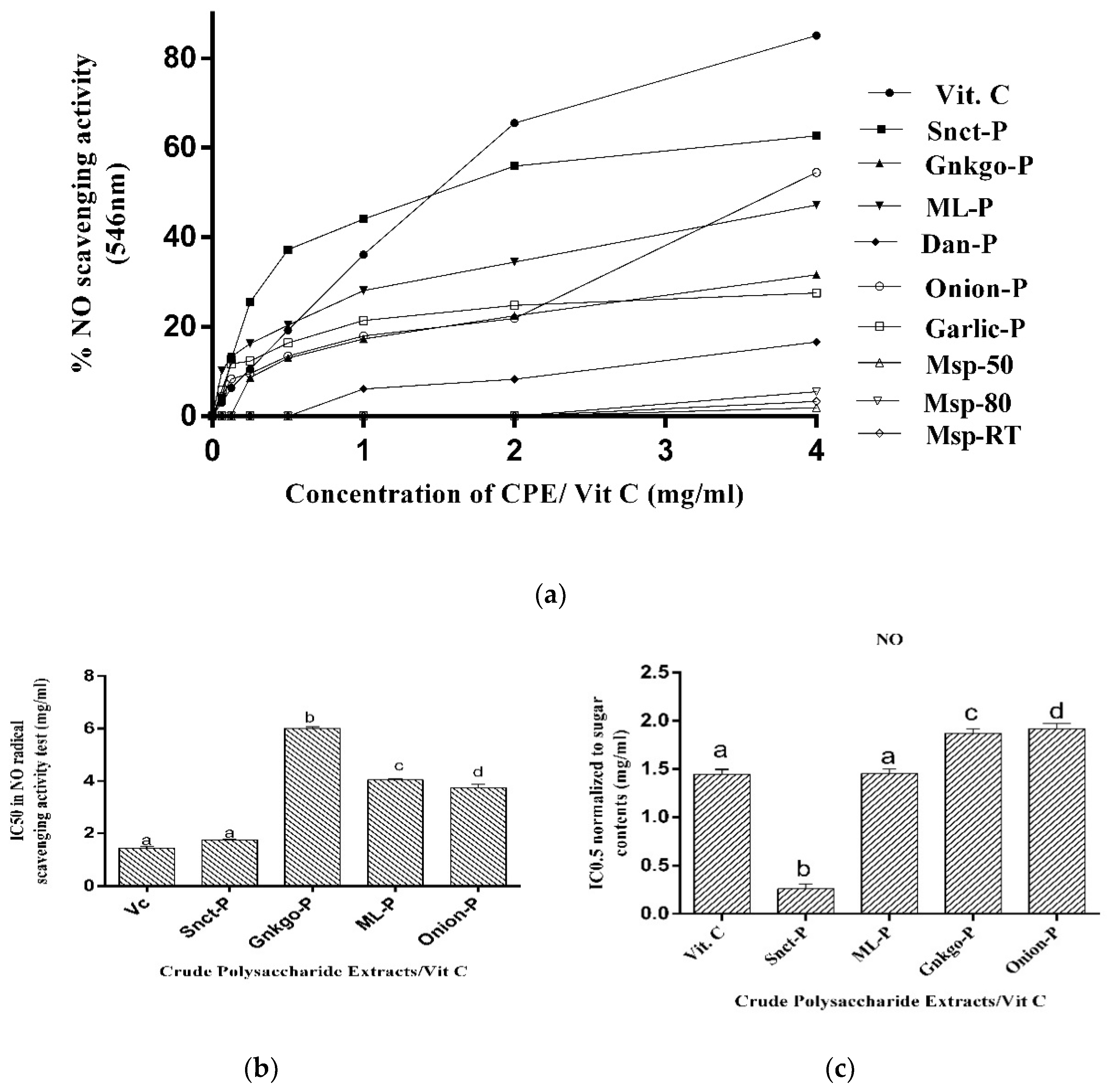
3.2.2. Determination of Nitric Oxide Radical-Scavenging Activity

Nitric oxide radical-scavenging activities of all the investigated plant polysaccharide extracts tested in the range of 0–4 mg/mL are presented in Figure 2. Snct-P, ML-P, Onion -P, and Gnkgo-P showed significantly higher NO-scavenging capacities (p < 0.05) than the other crude polysaccharides tested (Figure 2a). Figure 2b revealed IC50 values calculated for Snct-P, ML-P, Onion-P, and Gnkgo-P in the NO radical-scavenging activity test. As shown, the IC50 value of Snct-P was the lowest among the three crude polysaccharides presented (0.64 ± 0.01 mg/mL), followed by Onion-P and ML-P (3.74 ± 0.13, and 4.04 ± 0.05 mg/mL), respectively, while Gnkgo-P had lower NO-scavenging activity with a significantly higher IC50 (6.01 ± 0.04 mg/mL) than the remaining three polysaccharides presented (p < 0.05) (Figure 2b). At a concentration greater than 1.0 mg/mL, the NO-scavenging activity of Vit C (Figure 2a) revealed the highest % activity compared to the presented crude polysaccharides; however, they still possessed a significant NO-radical-reducing capacity in a dose-dependent manner. The IC50 normalized to total sugar was consistent in pattern with the IC50 values of NO-radical-reducing capacity of the crude polysaccharides (Figure 2c).
Figure 2. (a) Nitric oxide radical-scavenging activities of plant crude polysaccharides and Vit C as a control; (b) IC50 of plant crude polysaccharides and Vit C in Nitric oxide radical-scavenging activity test; (c) IC50 of the plant crude polysaccharides normalized to sugar contents and Vit C as a control. Results are illustrated as mean ± S.D from 4 measurements. IC50 results are significant at p < 0.05. Mean values with the same letters are not statistically different (p > 0.05); Mean values with different letters are statistically different (p < 0.05). Statistical analysis was carried out using one-way ANOVA and Bonferroni was used as a post hoc test.
Applsci 14 03683 g002

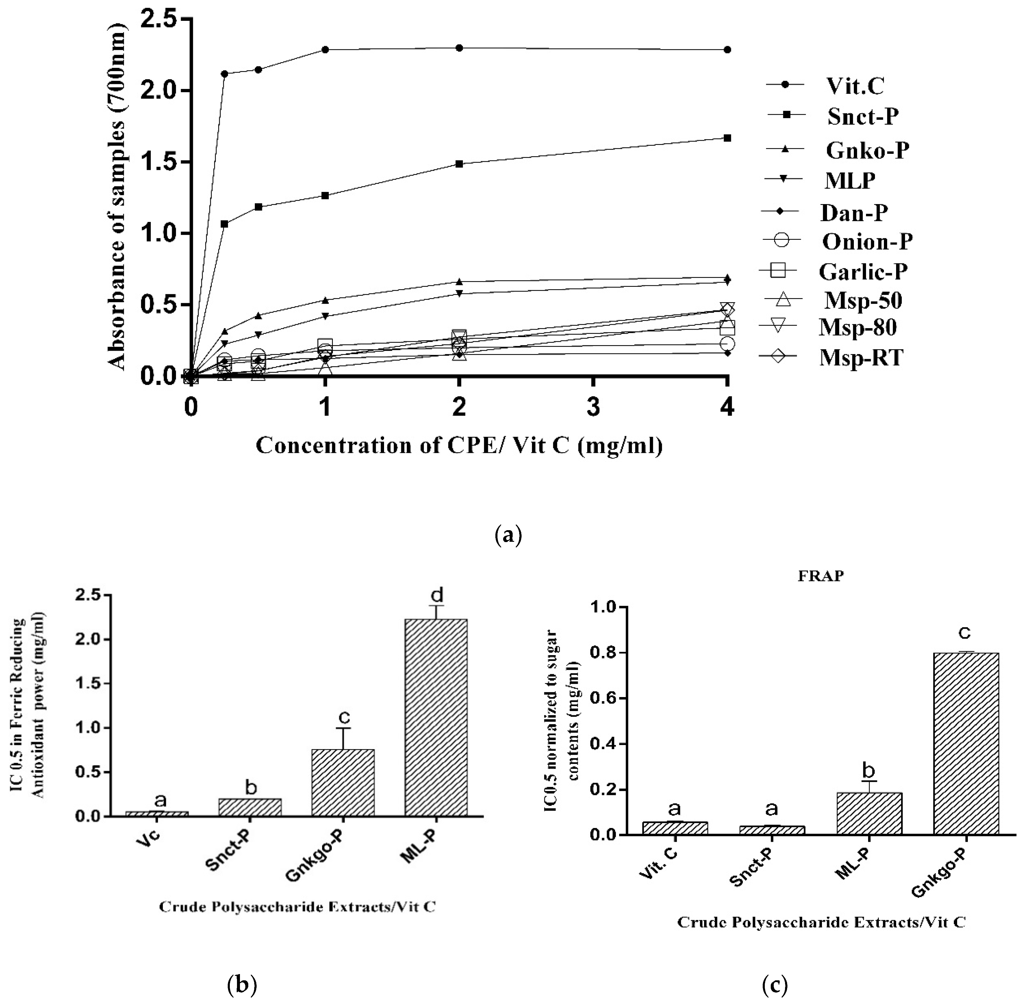
3.2.3. Determination of Ferric-Reducing Antioxidant Power (FRAP)

The absorbance given by the tested extracts and Vit C in the ferric-reducing power assay (y-axis) is plotted against concentrations on the x-axis in Figure 3. Snct-P, Gnkgo-P, and ML-P all showed significant (p < 0.05) absorbances in the Ferric-Reducing Antioxidant Power assay (Figure 3a); however, Snct-P showed the highest value among the tested plant polysaccharides. This is evident in the lowest IC0.5 value (Figure 3b) compared to the rest. However, all the plant polysaccharides showed significantly lower absorbances for ferric-reducing power compared to the positive control; still, this increase in a concentration-dependent manner (Figure 3a) is an indication that the plant polysaccharides have a certain ferric-reducing power and that the maximum capacity can be attained at higher concentrations. The IC0.5 values of FRAP normalized to total sugar were consistent in pattern with that of the crude polysaccharides (Figure 3c).
Figure 3. (a) Ferric-Reducing Antioxidant Power of plant crude polysaccharides and Vit C as a control; (b) IC0.5 of plant crude polysaccharides and Vit C in Ferric-Reducing Antioxidant Power test; (c) IC0.5 of the plant crude polysaccharides normalized to sugar content and Vit C as a control. Results are illustrated as mean ± S.D from 4 measurements. IC0.5 results are significant at p < 0.05. Mean values with the same letters are not statistically different (p > 0.05); Mean values with different letters are statistically different at p < 0.05. Statistical analysis was carried out using one-way ANOVA and Bonferroni was used as a post hoc test.
Applsci 14 03683 g003

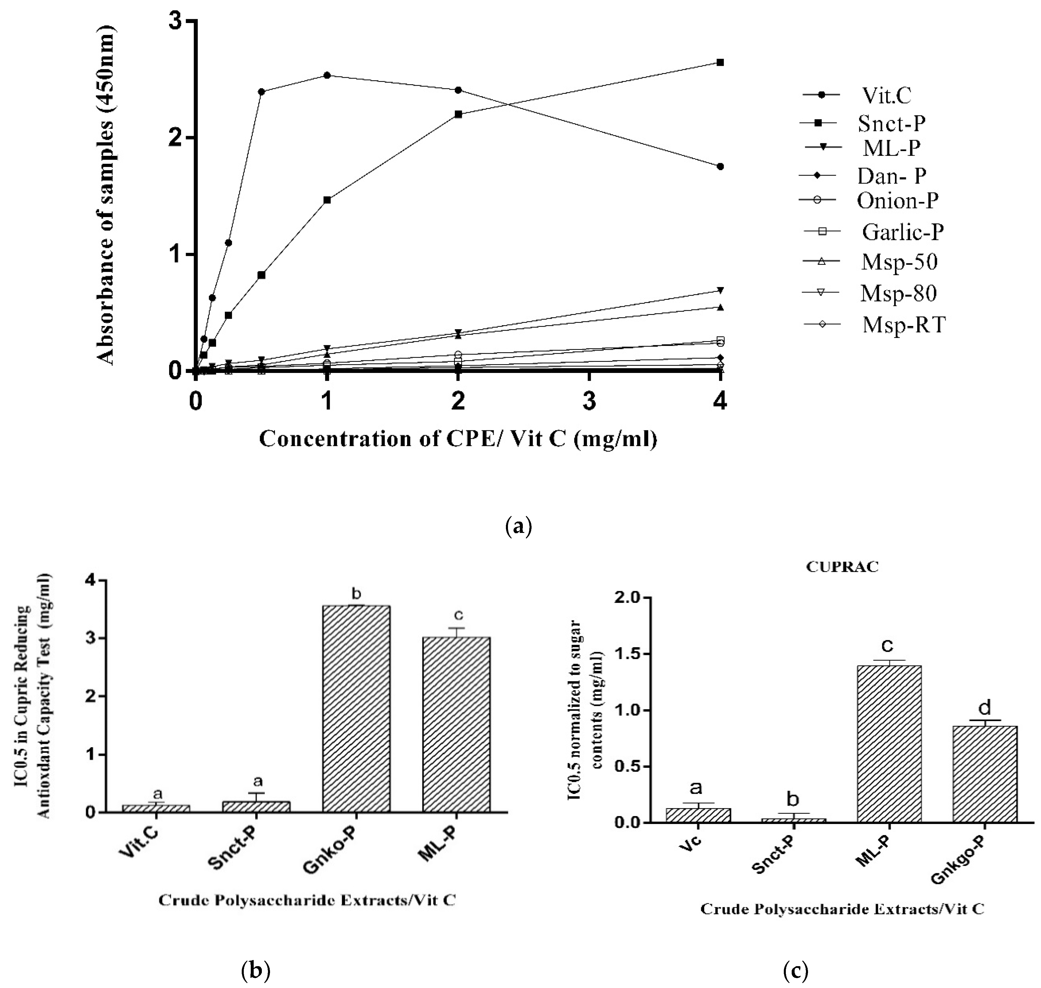
3.2.4. Determination of Cupric-Reducing Antioxidant Capacity (CUPRAC)

The absorbance shown by the tested extracts and Vit C in the Cupric-Reducing Antioxidant Capacity assay (y-axis) was plotted against concentrations on the x-axis, as shown in Figure 4. Snct-P, ML-P, and Gnkgo-P showed significant absorbance among all the tested crude polysaccharides. Snct-P revealed the highest absorbance (p < 0.05) among them; this is evident in the lowest IC0.5 value compared to the other polysaccharides (Figure 4b). All the plant polysaccharides showed significantly lower absorbances in the Cupric-Reducing Antioxidant Capacity assay compared to the Vit C; however, this absorbance increased with the concentration (Figure 4a), which shows that the plant crude polysaccharides possessed a cupric-reducing capacity and that the capacity can be enhanced at a higher concentration. The IC0.5 values of CUPRAC normalized to total sugar were consistent in pattern with that of the crude polysaccharides (Figure 4c).
Figure 4. (a) Cupric-Reducing Antioxidant Capacity of plant crude polysaccharides and (b) IC0.5 of plant crude polysaccharides in Cupric-Reducing Antioxidant Capacity test; (c) IC0.5 of the plant crude polysaccharides normalized to sugar contents. Results are illustrated as mean ± S.D from 4 measurements. IC0.5 results are significant at, p < 0.05. Mean values with the same letters are not statistically different (p > 0.05); Mean values with different letters are statistically different at p < 0.05. Statistical analysis was carried out using one-way ANOVA and Bonferroni was used as a post hoc test.
Applsci 14 03683 g004

3.3. Cytotoxic Activities of Plant Polysaccharides in MCF-7 Cells

Invasive breast ductal carcinoma, MCF-7, is one of the most common types of breast cancer; hence, it was used for the antiproliferative screening of the selected plant polysaccharides in this study by MTT. MCF-7 cells were incubated with different concentrations of the plant crude polysaccharide for 72 h (Figure 5a).
It was discovered that five of all the investigated plant crude polysaccharides (Ginkgo-P, Dan-P, ML-P, Onion-P, and Garlic-P) showed no significant inhibition of MCF-7 viability. Snct-P reduced MCF-7 cell viability to 55% at a 1000 µg/mL concentration, while only MSP-50 and MSP-80 showed significant antiproliferative activities at the concentrations of 0.25 to 1 mg/mL. MSP-RT showed significant antiproliferative activities at all the concentrations tested. The decrease in MCF-7 cell viability caused by the Moringa seed crude polysaccharide extracts occurred in a concentration-dependent manner. Sanctum crude polysaccharide extract (Snct-P) was cytotoxic at 1 mg/mL, the highest concentration presented in this study, while lower concentrations produced no significant effect. However, at a higher concentration of 2 mg/mL, there was observed increased cytotoxicity in sanctum polysaccharide; these data are not shown, which implies that Snct-P may show concentration-dependent cytotoxicity on the MCF-7 cells at concentrations higher than 1 mg/mL. The results of this study demonstrated that Moringa oleifera seed crude polysaccharides extracted through ethanol precipitation at 50% and 80% concentrations and room temperature were able to inhibit the growth of MCF-7 cells. The most significant antiproliferative activity against MCF-7 cells was exhibited by MSP-RT, with an IC50 of 0.061 mg/mL, as shown in Figure 5b and Table 3.
We, therefore, tested this extract on MRC-5 and Vero cell lines. The results showed that the MRC-5 and Vero cell lines were less responsive to MSP-RT than the MCF-7 cell line, with IC50 values 2.6 folds (0.157 mg/mL and 0.159 mg/mL) higher than what was observed in MCF-7 cells (Figure 5c and Table 4).

3.4. Effect of Plant Polysaccharide on MCF-7 Cell Proliferation

The MCF-7 cells treated with MSP-RT (μg/mL 0.0625 mg/mL) exhibited lower cell counts and a lower percentage of confluency when they were compared to the control in Figure 6 a,b. The results of % confluency and cell count follow the same trend as the % of cell viability.

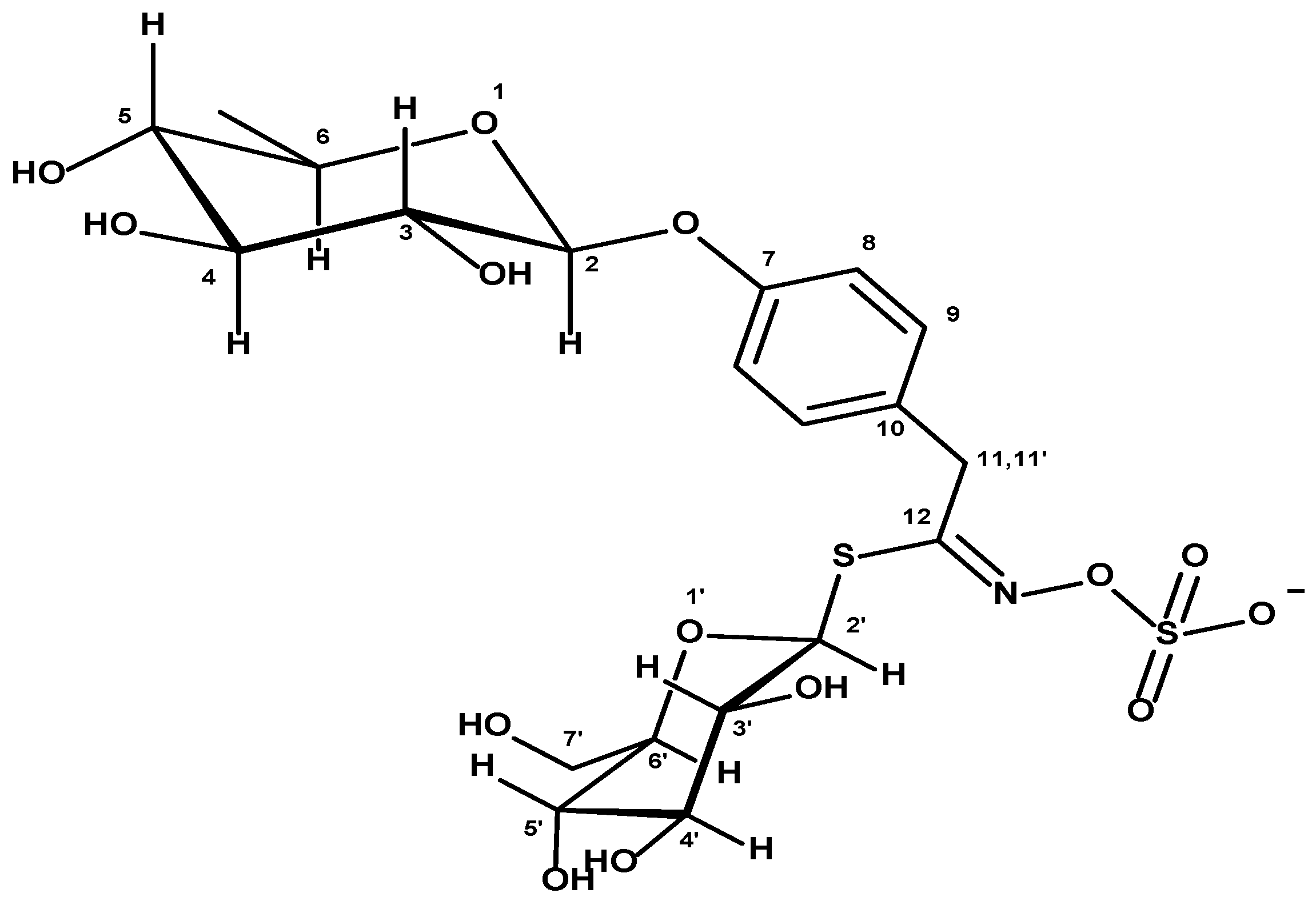
3.5. Elucidation of the MSP-RT-Abundant Compound Structure by Nuclear Magnetic Resonance Analysis

To determine the structure of the main extraction product contained in an MSP-RT crude polysaccharide extract in solution, 1H and 13C NMR spectra have been measured (see Supplementary Materials) and revealed a 4-α-rhamnopyranosyloxy-benzyl glucosinolate structure. The 1H and 13C NMR resonances were assigned unequivocally based on the combined information from the 1D and 2D NMR (gCOSY, gHSQC, and gHMBC) experiments. Coupling constants (1H-1H) were measured directly from resolution-enhanced 1D spectra and confirmed, when necessary, by homo-decoupling. gHSQC and gHMBC analysis allowed the assignment of the X regiochemistry. The chemical shift assignments in the proton/spectrum of the investigated compound were the starting point for the further analysis of the 1H and 13C NMR spectra. The proton 1H NMR of MSP-RT in the water solution is shown /in Figure S1 (see Supplementary Materials). The signal 1.25 (13C: 18.11 ppm) originated from the methyl group, and the signal 4.13 ppm (13C: 38.88 ppm) from CH2 residue. The signals in the proton spectrum lying in the 2–6 ppm (in 13C: 70–100 ppm) range were caused by protons (and carbons) of pyranose rings. Other signals (1H: 7.18 and 7.39 ppm; The 13C NMR of X is shown in Figure S2 (see Supplementary Materials). The signals 118.94 ppm, 130.84 ppm, 130.80 ppm, and 156.15 ppm were from the phenyl ring. The chemical shift of the carbon that came from the C=N group was 163.63 ppm. In the spectrum, there were also visible signals of unidentified compounds, but these were products occurring in small quantities. The structure of the abundant compound is presented in Figure 7.
In the mass spectrum of MSP-RT, crude polysaccharide extract peaks at m/z (ESI TOF MS ES-) 570.0203 were observed (Supplementary Data). The NMR and mass analyses confirm that the main product was 4-α-rhamnopyranosyloxy-benzyl glucosinolate.

4. Discussion

Naturally occurring chemicals originating from plants have become interesting candidates for cancer research due to their wide range of bioactive qualities and relative lack of toxicity compared to conventional chemotherapeutic drugs [9]. Through their antioxidant properties, bioactive substances originating from plants can have anticancer properties due to their ability to neutralize damaging reactive oxygen species (ROS) and reduce oxidative stress [10]. Plant polysaccharides have recently gained more attention due to their potential as antioxidants, having capacities to neutralize free radicals and potential free radicals through their side-chain glycosidic bonds and hydroxyl groups [15]. Surprisingly, despite their well-known health benefits, including anticancer capacities, there is a dearth of research regarding the anticancer properties of polysaccharides derived from plants in this study. The screening of antioxidative and antiproliferative activities of crude polysaccharides extracted from six different plants was carried out in this study. There were significant variations in the percentage of yield and chemical composition of the crude polysaccharides extracted from the plants. Generally, several factors may affect various constituents of plants, including species, plant sources, and other plant-inbuilt qualities that determine the release of different constituents into the solvents [35,36], even under similar laboratory conditions, as was the case in this study. The significant changes observed in the yields of the plant polysaccharides might be explained by different inherent qualities, differences in the source, species, and also various chemical constituents that have the capability of influencing the free release of polysaccharides and associated molecules into the extracting solvents. Physical or chemical methods were reported to greatly improve the yield of polysaccharide extraction as well as their total contents [37]. Some of the most commonly used methods for plant polysaccharide extraction include hot water extraction and sonication-assisted extraction methods [4,47,48], including various optimization methods involving parameters such as extraction temperature, time of incubation, water-to-sample ratio, and extraction solvent concentrations, among others [49,50]. The method reported in this study followed the usual protocol of defatting with a non-polar solvent (chloroform in this case) and depigmentation with ethanol, water extraction, and ethanol precipitation [35,36,51,52]. During this study, many of the parameters were optimized, including extraction temperature, time of incubation, water-to-sample ratio, and extraction solvent concentrations. However, since there were no significant differences among the crude polysaccharides obtained after varying different parameters, especially in the total sugar contents and cytotoxic effects of the tested extracts on MCF-7 cells, we decided to report the results obtained using the methods described in this study.
The variations noticed in the contents of protein and phenolics within the plant crude polysaccharides indicate the inbuilt complexity of these compounds. The significant protein content in some of the polysaccharide extracts could be associated with the presence of protein-containing sugars, which can be precipitated by ethanol. The extraction of both protein and sugars at the same time through ethanol suggests the complexity of the plant sample matrix. Ethanol generally possesses the capacity to selectively precipitate a wide range of compounds that have differing polarities, including sugars and proteins [53]. These extracted protein-containing sugars could be glycoproteins, glycopeptides, or other protein-sugar complexes, which might contribute to the significant increase in the protein content observed in this study in some of the extracts. Additionally, these molecules could play a function that is beyond the regular role of proteins and polysaccharides as independent molecules.
All the crude polysaccharides contain varying amounts of phenolics (p < 0.05), with significantly high amounts shown in Snct-P and ML-P. Low phenolic content was seen the most, especially in all Moringa seed crude polysaccharide extracts and Dan-P. There was a very strong correlation between the phenolic content and antioxidant activities [54]. The crude polysaccharides were obtained after the removal of ethanol-soluble phenolics; therefore, the significant phenolics obtained from Gnkgo-P, ML-P, and Snct-P may be regarded as water-soluble phenolics that are not significantly present in other crude polysaccharides. These might also be tightly bound to the polysaccharides as they precipitate together with the polysaccharides. The low phenolic contents in the crude polysaccharide extracts from other plants may be an indication that they are generally poor in phenolics or may be rich in ethanol-soluble phenolics that were removed during the first extraction with chloroform and ethanol. Aside from phenolics, sugars, and proteins that were quantified from these extracts, there may be other bioactive constituents that were not measured; these unmeasured components could also contribute to the overall chemical complexity of the bioactivity of these extracts. We acknowledge this as part of the limitation of this study. Future studies could focus on the complete chemical profiling of the other components. Antioxidants play a prospective role in the prevention of cancer and various other diseases. They are also responsible for the maintenance of homeostasis, hence the need for this study to screen the antioxidative abilities of the polysaccharides obtained from the selected plants. In the present study, the free radical-scavenging abilities of the plant polysaccharide extracts were analyzed by evaluating the DPPH and NO radical-scavenging activities as well as the Ferric-Reducing Antioxidant Power and Cupric-Reducing Antioxidant Capacity. DPPH is a known synthetic free radical for antioxidant assays. It is commonly used for the assessment of natural metabolites’ ability to scavenge free radicals, i.e., for estimation of their antioxidant capacities [55]. The results showed that Snct-P, Gnkgo-P, and ML-P had significant DPPH-scavenging activities when compared with other crude polysaccharides, while Snct-P had the highest activity among the three; this is evident by its lowest IC50 among the three. These three crude polysaccharides showed dose-dependent activities, which suggests that they possess radical-reducing capacities [55] with increasing doses and that they can act as hydrogen ion donors and free radical scavengers [56].
Nitric oxide plays a critical role in many physiological and pathological processes; it is essential for cellular signaling, has a short blood half-life of a few seconds, and is a strong vasodilator. Nitric oxide radicals become deleterious when they react with oxygen as they form nitrogen dioxide, which is a very toxic and reactive radical, thereby resulting in oxidative stress [57,58]. Consequently, the eradication of NO is favorable to living cells [12], and the ability of the plant crude polysaccharide to scavenge NO is measured by the NO-scavenging assay in this study. Snct-P, ML-P, Onion-P, and Gnkgo-P displayed significant NO radical-scavenging activity compared to the remaining plant crude polysaccharides. All these crude polysaccharides displayed NO radical-scavenging activities in a dose-dependent manner. This is an indication that these extracts could have the capability to prevent nitrogen dioxide production as their concentration increase, hence protecting against cellular damage [12]. Ferric-Reducing Antioxidant Power (FRAP) quantifies the capacity of a substance to reduce ferric ions (Fe3+) to ferrous (Fe2+) ions [59]. While ferric ions are not directly harmful, they do contribute to a Fenton reaction with hydrogen peroxide, which produces very reactive free radicals called hydroxide radicals (HO) [25]. For this reason, anything that lowers ferric ions will eventually be advantageous. From this study, all the plant polysaccharides showed significantly lower absorbances for ferric-reducing power compared to the positive control; however, the absorbances of Snct-P, Gnkgo-P, and ML-P increased significantly in a concentration-dependent manner compared to the remaining crude polysaccharides, which is an indication that these plant polysaccharides have certain ferric-reducing powers and that the maximum capacity can be attained at higher concentrations. At higher concentrations, these crude polysaccharides can significantly prevent the formation of hydroxyl radicals through the reduction of ferric ions [25], thereby contributing to the suppression of oxidative damage. The ability of polysaccharides to reduce ferric ions to ferrous ions is directly related to their ability to participate in electron transfer processes through the lone pair electrons within specific functional groups such as hydroxyl (-OH) and carbonyl (C=O) groups in their structures. These functional groups are able to donate electrons to ferric ions, reducing them to form ferrous ions. Also, polysaccharides, which contain specific monosaccharides with reducing properties, can also participate in the reduction of ferric ions to ferrous ions [60,61] Cupric-Reducing Antioxidant Capacity (CUPRAC) is used to measure the capacity of a compound to reduce cupric ions (Cu2+) to (Cu+) cuprous ions, thereby contributing to the redox balance of the cell [62,63]. All the plant polysaccharides showed significantly lower absorbances in the Cupric-Reducing Antioxidant Capacity assay compared to the Vit C; however, their absorbances increased with the concentration. Vit C showed a maximum absorbance in this assay at 1 mg, after which the absorbance began to reduce. This might be an indication of a diminished concentration of cupric ions in the reaction medium.
The antiproliferative capacities of the extracted plant crude polysaccharides against MCF-7 human adenocarcinoma cells, an invasive breast ductal carcinoma cell line regularly utilized in breast cancer research [64,65], were assessed. Antiproliferative activities of polysaccharides from various plants have been documented [66,67]; however, the antiproliferative screening studies for the majority of the plants investigated by us are limited. Polysaccharides from Ginkgo biloba and Taraxacum officinale were published to prevent the proliferation of breast and liver cancer cells, respectively [68,69]. However, in this study, there was no observed antiproliferative effect from the polysaccharides obtained from both plants. Various factors may be responsible for these unexpected results. Firstly, MCF-7 cell lines were used for this study, while in the reported studies, 4T1 and HepG2 cell lines were used [68,69]. Aside from this factor, the source of the plants may also be responsible for the difference in the effects obtained from these plant crude polysaccharides. Different parts of the Moringa species plants, especially the seeds, have been reported to show antiproliferative effects in many cancer cells [17,70,71], but the antiproliferative effects of extracted polysaccharides have not been documented. This study demonstrated that Moringa oleifera seed crude polysaccharides extracted through ethanol precipitation at 50% and 80% concentrations and room temperature were able to inhibit the growth of MCF-7 cells. The most significant antiproliferative activity against MCF-7 cells was exhibited by MSP-RT, with an IC50 of 0.061 mg/mL in this study. In this study, the MCF-7 cells treated with MSP-RT also exhibited lower cell counts and a lower percentage of confluency when they were compared to the control. This decrease may be an indication of cell proliferation inhibition, induced cell death, or decreased adhesion [72]. The results of % confluency and the cell count are consistent with the trend of percentage cell viability.
The ability of a drug to selectively inhibit cancer cells while being less cytotoxic to normal cells is a beneficial drug property in medical research; this property is especially useful in combatting drug resistance in cancer [73]. Regarding our results demonstrating the cytotoxic effect of MSP-RT towards MCF-7, we tested the cytotoxicity of MSP-RT towards MRC-5 and Vero cell lines. These two cell lines were derived from the fibroblast tissues of human fetal lungs [74] and from the monkey kidney epithelial cells, respectively [75]. They are regarded as normal cell lines [76]. This study showed that the MRC-5 and Vero cell lines were less responsive to MSP-RT than the MCF-7 cell lines, as IC50 values of 2.6 folds higher than that seen in MCF-7 cells were observed in these normal cells. As a result of this, Moringa seed crude polysaccharide (MSP-RT) extract may shield normal cells from the toxicity of chemotherapeutics while having no negative side effects on normal cells. It may also have antitumor and cancer-prevention properties [77,78] This is an indication that this crude polysaccharide extract may be promising as a valuable source for novel cancer therapy.
In the mass spectrum of MSP-RT crude polysaccharide extract peaks at m/z (ESI TOF MS ES-), 570.0203 can be observed (Supplementary Materials). The NMR and mass analyses confirm that the main product was 4-α-rhamnopyranosyloxy-benzyl glucosinolate. Glucosinolates are compounds synthesized naturally in Moringa oleifera and other members from Brassicales; these glucosinolates are converted into isothiocyanates when the plant tissues undergo some physical damage (trituration or mastication). These compounds from the plant tissues have been shown to have hypoglycemic, antioxidant, anti-inflammatory, and anticancer activities [79]. The mechanism of the cytotoxic activity of glucosinolates has been connected to their breakdown product isothiocyanates (ITC) [80,81]. The mechanism of cytotoxic action of ITC includes maintenance of low-systemic oxidative stress, cell-cycle progression inhibition, angiogenesis inhibition, and apoptotic induction in cancer cells [82].
The principal objective of the present study was the screening and evaluation of the antioxidative and antiproliferative properties of crude polysaccharide extracts obtained from selected plants. Natural compounds are being explored for medicinal purposes either to examine their potential as substitutions for toxic conventional chemotherapeutic agents or, better still, to assess their synergistic effect when used in combination with them.
Through the elucidation of antioxidative and antiproliferative potentials of the crude polysaccharide extracts from the plants in this study, we contributed to the ongoing examination of plant-based ingredients as viable candidates for future therapeutic progress. The variations observed in % yield, total protein, total sugar, and phenolics content among crude polysaccharide extracts from different plants reflect the diverse biochemical compositions inherent in each plant species, and these variations may explain why they exhibit different bioactivities. The differences in composition may arise from a combination of many factors. The observed variations in the antioxidative activities of these crude plant polysaccharide extracts indicate that they responded differently to different free radical models. Of all the crude polysaccharides, significant antioxidative capacities were exhibited by crude polysaccharides from Ocimum santum leaves, Moringa oleifera leaves, and Ginkgo biloba leaves, showing the highest significant activity in all the tested antioxidant models. It was also noted that the crude polysaccharide extracts with significantly higher phenolics than the others showed significant antioxidative activities, which is an indication that their antioxidative activities are associated with their phenolic component. The significant antiproliferative activities shown by only crude polysaccharide extract obtained from Moringa oleifera seeds and the lack of significant toxicity to the tested normal cells point out the potential of this plant as a valuable source for novel cancer therapy. Meanwhile, this display of antiproliferative activity without significant antioxidant effects indicates that distinct bioactive constituents may govern the antiproliferative mechanism, and this is independent of their contribution to antioxidative properties. NMR and mass analyses showed that the main product in the MSP-RT extract was 4-α-rhamnopyranosyloxy-benzyl glucosinolate; however, whether this compound is fully responsible for the antiproliferative activity of the crude polysaccharide extract remains to be elucidated.
The outcomes of this screening process open novel areas for future research.

5. Conclusions

In this study, we explored the antioxidant and antiproliferative properties of various plant crude polysaccharides obtained from six different plant materials. The crude polysaccharides exhibited different levels of antioxidative activities, ranging from very strong to poor; Snct-P showed the highest antioxidative activity, with the lowest IC50 and IC0.5 values of 0.076 ± 0.01 mg/mL, 0.64 ± 0.01 mg/mL, 0.20 ± 0.02 mg/mL, and 0.19 ± 0.07 mg/mL in DPPH, NO, FRAP, and CUPRAC assays, respectively. While most samples did not exhibit significant antiproliferative effects, Moringa oleifera seed polysaccharides displayed noteworthy activity without significant antioxidant effects. Further investigation with non-cancerous cells revealed Moringa oleifera seed crude polysaccharides to be at least in part selective in their antiproliferative activities between cancerous and non-cancerous cells. The structural analysis of these promising crude polysaccharides identified 4-α-rhamnopyranosyloxy-benzyl glucosinolate as a major compound. Further investigations are warranted to confirm whether this compound is fully responsible for the antiproliferative activity of the Moringa oleifera seed crude polysaccharide extract. To date, there is no report regarding the antiproliferative action of Moringa oleifera seed crude polysaccharides. Although the number of polysaccharides obtained from this extract is low, the fact that it is yielded through the general procedure for polysaccharide extraction recommends further optimization of the extraction method, which could potentially improve the yield of polysaccharides from this extract and provide an insight into its full potential and mechanism of antiproliferation.

Supplementary Materials

The following supporting information can be downloaded at https://www.mdpi.com/article/10.3390/app14093683/s1, Figure S1: 1H NMR spectrum of MSP-RT extract containing 4-α-rhamnopyranosyloxy-benzyl glucosinolate. Figure S2: 13C NMR spectrum of MSP-RT extract containing 4-α-rhamnopyranosyloxy-benzyl glucosinolate. Figure S3: Mass spectrum containing molecular peak m/z 570.0203 and confirming the structure in MSP-RT extract.

Author Contributions

Conceptualization, O.O.A., P.W. and J.C.; data curation, O.O.A., P.W. and J.C.; funding acquisition, J.C.; investigation, O.O.A., P.W. and H.K.; methodology, O.O.A., P.W., H.K, E.G. and J.C.; project administration, P.W. and J.C.; resources, P.W., H.K., E.G. and J.C.; writing—original draft, O.O.A.; writing—review and editing, O.O.A., P.W., H.K., E.G. and J.C. All authors have read and agreed to the published version of the manuscript.

Funding

This research received no external funding.

Institutional Review Board Statement

Not applicable.

Informed Consent Statement

Not applicable.

Data Availability Statement

The original contributions presented in the study are included in the article/Supplementary Material, further inquiries can be directed to the corresponding authors.

Acknowledgments

The research was supported by the Warsaw University of Technology within the Excellence Initiative: Research University (IDUB) program. The Mass Spectrometry analysis was performed in the Mass Spectrometry Facility at the Institute of Biochemistry and Biophysics, Polish Academy of Sciences.

Conflicts of Interest

The authors declare no conflicts of interest.

References

  1. Wilkinson, L.; Gathani, T. Understanding breast cancer as a global health concern. Brit. J. Radiol. 2022, 95, 20211033. [Google Scholar] [CrossRef] [PubMed]
  2. Feng, Y.; Spezia, M.; Huang, S.; Yuan, C.; Zeng, Z.; Zhang, L.; Ren, G. Breast cancer development and progression: Risk factors, cancer stem cells, signaling pathways, genomics, and molecular pathogenesis. Genes Dis. 2018, 5, 77–106. [Google Scholar] [CrossRef] [PubMed]
  3. Testa, U.; Castelli, G.; Pelosi, E. Breast cancer: A molecularly heterogenous disease needing subtype-specific treatments. Med. Sci. 2020, 8, 18. [Google Scholar] [CrossRef]
  4. Vuger, A.T.; Tiscoski, K.; Apolinario, T.; Cardoso, F. Anthracyclines in the treatment of early breast cancer friend or foe? Breast 2022, 65, 67–76. [Google Scholar] [CrossRef]
  5. Willson, M.L.; Burke, L.; Ferguson, T.; Ghersi, D.; Nowak, A.K.; Wilcken, N. Taxanes for adjuvant treatment of early breast cancer. Cochrane Database Syst. Rev. 2019, 9, 1–140. [Google Scholar] [CrossRef]
  6. Du, J.; Dong, Y.; Li, Y. Identification and prognostic value exploration of cyclophosphamide (Cytoxan)-centered chemotherapy response-associated genes in breast cancer. DNA Cell Biol. 2021, 40, 1356–1368. [Google Scholar] [CrossRef]
  7. Lin, C.; Cui, J.; Peng, Z.; Qian, K.; Wu, R.; Cheng, Y.; Yin, W. Efficacy of platinum-based and non-platinum-based drugs on triple-negative breast cancer: Meta-analysis. Eur. J. Med. Res. 2022, 27, 201. [Google Scholar] [CrossRef] [PubMed]
  8. Nguyen, S.M.; Pham, A.T.; Nguyen, L.M.; Cai, H.; Tran, T.V.; Shu, X.O.; Tran, H.T. Chemotherapy-Induced Toxicities and Their Associations with Clinical and Non-Clinical Factors among Breast Cancer Patients in Vietnam. Curr. Oncol. 2022, 29, 8269–8284. [Google Scholar] [CrossRef]
  9. Greenwell, M.; Rahman, P.K.S.M. Medicinal plants: Their use in anticancer treatment. Int. J. Pharm. Sci. Res. 2015, 6, 4103. [Google Scholar] [CrossRef] [PubMed]
  10. Stępień, A.E.; Trojniak, J.; Tabarkiewicz, J. Health-Promoting Properties: Anti-Inflammatory and Anticancer Properties of Sambucus nigra L. Flowers and Fruits. Molecules 2023, 28, 6235. [Google Scholar] [CrossRef]
  11. Ogunro, O.B.; Fakayode, A.E.; Batiha, G.E.S. Involvement of Antioxidants in the Prevention of Cellular Damage. In Importance of Oxidative Stress and Antioxidant System in Health and Disease; IntechOpen: London, UK, 2022; p. 10873. [Google Scholar] [CrossRef]
  12. He, L.; He, T.; Farrar, S.; Ji, L.; Liu, T.; Ma, X. Antioxidants maintain cellular redox homeostasis by elimination of reactive oxygen species. Cell Physiol. Biochem. 2017, 44, 532–553. [Google Scholar] [CrossRef]
  13. Didier, A.J.; Stiene, J.; Fang, L.; Watkins, D.; Dworkin, L.D.; Creeden, J.F. Antioxidant and Anti-Tumor Effects of Dietary Vitamins A, C, and E. Antioxidants 2023, 12, 632. [Google Scholar] [CrossRef] [PubMed]
  14. Pandey, K.B.; Rizvi, S.I. Plant polyphenols as dietary antioxidants in human health and disease. Oxidative Med. Cell. Longev. 2009, 2, 270–278. [Google Scholar] [CrossRef] [PubMed]
  15. Bai, L.; Xu, D.; Zhou, Y.M.; Zhang, Y.B.; Zhang, H.; Chen, Y.B.; Cui, Y.L. Antioxidant activities of natural polysaccharides and their derivatives for biomedical and medicinal applications. Antioxidants 2022, 11, 2491. [Google Scholar] [CrossRef]
  16. Jin, H.; Li, M.; Tian, F.; Yu, F.; Zhao, W. An Overview of Antitumour Activity of Polysaccharides. Molecules 2022, 27, 8083. [Google Scholar] [CrossRef]
  17. Wang, A.; Liu, Y.; Zeng, S.; Liu, Y.; Li, W.; Wu, D.; Wu, X.; Zou, L.; Chen, H. Dietary Plant Polysaccharides for Cancer Prevention: Role of Immune Cells and Gut Microbiota, Challenges and Perspectives. Nutrients 2023, 15, 3019. [Google Scholar] [CrossRef]
  18. Lemieszek, M.; Rzeski, W. Anticancer properties of polysaccharides isolated from fungi of the Basidiomycetes class. Contemp. Oncol. 2012, 16, 285–289. [Google Scholar] [CrossRef]
  19. Garcia, J.; Rodrigues, F.; Saavedra, M.J.; Nunes, F.M.; Marques, G. Bioactive polysaccharides from medicinal mushrooms: A review on their isolation, structural characteristics and antitumor activity. Food Biosci. 2022, 49, 101955. [Google Scholar] [CrossRef]
  20. Ying, Y.; Hao, W. Immunomodulatory function and anti-tumor mechanism of natural polysaccharides: A review. Front. Immunol. 2023, 14, 1147641. [Google Scholar] [CrossRef]
  21. Banerjee, S.; Parasramka, M.; Paruthy, S.B. Polysaccharides in cancer prevention: From bench to bedside. In Polysaccharides: Bioactivity and Biotechnology, 1st ed.; Ramawat, K.G., Mérillon, J.M., Eds.; Springer: Cham, Switzerland, 2015; pp. 2179–2214. [Google Scholar]
  22. Guo, R.; Chen, M.; Ding, Y.; Yang, P.; Wang, M.; Zhang, H.; Ma, H. Polysaccharides as potential anti-tumor biomacromolecules—A review. Front. Nutr. 2022, 9, 838179. [Google Scholar] [CrossRef]
  23. Chen, L.; He, C.; Zhou, M.; Long, J.; Li, L. Research Progress on the Mechanisms of Polysaccharides against Gastric Cancer. Molecules 2022, 27, 5828. [Google Scholar] [CrossRef] [PubMed]
  24. Satoh, Y.; Goi, T.; Nakazawa, T.; Kimura, Y.; Hirono, Y.; Katayama, K.; Yamaguchi, A. Polysaccharide K suppresses angiogenesis in colon cancer cells. Exp. Ther. Med. 2012, 4, 370–374. [Google Scholar] [CrossRef] [PubMed]
  25. Das, T.K.; Wati, M.R.; Fatima-Shad, K. Oxidative stress gated by Fenton and Haber Weiss reactions and its association with Alzheimer’s disease. Arch. Neurosci. 2015, 2, e60038. [Google Scholar] [CrossRef]
  26. Lis, B.; Olas, B. Pro-health activity of dandelion (Taraxacum officinale L.) and its food products–history and present. J. Funct. Foods 2019, 59, 40–48. [Google Scholar] [CrossRef]
  27. Kania-Dobrowolska, M.; Baraniak, J. Dandelion (Taraxacum officinale L.) as a source of biologically active compounds supporting the therapy of co-existing diseases in metabolic syndrome. Foods 2022, 11, 2858. [Google Scholar] [CrossRef] [PubMed]
  28. Bhattacharjya, D.; Adhikari, S.; Biswas, A.; Bhuimali, A.; Ghosh, P.; Saha, S. Ocimum phytochemicals and their potential impact on human health. Phytochem. Hum. Health 2019, 23, 1–26. [Google Scholar]
  29. Islam, Z.; Islam, S.M.; Hossen, F.; Mahtab-ul-Islam, K.; Hasan, M.R.; Karim, R. Moringa oleifera is a prominent source of nutrients with potential health benefits. Int. J. Food Sci. 2021, 2021, 6627265. [Google Scholar] [CrossRef] [PubMed]
  30. Mehwish, H.M.; Liu, G.; Rajoka, M.S.R.; Cai, H.; Zhong, J.; Song, X.; He, Z. Therapeutic potential of Moringa oleifera seed polysaccharide embedded silver nanoparticles in wound healing. Int. J. Biol. Macromol. 2021, 184, 144–158. [Google Scholar] [CrossRef]
  31. Kiple, K.F.; Ornelas, K.C. (Eds.) The Cambridge World History of Food; Cambridge University Press: Cambridge, UK, 2000; Volume 2. [Google Scholar]
  32. Santhosha, S.G.; Jamuna, P.; Prabhavathi, S.N. Bioactive components of garlic and their physiological role in health maintenance: A review. Food Biosci. 2013, 3, 59–74. [Google Scholar] [CrossRef]
  33. Arreola, R.; Quintero-Fabián, S.; López-Roa, R.I.; Flores-Gutiérrez, E.O.; Reyes-Grajeda, J.P.; Carrera-Quintanar, L.; Ortuño-Sahagún, D. Immunomodulation and anti-inflammatory effects of garlic compounds. J. Immunol. Res. 2015, 2015, 401630. [Google Scholar] [CrossRef]
  34. Kumari, N.; Kumar, M.; Lorenzo, J.M.; Sharma, D.; Puri, S.; Pundir, A.; Dhumal, S.; Bhuyan, D.J.; Jayanthy, G.; Selim, S.; et al. Onion and garlic polysaccharides: A review on extraction, characterization, bioactivity, and modifications. Int. J. Biol. Macromol. 2022, 219, 1047–1061. [Google Scholar] [CrossRef] [PubMed]
  35. Jesumani, V.; Du, H.; Pei, P.; Aslam, M.; Huang, N. Comparative study on skin protection activity of polyphenol-rich extract and polysaccharide-rich extract from Sargassum vachellianum. PLoS ONE 2020, 15, e0227308. [Google Scholar] [CrossRef] [PubMed]
  36. Dare, C.A.; Oyedapo, O.O.; Akinlalu, A.O.; Komolafe, I.J.; Fajobi, A.O.; Ogunsusi, M. Genotoxic Activities of Polysaccharides from Cotyledon and Coat of Fermented and Unfermented Annona squamosa L. Seed. Egypt. Acad. J. Biol. Sci. H. Bot. 2021, 12, 189–207. [Google Scholar] [CrossRef]
  37. Wu, H.; Shang, H.; Guo, Y.; Zhang, H.; Wu, H. Comparison of different extraction methods of polysaccharides from cup plant (Silphium perfoliatum L.). Process Biochem. 2020, 90, 241–248. [Google Scholar] [CrossRef]
  38. DuBois, M.; Gilles, K.A.; Hamilton, J.K.; Rebers, P.T.; Smith, F. Colorimetric method for determination of sugars and related substances. Anal. Chem. 1956, 28, 350–356. [Google Scholar] [CrossRef]
  39. Bradford, M.M. A rapid and sensitive method for the quantitation of microgram quantities of protein utilizing the Principle of protein-dye binding. Anal Biochem. 1976, 72, 248–254. [Google Scholar] [CrossRef] [PubMed]
  40. Akyüz, M. Nutritive value, flavonoid content, and radical scavenging activity of the truffle (Terfezia Boudieri Chatin). J. Soil Sci. Plant Nutr. 2013, 13, 143–151. [Google Scholar] [CrossRef]
  41. Kury, L.T.A.; Taha, Z.; Talib, W.H. Immunomodulatory and anticancer activities of Hyacinthus L.: An in vitro and in vivo study. Plants 2021, 10, 617. [Google Scholar] [CrossRef] [PubMed]
  42. Bouhlali, E.D.T.; Alem, C.; Zegzouti, Y.F. Antioxidant and anti-hemolytic activities of phenolic constituents of six Moroccan date fruit (Phoenix dactylifera L.) syrups. Indian J. Biotechnol. 2016, 12, 45–52. [Google Scholar]
  43. Marcocci, L.; Maguire, J.J.; Droyl-lefaix, M.T.; Packer, L. The nitric oxide scavenging properties of Ginkgo biloba extract. Biochem. Biophys. Res. Commun. 1994, 201, 748–755. [Google Scholar] [CrossRef]
  44. Etim, O.E.; Ekanem, S.E.; Sam, S.M. In vitro antioxidant activity and nitric oxide scavenging activity of Citrullus lanatus seeds. In Vitro 2013, 3, 126–132. [Google Scholar]
  45. Oyaizu, M. Studies on products of browning reaction: Antioxidative activities of products of browning reaction prepared from glucosamine. Jpn. J. Nutr. Diet. 1986, 44, 307–315. [Google Scholar] [CrossRef]
  46. Apak, R.; Güçlü, K.; Özyürek, M.; Burcu Bektas, B.; Bener, M. Cupric ion reducing antioxidant capacity assay for food antioxidants: Vitamins, polyphenolics, and flavonoids in food extracts. In Advanced Protocols in Oxidative Stress; Humana Press: Totowa, NJ, USA, 2008; pp. 163–193. [Google Scholar] [CrossRef]
  47. Niyigaba, T.; Liu, D.; Habimana, J.D.D. The extraction, functionalities, and applications of plant polysaccharides in fermented foods: A review. Foods 2021, 10, 3004. [Google Scholar] [CrossRef]
  48. Ahmad, M.M.; Chatha, S.A.S.; Iqbal, Y.; Hussain, A.I.; Khan, I.; Xie, F. Recent trends in extraction, purification, and antioxidant activity evaluation of plant leaf-extract polysaccharides. Biofuels Bioprod. Biorefin. 2022, 16, 1820–1848. [Google Scholar] [CrossRef]
  49. Cai, L.; Chen, B.; Yi, F.; Zou, S. Optimization of extraction of polysaccharide from dandelion root by response surface methodology: Structural characterization and antioxidant activity. Int. J. Biol. Macromol. 2019, 140, 907–919. [Google Scholar] [CrossRef] [PubMed]
  50. Yang, B.; Jiang, Y.; Zhao, M.; Shi, J.; Wang, L. Effects of ultrasonic extraction on the physical and chemical properties of polysa-ccharides from longan fruit pericarp. Polym. Degrad. Stab. 2008, 93, 268–272. [Google Scholar] [CrossRef]
  51. Zhou, S.; Huang, G.; Huang, H. Extraction, derivatization and antioxidant activities of onion polysaccharide. Food Chem. 2022, 388, 133000. [Google Scholar] [CrossRef]
  52. Awad, A.M.; Kumar, P.; Ismail-Fitry, M.R.; Jusoh, S.; Ab Aziz, M.F.; Sazili, A.Q. Green extraction of bioactive compounds from plant biomass and their application in meat as a natural antioxidant. Antioxidants 2021, 10, 1465. [Google Scholar] [CrossRef]
  53. Aryal, S.; Baniya, M.K.; Danekhu, K.; Kunwar, P.; Gurung, R.; Koirala, N. Total phenolic content, flavonoid content and antioxidant potential of wild vegetables from Western Nepal. Plants 2019, 8, 96. [Google Scholar] [CrossRef]
  54. Baliyan, S.; Mukherjee, R.; Priyadarshini, A.; Vibhuti, A.; Gupta, A.; Pandey, R.P.; Chang, C.M. Determination of antioxidants by DPPH radical scavenging activity and quantitative phytochemical analysis of Ficus religiosa. Molecules 2022, 27, 1326. [Google Scholar] [CrossRef]
  55. Tokumaru, O.; Shuto, Y.; Ogata, K.; Kamibayashi, M.; Bacal, K.; Takei, H.; Kitano, T. Dose-dependency of multiple free radical-scavenging activity of edaravone. J. Surg. Res. 2018, 228, 147–153. [Google Scholar] [CrossRef] [PubMed]
  56. Adewale, O.O.; Oyelola, R.F.; Oladele, J.O.; Agbaje, W.B. Assessing the ability of Polysaccharides extracted from Date Palm Fruit to salvage Wistar rats from cisplatin-linked hepatic damage. Pharmacol. Res.- Mod. Chin. Med. 2024, 11, 100400. [Google Scholar] [CrossRef]
  57. Al-Amiery, A.A.; Al-Majedy, Y.K.; Kadhum, A.A.H.; Mohamad, A.B. Hydrogen peroxide scavenging activity of novel coumarins synthesized using different approaches. PLoS ONE 2015, 10, e0132175. [Google Scholar] [CrossRef]
  58. Lalhminghlui, K.; Jagetia, G.C. Evaluation of the free-radical scavenging and antioxidant activities of Chilauni, Schima wallichii Korth in vitro. Future Sci. OA 2018, 4, FSO272. [Google Scholar] [CrossRef] [PubMed]
  59. Benzie, I.F.; Devaki, M. The ferric reducing/antioxidant power (FRAP) assay for non-enzymatic antioxidant capacity: Concepts, procedures, limitations and applications. In Measurement of Antioxidant Activity & Capacity: Recent Trends and Applications; Wiley: Hoboken, NJ, USA, 2018; pp. 77–106. [Google Scholar] [CrossRef]
  60. Ahmad, M.M. Characterization and antioxidant activities of polysaccharides extracted from flageolet bean pods waste. Curr. Res. Green Sustain. Chem. 2021, 4, 100154. [Google Scholar] [CrossRef]
  61. Gao, J.; Zhang, T.; Jin, Z.Y.; Xu, X.M.; Wang, J.H.; Zha, X.Q.; Chen, H.Q. Structural characterisation, physicochemical properties and antioxidant activity of polysaccharide from Lilium lancifolium Thunb. Food Chem. 2015, 169, 430–438. [Google Scholar] [CrossRef] [PubMed]
  62. Munteanu, I.G.; Apetrei, C. Analytical methods used in determining antioxidant activity: A review. Int. J. Mol. Sci. 2021, 22, 3380. [Google Scholar] [CrossRef] [PubMed]
  63. Silvestrini, A.; Meucci, E.; Ricerca, B.M.; Mancini, A. Total Antioxidant Capacity: Biochemical aspects and clinical significance. Int. J. Mol. Sci. 2023, 24, 10978. [Google Scholar] [CrossRef]
  64. Camarillo, I.G.; Xiao, F.; Madhivanan, S.; Salameh, T.; Nichols, M.; Reece, L.M.; Sundararajan, R. Low and high voltage electrochemotherapy for breast cancer: An in vitro model study. In Electroporation-Based Therapies for Cancer, 1st ed.; Sundararajan, R., Ed.; Elsevier: Amsterdam, The Netherlands, 2014; pp. 55–102. [Google Scholar]
  65. Ghoderao, P.; Sahare, S.; Kulkarni, A.A.; Bhave, T. Paclitaxel-conjugated magnetic carbon nanotubes induce apoptosis in breast cancer cells and breast cancer stem cells in vitro. In Paclitaxel; Academic Press: Cambridge, MA, USA, 2022; pp. 309–331. [Google Scholar] [CrossRef]
  66. Patel, M.K.; Tanna, B.; Gupta, H.; Mishra, A.; Jha, B. Physicochemical, scavenging and anti-proliferative analyses of polysaccharides extracted from psyllium (Plantago ovata Forssk) husk and seeds. Int. J. Biol. Macromol. 2019, 133, 190–201. [Google Scholar] [CrossRef]
  67. Chang, T.; Li, H.; Lv, H.; Tan, M.; Hou, S.; Liu, X.; Zhao, B. Extraction, Physicochemical Properties, Anti-Aging, and Antioxidant Activities of Polysaccharides from Industrial Hemp Residues. Molecules 2022, 27, 5746. [Google Scholar] [CrossRef]
  68. Chang, P.; XU, Y.; Zhou, D.Y.; WU, J.D. Effects of polysaccharides from Ginkgo biloba on the proliferation of 4T1 breast cancer cells and expression of GLUT family genes. Chin. Pharmacol. Bull. 2018, 34, 1301–1307. [Google Scholar]
  69. Ren, F.; Wu, K.; Yang, Y.; Yang, Y.; Wang, Y.; Li, J. Dandelion polysaccharide exerts anti-angiogenesis effect on hepatocellular carcinoma by regulating VEGF/HIF-1α expression. Front. Pharmacol. 2020, 11, 460. [Google Scholar] [CrossRef]
  70. Adebayo, I.A.; Arsad, H.; Samian, M.R. Antiproliferative effect on breast cancer (MCF7) of Moringa oleifera seed extracts. Afr. J. Tradit. Complement. Altern. Med. 2017, 14, 282–287. [Google Scholar] [CrossRef]
  71. Ramarao, K.D.R.; Somasundram, C.; Razali, Z.; Kunasekaran, W.; Jin, T.L.; Musa, S.; Achari, V.M. Antiproliferative effects of dried Moringa oleifera leaf extract on human Wharton’s Jelly mesenchymal stem cells. PLoS ONE 2022, 17, e0274814. [Google Scholar] [CrossRef] [PubMed]
  72. Richards, M.; Phoon, C.W.; Goh, G.T.W.; Seng, E.K.; Guo, X.M.; Tan, C.M.F.; Lee, J.M.K. A new class of pluripotent stem cell cytotoxic small molecules. PLoS ONE 2014, 9, e85039. [Google Scholar] [CrossRef] [PubMed]
  73. Konishi, K.; Yamaji, T.; Sakuma, C.; Kasai, F.; Endo, T.; Kohara, A.; Osada, N. Whole- Genome Sequencing of Vero E6 (VEROC1008) and Comparative Analysis of Four Vero Cell Sublines. Front. Genet. 2022, 13, 801382. [Google Scholar] [CrossRef] [PubMed]
  74. Abdi Goushbolagh, N.; Farhood, B.; Astani, A.; Nikfarjam, A.; Kalantari, M.; Zare, M.H. Quantitative cytotoxicity, cellular uptake and radioprotection effect of cerium oxide nanoparticles in MRC-5 normal cells and MCF-7 cancerous cells. BioNanoScience 2018, 8, 769–777. [Google Scholar] [CrossRef]
  75. Swanson, S.K.; Mento, S.J.; Weeks-Levy, C.; Brock, B.D.; Kowal, K.J.; Wallace, R.E.; Cano, F.R. Characterization of Vero cells. J. Biol. Stand. 1988, 16, 311–320. [Google Scholar] [CrossRef]
  76. Shakeri, M.; Tayer, A.H.; Shakeri, H.; Jahromi, A.S.; Moradzadeh, M.; Hojjat-Farsangi, M. Toxicity of saffron extracts on cancer and normal cells: A review article. Asian Pac. J. Cancer Prev. 2020, 21, 1867. [Google Scholar] [CrossRef]
  77. Komura, M.; Sato, T.; Yoshikawa, H.; Nitta, N.A.; Suzuki, Y.; Koike, K.; Takahashi, K. Propylene glycol, a component of electronic cigarette liquid, damages epithelial cells in human small airways. Respir. Res. 2022, 23, 216. [Google Scholar] [CrossRef]
  78. Wang, X.; Zhang, H.; Chen, X. Drug resistance and combating drug resistance in cancer. Cancer Drug Resist. 2019, 2, 141. [Google Scholar] [CrossRef] [PubMed]
  79. Lopez-Rodriguez, N.A.; Gaytán-Martínez, M.; de la Luz Reyes-Vega, M.; Loarca-Piña, G. Glucosinolates and isothiocyanates from Moringa oleifera: Chemical and biological approaches. Plant Foods Hum. Nutr. 2020, 75, 447–457. [Google Scholar] [CrossRef]
  80. Bell, L.; Oloyede, O.O.; Lignou, S.; Wagstaff, C.; Methven, L. Taste and flavor perceptions of glucosinolates, isothiocyanates, and related compounds. Mol. Nutr. Food Res. 2018, 62, 1700990. [Google Scholar] [CrossRef] [PubMed]
  81. Zhou, Q.; Tang, H.; Jia, X.; Zheng, C.; Huang, F.; Zhang, M. Distribution of glucosinolate and pungent odors in rapeseed oils from raw and microwaved seeds. Int. J. Food Prop. 2018, 21, 2296–2308. [Google Scholar] [CrossRef]
  82. Sanlier, N.; Guler, S.M. The benefits of Brassica vegetables on human health. J. Hum. Health Res. 2018, 1, 1–13. [Google Scholar]
Figure 5. (a) Cytotoxic activities of crude polysaccharides extracted from selected plants (Snct-P, Gnkgo-P, ML-P, Dan-P, Onion-P, Garlic-P, MSP-50, MSP-80 and MSP-RT on human adenocarcinoma cell line MCF-7. MCF-7 cells were subjected to 0, 0.0625, 0.125, 0.25, 0.5, and 1 mg/mL of the crude polysaccharides from the selected plants for 72 h. Results are illustrated as mean ± S.D of 4 measurements. Results are significant at * p < 0.05 compared with control. Statistical analysis was carried out using one-way ANOVA and t-tests. (b) Dose-response curves for MSP-50, MSP-80, and MSP-RT on MCF-7 cells. (c) Dose-response curves for MSP-RT on MRC-5 and Vero Cell lines.
Applsci 14 03683 g005
Figure 6. Effect of Moringa seed polysaccharide obtained at room temperature (MSP-RT) on % confluency (a) and cell count (b).
Applsci 14 03683 g006
Figure 7. The structure of the main compound in MSP-RT extract: 4-α-rhamnopyranosyloxy-benzyl glucosinolate (for NMR data, see Supplementary Materials).
Applsci 14 03683 g007
Table 1. Information concerning the selected plants used for this study.
SamplesPlant
Species
Where They Were
Obtained
Part of Plant UsedForm of Plant Used
Snct-POcimum sanctumEco-Herba, PolandLeavesDried leaves
ML-PMoringa oleiferaOsun State, NigeriaLeavesDried leaves
Ginkgo-PGinkgo bilobaEco-Herba, PolandLeavesDried leaves
Onion-PAllium cepaLocal market, PolandBulbsFresh bulbs
Garlic-PAllium sativumLocal market, PolandLeavesDried leaves
Dan-PT. officinaleEco-Herba, PolandRootsDried roots
MSP-50Moringa oleiferaOsun State, NigeriaSeedsDried seeds
MSP-80Moringa oleiferaOsun State, NigeriaSeedsDried seeds
MSP-RTMoringa oleiferaOsun State, NigeriaSeedsDried seeds
Table 2. Percentage yield and the chemical composition of selected plant Polysaccharide extracts.
Samples* Yield (%)Total Sugar
Content (%)
Protein
Content (%)
Phenolic
Content (%)
Snct-P 13.28 ± 0.78 a17.70 ± 0.03 a9.27 ± 0.30 a 16.50 ± 0.56 a
ML-P 8.23 ± 1.1 a 46.14 ± 1.16 c11.81 ± 0.30 ac 18.44 ± 0.80 c,b
Gnkgo-P 7.11 ± 0.45 a,d24.08 ± 1.88 d1.45 ± 0.07 d,b 7.78 ± 1.43 d,b
* Onion-P 0.60 ± 0.0551.19 ± 0.72 e2.19 ± 0.30 e,b 1.51 ± 0.03 e,b,d
Garlic-P 9.39 ± 0.57 a24.71 ± 2.42 f0.67 ± 0.01 f,b 3.18 ± 0.15 f,b,d,e
Dan-P 27.88 ± 4.56 g83.78 ± 1.28 g0.15 ± 0.01 g,b 1.26 ± 0.02 g,d,e
MSP-50 6.41 ± 0.89 h 6.30 ± 1.45 h,i31.76 ± 1.70 h 2.18 ± 0.01 h,b,c,d
MSP-80 11.09 ± 0.31 a,h,i7.86 ± 0.38 i32.89 ± 0.70 i,h 2.68 ± 0.02 i,c
MSP-RT 13.97 ± 1.22 i9.00 ± 0.89 j,i34.38 ± 0.22 j,h 2.35 ± 0.02 j,c,d
Yield (%) is calculated as a mass of obtained crude polysaccharides relative to the mass of plant powder used for extraction, except * Onion-P where fresh bulb mass used for extraction was taken for calculation. Crude polysaccharides with the same subscript letters within the same column are not significantly different from one another at p < 0.05, while those with different letters within the same column are significantly different from one another at p < 0.05.
Table 3. IC50 and regression parameters of MSP-50, MSP-80 and MSP-RT in MCF-7 cell line.
Crude Polysaccharides
MSP-50MSP-80MSP-RT
IC500.34150.34110.06073
R20.99450.9960.9929
Table 4. IC50 and regression parameters of MSP-RT in MRC-5 and Vero cell lines.
Cell Line
MRC-5Vero
IC500.15740.1594
R20.99890.9626
Disclaimer/Publisher’s Note: The statements, opinions and data contained in all publications are solely those of the individual author(s) and contributor(s) and not of MDPI and/or the editor(s). MDPI and/or the editor(s) disclaim responsibility for any injury to people or property resulting from any ideas, methods, instructions or products referred to in the content.

