Article

Utility of Clinical Next Generation Sequencing Tests in KIT/PDGFRA/SDH Wild-Type Gastrointestinal Stromal Tumors

1
Division of Cancer Medicine, The University of Texas MD Anderson Cancer Center, Houston, TX 77030, USA
2
Department of Sarcoma Medical Oncology, The University of Texas MD Anderson Cancer Center, Houston, TX 77030, USA
3
Department of Pathology, Division of Pathology & Laboratory Medicine, The University of Texas MD Anderson Cancer Center, Houston, TX 77030, USA
*
Author to whom correspondence should be addressed.
Cancers 2024, 16(9), 1707; https://doi.org/10.3390/cancers16091707
Submission received: 15 March 2024 / Revised: 18 April 2024 / Accepted: 23 April 2024 / Published: 27 April 2024

Abstract

:

Simple Summary

Most gastrointestinal stromal tumors (GISTs) are driven by activating mutations in KIT and PDGFRA or alterations in the succinate dehydrogenase (SDH) complex. A small fraction of GISTs lack alterations in KIT, PDGFRA, and the SDH complex, so-called “triple-negative” GISTs. We assessed clinical genomic sequencing, treatment, and survival outcomes in a cohort of 20 triple-negative GISTs. Genomic alterations were most commonly seen in the RAS/RAF/MAPK pathway and the DNA damage response pathway. Compared to KIT/PDGFRA mutant GIST, limited benefit was observed with imatinib in triple-negative GIST. In-depth molecular profiling can be helpful in identifying driver mutations and guiding therapy.

Abstract

Objective: The vast majority of gastrointestinal stromal tumors (GISTs) are driven by activating mutations in KIT, PDGFRA, or components of the succinate dehydrogenase (SDH) complex (SDHA, SDHB, SDHC, and SDHD genes). A small fraction of GISTs lack alterations in KIT, PDGFRA, and SDH. We aimed to further characterize the clinical and genomic characteristics of these so-called “triple-negative” GISTs. Methods: We extracted clinical and genomic data from patients seen at MD Anderson Cancer Center with a diagnosis of GIST and available clinical next generation sequencing data to identify “triple-negative” patients. Results: Of the 20 patients identified, 11 (55.0%) had gastric, 8 (40.0%) had small intestinal, and 1 (5.0%) had rectal primary sites. In total, 18 patients (90.0%) eventually developed recurrent or metastatic disease, and 8 of these presented with de novo metastatic disease. For the 13 patients with evaluable response to imatinib (e.g., neoadjuvant treatment or for recurrent/metastatic disease), the median PFS with imatinib was 4.4 months (range 0.5–191.8 months). Outcomes varied widely, as some patients rapidly developed progressive disease while others had more indolent disease. Regarding potential genomic drivers, four patients were found to have alterations in the RAS/RAF/MAPK pathway: two with a BRAF V600E mutation and two with NF1 loss-of-function (LOF) mutations (one deletion and one splice site mutation). In addition, we identified two with TP53 LOF mutations, one with NTRK3 fusion (ETV6-NTRK3), one with PTEN deletion, one with FGFR1 gain-of-function (GOF) mutation (K654E), one with CHEK2 LOF mutation (T367fs*), one with Aurora kinase A fusion (AURKA-CSTF1), and one with FANCA deletion. Patients had better responses with molecularly targeted therapies than with imatinib. Conclusions: Triple-negative GISTs comprise a diverse cohort with different driver mutations. Compared to KIT/PDGFRA-mutant GIST, limited benefit was observed with imatinib in triple-negative GIST. In depth molecular profiling can be helpful in identifying driver mutations and guiding therapy.

1. Introduction

Gastrointestinal stromal tumors (GISTs) are the most common mesenchymal neoplasms of the gastrointestinal tract and arise from the interstitial cells of Cajal [1]; however they are relatively uncommon, with an estimated incidence of approximately 7 cases per million people per year in the United States [2]. The vast majority of GISTs are driven by activating mutations in KIT or PDGFRA [3]. About 10–15% do not have an alteration in one of these two genes, and they are generally less sensitive to currently approved tyrosine kinase inhibitors (TKIs) [4,5,6]. Previous reports have some shed light on alternate oncogenic drivers in this subset of so-called “wild-type” GIST that are more prevalent in young females and associated with multifocal but indolent disease, gastric primary, and lack of response to traditional KIT/PDGFRA TKIs such as imatinib [7].
The next most common alterations after KIT and PDGFRA are in SDHA, SDHB, SDHC, and SDHD genes, components of the succinate dehydrogenase (SDH) complex that catalyzes the oxidation of succinate to fumarate in the tricarboxylic acid cycle [8,9,10]. SDH deficient GIST accounts for 7–10% of all GIST [11,12]. SDH deficiency leads to succinate accumulation, which inhibits prolyl hydroxylases, leading to accumulation of the hypoxia-inducible factor 1 alpha (HIF-1α) [13]. SDH-deficient GISTs are more commonly seen in pediatric patients and can be associated with either the Carney triad (GIST, paraganglioma, pulmonary chondroma) characterized by epigenetic silencing of the SDHC gene by the promoter hypermethylation or Carney–Stratakis syndrome (GIST and paraganglioma), which are caused by germline mutations in SDH [14,15,16,17].
There have been small case series and case reports of GIST lacking alterations in KIT, PDGFRA, and SDH (“triple-negative GIST”). One of the more common alterations in this cohort is in the RAS/RAF/MAPK pathway [18,19,20,21]. For example, some have BRAF mutations, which are often the classic V600E gain-of-function (GOF) mutation that is seen frequently in melanoma and other cancer types [22,23], and this is often mutually exclusive with KIT/PDGFRA mutations [24]. The next-most-common alteration is loss-of-function (LOF) mutations of NF1 [18,20,21,25], a GTPase-activating protein that promotes the hydrolysis of Ras-bound GTP, thereby acting as a negative regulator of the RAS/RAF/MAPK pathway; NF1 LOF mutation results in RAS/RAF/MAPK pathway activation. Alterations in DNA damage repair genes have also been reported [21]. Other studies have shown that about 20% of tumors thought to be KIT/PDGFRA/SDH wild-type end up having cryptic pathogenic KIT mutations [20]. However, most of these studies to date have used targeted sequencing of a limited number of genes, and how these triple-negative GISTs behave clinically is not well understood. In this study, we aimed to characterize the genomic landscape of triple-negative GIST using clinical next generation sequencing with larger gene panels and the associated treatment and outcome data.

2. Materials and Methods

2.1. Study Design

This is a retrospective study of all patients treated at MD Anderson between 1 January 2015 and 31 December 2023 with a diagnosis of GIST and available clinical next-generation sequencing data. We included those that lacked alterations in KIT and PDGFRA and either (1) retained expression of SDHB by immunohistochemistry (15 patients) or (2) had wild-type SDHB according to a sequencing assay (5 patients had only SDH mutation testing and not SDHB immunohistochemistry). The following clinical data were collected by retrospective chart review: age, gender, race, ethnicity, date of birth, date of diagnosis, vital status, tumor grade, tumor mitotic rate, tumor stage, surgery date, systemic therapies and dates of treatment, response to treatment, personal history of cancer, family history of cancer, and results of next generation sequencing tests. Response to treatment was based on the treating clinician’s documentation in the patients’ charts.

2.2. Clinical Genomic Sequencing

Clinical sequencing was available from multiple platforms. Twelve patients had sequencing with an MD Anderson platform. Five patients had BostonGene, four had FoundationOne®, one patient had Caris, one patient had NeoTYPE®, one patient had Endeavor, and two had outside institution panels. Five patients had multiple panels: one patient had BostonGene, FoundationOne®, and MD Anderson panels; one had FoundationOne® and MD Anderson panels; one had Caris and BostonGene; one had NeoTYPE® and BostonGene; and one had Endeavor and BostonGene. The MD Anderson platform has evolved to include more genes over time. The latest is the Mutation Analysis Precision Panel (MAPP, utilized for three patients) is an MD Anderson Molecular Diagnostic Laboratory (MDL)-developed and -validated Illumina hybrid capture-based assay which can detect mutations and sequence variants in 610 genes, copy number variants in 583 genes, select gene rearrangements in 34 genes, and select immune oncology signatures, including microsatellite instability (MSI) and tumor mutational burden (TMB); it compares these to matched non-tumor tissue to filter out single nucleotide polymorphisms and germline variants. The Solid Tumor Genomics Assay 2018 (STGA 2018, five patients) is an AmpliSeq chemistry-based ion torrent assay, which utilizes a germline control sample and was first implemented in 2018. It assesses mutations in 134 genes and selected copy number variations in 47 genes and compares to matched non-tumor tissue [26]. The CMS50 panel (four patients) also uses an AmpliSeq chemistry-based ion torrent assay, though it does not utilize a germline control. It was first implemented in 2012 and assessed 50 genes using PCR-based sequencing [27]. The MD Anderson Precision Oncology Decision Support (PODS) tool was used to assess for clinical actionability of genomic alterations [28].

2.3. Survival Analyses and Statistics

Statistical analyses were performed using GraphPad Prism (version 9.5.0 or higher, RRID:SCR_002798) and R (version 4.2.2 or higher).
Overall survival (OS) was calculated from the date of first histologic diagnosis (either pre-treatment biopsy or surgical pathology) to death or the latest follow-up. Recurrence-free survival (RFS) was calculated in patients with initially localized disease from the date of surgery of the primary tumor to the date of recurrence or the latest follow-up. Progression-free survival (PFS) was calculated in patients with metastatic disease from the start of therapy to the date of progression or the latest follow-up.

2.4. Data Availability

De-identified data generated in this study are available upon request from the corresponding author.

2.5. Ethics

This study was approved by the University of Texas MD Anderson Cancer Center Institutional Review Board (protocols 2022-0278 and LAB04-0890) and was conducted in accordance with the U.S. Common Rule. Clinical and genomic data were obtained following signed informed consent onto prospective institutional protocols or under retrospective review protocols with a limited waiver of authorization.

3. Results

3.1. Clinical Characteristics of Triple-Negative GIST Cohort

We identified 20 patients with GIST that lacked alterations in KIT, PDGFRA, and the SDH complex, so called “triple-negative GIST.” Patient characteristics are summarized in Table 1. Median age of diagnosis was 48 years (mean 45.3 years; Figure 1A), and twelve patients (60.0%) were female. The median tumor size was 7 cm (mean 9.6 cm; Figure 1B). The median mitotic index was 15 (per 50 hpf or 5 mm2), and the mean was 31.8 (Figure 1C). In total, 11 (55.0%) triple-negative GISTs were gastric, 8 (40.0%) were small intestinal, and 1 (5.0%) was rectal (Figure 1D); 8 patients (40.0%) presented with de novo metastatic disease (i.e., stage IV; Figure 1E); and 19 patients total (95.0%) eventually developed recurrent or metastatic disease versus 1 (5.0%) with localized disease at the time of most recent follow-up (median follow-up of 66.4 months from the time of diagnosis). Of the 19 with recurrent or metastatic disease, 8 had local relapse in the abdomen or pelvis, 9 had metastasis to the liver, 5 to the omentum or peritoneum, 1 to the pancreas, and 1 to the spleen.

3.2. The Genomics of Triple-Negative GIST

All 20 cases have had clinical genomic sequencing to reveal a potential driver (Table 1, Figure 2A). The mean and median number of somatic mutations identified were 2.0 and 1, respectively (range 0–8; Figure 2B). The most commonly altered genes were NF1, BRAF, FAM123B, and PIK3CA (Figure 2C). Regarding hypothesized drivers identified by sequencing (Figure 2D), four patients were found to have alterations in the RAS/RAF/MAPK pathway: two with a BRAF V600E GOF mutation, two with NF1 LOF mutations (1 deletion and 1 splice site mutation), and two with TP53 LOF mutations. However, one of the patients with a TP53 mutation (patient 15) also had three other potential driver mutations: NF2 c.517-1G > A (splice site mutation listed as pathogenic in ClinVar and predicted to result in loss of protein function) and amplification of FGF3, FGF19, and CCND1 (all listed as likely oncogenic by OncoKB). There was one other case with a likely non-pathogenic NF1 mutation (H2457R). In addition, one tumor each had a NTRK3 fusion (ETV6-NTRK3), a PTEN deletion, an FGFR1 GOF mutation (K654E), a CHEK2 LOF mutation (T367fs*), and an Aurora kinase A fusion (AURKA-CSTF1). Two patients had PIK3CA mutations (both I391M), though this alteration has not been identified as a driver mutation. Sequencing of the remaining seven patients (45.0%) did not identify an obvious driver mutation.

3.3. Response to Treatment

Regarding treatment, 13 (65.0%) had surgery as the initial treatment, while 7 (35.0%) had medical therapy as the initial treatment. Of the eight patients with de novo metastatic disease, four initially started with imatinib and three developed progressive disease after 2.0, 4.2, and 4.4 months. One is still on imatinib 11 years after initial diagnosis. The remaining four patients with stage IV disease had metastatic disease discovered at the time of initial surgery by surgical pathology or were undergoing surgery for oligometastatic disease to render them disease-free.
All 20 patients were treated with imatinib, the most common first-line therapy for GIST, at some point in their treatment course. The mean number of lines of therapies was 3.0 (median 2, range 1–9). For the 13 patients with evaluable response to imatinib (e.g., neoadjuvant treatment or for recurrent/metastatic disease), median PFS from the start of therapy with imatinib was 4.4 months (range 0.5–129.4 months; Figure 3A).
The results of clinical sequencing testing led to treatment with a molecularly targeted treatment for which there is an FDA-approved drug in three patients (Figure 3A). Two patients with BRAF V600E mutation have both been treated with a combination of BRAF and MEK inhibitors. One patient has been on this therapy for 34.2 months, has had a partial response, and treatment is ongoing; unfortunately, the other patient had to stop therapy after two months due to toxicity. The patient with ETV6-NTRK3 fusion was treated with larotrectinib for 28.7 months prior to progression, likely due to an acquired NTRK3 gatekeeper mutation (F617L) rendering resistance to larotrectinib [29]. This patient was then transitioned to a second-generation NTRK inhibitor, selitrectinib, and was on this for 16.5 months prior to progression. In addition to these, Figure 3B displays the timeline of treatments for all patients in the cohort arranged by the purported driver.

3.4. Survival Outcomes

Follow-up data were available for a median of 66.4 months from the time of diagnosis, which was defined as the date of pathologic confirmation of GIST. Regarding clinical outcomes, the median OS was 301.2 months (Figure 4A). For the 11 patients initially treated with curative intent, the median recurrence-free survival was 57.4 months (Figure 4B). For the 10 patients with localized disease initially that later developed recurrent/metastatic disease, the median time to recurrence/metastasis from the initiation of treatment was 43.3 months (range 1.1–509.9 months, mean 105.4 months). For patients with advanced/metastatic disease, the median PFS with first-line systemic treatment was 23.1 months (Figure 4C). The median OS for patients that presented with de novo metastatic disease (i.e., stage IV) was 66.8 months, and the median OS for patients that ever developed metastatic disease was 301 months.
We next looked at whether the specific driver mutation was associated with differences in outcomes. The two patients in our cohort with TP53 mutation had notably worse outcomes. One patient had OS of 3.7 and PFS of 1.1 months on imatinib, while the second patient had OS of 7.9 months and PFS of 3.8 months on imatinib. In the patients with the best outcomes (either no recurrence/progression or recurrence/progression 5 years or more after diagnosis, n = 8), there was no obvious association with genomic driver; one with FGFR1 GOF mutation, one with BRAF GOF mutation, one with CHEK2 LOF mutation, one with FANCA deletion, and the remaining four without identified drivers.

3.5. Review of Literature on Triple-Negative GIST

Lastly, we reviewed the literature for all cases of triple-negative GIST (Supplemental Table S1). Including the patients reported in our cohort, we identified a total of 112 cases. The mean and median ages of diagnosis were 54.7 and 56.5 years, respectively. There were 49 females (51.0% of cases with sex reported) and 47 males (48.9%). There was a predilection for small intestinal GIST (64.9% of cases with primary site listed) compared to gastric (22.3%), colorectal (4.3%), and peritoneal/retroperitoneal (5.3%). The mean and median tumor sizes were 7.4 cm and 6.5 cm, respectively. The mean and median mitotic rates were 23.1 and 8 per 50 hpf or 5 mm2 (range <5 to 160), respectively. BRAF mutations (33 cases, 29.5%), NF1 LOF mutations (24 cases, 21.4%), and FGFR1 pathway alterations (13 cases, 11.6%) were the most common alterations. Other reported alterations in more than one case included NTRK3 fusion and TP53 LOF mutation.

4. Discussion

In this study we identified 20 cases of so-called “triple-negative GIST”, which are those that are KIT WT, PDGFRA WT, and with intact SDH complex. Comparing patient characteristics of our triple-negative GIST cohort to all GISTs, our cohort was younger with an average age of 45 years versus 64 in other cohorts [30]. Our triple-negative cohort was more likely to present with stage IV disease (35% with stage IV versus 17% in other studies of all GIST) [30]. Regarding genomics, in our cohort 4 patients had an alteration in the RAS/RAF/MAPK pathway (2 with BRAF V600E mutation and 2 with NF1 alterations). Two had TP53 LOF mutations, though one of these also had additional potential driver mutations (NF2 splice site mutation and amplification of CCND1, FGF3, and FGF19). Among the rest, one tumor each had a ETV6-NTRK3 fusion, a PTEN deletion, an FGFR1 gain-of-function mutation, a CHEK2 LOF mutation (T367fs*), an AURKA-CSTF1 fusion, and a FANCA deletion. The remaining 8 did not have an identifiable genomic driver. Following KIT, PDGFRA, and SDH alterations, the next most common alterations reported in the literature are RAS/RAF/MAPK pathway alterations, which is consistent with our findings. This pathway is most often altered by BRAF V600E mutation or NF1 LOF [18,19,20,23,31,32,33,34,35]. The term “quadruple-negative GIST” has been used in the literature to describe GISTs that have wild-type KIT and PDGFRA, intact SDH complex, and unaltered RAS/RAF/MAPK pathway [18,36]. Of the 20 patients in our cohort, 16 (80%) could be described as “quadruple-negative”.
Outside of RAS/RAF/MAPK pathway alterations, one of the next-most-commonly altered pathways is the FGFR1 pathway [37]. Multiple studies have identified different alterations in this pathway. Some studies have identified GOF mutations in the FGFR1 receptor itself [38,39], including the current study. FGFR1 fusions have also been identified [39]. FGF3, FGF4, and FGF19 are adjacent genes on chromosome 11q13, are frequently co-amplified and are known to be ligands for FGFR1 [40]. FGF4 amplification has been reported in wild-type GIST [41], and our study identified FGF3 and FGF19 amplification in one case. Taken together, there appear to be multiple mechanisms leading to increased FGFR1 activity as a driver in a sizable fraction of quadruple-negative GISTs [37]. Interestingly, SDH-deficient GISTs have also been shown to upregulate FGF4 expression via DNA-hypermethylation-mediated disruption of an insulator that normally separates a super-enhancer from the FGF4 gene [42]. Concordantly, an SDH-deficient patient-derived xenograft model was sensitive to FGFR inhibition [42]. Interestingly, regorafenib is a TKI approved for GIST that also targets FGFR and has shown benefit in SDH-deficient GIST [43]. Therefore, the FGFR pathway appears to be important for the pathogenesis of wild-type GIST and may be an important therapeutic target.
There are a number of rarer drivers that have been identified. Our study and others have identified NTRK3 fusions, which are readily targetable with NTRK TKIs such as entrectinib and larotrectinib [39,44]. One study identified a mutation in the E3 ubiquitin ligase CBL, a KIT-PDGFRA fusion, and an ARID1A mutation as potential drivers of wild-type GIST [19]. Another study performed whole exome sequencing of nine cases of quadruple-negative GIST, finding somatic oncogenic mutations in TP53, MEN1, MAX, FGFR1, CHD4, and CTDNN2 [38]. Another study of two cases of quadruple-negative GIST used transcriptomics and found that these two cases had a distinct gene expression profile from other GISTs characterized by overexpression of CALCRL, COL22A1, NTRK2, CDK6, and ERG [36]. A final study of 72 quadruple-negative GISTs in China showed that 27.78% and 25% had TP53 and RB1 mutations, respectively [45]. There were also mutations identified in many other genes, including ALK, CCNE1, MYC, PIK3CA, POLE, and PTEN, among others, though it is unclear whether or not these represent driver mutations. Our review of the literature and analysis of all these aforementioned cases of triple-negative and quadruple-negative GIST suggests that BRAF V600E, NF1 LOF mutations, and FGFR1 pathway GOF alterations are the most common drivers of triple-negative GIST.
In our study, advanced/metastatic triple-negative GIST was associated with better survival than other GISTs with an OS of 301.2 months (~25 years) compared to a contemporary overall survival of approximately 5 years for all GISTs who get multiple TKIs [46]. However, while most triple-negative GIST patients in our cohort had superior outcomes, there was a small number that had rapidly progressive disease, and one patient died from their disease 3.7 months after diagnosis. The two patients in our cohort with TP53 mutation had notably worse outcomes (OS 3.7 and 7.9 months, PFS 1.1 and 3.8 months, respectively). It appears that triple-negative GIST patients generally have better survival outcomes compared to all GIST cases, though there is considerable heterogeneity.
Regarding treatment response to approved TKIs, our cohort demonstrated lower response rates than all GIST at-large. This is consistent with previous reports that have demonstrated lack of response to traditional KIT/PDGFRA TKIs in wild-type GIST [7]. Ours and prior studies highlight the importance of upfront sequencing to identify the driver, and if no alterations in KIT or PDGFRA are discovered to delve further with more comprehensive sequencing. There may also be benefit to comprehensive sequencing in KIT-mutant GIST after progression on imatinib, as some may have targetable resistance alterations. For example, CDKN2A deletion is a reported mechanism of resistance to imatinib and often co-occurs with deletion of an adjacent gene, MTAP, which may confer sensitivity to PRMT5 inhibitors and anti-folates [47,48,49]. Identification of BRAF mutations, one of the most common alterations in triple-negative GIST, is important to guide therapy. Based on data from the present study and others, BRAF-mutant GIST does not respond well to KIT TKIs but does respond well to combination BRAF and MEK inhibition, as is seen in other BRAF-driven cancers, such as melanoma. Another common alteration seen in triple-negative GIST is LOF mutation in NF1, which similarly serves to activate the RAS/RAF/MAPK pathway. An interesting future area of investigation will be to see if combination BRAF and MEK inhibition is effective in triple-negative GIST with NF1 LOF mutation. Alterations in other pathways are much less common, but one worth mentioning is the DNA damage response pathway. In our cohort, we identified two patients with alterations in the DNA damage response genes CHEK2 and FANCA. Two other studies have also identified alterations in DNA damage response genes in GIST [45,50]. It will be interesting to see if GISTs with alterations in DNA damage response genes respond to poly (ADP-ribose) polymerase (PARP) inhibitors. Lastly, given that NTRK fusions are occasionally seen in these triple-negative GISTs, and tumors harboring these fusions tend to respond well to NTRK inhibitors such as larotrectinib, it is important to utilize a sequencing assay with the ability to detect NTRK fusions.
This study has several limitations. First, this is a small retrospective analysis, and certain data, such as patient outcomes, may be prone to confounding and bias, which may make it difficult to generalize these findings to all wild-type GISTs. Second, we are bound by the limitations of the sequencing panels to detect driver mutations, and the panels were not completely overlapping, though common drivers identified were included in all the panels. The altered genes identified were not tested directly as specific drivers of the tumor but were classified as such based on data reported in the literature. However, the data from this cohort are comparable to what is reported in the literature and can serve as a guide to approaching this rare but challenging-to-treat population. Third, most but not all patients had SDHB immunohistochemistry performed, and we cannot exclude the possibility that those without SDHB immunohistochemistry may have had epigenetic silencing of the SDHB locus, which would not have been ascertained with sequencing. However, we identified driver mutations for most of these (one with NTRK3 fusion, one with BRAF V600E mutation, and two with TP53 mutation).

5. Conclusions

In summary, we report our experience with 20 patients with triple-negative GIST. Survival outcomes were superior but response to targeted TKIs in this cohort was inferior compared to KIT/PDGFRA-mutated GIST. Molecular profiling did reveal actionable alterations in a fraction of the triple-negative GIST cohort, and treatment with molecularly matched treatments resulted in greater clinical benefit than typical TKIs. In patients with no identifiable alterations on next-generation sequencing panels, a more comprehensive genetic and epigenetic evaluation might be worth pursuing to establish unidentified mechanisms of oncogenesis.

Supplementary Materials

The following supporting information can be downloaded at: https://www.mdpi.com/article/10.3390/cancers16091707/s1, Table S1. Review of all the cases of triple or quadruple negative GIST reported in the literature.

Author Contributions

R.A.D.: Conceptualization, data curation, formal analysis, writing—original draft, writing—review and editing. C.P.J.: Data curation, writing—review and editing. E.S.U.: Data curation, writing—review and editing. P.S.B.: Data curation, writing—review and editing. R.K.Y.: Writing—review and editing. R.R.: Data curation, writing—review and editing. M.A.Z.: Data curation, writing—review and editing. A.P.C.: Data curation, writing—review and editing. D.M.A.: Data curation, writing—review and editing. V.R.: Data curation, writing—review and editing. E.F.N.H.: Data curation, writing—review and editing. M.S.N.: Data curation, writing—review and editing. S.P.: Data curation, writing—review and editing. W.-L.W.: Data curation, writing—review and editing. A.J.L.: Data curation, writing—review and editing; N.S.: Conceptualization, resources, supervision, investigation, methodology, data curation, formal analysis, writing—original draft, writing—review and editing. All authors have read and agreed to the published version of the manuscript.

Funding

The authors acknowledge support from the MD Anderson Cancer Center Support Grant (P30 CA016672). RAD is supported by NIH grant T32 CA009666.

Institutional Review Board Statement

This study was approved by the University of Texas MD Anderson Cancer Center Institutional Review Board (protocol 2022-0278 approved 12/9/22 and protocol LAB04-0890 initially approved 4/19/05 and most recently 2/16/24) and was conducted in accordance with the U.S. Common Rule. Clinical and genomic data were obtained following signed informed consent onto prospective institutional protocols or under retrospective review protocols with a limited waiver of authorization.

Informed Consent Statement

Patient consent was waived due to use of de-identified data.

Data Availability Statement

De-identified data generated in this study are available upon request from the corresponding author.

Acknowledgments

The authors thank Amanda Dann for critical review of the manuscript.

Conflicts of Interest

R.A.D. reports no conflicts of interest. C.P.J. reports no conflicts of interest. E.S.U. reports no conflicts of interest. P.S.B. reports no conflicts of interest. R.K.Y. reports no conflicts of interest. R.R. reports no conflicts of interest. M.A.Z. reports no conflicts of interest. A.P.C. reports research funding from Eli Lilly, Epicentrx, Chordoma Foundation, Inhibrx, NantPharma, NCI, and Roche; consultancy or advisory board participation for Aadi Biosciences, Applied Clinical Intelligence (DSMB) Deciphera, Inhibrx; and other relationships with Medscape. D.M.A. reports no conflicts of interest. V.R. reports no conflicts of interest. E.F.N.H. reports no conflicts of interest. M.S.N. reports no conflicts of interest. S.P. reports no conflicts of interest. W.-L.W. reports no conflicts of interest. A.J.L. reports consulting and/or advisory board relationships with the following entities: AbbVie, Adaptimmune, AJCC, Astra-Zeneca, Bain Capital, Bayer, Bio-AI Health, BMS, CAP, Caris, Deciphera, Elsevier, Foghorn Therapeutics, Gothams, GSK, Illumina, Invitae/Archer DX, Iterion Therapeutics, Merck, Novartis, Nucleai, OncoKB (MSKCC), Paige, Pfizer, Regeneron, Roche/Genentech, SpringerNature, SpringWorks, Tempus, ThermoFisher, USCAP. N.S. reports consulting and/or advisory board relationships with the following entities: Deciphera, Adaptimmune, Boehringer Ingelheim.

Abbreviations

GISTgastrointestinal stromal tumor
GOFgain-of-function
LOFloss-of-function
OSoverall survival
PFSprogression-free survival
SDHsuccinate dehydrogenase
TKItyrosine kinase inhibitor

References

  1. Kindblom, L.G.; Remotti, H.E.; Aldenborg, F.; Meis-Kindblom, J.M. Gastrointestinal Pacemaker Cell Tumor (GIPACT): Gastrointestinal Stromal Tumors Show Phenotypic Characteristics of the Interstitial Cells of Cajal. Am. J. Pathol. 1998, 152, 1259–1269. [Google Scholar]
  2. Patel, N.; Benipal, B. Incidence of Gastrointestinal Stromal Tumors in the United States from 2001–2015: A United States Cancer Statistics Analysis of 50 States. Cureus 2019, 11, e4120. [Google Scholar] [CrossRef]
  3. Corless, C.L.; Fletcher, J.A.; Heinrich, M.C. Biology of Gastrointestinal Stromal Tumors. J. Clin. Oncol. 2004, 22, 3813–3825. [Google Scholar] [CrossRef]
  4. Heinrich, M.C.; Corless, C.L.; Demetri, G.D.; Blanke, C.D.; von Mehren, M.; Joensuu, H.; McGreevey, L.S.; Chen, C.-J.; Van den Abbeele, A.D.; Druker, B.J.; et al. Kinase Mutations and Imatinib Response in Patients with Metastatic Gastrointestinal Stromal Tumor. J. Clin. Oncol. 2003, 21, 4342–4349. [Google Scholar] [CrossRef]
  5. Heinrich, M.C.; Owzar, K.; Corless, C.L.; Hollis, D.; Borden, E.C.; Fletcher, C.D.M.; Ryan, C.W.; von Mehren, M.; Blanke, C.D.; Rankin, C.; et al. Correlation of Kinase Genotype and Clinical Outcome in the North American Intergroup Phase III Trial of Imatinib Mesylate for Treatment of Advanced Gastrointestinal Stromal Tumor: CALGB 150105 Study by Cancer and Leukemia Group B and Southwest Oncology Group. J. Clin. Oncol. 2008, 26, 5360–5367. [Google Scholar] [CrossRef]
  6. Debiec-Rychter, M.; Sciot, R.; Le Cesne, A.; Schlemmer, M.; Hohenberger, P.; van Oosterom, A.T.; Blay, J.-Y.; Leyvraz, S.; Stul, M.; Casali, P.G.; et al. KIT Mutations and Dose Selection for Imatinib in Patients with Advanced Gastrointestinal Stromal Tumours. Eur. J. Cancer 2006, 42, 1093–1103. [Google Scholar] [CrossRef]
  7. Pappo, A.S.; Janeway, K.A. Pediatric Gastrointestinal Stromal Tumors. Hematol. Oncol. Clin. N. Am. 2009, 23, 15–34. [Google Scholar] [CrossRef]
  8. Pantaleo, M.A.; Nannini, M.; Corless, C.L.; Heinrich, M.C. Quadruple Wild-Type (WT) GIST: Defining the Subset of GIST That Lacks Abnormalities of KIT, PDGFRA, SDH, or RAS Signaling Pathways. Cancer Med. 2015, 4, 101–103. [Google Scholar] [CrossRef]
  9. Belinsky, M.G.; Rink, L.; von Mehren, M. Succinate Dehydrogenase Deficiency in Pediatric and Adult Gastrointestinal Stromal Tumors. Front. Oncol. 2013, 3, 117. [Google Scholar] [CrossRef]
  10. Janeway, K.A.; Kim, S.Y.; Lodish, M.; Nosé, V.; Rustin, P.; Gaal, J.; Dahia, P.L.M.; Liegl, B.; Ball, E.R.; Raygada, M.; et al. Defects in Succinate Dehydrogenase in Gastrointestinal Stromal Tumors Lacking KIT and PDGFRA Mutations. Proc. Natl. Acad. Sci. USA 2011, 108, 314–318. [Google Scholar] [CrossRef]
  11. Mason, E.F.; Hornick, J.L. Conventional Risk Stratification Fails to Predict Progression of Succinate Dehydrogenase-Deficient Gastrointestinal Stromal Tumors: A Clinicopathologic Study of 76 Cases. Am. J. Surg. Pathol. 2016, 40, 1616–1621. [Google Scholar] [CrossRef]
  12. Gill, A.J.; Chou, A.; Vilain, R.; Clarkson, A.; Lui, M.; Jin, R.; Tobias, V.; Samra, J.; Goldstein, D.; Smith, C.; et al. Immunohistochemistry for SDHB Divides Gastrointestinal Stromal Tumors (GISTs) into 2 Distinct Types. Am. J. Surg. Pathol. 2010, 34, 636–644. [Google Scholar] [CrossRef]
  13. Pollard, P.J.; Brière, J.J.; Alam, N.A.; Barwell, J.; Barclay, E.; Wortham, N.C.; Hunt, T.; Mitchell, M.; Olpin, S.; Moat, S.J.; et al. Accumulation of Krebs Cycle Intermediates and Over-Expression of HIF1alpha in Tumours Which Result from Germline FH and SDH Mutations. Hum. Mol. Genet. 2005, 14, 2231–2239. [Google Scholar] [CrossRef]
  14. McWhinney, S.R.; Pasini, B.; Stratakis, C.A. International Carney Triad and Carney-Stratakis Syndrome Consortium Familial Gastrointestinal Stromal Tumors and Germ-Line Mutations. N. Engl. J. Med. 2007, 357, 1054–1056. [Google Scholar] [CrossRef]
  15. Stratakis, C.A.; Carney, J.A. The Triad of Paragangliomas, Gastric Stromal Tumours and Pulmonary Chondromas (Carney Triad), and the Dyad of Paragangliomas and Gastric Stromal Sarcomas (Carney-Stratakis Syndrome): Molecular Genetics and Clinical Implications. J. Intern. Med. 2009, 266, 43–52. [Google Scholar] [CrossRef]
  16. Pasini, B.; McWhinney, S.R.; Bei, T.; Matyakhina, L.; Stergiopoulos, S.; Muchow, M.; Boikos, S.A.; Ferrando, B.; Pacak, K.; Assie, G.; et al. Clinical and Molecular Genetics of Patients with the Carney-Stratakis Syndrome and Germline Mutations of the Genes Coding for the Succinate Dehydrogenase Subunits SDHB, SDHC, and SDHD. Eur. J. Hum. Genet. 2008, 16, 79–88. [Google Scholar] [CrossRef]
  17. Killian, J.K.; Miettinen, M.; Walker, R.L.; Wang, Y.; Zhu, Y.J.; Waterfall, J.J.; Noyes, N.; Retnakumar, P.; Yang, Z.; Smith, W.I.; et al. Recurrent Epimutation of SDHC in Gastrointestinal Stromal Tumors. Sci. Transl. Med. 2014, 6, 268ra177. [Google Scholar] [CrossRef]
  18. Gasparotto, D.; Rossi, S.; Polano, M.; Tamborini, E.; Lorenzetto, E.; Sbaraglia, M.; Mondello, A.; Massani, M.; Lamon, S.; Bracci, R.; et al. Quadruple-Negative GIST Is a Sentinel for Unrecognized Neurofibromatosis Type 1 Syndrome. Clin. Cancer Res. 2017, 23, 273–282. [Google Scholar] [CrossRef]
  19. Boikos, S.A.; Pappo, A.S.; Killian, J.K.; LaQuaglia, M.P.; Weldon, C.B.; George, S.; Trent, J.C.; von Mehren, M.; Wright, J.A.; Schiffman, J.D.; et al. Molecular Subtypes of KIT/PDGFRA Wild-Type Gastrointestinal Stromal Tumors: A Report From the National Institutes of Health Gastrointestinal Stromal Tumor Clinic. JAMA Oncol. 2016, 2, 922–928. [Google Scholar] [CrossRef]
  20. Astolfi, A.; Indio, V.; Nannini, M.; Saponara, M.; Schipani, A.; De Leo, A.; Altimari, A.; Vincenzi, B.; Comandini, D.; Grignani, G.; et al. Targeted Deep Sequencing Uncovers Cryptic KIT Mutations in KIT/PDGFRA/SDH/RAS-P Wild-Type GIST. Front. Oncol. 2020, 10, 504. [Google Scholar] [CrossRef]
  21. Trent, J.C.; Gomez-Peregrina, D.; Elliott, A.; Boikos, S.A.; Florou, V. Multi-Omic Characterization of Gastrointestinal Stromal Tumor (GIST) in a Large Real-World Patient Cohort. J. Clin. Oncol. 2023, 41, 11522. [Google Scholar] [CrossRef]
  22. Agaram, N.P.; Wong, G.C.; Guo, T.; Maki, R.G.; Singer, S.; Dematteo, R.P.; Besmer, P.; Antonescu, C.R. Novel V600E BRAF Mutations in Imatinib-Naive and Imatinib-Resistant Gastrointestinal Stromal Tumors. Genes Chromosomes Cancer 2008, 47, 853–859. [Google Scholar] [CrossRef]
  23. Daniels, M.; Lurkin, I.; Pauli, R.; Erbstösser, E.; Hildebrandt, U.; Hellwig, K.; Zschille, U.; Lüders, P.; Krüger, G.; Knolle, J.; et al. Spectrum of KIT/PDGFRA/BRAF Mutations and Phosphatidylinositol-3-Kinase Pathway Gene Alterations in Gastrointestinal Stromal Tumors (GIST). Cancer Lett. 2011, 312, 43–54. [Google Scholar] [CrossRef]
  24. Rossi, S.; Sbaraglia, M.; Dell’Orto, M.C.; Gasparotto, D.; Cacciatore, M.; Boscato, E.; Carraro, V.; Toffolatti, L.; Gallina, G.; Niero, M.; et al. Concomitant KIT/BRAF and PDGFRA/BRAF Mutations Are Rare Events in Gastrointestinal Stromal Tumors. Oncotarget 2016, 7, 30109–30118. [Google Scholar] [CrossRef]
  25. Belinsky, M.G.; Rink, L.; Cai, K.Q.; Capuzzi, S.J.; Hoang, Y.; Chien, J.; Godwin, A.K.; von Mehren, M. Somatic Loss of Function Mutations in Neurofibromin 1 and MYC Associated Factor X Genes Identified by Exome-Wide Sequencing in a Wild-Type GIST Case. BMC Cancer 2015, 15, 887. [Google Scholar] [CrossRef]
  26. Luthra, R.; Patel, K.P.; Routbort, M.J.; Broaddus, R.R.; Yau, J.; Simien, C.; Chen, W.; Hatfield, D.Z.; Medeiros, L.J.; Singh, R.R. A Targeted High-Throughput Next-Generation Sequencing Panel for Clinical Screening of Mutations, Gene Amplifications, and Fusions in Solid Tumors. J. Mol. Diagn. 2017, 19, 255–264. [Google Scholar] [CrossRef]
  27. Singh, R.R.; Patel, K.P.; Routbort, M.J.; Reddy, N.G.; Barkoh, B.A.; Handal, B.; Kanagal-Shamanna, R.; Greaves, W.O.; Medeiros, L.J.; Aldape, K.D.; et al. Clinical Validation of a Next-Generation Sequencing Screen for Mutational Hotspots in 46 Cancer-Related Genes. J. Mol. Diagn. 2013, 15, 607–622. [Google Scholar] [CrossRef]
  28. Chen, K.; Meric-Bernstam, F.; Zhao, H.; Zhang, Q.; Ezzeddine, N.; Tang, L.-Y.; Qi, Y.; Mao, Y.; Chen, T.; Chong, Z.; et al. Clinical Actionability Enhanced through Deep Targeted Sequencing of Solid Tumors. Clin. Chem. 2015, 61, 544–553. [Google Scholar] [CrossRef]
  29. Hong, D.S.; Bauer, T.M.; Lee, J.J.; Dowlati, A.; Brose, M.S.; Farago, A.F.; Taylor, M.; Shaw, A.T.; Montez, S.; Meric-Bernstam, F.; et al. Larotrectinib in Adult Patients with Solid Tumours: A Multi-Centre, Open-Label, Phase I Dose-Escalation Study. Ann. Oncol. 2019, 30, 325–331. [Google Scholar] [CrossRef]
  30. Yang, D.Y.; Wang, X.; Yuan, W.J.; Chen, Z.H. Metastatic Pattern and Prognosis of Gastrointestinal Stromal Tumor (GIST): A SEER-Based Analysis. Clin. Transl. Oncol. 2019, 21, 1654–1662. [Google Scholar] [CrossRef]
  31. Miranda, C.; Nucifora, M.; Molinari, F.; Conca, E.; Anania, M.C.; Bordoni, A.; Saletti, P.; Mazzucchelli, L.; Pilotti, S.; Pierotti, M.A.; et al. KRAS and BRAF Mutations Predict Primary Resistance to Imatinib in Gastrointestinal Stromal Tumors. Clin. Cancer Res. 2012, 18, 1769–1776. [Google Scholar] [CrossRef]
  32. Falchook, G.S.; Trent, J.C.; Heinrich, M.C.; Beadling, C.; Patterson, J.; Bastida, C.C.; Blackman, S.C.; Kurzrock, R. BRAF Mutant Gastrointestinal Stromal Tumor: First Report of Regression with BRAF Inhibitor Dabrafenib (GSK2118436) and Whole Exomic Sequencing for Analysis of Acquired Resistance. Oncotarget 2013, 4, 310–315. [Google Scholar] [CrossRef]
  33. Zheng, S.; Huang, K.; Pan, Y.; Zhou, Y.; Pan, S.; Li, X.; Jia, J.; Zheng, X.; Tao, D. KIT and BRAF Heterogeneous Mutations in Gastrointestinal Stromal Tumors after Secondary Imatinib Resistance. Gastric Cancer 2015, 18, 796–802. [Google Scholar] [CrossRef]
  34. Agaimy, A.; Terracciano, L.M.; Dirnhofer, S.; Tornillo, L.; Foerster, A.; Hartmann, A.; Bihl, M.P. V600E BRAF Mutations Are Alternative Early Molecular Events in a Subset of KIT/PDGFRA Wild-Type Gastrointestinal Stromal Tumours. J. Clin. Pathol. 2009, 62, 613–616. [Google Scholar] [CrossRef]
  35. Hostein, I.; Faur, N.; Primois, C.; Boury, F.; Denard, J.; Emile, J.-F.; Bringuier, P.-P.; Scoazec, J.-Y.; Coindre, J.-M. BRAF Mutation Status in Gastrointestinal Stromal Tumors. Am. J. Clin. Pathol. 2010, 133, 141–148. [Google Scholar] [CrossRef]
  36. Nannini, M.; Astolfi, A.; Urbini, M.; Indio, V.; Santini, D.; Heinrich, M.C.; Corless, C.L.; Ceccarelli, C.; Saponara, M.; Mandrioli, A.; et al. Integrated Genomic Study of Quadruple-WT GIST (KIT/PDGFRA/SDH/RAS Pathway Wild-Type GIST). BMC Cancer 2014, 14, 685. [Google Scholar] [CrossRef]
  37. Astolfi, A.; Pantaleo, M.A.; Indio, V.; Urbini, M.; Nannini, M. The Emerging Role of the FGF/FGFR Pathway in Gastrointestinal Stromal Tumor. Int. J. Mol. Sci. 2020, 21, 3313. [Google Scholar] [CrossRef]
  38. Pantaleo, M.A.; Urbini, M.; Indio, V.; Ravegnini, G.; Nannini, M.; De Luca, M.; Tarantino, G.; Angelini, S.; Gronchi, A.; Vincenzi, B.; et al. Genome-Wide Analysis Identifies MEN1 and MAX Mutations and a Neuroendocrine-Like Molecular Heterogeneity in Quadruple WT GIST. Mol. Cancer Res. 2017, 15, 553–562. [Google Scholar] [CrossRef]
  39. Shi, E.; Chmielecki, J.; Tang, C.-M.; Wang, K.; Heinrich, M.C.; Kang, G.; Corless, C.L.; Hong, D.; Fero, K.E.; Murphy, J.D.; et al. FGFR1 and NTRK3 Actionable Alterations in “Wild-Type” Gastrointestinal Stromal Tumors. J. Transl. Med. 2016, 14, 339. [Google Scholar] [CrossRef]
  40. Ferguson, H.R.; Smith, M.P.; Francavilla, C. Fibroblast Growth Factor Receptors (FGFRs) and Noncanonical Partners in Cancer Signaling. Cells 2021, 10, 1201. [Google Scholar] [CrossRef]
  41. Urbini, M.; Indio, V.; Tarantino, G.; Ravegnini, G.; Angelini, S.; Nannini, M.; Saponara, M.; Santini, D.; Ceccarelli, C.; Fiorentino, M.; et al. Gain of FGF4 Is a Frequent Event in KIT/PDGFRA/SDH/RAS-P WT GIST. Genes Chromosomes Cancer 2019, 58, 636–642. [Google Scholar] [CrossRef]
  42. Flavahan, W.A.; Drier, Y.; Johnstone, S.E.; Hemming, M.L.; Tarjan, D.R.; Hegazi, E.; Shareef, S.J.; Javed, N.M.; Raut, C.P.; Eschle, B.K.; et al. Altered Chromosomal Topology Drives Oncogenic Programs in SDH-Deficient GISTs. Nature 2019, 575, 229–233. [Google Scholar] [CrossRef] [PubMed]
  43. Ben-Ami, E.; Barysauskas, C.M.; von Mehren, M.; Heinrich, M.C.; Corless, C.L.; Butrynski, J.E.; Morgan, J.A.; Wagner, A.J.; Choy, E.; Yap, J.T.; et al. Long-Term Follow-up Results of the Multicenter Phase II Trial of Regorafenib in Patients with Metastatic and/or Unresectable GI Stromal Tumor after Failure of Standard Tyrosine Kinase Inhibitor Therapy. Ann. Oncol. 2016, 27, 1794–1799. [Google Scholar] [CrossRef]
  44. Brenca, M.; Rossi, S.; Polano, M.; Gasparotto, D.; Zanatta, L.; Racanelli, D.; Valori, L.; Lamon, S.; Dei Tos, A.P.; Maestro, R. Transcriptome Sequencing Identifies ETV6-NTRK3 as a Gene Fusion Involved in GIST. J. Pathol. 2016, 238, 543–549. [Google Scholar] [CrossRef]
  45. Wang, S.; Sun, R.Z.; Han, Q.; Wang, S.Y.; Wang, E.H.; Yang, L. Genomic Study of Chinese Quadruple-Negative GISTs Using next-Generation Sequencing Technology. Appl. Immunohistochem. Mol. Morphol. 2021, 29, 34–41. [Google Scholar] [CrossRef]
  46. Call, J.W.; Wang, Y.; Montoya, D.; Scherzer, N.J.; Heinrich, M.C. Survival in Advanced GIST Has Improved over Time and Correlates with Increased Access to Post-Imatinib Tyrosine Kinase Inhibitors: Results from Life Raft Group Registry. Clin. Sarcoma Res. 2019, 9, 4. [Google Scholar] [CrossRef] [PubMed]
  47. Huang, H.-Y.; Li, S.-H.; Yu, S.-C.; Chou, F.-F.; Tzeng, C.-C.; Hu, T.-H.; Uen, Y.-H.; Tian, Y.-F.; Wang, Y.-H.; Fang, F.-M.; et al. Homozygous Deletion of MTAP Gene as a Poor Prognosticator in Gastrointestinal Stromal Tumors. Clin. Cancer Res. 2009, 15, 6963–6972. [Google Scholar] [CrossRef] [PubMed]
  48. Kryukov, G.V.; Wilson, F.H.; Ruth, J.R.; Paulk, J.; Tsherniak, A.; Marlow, S.E.; Vazquez, F.; Weir, B.A.; Fitzgerald, M.E.; Tanaka, M.; et al. MTAP Deletion Confers Enhanced Dependency on the PRMT5 Arginine Methyltransferase in Cancer Cells. Science 2016, 351, 1214–1218. [Google Scholar] [CrossRef]
  49. Alhalabi, O.; Chen, J.; Zhang, Y.; Lu, Y.; Wang, Q.; Ramachandran, S.; Tidwell, R.S.; Han, G.; Yan, X.; Meng, J.; et al. MTAP Deficiency Creates an Exploitable Target for Antifolate Therapy in 9p21-Loss Cancers. Nat. Commun. 2022, 13, 1797. [Google Scholar] [CrossRef]
  50. Liu, T.-T.; Li, C.-F.; Tan, K.-T.; Jan, Y.-H.; Lee, P.-H.; Huang, C.-H.; Yu, S.-C.; Tsao, C.-F.; Wang, J.-C.; Huang, H.-Y. Characterization of Aberrations in DNA Damage Repair Pathways in Gastrointestinal Stromal Tumors: The Clinicopathologic Relevance of γH2AX and 53BP1 in Correlation with Heterozygous Deletions of CHEK2, BRCA2, and RB1. Cancers 2022, 14, 1787. [Google Scholar] [CrossRef]
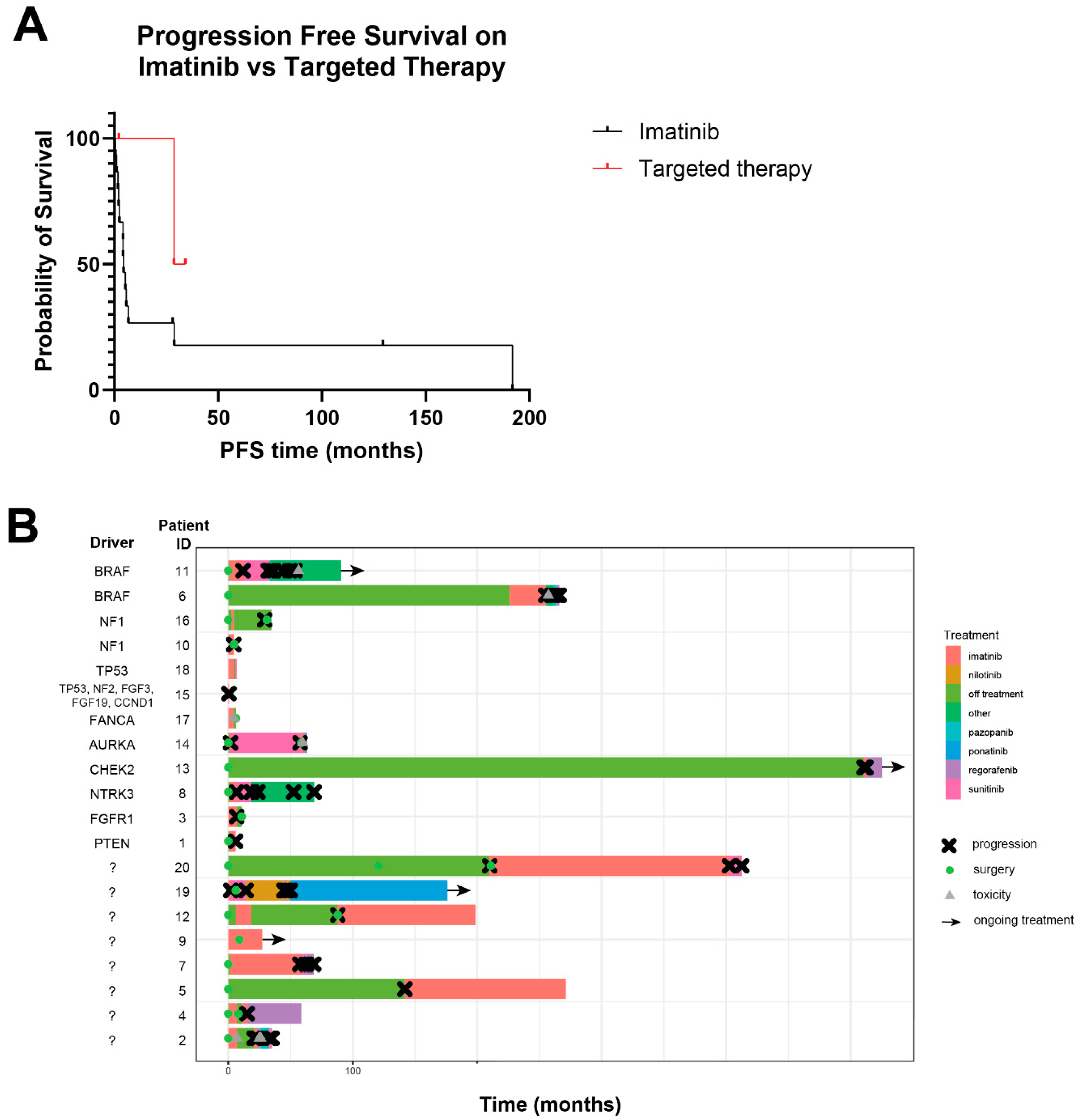
Figure 1. Clinical characteristics and outcomes in triple-negative GIST. (A) Distribution of age at diagnosis. (B) Distribution of tumor size at diagnosis. (C) Distribution of mitotic count from original biopsy or resected specimen. In (AC), bars represent means ± SD. (D) Anatomic distribution of triple-negative GIST cases. (E) On the left in colored bars are the initial disease stage. Black and gray bars on the right indicate the percent of patients that remained with localized disease versus those that developed recurrent or metastatic disease. Bars represent percentages plus standard error of proportion.
Cancers 16 01707 g001
Figure 2. Genomics of triple-negative GIST. (A) Map of the genomic alterations in triple-negative GIST patients. Each row represents a patient. Each column represents a clinical feature (left 3 columns) or gene, as indicated. White boxes indicate that the gene was profiled but that no alteration was found, and gray boxes indicate that the gene was not profiled. (B) Number of somatic mutations detected by clinical sequencing assays. Each dot represents a single tumor, and bars represent mean ± SD. (C) Distribution of the most commonly altered genes in the cohort. (D) Percentage of tumors with each hypothesized driver mutation. In (C,D), percentages plus standard error of proportion are plotted.
Cancers 16 01707 g002
Figure 3. Response to treatment in triple-negative GIST. (A) Progression-free survival while on imatinib (n = 15 patients) versus molecularly matched treatments (n = 3 patients). p = 0.21. (B) Swimmer’s plot showing timeline of indicated therapies. Purported driver mutation is shown in the column on the left.
Cancers 16 01707 g003
Figure 4. Survival outcomes in triple-negative GIST. Overall survival (A), recurrence-free survival (B), and progression-free survival (C) of patients with triple- negative GIST. Recurrence-free survival was calculated in patients with initially localized disease from the date of histologic diagnosis to the date of recurrence, death, or the latest follow-up. Progression-free survival was calculated from the start of therapy to the date of recurrence, death, or the latest follow-up.
Cancers 16 01707 g004
Table 1. Clinical characteristics of triple-negative GIST cohort.
IDAge at DiagnosisPrimary SiteStage at Diagnosis (AJCC 8th)Site of Recurrence/MetastasisImatinib Received in Which Setting?PFS on Imatinib (Months)Time to Relapse from First Therapy (Months)DriverSequencing Platform
141GastricIIIBLocalAdjuvantNA4.1PTEN deletionNeotype, Boston Gene
268Small intestineIVPelvisAdjuvantNA2.4?MDA
338Small intestineIANANeoadjuvant6.7NAFGFR1 K654EMDA
421GastricIBLocal, liverNeoadjuvant and adjuvant4.17.1?Boston Gene, Foundation One, MDA
517GastricIVLiverRecurrent/metastatic129.4141.8?MDA
612Gastric?Right psoas, retroperitoneumRecurrent/metastatic28.8226.2BRAF V600EMDA
757Small intestineIBLiverAdjuvantNA57.4?MDA
844GastricIVLocal, liver, spleenRecurrent/metastatic5.76.6ETV6-NTRK3 fusionFoundation One, MDA
937GastricIVOmentumNeoadjuvant28NA?OSI
1072Small intestineIVPancreasNeoadjuvant5.24.4NF1 deletionMDA
1158Small intestineIIIBBladder, abdomenAdjuvantNA10.8BRAF V600EMDA
1248Small intestineIIIALocalAdjuvantNA88?MDA
1314Gastric?Liver, lungRecurrent/metastatic1.7509.9CHEK2 frameshiftFoundation One
1473Small intestineIVOmentum, lymph nodesRecurrent/metastatic11AURKA-CSTF1 fusionFoundation One
1550Gastric?Local, liver, pancreas, periportal region, duodenum, omentumNeoadjuvant0.52.2NF2 splice site mutation, FGF3/FGF19 amplification, CCND1 amplification, TP53 frameshiftMDA
1658GastricIILocalAdjuvantNA29.2NF1 splice site mutationMDA
1754RectumIIIAPeritoneum, ovaryNeoadjuvant4.44.4FANCA lossBoston Gene
1853Small intestineIVLiverRecurrent/metastatic4.24.2TP53 missense and deletionCaris, Boston Gene
1925GastricIVLiverRecurrent/metastatic2.02.0 ?OSI panel
2011Gastric?Liver, omentumRecurrent/metastatic191.8120.7 ?Endeavor, Boston Gene
MDA = MD Anderson in-house test; OSI = outside institution; ? indicates that no potential driver alteration was identified.
Disclaimer/Publisher’s Note: The statements, opinions and data contained in all publications are solely those of the individual author(s) and contributor(s) and not of MDPI and/or the editor(s). MDPI and/or the editor(s) disclaim responsibility for any injury to people or property resulting from any ideas, methods, instructions or products referred to in the content.

