Article

Systematic Assessment of Small RNA Profiling in Human Extracellular Vesicles

1
Department of Biostatistics, Vanderbilt University Medical Center, Nashville, TN 37232, USA
2
Center for Quantitative Sciences, Vanderbilt University Medical Center, Nashville, TN 37232, USA
3
Department of Cell and Developmental Biology, Vanderbilt University School of Medicine, Nashville, TN 37232, USA
4
Center for Extracellular Vesicle Research, Vanderbilt University School of Medicine, Nashville, TN 37232, USA
5
Department of Medicine, Vanderbilt University Medical Center, Nashville, TN 37232, USA
6
Department of Biological Sciences, Vanderbilt University, Nashville, TN 37232, USA
7
Department of Pathology, Microbiology and Immunology, Vanderbilt University Medical Center, Nashville, TN 37232, USA
*
Authors to whom correspondence should be addressed.
These authors contributed equally to this work.
Cancers 2023, 15(13), 3446; https://doi.org/10.3390/cancers15133446
Submission received: 23 May 2023 / Revised: 22 June 2023 / Accepted: 28 June 2023 / Published: 30 June 2023

Abstract

:

Simple Summary

Small RNA sequencing has been widely used for characterizing the landscape of small non-coding RNAs—the most abundant cargo in extracellular vesicles (EVs). Here, we performed a systematic assessment of the quality, technical, and potential biological biases introduced by different EV isolation methods and the enrichment of specific, small RNA biotypes in EVs. The findings in this study guide the quality control of EV small RNA-seq and the selection of EV isolation techniques and enhance the interpretation of small RNA contents and the preferential loading of specific RNA biotypes into EVs.

Abstract

Motivation: Extracellular vesicles (EVs) are produced and released by most cells and are now recognized to play a role in intercellular communication through the delivery of molecular cargo, including proteins, lipids, and RNA. Small RNA sequencing (small RNA-seq) has been widely used to characterize the small RNA content in EVs. However, there is a lack of a systematic assessment of the quality, technical biases, RNA composition, and RNA biotypes enrichment for small RNA profiling of EVs across cell types, biofluids, and conditions. Methods: We collected and reanalyzed small RNA-seq datasets for 2756 samples from 83 studies involving 55 with EVs only and 28 with both EVs and matched donor cells. We assessed their quality by the total number of reads after adapter trimming, the overall alignment rate to the host and non-host genomes, and the proportional abundance of total small RNA and specific biotypes, such as miRNA, tRNA, rRNA, and Y RNA. Results: We found that EV extraction methods varied in their reproducibility in isolating small RNAs, with effects on small RNA composition. Comparing proportional abundances of RNA biotypes between EVs and matched donor cells, we discovered that rRNA and tRNA fragments were relatively enriched, but miRNAs and snoRNA were depleted in EVs. Except for the export of eight miRNAs being context-independent, the selective release of most miRNAs into EVs was study-specific. Conclusion: This work guides quality control and the selection of EV isolation methods and enhances the interpretation of small RNA contents and preferential loading in EVs.

Graphical Abstract

1. Introduction

Extracellular vesicles (EVs) are lipid-bound particles that are secreted from cells into the extracellular environment. Although EVs were initially considered to be part of a waste-removal mechanism to discard unwanted cellular materials [1], increasing evidence suggests that EVs play a fundamental and evolutionarily conserved role in cellular communication by delivering molecular cargo [2,3,4,5,6,7,8,9]. EVs carry a range of molecular cargo, including lipids, DNA, RNA, and proteins, which can be taken up by recipient cells and alter their gene expression and function [8,10,11]. Decoding EV cargo is important since cargo composition reflects the functional state of donor cells, and the alteration of cargo is associated with homeostasis and disease progression [12,13,14,15,16].
Among the variety of molecular cargo in EVs, small non-coding RNAs are the most abundant, including miRNAs, rRNA fragments, tRNA fragments, Y RNAs, snRNAs, and snoRNAs [1,17]. Among these, miRNAs are the best characterized in post-transcriptional alteration of gene expression in recipient cells, affecting cellular responses to stress and inflammation and driving disease progression [18]. As an example, miR-155 in adipocyte-derived microvesicles mediates M1 macrophage polarization, which reciprocally regulates insulin signaling and glucose uptake in adipocytes and thus causes chronic inflammation and local insulin resistance [19,20]. In addition, small non-coding RNAs in EVs can serve as biomarkers and therapeutic targets in a variety of diseases, including HIV, cancer, cardiovascular disease, Type 1 and Type 2 Diabetes, COVID-19, glioma, and neurological disorders [21,22,23,24,25,26,27]. miR-486-3p is a good example of a circulating biomarker, as it has been reported to distinguish glioblastoma from lower-grade astrocytoma [28]. Therapeutically, the use of EVs to deliver miR-219a-5p might be a feasible and promising strategy to induce remyelination in multiple sclerosis patients, since the overexpression of miR-219a-5p in EVs induced more differentiation of oligodendrocyte precursor cells than liposomes and polymeric nanoparticles [29]. These findings highlight the need for new technologies for the association of biomarkers with a specific exosome subtype and the exosome subtype to a particular function and/or group of functions [26,27,30].
Small RNA sequencing has been widely used for characterizing small RNA landscapes in EVs [29,31,32,33,34,35,36,37,38,39,40,41,42,43]. Depending on the size of the vesicle and the purification strategy, some studies have revealed a large overlap and good correlation between EV and cellular small RNA, whereas other studies have demonstrated selective miRNA exports [34,35,36,37,40,42,43]. Although these findings shed light on small RNAs in EVs, they are limited to certain cell types or conditions. Several EV repositories have been developed, including exoRBase [44], EVmiRNA [45], and EVAtlas [46], which provide rich resources of long RNA and small RNA profiling in EVs. However, there is a lack of systematic analyses to provide a global picture of the quality and preferential loadings of small RNAs in EVs. The Extracellular RNA Communication Consortium (ERCC) led an effort to build a reference catalog of extracellular RNA in five human biofluids covering twenty-three health conditions [47]. However, the samples were collected primarily from cell-free biofluids and contained combinations of vesicular and non-vesicular particles, which resulted in significant variability between and within studies [48]. Here, we focused on small RNAs within membrane-bound EVs. We collected and reanalyzed all the publicly available EV small RNA-seq datasets from biofluids, cell lines, and primary cell cultures. We performed a systematic assessment of the quality, technical, and potential biological biases introduced by different EV isolation methods and specific biotype enrichment in EVs compared to matched cellular levels. Our findings not only guide quality control and the selection of EV isolation methods but also enhance the interpretation of small RNA contents and preferential loading of specific RNA biotypes from individual studies as well as across studies.

2. Materials and Methods

2.1. Data Sources

We used the terms “(exosome OR exosomes OR ectosomes OR microvesicles OR microvesicles OR “EV” OR “extracellular RNA” OR (extracellular AND vesicle) OR (extracellular AND vesicles)) AND “Homo sapiens”[porgn] AND “gse”[Filter]) NOT “exosome complex” NOT “nuclear exosome” NOT “RNA exosome” AND “high throughput sequencing”[PTYP]“ to search the Gene Expression Omnibus (GEO) and the Sequence Read Archive (SRA). In addition, we reviewed all datasets deposited in the ERCC and publications listed on vendor websites selling exosome extraction kits. We downloaded and manually checked all publicly available datasets from the GEO, SRA, and ERCC. We only kept small RNA-seq datasets from human EVs and removed those from non-vesicles, non-small RNA-seq, or other organisms. To ensure EVs are enriched, we only kept studies where EVs were isolated using either commercial exosome extraction kits, such as Exo-Quick, ExoEasy, qEV, and total exosome isolation kit (Thermo, Waltham, MA, USA), or differential ultracentrifugation, which have been proven to isolate high-yield and high-purity EVs [43,49,50,51,52]. Besides, most studies further verified the presence of EVs using several methods, including Western blotting of EV-enriched proteins (such as CD63, CD81, and CD9) [53,54,55,56,57] and non-EV-enriched proteins [53,55,58,59,60], qualitative (such as electron microscopy and atomic force microscopy) and quantitative methods (such as nanoparticle tracking analysis, dynamic light scattering, tunable resistive pulse sensing, and high-resolution flow cytometry) [53,55,57,58,59,61], and a wide-field and a close-up electron microscopy image [55,61,62]. Notably, each EV isolation method is prone to isolating some contaminants. This study analyzes the presence of different small RNA species in different EV-enriched isolates (or fractions) from different methods. After filtering, 2756 samples from 83 studies including 55 studies with EVs only and 28 studies with both EVs and matched donor cells, were kept for downstream analysis. We pulled experimental metadata from the original source and then conducted manual curation to try to standardize the information, such as extraction methods and small RNA preparation kits.

2.2. Small RNA-Seq Data Analysis

We processed and analyzed all the raw sequencing data uniformly. Since a variety of commercial and customized small RNA library preparation kits were used, different adapter patterns were included in raw reads because adaptor sequences are not always available. To streamline the analysis, we developed a Python package, FindAdapt, for fast, accurate, and automatic detection of adapter patterns without any prior information (https://github.com/chc-code/findadapt, accessed on 23 October 2022). The adapter patterns identified by FindAdapt, including adapter sequences and random bases at 5′ and 3′ ends, were provided to our standard small RNA-seq analysis pipeline, TIGER, for adapter trimming, reads mapping to host and non-host genomes, and quantification and differential expression of a variety of small RNA biotypes [63]. Briefly, Cutadapt (v2.10) [64] was used to trim 3′ adapters and random 5′ and 3′ bases. Reads with fewer than 16 nucleotides were designated as “too short” and discarded. Quality control on both raw reads and adaptor-trimmed reads was evaluated using FastQC (v0.11.9) (www.bioinformatics.babraham.ac.uk/projects/fastqc (accessed on 23 October 2022)). After trimming, reads were first mapped to a customized database built from the host genome (GENCODE GRCh37.p13) and from known sequences of host mature transcripts curated in specific library databases (such as for miRNAs in miRbase and tRNAs in GtRNAdb2) by Bowtie1 (v1.3.0) [65], allowing one mismatch. Mapped reads were assigned to different classes of annotated small RNAs, including miRNA, tRNA, rRNA, snRNA, snoRNA, Y RNA, and lincRNA. Unmapped reads longer than 19 nucleotides were then aligned to non-host genomes in parallel, including exogenous structural RNA databases and curated exogenous genome databases (bacteria, fungal, algae, and viral), allowing no mismatches. Raw counts and normalized counts per million total counts (CPM) were reported for each small RNA.

2.3. Small RNA-Seq Data Normalization and Comparison

The proportion of total mapped reads and host genome reads was calculated by the number of total mapped reads and host genome reads divided by the total number of reads after trimming. Small RNA abundance was determined by the number of small RNA reads divided by the number of host genome reads. The abundance of each type of small RNA was determined by the number of reads that mapped to that type of small RNA relative to the total number of small RNA reads. To reduce the potential bias from characteristic compositions of different EV isolation methods, three-way ANOVA with the EV isolation method as a confounding variable was used to estimate the significance of enrichment or depletion of RNA biotypes in EV compared to cells. The expression of miRNAs was normalized to the median value across all samples to identify highly expressed miRNAs in both EVs and cells, as well as EV-specific miRNAs. For each study with matched cell and EV samples, DESeq2 [66] was used to detect differentially expressed miRNAs with the EV isolation method as a confounding variable. miRNAs with FDR < 0.05 and an absolute value of fold change > 1.5 were selected to be significantly differential.

3. Results

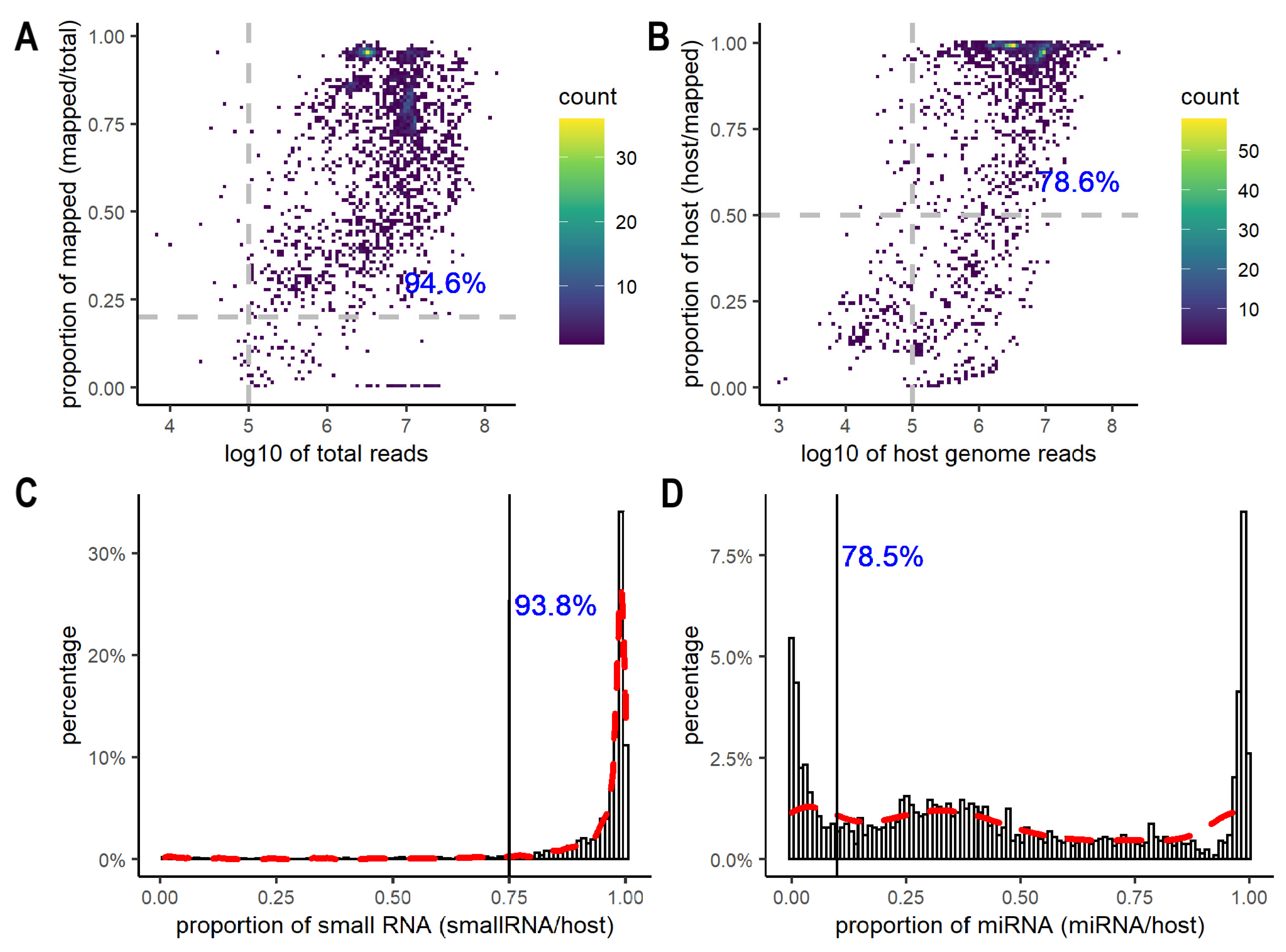
3.1. A Global View of Quality in EV Small RNA Sequencing

After excluding 513 samples from the donor cells, we assessed the quality of 2243 EV datasets using the total number of reads after adapter trimming, the overall mapping rates to both the host and non-host genomes, the total number of reads and the mapping rates to the host genome only, and the proportion of small RNA and miRNA reads (Supplementary Table S1). After adapter trimming, most (94.6%) datasets had more than 100,000 reads and greater than 20% mapping rates when aligned to both host and non-host genomes (Figure 1A, Supplementary Table S1). Data with less than 100,000 reads or 20% mapping rates were flagged and excluded from downstream analyses. Most of the excluded samples came from GSE100467 (38 samples), GSE148576 (38 samples), and GSE115572 (24 samples), all of which were large-scale studies (>100 samples) from blood or plasma. Since other samples from the same experimental series achieved a decent number of reads and mapping rates, caution needs to be taken for analyzing and interpreting those samples with extremely low numbers of reads and mapping rates. Besides examining the overall mapping rates to both host and non-host genomes, it is important to consider the number and the percentage of reads aligned to the host genome only. The ERC Consortium quality control for small RNA-seq data requires a minimum of 100,000 reads mapped to the host genome, and the percentage of the host genome reads greater than 50% [48]. Among the 2120 datasets, 1963 datasets had more than 100,000 reads mapped to the host genome; 1681 contained greater than 50% of host genome reads, and 78.6% of the datasets met both criteria (Figure 1B, Supplementary Table S1). In addition, we further evaluated the percentage of small RNA and miRNA in the host genome reads (Figure 1C,D). As expected, most host genome reads were mapped to small RNAs. Small RNAs accounted for greater than 75% of the host genome reads in 93.8% of the datasets (Figure 1C). In comparison, miRNAs showed a wide range of distribution across the datasets, with some having a very low fraction of miRNAs (<10%), while others contained a high percentage of miRNA reads (>80%) (Figure 1D). Overall, 78.5% of datasets had miRNA reads constituting greater than 10% of the host genome reads (Figure 1D, Supplementary Table S1). We decided to exclude the experimental series where more than half of the samples had either <75% small RNAs or <10% miRNA reads since they were outliers with high sparsity in miRNAs and other non-coding RNAs.
We further explored the median and the interquartile range (IQR) of small RNA proportions within each study. The IQR is a measure of variability, and a high IQR suggests high variability in the study. Most studies obtained high median values (>0.9) and small IQRs (<0.06), indicating high abundances and low variability in small RNA content (Figure S1). Several studies, however, showed a high IQR in small RNA content (Figure S1). If high variability arose partly from replicates in these studies, caution and additional evaluation are needed to reanalyze and reuse these studies due to low consistency and replicability. After grouping studies by the donor source, we found that EVs from biofluids had significantly higher median values of small RNA proportions than those from cell lines and primary cell cultures (Figure S1A, t-test: p = 2.37 × 10⁻⁵/0.02). EVs from urine obtained a larger IQR in small RNA proportions than EVs from other biofluids (Figure S1B, t-test: p = 0.003).
There are six commonly used extraction methods: Exo-Quick, ExoEasy, ExoRNeasy, qEV, total exosome isolation kit (Thermo), and differential ultracentrifugation. Sorting studies by EV extraction methods, we found that differential ultracentrifugation generally resulted in significantly larger IQRs in small RNA proportions than other methods, except total exosome isolation kit (Figure 2A, t-test: p = 0.006/0.002/0.03/0.008/0.26, differential ultracentrifugation compared to Exo-Quick kit, ExoEasy, ExoRNeasy, qEV, and total exosome isolation kit, respectively). To reduce the potential bias introduced by different experimental conditions and small RNA extraction methods, we focused on studies of plasma only and further narrowed down to those using miRNeasy kits for RNA purification. Comparing the variability after restricting to these studies resulted in the same conclusion: that differential ultracentrifugation purification consistently showed higher variability and lower replicability in small RNA proportions within each study (Figure 2B,C). These findings suggest that differential ultracentrifugation may not be as reproducible as other methods for small RNA isolation, which is consistent with previous findings [67,68]. Besides high variability in small RNA proportions within each study, differential ultracentrifugation showed high variability across studies as well, as indicated by the high variation of median values (Figure 2). The high variability across studies might be due to differences in ultracentrifugation equipment, protocols, centrifugation rotors, times, speeds, and whether iodixanol cushions were used [69,70].

3.2. Small RNA Composition in EVs

There are a variety of small RNA biotypes found in EVs [23,47,71]. We investigated the relative abundance of eight small RNA biotypes across EV studies from plasma, including miRNAs, tRNA fragments, rRNA fragments, Y RNAs, snRNA fragments, snoRNA fragments, mt-tRNA fragments, and miscellaneous RNAs (misc-RNAs). MiRNAs were the most abundant RNA in plasma EVs, with a median proportion value of 39.6%. Besides miRNAs, tRNA fragments, rRNA fragments, and Y RNAs also contributed to a significant portion of small RNAs (Figure 3A and Figure S2). Y RNAs were the second most abundant small RNA (median: 15.9%), followed by rRNA fragments (median: 10.5%) and tRNA fragments (median: 3.2%). In comparison, the proportions of snRNA fragments, snoRNA fragments, mt-tRNA fragments, and misc-RNAs were very low (<1%) (Figure S3). The relative representation of these different small RNAs was highly variable across different EV samples (Figure S2). Interestingly, different EV extraction methods seemed to capture different small RNA compositions. This is consistent with previous studies, which reported that different EV extraction methods have characteristic compositions, that is, EVs with different degrees of other small RNA carriers [8,10,72]. Differential ultracentrifugation seemed to capture more misc-RNAs and snoRNA fragments (Figure S3). In plasma EV samples, Exo-Quick and ExoRNeasy kits captured more miRNAs compared to rRNA fragments (Figure 3B and Figure S2). Eight plasma studies using the Exo-Quick kit and three studies using ExoRNeasy all showed median values of log2-transformed ratios of miRNA/rRNA greater than 1 (Figure 3B). This is consistent with a recent study that ranked Exo-Quick as the top/second for detecting the EV miRNA markers [73]. ExoRNeasy and the total exosome isolation kit seemed to enrich for Y RNAs compared to tRNA fragments (Figure 3C and Figure S2). Six studies using the two kits all showed high Y RNA/tRNA fragment ratios (Figure 3C).

3.3. Enrichment of RNA Biotypes in EVs

We analyzed 28 studies that profiled small RNAs from both EVs and their donor cells, which allowed us to explore the preferential enrichment of small RNA biotypes in EVs. Of the 28 studies, we further focused on 15 studies with EVs released from cell lines because they were more biologically homogenous compared to biofluids and primary cell cultures. From these studies, we observed a consistent RNA enrichment preference regardless of EV isolation methods (Figure 4). The overall miRNA proportion in EV was significantly lower than their matched cellular levels (p = 7.56 × 10−64, three-way ANOVA with the EV isolation method as a confounding variable; Figure 4A). In addition, the miRNA content across these EV samples was generally more variable than that observed in the donor cells. Similarly, snoRNA fragments were detected at significantly lower abundance in EVs compared to matched cellular levels, except for two studies (GSE143613 and GSE85761) (p = 4.19 × 10⁻²¹, three-way ANOVA; Figure 4B). These findings suggest that miRNAs and snoRNA fragments are more likely to be retained in cells than transported into EVs. In contrast, rRNA fragments and tRNA fragments showed significantly higher levels in EVs compared to their cellular levels (p = 1.58 × 10⁻³⁰ and p = 4.67 × 10⁻²⁵, respectively, three-way ANOVA; Figure 4C,D), suggesting they are more likely to be transported into EVs.
Focusing on specific miRNAs, we found that 15 miRNAs were abundant in both EVs and donor cells (Figure 5A), including the hsa-let-7 family, miR-26a-5p, miR-30d-5p, miR-25-3p, and miR-21-5p. It is well known that let-7 family members are the most abundant among all miRNAs in the cell [74,75,76], and we found that these miRNAs were highly abundant in EVs as well. In addition, we discovered nine miRNAs that were highly abundant in EVs but not in the cells, including hsa-miR-451a, hsa-miR-486-5p, hsa-miR-122-5p, hsa-miR-146a-5p, hsa-miR-199a-3p; hsa-miR-199b-3p, hsa-miR-143-3p, hsa-miR-144-3p, hsa-miR-21-5p, and hsa-miR-223-5p (Figure 5A). Differential expression with the EV isolation method as a confounding variable also demonstrated that the nine miRNAs were significantly enriched in EVs compared to the cells (log2FC > 3 and FDR < 0.01), indicating that their high abundance was not likely to be biased by different EV isolation methods. Among the nine miRNAs, mir-451a and miR-144 have been previously reported to show higher levels in human EVs compared to cellular levels [35].
We calculated the differential expression of miRNAs between EVs and matched cellular levels. Eight miRNAs were found to be preferentially exported into EVs in a context-independent way (Figure 5B highlighted in cyan), and ten other miRNAs were more enriched in EVs in all except three datasets (GSE165323, GSE143613, and GSE85761) (Figure 5B highlighted in magenta). These miRNAs include hsa-miR-199a-3p; hsa-miR-199b-3p, and miR-320 family (miR-320a-3p, miR-320b, miR-320c, and miR-320d). Previously, miR-320b was found to be abundant in exosomes and underrepresented in matched donor cells [43]. Our findings validated that report and suggested that sorting of the miR-320 family members into EVs might be more general. In contrast, seven miRNAs were enriched in cells compared to matched EVs (Figure 5B, highlighted in blue). Although several miRNAs showed an overall preference to be either loaded into EVs or retained in cells, the selective sorting of miRNAs into EVs was mostly study-specific. For instance, seven miRNAs were enriched only in EVs derived from breast cancer cell lines and adipose MSCs (Figure 5B, highlighted in green), one of which was miR-100-5p. miR-100-5p has been reported to inhibit the proliferation, migration, and invasion of breast cancer cells [77]. Five miRNAs including miR-93-5p, miR-125a-5p, miR-1306-5p, miR-147b-3p, and miR-190a-5p were selectively retained in cells only in colorectal cancer cell lines (Figure 5B, highlighted in lime). All of them were abnormally expressed in colorectal cancer [78,79,80,81,82,83]. Selective sorting of functionally important miRNAs into EVs derived from specific cancer cell types might be part of molecular signaling pathways that drive cancer initiation/progression.

4. Discussion

An increasing number of studies on EV small RNA profiling have now been published across different cell types and conditions. Here, we provide a comprehensive assessment and comparison of all the publicly available datasets across 83 studies involving a variety of diseases and conditions. We analyzed the overall quality, including the total number of reads, the overall mapping rates to both host and non-host genomes, as well as the mapping rates to the host genome only, and the small RNA and miRNA proportions. The majority of small RNA reads in EVs were derived from miRNAs, Y RNAs, and fragments of rRNA and tRNA. Full-length miRNAs were present in most samples but contributed only a small portion of the overall reads and are highly variable across studies. We also evaluated the technical and potential biological biases introduced by different EV isolation methods. Notably, we found that differential ultracentrifugation had high variability within each study and across studies on small RNA proportions. Together, the analysis of such a large-scale collection provides a global picture of quality in EV small RNA-seq experiments and helps with future quality control and selection of EV isolation methods.
EV isolates have characteristic compositions. In other words, they are usually enriched in EV and to a different degree in other small RNA carriers that vary according to the isolation principle/method used [8,10,72]. Each of the common EV isolation methods is prone to isolating some contaminants; for example, Exo-Quick and total exosome isolation are likely to obtain proteins by PEG-precipitation, while SEC from plasma usually includes a certain proportion of lipoproteins [72,84,85]. For differential ultracentrifugation, the greater variability in reproducibility of isolating small RNAs could come from the fact that ultracentrifugation succeeds in purifying the EV better than other methods [86]. Also, ultracentrifugation has more equipment, protocol, and operator-dependent variation, hence the variability [10]. Although our analysis tried to reduce the bias from different EV isolation methods, we still need to be cautious about the results interpretation.
Our study showed low variability in the total small RNA proportions but high variations in the small RNA compositions. The high variability of small RNA compositions across studies might be driven by multiple factors, including experimental conditions, technical factors such as isolation methods, and inherent EV heterogeneity [47,87,88,89]. Given these confounding factors, it is challenging to characterize cargo within specific EVs because of differences in the biogenesis of different classes of EVs due to heterogeneity in size and composition [90]. EVs can be classified by size into exosomes (30–150 nm), microvesicles (100–1000 nm), large oncosomes (1000–10,000 nm), and apoptotic bodies (100–5000 nm), but different studies have used different size ranges in their definition of these particles [91,92]. To make things worse, there is no standard terminology for EV types. Some studies use “exosome” as a generic descriptor of EVs, while others define “exosome” to be EVs strictly originating within the endosomal system [93]. This is compounded by the fact that different names have been used even when the same isolation method was used. For example, some studies using Exo-Quick kits refer to the resulting particles as EVs, while others refer to these particles as exosomes [94,95,96,97,98,99,100,101]. The International Society of Extracellular Vesicles (ISEVs) has recommended standardized nomenclature in which “exosomes” are only used when derived from multivesicular bodies as part of the endosomal system, whereas EVs should be used when the mode of biogenesis is unclear [102,103]. For ongoing retrospective analyses of EVs, adherence to the ISEV standards and methodology is recommended to reduce sources of pre-analytical variability and allow proper comparison between studies [104].
In addition to pre-analytical variability, there are also downstream analytical biases driven by the choice of computational methods and assumptions, for example, how to normalize data for accurate comparison. A common assumption in most normalization methods is that the amount of total input RNA content is the same across experimental conditions. However, when comparing small RNA biotypes between cells and EVs, that assumption might not always be true. For example, Sork et al. (2018) found that parental cells expressed abundant levels of small RNA, while the small RNA content in EVs was modest and highly variable across samples [35]. If the total small RNA content is significantly different across conditions, normalization methods could introduce computational bias and lead to wrong conclusions. A way around this problem is to use spike-in controls to create a standard baseline measurement, which would enable accurate measurement of biological differences in total small RNA content between samples.
Despite the challenges above, we sought to determine global enrichment for each small RNA biotype. We found that miRNAs and snoRNA fragments are generally more likely to be retained in cells rather than exported into EVs, while rRNA fragments and tRNA fragments are more likely to be released into EVs. This finding may provide a clue toward understanding the selective sorting of small RNAs into EVs. miRNAs are typically associated with Ago2 within RISC complexes, but eukaryotic cells express multiple Ago proteins. Differential association with Ago proteins, as is observed in plants [105,106], could regulate differential export. Here, we identified several miRNAs exported under most conditions, whereas most miRNAs display context-dependent sorting. One possibility for context-dependent sorting is differential association with specific RNA-binding proteins, which are able to enter EVs and potentially carry their RNA cargo [90,107,108,109,110,111,112,113]. Integration of proteomic and small RNA profiling will help uncover the relationships between specific RBPs and exported miRNAs. A map of RBPs and their binding targets from ENCODE eCLIP data [114,115], computational prediction [116], or enriched sequence motifs should further confirm the connection.

5. Conclusions

We analyzed the overall quality of all the publicly available small RNA-seq data on EVs. We found that small RNA reads in EVs were mainly derived from miRNAs, Y RNAs, and fragments of rRNA and tRNA. We further evaluated the technical and potential biological biases introduced by different EV isolation techniques in terms of small RNA proportions. The analysis presents a global picture of quality in EV small RNA-seq experiments and guides future quality control and selection of EV isolation methods.
We further studied the preferential enrichment of small RNA biotypes in EVs compared to their cellular levels. We revealed that miRNAs and snoRNA fragments are more likely to be retained in cells, while rRNA fragments and tRNA fragments are more likely to be transported into EVs. Although several miRNAs showed an overall preference to be either loaded into EVs or retained in cells, selective sorting of miRNAs into EVs was mostly study-specific. Together, our findings shed light on the preferential loading of specific RNA biotypes into EVs.

Supplementary Materials

The following supporting information can be downloaded at https://www.mdpi.com/article/10.3390/cancers15133446/s1. Table S1: Alignment matrix and other information for each sample; Figure S1: Proportion of host small RNA relative to host genome for (A) all studies colored by the type of donors: cell line, primary cell culture, and biofluids, and (B) biofluid studies colored by source; Figure S2: Proportion of miRNA, rRNA fragments, tRNA fragments, and Y RNA fragments relative to host total small RNA colored by EV extraction methods; Figure S3: Proportion of misc-RNA, mt-tRNA fragments, snRNA fragments, and snoRNA fragments relative to host total small RNA colored by EV extraction methods.

Author Contributions

Conceptualization, Q.L. and J.W.; methodology, Q.L., J.W. and H.-C.C.; formal analysis, J.W. and H.-C.C.; investigation, Q.L. and Y.S.; data curation, J.W., H.-C.C. and Q.S.; writing—original draft preparation, J.W.; writing—review and editing, Q.L., T.R.D., R.J.C., J.G.P. and A.M.W.; supervision, Q.L. All authors have read and agreed to the published version of the manuscript.

Funding

This research was funded by the National Cancer Institute grants (U2C CA233291, U54 CA217450, P01CA229123, and U54 CA274367), the National Institutes of Health (P01 AI139449), and the Cancer Center Support Grant (P30CA068485).

Institutional Review Board Statement

Not applicable.

Informed Consent Statement

Not applicable.

Data Availability Statement

The data presented in this study are available in Table S1.

Conflicts of Interest

The authors declare no conflict of interest.

References

  1. Clancy, J.L.; Patel, H.R.; Hussein, S.M.I.; Tonge, P.D.; Cloonan, N.; Corso, A.J.; Li, M.; Lee, D.-S.; Shin, J.-Y.; Wong, J.J.L.; et al. Small RNA changes en route to distinct cellular states of induced pluripotency. Nat. Commun. 2014, 5, 5522. [Google Scholar] [CrossRef] [Green Version]
  2. Gross, N.; Kropp, J.; Khatib, H. MicroRNA Signaling in Embryo Development. Biology 2017, 6, 34. [Google Scholar] [CrossRef] [PubMed]
  3. Cruz, L.; Romero, J.A.A.; Iglesia, R.P.; Lopes, M.H. Extracellular Vesicles: Decoding a New Language for Cellular Communication in Early Embryonic Development. Front. Cell Dev. Biol. 2018, 6, 94. [Google Scholar] [CrossRef] [PubMed]
  4. Baj-Krzyworzeka, M.; Szatanek, R.; Węglarczyk, K.; Baran, J.; Urbanowicz, B.; Brański, P.; Ratajczak, M.Z.; Zembala, M. Tumour-derived microvesicles carry several surface determinants and mRNA of tumour cells and transfer some of these determinants to monocytes. Cancer Immunol. Immunother. 2006, 55, 808–818. [Google Scholar] [CrossRef]
  5. Pegtel, D.M.; Cosmopoulos, K.; Thorley-Lawson, D.A.; van Eijndhoven, M.A.; Hopmans, E.S.; Lindenberg, J.L.; de Gruijl, T.D.; Würdinger, T.; Middeldorp, J.M. Functional delivery of viral miRNAs via exosomes. Proc. Natl. Acad. Sci. USA 2010, 107, 6328–6333. [Google Scholar] [CrossRef] [Green Version]
  6. Ratajczak, J.; Wysoczynski, M.; Hayek, F.; Janowskawieczorek, A.; Ratajczak, M.Z. Membrane-derived microvesicles: Important and underappreciated mediators of cell-to-cell communication. Leukemia 2006, 20, 1487–1495. [Google Scholar] [CrossRef] [PubMed] [Green Version]
  7. Skog, J.; Würdinger, T.; Van Rijn, S.; Meijer, D.H.; Gainche, L.; Curry, W.T., Jr.; Carter, B.S.; Krichevsky, A.M.; Breakefield, X.O. Glioblastoma microvesicles transport RNA and proteins that promote tumour growth and provide diagnostic biomarkers. Nat. Cell Biol. 2008, 10, 1470–1476. [Google Scholar] [CrossRef] [PubMed]
  8. Yanez-Mo, M.; Siljander, P.R.-M.; Andreu, Z.; Bedina Zavec, A.; Borràs, F.E.; Buzas, E.I.; Buzas, K.; Casal, E.; Cappello, F.; Carvalho, J.; et al. Biological properties of extracellular vesicles and their physiological functions. J. Extracell. Vesicles 2015, 4, 27066. [Google Scholar] [CrossRef] [Green Version]
  9. Chetty, V.K.; Ghanam, J.; Anchan, S.; Reinhardt, K.; Brenzel, A.; Gelléri, M.; Cremer, C.; Grueso-Navarro, E.; Schneider, M.; von Neuhoff, N.; et al. Efficient Small Extracellular Vesicles (EV) Isolation Method and Evaluation of EV-Associated DNA Role in Cell-Cell Communication in Cancer. Cancers 2022, 14, 2068. [Google Scholar] [CrossRef]
  10. Zaborowski, M.P.; Balaj, L.; Breakefield, X.O.; Lai, C.P. Extracellular Vesicles: Composition, Biological Relevance, and Methods of Study. Bioscience 2015, 65, 783–797. [Google Scholar] [CrossRef] [Green Version]
  11. Bebelman, M.P.; Smit, M.J.; Pegtel, D.M.; Baglio, S.R. Biogenesis and function of extracellular vesicles in cancer. Pharmacol. Ther. 2018, 188, 1–11. [Google Scholar] [CrossRef] [PubMed]
  12. Yim, K.H.W.; Al Hrout, A.; Borgoni, S.; Chahwan, R. Extracellular Vesicles Orchestrate Immune and Tumor Interaction Networks. Cancers 2020, 12, 3696. [Google Scholar] [CrossRef]
  13. Cappariello, A.; Rucci, N. Tumour-Derived Extracellular Vesicles (EVs): A Dangerous “Message in A Bottle” for Bone. Int. J. Mol. Sci. 2019, 20, 4805. [Google Scholar] [CrossRef] [PubMed] [Green Version]
  14. Georgievski, A.; Michel, A.; Thomas, C.; Mlamla, Z.; de Barros, J.-P.P.; Lemaire-Ewing, S.; Garrido, C.; Quéré, R. Acute lymphoblastic leukemia-derived extracellular vesicles affect quiescence of hematopoietic stem and progenitor cells. Cell Death Dis. 2022, 13, 337. [Google Scholar] [CrossRef]
  15. Xiao, Y.; Zheng, L.; Zou, X.; Wang, J.; Zhong, J.; Zhong, T. Extracellular vesicles in type 2 diabetes mellitus: Key roles in pathogenesis, complications, and therapy. J. Extracell. Vesicles 2019, 8, 1625677. [Google Scholar] [CrossRef] [PubMed] [Green Version]
  16. Zhang, Y.; Shi, L.; Mei, H.; Zhang, J.; Zhu, Y.; Han, X.; Zhu, D. Inflamed macrophage microvesicles induce insulin resistance in human adipocytes. Nutr. Metab. 2015, 12, 21. [Google Scholar] [CrossRef] [PubMed] [Green Version]
  17. Galagali, H.; Kim, J.K. The multifaceted roles of microRNAs in differentiation. Curr. Opin. Cell Biol. 2020, 67, 118–140. [Google Scholar] [CrossRef]
  18. Yu, X.; Odenthal, M.; Fries, J.W.U. Exosomes as miRNA Carriers: Formation-Function-Future. Int. J. Mol. Sci. 2016, 17, 2028. [Google Scholar] [CrossRef] [Green Version]
  19. Polina, E.R.; Oliveira, F.M.; Sbruzzi, R.C.; Crispim, D.; Canani, L.H.; Santos, K.G. Gene polymorphism and plasma levels of miR-155 in diabetic retinopathy. Endocr. Connect. 2019, 8, 1591–1599. [Google Scholar] [CrossRef] [Green Version]
  20. Zhang, Y.; Mei, H.; Chang, X.; Chen, F.; Zhu, Y.; Han, X. Adipocyte-derived microvesicles from obese mice induce M1 macrophage phenotype through secreted miR-155. J. Mol. Cell Biol. 2016, 8, 505–517. [Google Scholar] [CrossRef] [Green Version]
  21. Okoye, I.; Xu, L.; Oyegbami, O.; Shahbaz, S.; Pink, D.; Gao, P.; Sun, X.; Elahi, S. Plasma Extracellular Vesicles Enhance HIV-1 Infection of Activated CD4(+) T Cells and Promote the Activation of Latently Infected J-Lat10.6 Cells via miR-139-5p Transfer. Front. Immunol. 2021, 12, 697604. [Google Scholar] [CrossRef]
  22. Peruzzotti-Jametti, L.; Bernstock, J.D.; Willis, C.M.; Manferrari, G.; Rogall, R.; Fernandez-Vizarra, E.; Williamson, J.C.; Braga, A.; van den Bosch, A.; Leonardi, T.; et al. Neural stem cells traffic functional mitochondria via extracellular vesicles. PLoS Biol. 2021, 19, e3001166. [Google Scholar] [CrossRef] [PubMed]
  23. Chettimada, S.; Lorenz, D.R.; Misra, V.; Wolinsky, S.M.; Gabuzda, D. Small RNA sequencing of extracellular vesicles identifies circulating miRNAs related to inflammation and oxidative stress in HIV patients. BMC Immunol. 2020, 21, 57. [Google Scholar] [CrossRef] [PubMed]
  24. Xavier, C.P.R.; Caires, H.R.; Barbosa, M.A.G.; Bergantim, R.; Guimarães, J.E.; Vasconcelos, M.H. The Role of Extracellular Vesicles in the Hallmarks of Cancer and Drug Resistance. Cells 2020, 9, 1141. [Google Scholar] [CrossRef] [PubMed]
  25. De Toro, J.; Herschlik, L.; Waldner, C.; Mongini, C. Emerging roles of exosomes in normal and pathological conditions: New insights for diagnosis and therapeutic applications. Front. Immunol. 2015, 6, 203. [Google Scholar] [CrossRef] [Green Version]
  26. Mimmi, S.; Zimbo, A.M.; Rotundo, S.; Cione, E.; Nisticò, N.; Aloisio, A.; Maisano, D.; Tolomeo, A.M.; Dattilo, V.; Lionello, R.; et al. SARS-CoV-2 spike protein-guided exosome isolation facilitates detection of potential miRNA biomarkers in COVID-19 infections. Clin. Chem. Lab. Med. 2023. [Google Scholar] [CrossRef]
  27. Luo, H.; Zhang, H.; Mao, J.; Cao, H.; Tao, Y.; Zhao, G.; Zhang, Z.; Zhang, N.; Liu, Z.; Zhang, J.; et al. Exosome-based nanoimmunotherapy targeting TAMs, a promising strategy for glioma. Cell Death Dis. 2023, 14, 235. [Google Scholar] [CrossRef]
  28. Hallal, S.; Khani, S.E.; Wei, H.; Lee, M.Y.T.; Sim, H.-W.; Sy, J.; Shivalingam, B.; Buckland, M.E.; Alexander-Kaufman, K.L. Deep Sequencing of Small RNAs from Neurosurgical Extracellular Vesicles Substantiates miR-486-3p as a Circulating Biomarker that Distinguishes Glioblastoma from Lower-Grade Astrocytoma Patients. Int. J. Mol. Sci. 2020, 21, 4954. [Google Scholar] [CrossRef]
  29. Osorio-Querejeta, I.; Carregal-Romero, S.; Ayerdi-Izquierdo, A.; Mäger, I.; Nash, L.A.; Wood, M.; Egimendia, A.; Betanzos, M.; Alberro, A.; Iparraguirre, L.; et al. MiR-219a-5p Enriched Extracellular Vesicles Induce OPC Differentiation and EAE Improvement More Efficiently Than Liposomes and Polymeric Nanoparticles. Pharmaceutics 2020, 12, 186. [Google Scholar] [CrossRef] [Green Version]
  30. Raghavan, K.S.; Francescone, R.; Franco-Barraza, J.; Gardiner, J.C.; Vendramini-Costa, D.B.; Luong, T.; Pourmandi, N.; Andren, A.; Kurimchak, A.; Ogier, C.; et al. NetrinG1(+) cancer-associated fibroblasts generate unique extracellular vesicles that support the survival of pancreatic cancer cells under nutritional stress. Cancer Res. Commun. 2022, 2, 1017–1036. [Google Scholar] [CrossRef]
  31. Bydak, B.; Pierdoná, T.M.; Seif, S.; Sidhom, K.; Obi, P.O.; Labouta, H.I.; Gordon, J.W.; Saleem, A. Characterizing Extracellular Vesicles and Particles Derived from Skeletal Muscle Myoblasts and Myotubes and the Effect of Acute Contractile Activity. Membranes 2022, 12, 464. [Google Scholar] [CrossRef] [PubMed]
  32. Iparraguirre, L.; Alberro, A.; Hansen, T.B.; Castillo-Triviño, T.; Muñoz-Culla, M.; Otaegui, D. Profiling of Plasma Extracellular Vesicle Transcriptome Reveals That circRNAs Are Prevalent and Differ between Multiple Sclerosis Patients and Healthy Controls. Biomedicines 2021, 9, 1850. [Google Scholar] [CrossRef] [PubMed]
  33. Godakumara, K.; Ord, J.; Lättekivi, F.; Dissanayake, K.; Viil, J.; Boggavarapu, N.R.; Faridani, O.R.; Jääger, K.; Velthut-Meikas, A.; Jaakma, Ü.; et al. Trophoblast derived extracellular vesicles specifically alter the transcriptome of endometrial cells and may constitute a critical component of embryo-maternal communication. Reprod. Biol. Endocrinol. 2021, 19, 115. [Google Scholar] [CrossRef] [PubMed]
  34. Kaur, S.; Abu-Shahba, A.G.; Paananen, R.O.; Hongisto, H.; Hiidenmaa, H.; Skottman, H.; Seppänen-Kaijansinkko, R.; Mannerström, B. Small non-coding RNA landscape of extracellular vesicles from human stem cells. Sci. Rep. 2018, 8, 15503. [Google Scholar] [CrossRef] [Green Version]
  35. Sork, H.; Corso, G.; Krjutskov, K.; Johansson, H.J.; Nordin, J.Z.; Wiklander, O.P.B.; Lee, Y.X.F.; Westholm, J.O.; Lehtiö, J.; Wood, M.J.A.; et al. Heterogeneity and interplay of the extracellular vesicle small RNA transcriptome and proteome. Sci. Rep. 2018, 8, 10813. [Google Scholar] [CrossRef] [Green Version]
  36. Lässer, C.; Shelke, G.V.; Yeri, A.; Kim, D.-K.; Crescitelli, R.; Raimondo, S.; Sjöstrand, M.; Gho, Y.S.; Van Keuren Jensen, K.; Lötvall, J. Two distinct extracellular RNA signatures released by a single cell type identified by microarray and next-generation sequencing. RNA Biol. 2017, 14, 58–72. [Google Scholar] [CrossRef]
  37. Lunavat, T.R.; Cheng, L.; Kim, D.-K.; Bhadury, J.; Jang, S.C.; Lässer, C.; Sharples, R.A.; López, M.D.; Nilsson, J.; Gho, Y.S.; et al. Small RNA deep sequencing discriminates subsets of extracellular vesicles released by melanoma cells—Evidence of unique microRNA cargos. RNA Biol. 2015, 12, 810–823. [Google Scholar] [CrossRef] [Green Version]
  38. Van Balkom, B.W.; Eisele, A.S.; Pegtel, D.M.; Bervoets, S.; Verhaar, M.C. Quantitative and qualitative analysis of small RNAs in human endothelial cells and exosomes provides insights into localized RNA processing, degradation and sorting. J. Extracell. Vesicles 2015, 4, 26760. [Google Scholar] [CrossRef]
  39. Tosar, J.P.; Gámbaro, F.; Sanguinetti, J.; Bonilla, B.; Witwer, K.W.; Cayota, A. Assessment of small RNA sorting into different extracellular fractions revealed by high-throughput sequencing of breast cell lines. Nucleic Acids Res. 2015, 43, 5601–5616. [Google Scholar] [CrossRef] [Green Version]
  40. Bellingham, S.A.; Coleman, B.M.; Hill, A.F. Small RNA deep sequencing reveals a distinct miRNA signature released in exosomes from prion-infected neuronal cells. Nucleic Acids Res. 2012, 40, 10937–10949. [Google Scholar] [CrossRef] [Green Version]
  41. Guduric-Fuchs, J.; O’connor, A.; Camp, B.; O’Neill, C.L.; Medina, R.J.; Simpson, D.A. Selective extracellular vesicle-mediated export of an overlapping set of microRNAs from multiple cell types. BMC Genom. 2012, 13, 357. [Google Scholar] [CrossRef] [PubMed] [Green Version]
  42. Nolte-’t Hoen, E.N.M.; Buermans, H.P.J.; Waasdorp, M.; Stoorvogel, W.; Wauben, M.H.M.; ’t Hoen, P.A.C. Deep sequencing of RNA from immune cell-derived vesicles uncovers the selective incorporation of small non-coding RNA biotypes with potential regulatory functions. Nucleic Acids Res. 2012, 40, 9272–9285. [Google Scholar] [CrossRef] [Green Version]
  43. Cha, D.J.; Franklin, J.L.; Dou, Y.; Liu, Q.; Higginbotham, J.N.; Demory Beckler, M.; Weaver, A.M.; Vickers, K.; Prasad, N.; Levy, S.; et al. KRAS-dependent sorting of miRNA to exosomes. Elife 2015, 4, e07197. [Google Scholar] [CrossRef]
  44. Lai, H.; Li, Y.; Zhang, H.; Hu, J.; Liao, J.; Su, Y.; Li, Q.; Chen, B.; Li, C.; Wang, Z.; et al. exoRBase 2.0: An atlas of mRNA, lncRNA and circRNA in extracellular vesicles from human biofluids. Nucleic Acids Res. 2021, 50, D118–D128. [Google Scholar] [CrossRef]
  45. Liu, T.; Zhang, Q.; Zhang, J.; Li, C.; Miao, Y.-R.; Lei, Q.; Li, Q.; Guo, A.-Y. EVmiRNA: A database of miRNA profiling in extracellular vesicles. Nucleic Acids Res. 2018, 47, D89–D93. [Google Scholar] [CrossRef] [PubMed] [Green Version]
  46. Liu, C.-J.; Xie, G.-Y.; Miao, Y.-R.; Xia, M.; Wang, Y.; Lei, Q.; Zhang, Q.; Guo, A.-Y. EVAtlas: A comprehensive database for ncRNA expression in human extracellular vesicles. Nucleic Acids Res. 2021, 50, D111–D117. [Google Scholar] [CrossRef] [PubMed]
  47. Murillo, O.D.; Thistlethwaite, W.; Rozowsky, J.; Subramanian, S.L.; Lucero, R.; Shah, N.; Jackson, A.R.; Srinivasan, S.; Chung, A.; Laurent, C.D.; et al. exRNA Atlas Analysis Reveals Distinct Extracellular RNA Cargo Types and Their Carriers Present across Human Biofluids. Cell 2019, 177, 463–477.e15. [Google Scholar] [CrossRef] [Green Version]
  48. Rozowsky, J.; Kitchen, R.R.; Park, J.J.; Galeev, T.R.; Diao, J.; Warrell, J.; Thistlethwaite, W.; Subramanian, S.L.; Milosavljevic, A.; Gerstein, M. exceRpt: A Comprehensive Analytic Platform for Extracellular RNA Profiling. Cell Syst. 2019, 8, 352–357.e3. [Google Scholar] [CrossRef] [Green Version]
  49. Sohel, M.H.; Hoelker, M.; Noferesti, S.S.; Salilew-Wondim, D.; Tholen, E.; Looft, C.; Rings, F.; Uddin, M.J.; Spencer, T.E.; Schellander, K.; et al. Exosomal and Non-Exosomal Transport of Extra-Cellular microRNAs in Follicular Fluid: Implications for Bovine Oocyte Developmental Competence. PLoS ONE 2013, 8, e78505. [Google Scholar] [CrossRef] [Green Version]
  50. Hartman, Z.C.; Wei, J.; Glass, O.K.; Guo, H.; Lei, G.; Yang, X.-Y.; Osada, T.; Hobeika, A.; Delcayre, A.; Le Pecq, J.-B.; et al. Increasing vaccine potency through exosome antigen targeting. Vaccine 2011, 29, 9361–9367. [Google Scholar] [CrossRef] [Green Version]
  51. Da Silveira, J.C.; Veeramachaneni, D.R.; Winger, Q.A.; Carnevale, E.M.; Bouma, G.J. Cell-secreted vesicles in equine ovarian follicular fluid contain miRNAs and proteins: A possible new form of cell communication within the ovarian follicle. Biol. Reprod. 2012, 86, 71. [Google Scholar] [CrossRef]
  52. Higginbotham, J.N.; Beckler, M.D.; Gephart, J.D.; Franklin, J.L.; Bogatcheva, G.; Kremers, G.-J.; Piston, D.W.; Ayers, G.D.; McConnell, R.E.; Tyska, M.J.; et al. Amphiregulin exosomes increase cancer cell invasion. Curr. Biol. 2011, 21, 779–786. [Google Scholar] [CrossRef] [PubMed] [Green Version]
  53. Menon, R.; Debnath, C.; Lai, A.; Guanzon, D.; Bhatnagar, S.; Kshetrapal, P.K.; Sheller-Miller, S.; Salomon, C.; The Garbhini Study Team. Circulating Exosomal miRNA Profile During Term and Preterm Birth Pregnancies: A Longitudinal Study. Endocrinology 2019, 160, 249–275. [Google Scholar] [CrossRef] [Green Version]
  54. Anastasi, F.; Masciandaro, S.M.; Del Carratore, R.; Dell’anno, M.T.; Signore, G.; Falleni, A.; McDonnell, L.A.; Bongioanni, P. Proteomics Profiling of Neuron-Derived Small Extracellular Vesicles from Human Plasma: Enabling Single-Subject Analysis. Int. J. Mol. Sci. 2021, 22, 2951. [Google Scholar] [CrossRef]
  55. Zhao, F.; Xu, Y.; Liu, N.; Lv, D.; Chen, Y.; Liu, Z.; Jin, X.; Xiao, M.; Lavillette, D.; Zhong, J.; et al. Extracellular vesicles from Zika virus-infected cells display viral E protein that binds ZIKV-neutralizing antibodies to prevent infection enhancement. EMBO J. 2023, 42, e112096. [Google Scholar] [CrossRef]
  56. Lapitz, A.; Azkargorta, M.; Milkiewicz, P.; Olaizola, P.; Zhuravleva, E.; Grimsrud, M.M.; Schramm, C.; Arbelaiz, A.; O’Rourke, C.J.; La Casta, A.; et al. Liquid biopsy-based protein biomarkers for risk prediction, early diagnosis and prognostication of cholangiocarcinoma. J. Hepatol. 2023, 79, 93–108. [Google Scholar] [CrossRef] [PubMed]
  57. Mehanny, M.; Kroniger, T.; Koch, M.; Hoppstädter, J.; Becher, D.; Kiemer, A.K.; Lehr, C.; Fuhrmann, G. Yields and Immunomodulatory Effects of Pneumococcal Membrane Vesicles Differ with the Bacterial Growth Phase. Adv. Healthc. Mater. 2021, 11, e2101151. [Google Scholar] [CrossRef] [PubMed]
  58. Tsang, E.K.; Abell, N.S.; Li, X.; Anaya, V.; Karczewski, K.J.; Knowles, D.A.; Sierra, R.G.; Smith, K.S.; Montgomery, S.B. Small RNA Sequencing in Cells and Exosomes Identifies eQTLs and 14q32 as a Region of Active Export. G3 2017, 7, 31–39. [Google Scholar] [CrossRef] [Green Version]
  59. Au Yeung, C.L.; Co, N.-N.; Tsuruga, T.; Yeung, T.-L.; Kwan, S.Y.; Leung, C.S.; Li, Y.; Lu, E.S.; Kwan, K.; Wong, K.-K.; et al. Exosomal transfer of stroma-derived miR21 confers paclitaxel resistance in ovarian cancer cells through targeting APAF1. Nat. Commun. 2016, 7, 11150. [Google Scholar] [CrossRef] [Green Version]
  60. Fallen, S.; Baxter, D.; Wu, X.; Kim, T.; Shynlova, O.; Lee, M.Y.; Scherler, K.; Lye, S.; Hood, L.; Wang, K. Extracellular vesicle RNAs reflect placenta dysfunction and are a biomarker source for preterm labour. J. Cell. Mol. Med. 2018, 22, 2760–2773. [Google Scholar] [CrossRef] [Green Version]
  61. Truong, G.; Guanzon, D.; Kinhal, V.; Elfeky, O.; Lai, A.; Longo, S.; Nuzhat, Z.; Palma, C.; Scholz-Romero, K.; Menon, R.; et al. Oxygen tension regulates the miRNA profile and bioactivity of exosomes released from extravillous trophoblast cells—Liquid biopsies for monitoring complications of pregnancy. PLoS ONE 2017, 12, e0174514. [Google Scholar] [CrossRef] [Green Version]
  62. Hannafon, B.N.; Carpenter, K.J.; Berry, W.L.; Janknecht, R.; Dooley, W.C.; Ding, W.-Q. Exosome-mediated microRNA signaling from breast cancer cells is altered by the anti-angiogenesis agent docosahexaenoic acid (DHA). Mol. Cancer 2015, 14, 133. [Google Scholar] [CrossRef] [Green Version]
  63. Allen, R.M.; Zhao, S.; Solano, M.A.R.; Zhu, W.; Michell, D.L.; Wang, Y.; Shyr, Y.; Sethupathy, P.; Linton, M.F.; Graf, G.A.; et al. Bioinformatic analysis of endogenous and exogenous small RNAs on lipoproteins. J. Extracell. Vesicles 2018, 7, 1506198. [Google Scholar] [CrossRef] [PubMed] [Green Version]
  64. Martin, M. Cutadapt removes adapter sequences from high-throughput sequencing reads. EMBnet J. 2011, 17, 3. [Google Scholar] [CrossRef]
  65. Langmead, B.; Trapnell, C.; Pop, M.; Salzberg, S.L. Ultrafast and memory-efficient alignment of short DNA sequences to the human genome. Genome Biol. 2009, 10, R25. [Google Scholar] [CrossRef] [Green Version]
  66. Love, M.I.; Huber, W.; Anders, S. Moderated estimation of fold change and dispersion for RNA-seq data with DESeq2. Genome Biol. 2014, 15, 550. [Google Scholar] [CrossRef] [PubMed] [Green Version]
  67. Caradec, J.; Kharmate, G.; Hosseini-Beheshti, E.; Adomat, H.; Gleave, M.; Guns, E. Reproducibility and efficiency of serum-derived exosome extraction methods. Clin. Biochem. 2014, 47, 1286–1292. [Google Scholar] [CrossRef] [PubMed]
  68. Helwa, I.; Cai, J.; Drewry, M.D.; Zimmerman, A.; Dinkins, M.B.; Khaled, M.L.; Seremwe, M.; Dismuke, W.M.; Bieberich, E.; Stamer, W.D.; et al. A Comparative Study of Serum Exosome Isolation Using Differential Ultracentrifugation and Three Commercial Reagents. PLoS ONE 2017, 12, e0170628. [Google Scholar] [CrossRef] [Green Version]
  69. Ayala-Mar, S.; Donoso-Quezada, J.; Gallo-Villanueva, R.C.; Perez-Gonzalez, V.H.; González-Valdez, J. Recent advances and challenges in the recovery and purification of cellular exosomes. Electrophoresis 2019, 40, 3036–3049. [Google Scholar] [CrossRef] [Green Version]
  70. Livshits, M.A.; Khomyakova, E.; Evtushenko, E.G.; Lazarev, V.N.; Kulemin, N.A.; Semina, S.E.; Generozov, E.V.; Govorun, V.M. Isolation of exosomes by differential centrifugation: Theoretical analysis of a commonly used protocol. Sci. Rep. 2015, 5, 17319. [Google Scholar] [CrossRef] [Green Version]
  71. Fiskaa, T.; Knutsen, E.; Nikolaisen, M.A.; Jørgensen, T.E.; Johansen, S.D.; Perander, M.; Seternes, O.M. Distinct Small RNA Signatures in Extracellular Vesicles Derived from Breast Cancer Cell Lines. PLoS ONE 2016, 11, e0161824. [Google Scholar] [CrossRef] [Green Version]
  72. Doyle, L.; Wang, M. Overview of Extracellular Vesicles, Their Origin, Composition, Purpose, and Methods for Exosome Isolation and Analysis. Cells 2019, 8, 727. [Google Scholar] [CrossRef] [PubMed] [Green Version]
  73. Ter-Ovanesyan, D.; Norman, M.; Lazarovits, R.; Trieu, W.; Lee, J.; Church, G.M.; Walt, D.R. Framework for rapid comparison of extracellular vesicle isolation methods. Elife 2021, 10, e70725. [Google Scholar] [CrossRef] [PubMed]
  74. Wen, K.; Ni, K.; Guo, J.; Bu, B.; Liu, L.; Pan, Y.; Li, J.; Luo, M.; Deng, L. MircroRNA Let-7a-5p in Airway Smooth Muscle Cells is Most Responsive to High Stretch in Association With Cell Mechanics Modulation. Front. Physiol. 2022, 13, 830406. [Google Scholar] [CrossRef] [PubMed]
  75. Polikepahad, S.; Knight, J.M.; Naghavi, A.O.; Oplt, T.; Creighton, C.J.; Shaw, C.; Benham, A.L.; Kim, J.; Soibam, B.; Harris, R.A.; et al. Proinflammatory role for let-7 microRNAS in experimental asthma. J. Biol. Chem. 2010, 285, 30139–30149. [Google Scholar] [CrossRef] [Green Version]
  76. Chen, S.-Y.; Wang, Y.; Telen, M.J.; Chi, J.-T. The genomic analysis of erythrocyte microRNA expression in sickle cell diseases. PLoS ONE 2008, 3, e2360. [Google Scholar]
  77. Xie, H.; Xiao, R.; He, Y.; He, L.; Xie, C.; Chen, J.; Hong, Y. MicroRNA-100 inhibits breast cancer cell proliferation, invasion and migration by targeting FOXA1. Oncol. Lett. 2021, 22, 816. [Google Scholar] [CrossRef]
  78. Wang, W.; Zhang, J.; Fan, Y.; Zhang, L. MiR-1306-5p predicts favorable prognosis and inhibits proliferation, migration, and invasion of colorectal cancer cells via PI3K/AKT/mTOR pathway. Cell Cycle 2022, 21, 1491–1501. [Google Scholar] [CrossRef]
  79. Svoboda, M.; Sana, J.; Fabian, P.; Kocakova, I.; Gombosova, J.; Nekvindova, J.; Radova, L.; Vyzula, R.; Slaby, O. MicroRNA expression profile associated with response to neoadjuvant chemoradiotherapy in locally advanced rectal cancer patients. Radiat. Oncol. 2012, 7, 195. [Google Scholar] [CrossRef] [Green Version]
  80. Imedio, L.; Cristóbal, I.; Rubio, J.; Santos, A.; Rojo, F.; García-Foncillas, J. MicroRNAs in Rectal Cancer: Functional Significance and Promising Therapeutic Value. Cancers 2020, 12, 2040. [Google Scholar] [CrossRef]
  81. Ning, X.; Wang, C.; Zhang, M.; Wang, K. Ectopic Expression of miR-147 Inhibits Stem Cell Marker and Epithelial-Mesenchymal Transition (EMT)-Related Protein Expression in Colon Cancer Cells. Oncol. Res. 2019, 27, 399–406. [Google Scholar] [CrossRef]
  82. Zhu, F.; Wei, J.; He, D.; He, J.; Liu, L.; Hou, H.; Shi, H.; Jin, S.; Li, J.; Shi, X.; et al. The miRNA125a-5p and miRNA125b-1-5p cluster induces cell invasion by down-regulating DDB2-reduced epithelial-to-mesenchymal transition (EMT) in colorectal cancer. J. Gastrointest. Oncol. 2022, 13, 3112–3122. [Google Scholar] [CrossRef] [PubMed]
  83. Mullany, L.E.; Slattery, M.L. The functional role of miRNAs in colorectal cancer: Insights from a large population-based study. Cancer Biol. Med. 2019, 16, 211–219. [Google Scholar] [PubMed] [Green Version]
  84. De Sousa, K.P.; Rossi, I.; Abdullahi, M.; Ramirez, M.I.; Stratton, D.; Inal, J.M. Isolation and characterization of extracellular vesicles and future directions in diagnosis and therapy. Wiley Interdiscip Rev. Nanomed. Nanobiotechnol. 2023, 15, e1835. [Google Scholar] [CrossRef] [PubMed]
  85. Liangsupree, T.; Multia, E.; Riekkola, M.-L. Modern isolation and separation techniques for extracellular vesicles. J. Chromatogr. A 2021, 1636, 461773. [Google Scholar] [CrossRef]
  86. Carnino, J.M.; Lee, H.; Jin, Y. Isolation and characterization of extracellular vesicles from Broncho-alveolar lavage fluid: A review and comparison of different methods. Respir. Res. 2019, 20, 240. [Google Scholar] [CrossRef] [Green Version]
  87. Jeppesen, D.K.; Fenix, A.M.; Franklin, J.L.; Higginbotham, J.N.; Zhang, Q.; Zimmerman, L.J.; Liebler, D.C.; Ping, J.; Liu, Q.; Evans, R.; et al. Reassessment of Exosome Composition. Cell 2019, 177, 428–445.e18. [Google Scholar] [CrossRef] [Green Version]
  88. Driedonks, T.; Twilhaar, M.K.N.; Hoen, E.N.M.N. Technical approaches to reduce interference of Fetal calf serum derived RNA in the analysis of extracellular vesicle RNA from cultured cells. Extracell. Vesicles 2019, 8, 1552059. [Google Scholar] [CrossRef] [Green Version]
  89. Van Deun, J.; Mestdagh, P.; Sormunen, R.; Cocquyt, V.; Vermaelen, K.; Vandesompele, J.; Bracke, M.; De Wever, O.; Hendrix, A. The impact of disparate isolation methods for extracellular vesicles on downstream RNA profiling. J. Extracell. Vesicles 2014, 3, 24858. [Google Scholar] [CrossRef] [Green Version]
  90. Dixson, A.C.; Dawson, T.R.; Di Vizio, D.; Weaver, A.M. Context-specific regulation of extracellular vesicle biogenesis and cargo selection. Nat. Rev. Mol. Cell Biol. 2023, 24, 454–476. [Google Scholar] [CrossRef]
  91. Romani, P.; Brian, I.; Santinon, G.; Pocaterra, A.; Audano, M.; Pedretti, S.; Mathieu, S.; Forcato, M.; Bicciato, S.; Manneville, J.-B.; et al. Extracellular matrix mechanical cues regulate lipid metabolism through Lipin-1 and SREBP. Nature 2019, 21, 338–347. [Google Scholar] [CrossRef] [PubMed] [Green Version]
  92. Zhao, Z.; Wijerathne, H.; Godwin, A.K.; Soper, S.A. Isolation and analysis methods of extracellular vesicles (EVs). Extracell. Vesicles Circ. Nucleic Acids 2021, 2, 80–103. [Google Scholar]
  93. Witwer, K.W.; Théry, C. Extracellular vesicles or exosomes? On primacy, precision, and popularity influencing a choice of nomenclature. J. Extracell. Vesicles 2019, 8, 1648167. [Google Scholar] [CrossRef] [PubMed]
  94. Slonchak, A.; Clarke, B.; Mackenzie, J.; Amarilla, A.A.; Setoh, Y.X.; Khromykh, A.A. West Nile virus infection and interferon alpha treatment alter the spectrum and the levels of coding and noncoding host RNAs secreted in extracellular vesicles. BMC Genom. 2019, 20, 474. [Google Scholar] [CrossRef] [PubMed] [Green Version]
  95. Kaur, S.; Elkahloun, A.G.; Arakelyan, A.; Young, L.; Myers, T.G.; Otaizo-Carrasquero, F.; Wu, W.; Margolis, L.; Roberts, D.D. CD63, MHC class 1, and CD47 identify subsets of extracellular vesicles containing distinct populations of noncoding RNAs. Sci. Rep. 2018, 8, 2577. [Google Scholar] [CrossRef] [PubMed] [Green Version]
  96. Yuan, T.; Huang, X.; Woodcock, M.; Du, M.; Dittmar, R.; Wang, Y.; Tsai, S.; Kohli, M.; Boardman, L.; Patel, T.; et al. Plasma extracellular RNA profiles in healthy and cancer patients. Sci. Rep. 2016, 6, 19413. [Google Scholar] [CrossRef] [Green Version]
  97. Liu, T.; Du, L.-T.; Wang, Y.-S.; Gao, S.-Y.; Li, J.; Li, P.-L.; Sun, Z.-W.; Binang, H.; Wang, C.-X. Development of a Novel Serum Exosomal MicroRNA Nomogram for the Preoperative Prediction of Lymph Node Metastasis in Esophageal Squamous Cell Carcinoma. Front. Oncol. 2020, 10, 573501. [Google Scholar] [CrossRef]
  98. Nair, V.; Ge, Y.; Li, S.; Pincas, H.; Jain, N.; Seenarine, N.; Amper, M.A.S.; Goodpaster, B.H.; Walsh, M.J.; Coen, P.; et al. Sedentary and Trained Older Men Have Distinct Circulating Exosomal microRNA Profiles at Baseline and in Response to Acute Exercise. Front. Physiol. 2020, 11, 605. [Google Scholar] [CrossRef]
  99. Parimon, T.; Brauer, R.; Schlesinger, S.Y.; Xie, T.; Jiang, D.; Ge, L.; Huang, Y.; Birkland, T.P.; Parks, W.C.; Habiel, D.M.; et al. Syndecan-1 Controls Lung Tumorigenesis by Regulating miRNAs Packaged in Exosomes. Am. J. Pathol. 2018, 188, 1094–1103. [Google Scholar] [CrossRef] [PubMed] [Green Version]
  100. Du, M.; Giridhar, K.V.; Tian, Y.; Tschannen, M.R.; Zhu, J.; Huang, C.C.; Kilari, D.; Kohli, M.; Wang, L. Plasma exosomal miRNAs-based prognosis in metastatic kidney cancer. Oncotarget 2017, 8, 63703–63714. [Google Scholar] [CrossRef] [PubMed] [Green Version]
  101. Huang, X.; Yuan, T.; Tschannen, M.; Sun, Z.; Jacob, H.; Du, M.; Liang, M.; Dittmar, R.L.; Liu, Y.; Liang, M.; et al. Characterization of human plasma-derived exosomal RNAs by deep sequencing. BMC Genom. 2013, 14, 319. [Google Scholar] [CrossRef] [Green Version]
  102. Pérez-Boza, J.; Lion, M.; Struman, I. Exploring the RNA landscape of endothelial exosomes. RNA 2018, 24, 423–435. [Google Scholar] [CrossRef] [PubMed] [Green Version]
  103. Théry, C.; Witwer, K.W.; Aikawa, E.; Alcaraz, M.J.; Anderson, J.D.; Andriantsitohaina, R.; Antoniou, A.; Arab, T.; Archer, F.; Atkin-Smith, G.K.; et al. Minimal information for studies of extracellular vesicles 2018 (MISEV2018): A position statement of the International Society for Extracellular Vesicles and update of the MISEV2014 guidelines. J. Extracell. Vesicles 2018, 7, 1535750. [Google Scholar] [CrossRef] [PubMed] [Green Version]
  104. Consortium, E.-T.; Mestdagh, P.; Agostinis, P.; Akay, Ö.; Anand, S.; Anckaert, J.; Martinez, Z.A.; Baetens, T.; Beghein, E.; Bertier, L.; et al. EV-TRACK: Transparent reporting and centralizing knowledge in extracellular vesicle research. Nat. Methods 2017, 14, 228–232. [Google Scholar] [CrossRef] [PubMed]
  105. Kim, V.N. Sorting out small RNAs. Cell 2008, 133, 25–26. [Google Scholar] [CrossRef] [Green Version]
  106. Czech, B.; Hannon, G.J. Small RNA sorting: Matchmaking for Argonautes. Nat. Rev. Genet. 2011, 12, 19–31. [Google Scholar] [CrossRef]
  107. He, B.; Cai, Q.; Qiao, L.; Huang, C.-Y.; Wang, S.; Miao, W.; Ha, T.; Wang, Y.; Jin, H. RNA-binding proteins contribute to small RNA loading in plant extracellular vesicles. Nat. Plants 2021, 7, 342–352. [Google Scholar] [CrossRef] [PubMed]
  108. Wozniak, A.L.; Adams, A.; King, K.E.; Dunn, W.; Christenson, L.K.; Hung, W.-T.; Weinman, S.A. The RNA binding protein FMR1 controls selective exosomal miRNA cargo loading during inflammation. J. Cell Biol. 2020, 219, e201912074. [Google Scholar] [CrossRef]
  109. Statello, L.; Maugeri, M.; Garre, E.; Nawaz, M.; Wahlgren, J.; Papadimitriou, A.; Lundqvist, C.; Lindfors, L.; Collén, A.; Sunnerhagen, P.; et al. Identification of RNA-binding proteins in exosomes capable of interacting with different types of RNA: RBP-facilitated transport of RNAs into exosomes. PLoS ONE 2018, 13, e0195969. [Google Scholar] [CrossRef] [Green Version]
  110. Villarroya-Beltri, C.; Gutiérrez-Vázquez, C.; Sánchez-Cabo, F.; Pérez-Hernández, D.; Vázquez, J.; Martin-Cofreces, N.; Martinez-Herrera, D.J.; Pascual-Montano, A.; Mittelbrunn, M.; Sánchez-Madrid, F. Sumoylated hnRNPA2B1 controls the sorting of miRNAs into exosomes through binding to specific motifs. Nat. Commun. 2013, 4, 2980. [Google Scholar] [CrossRef] [Green Version]
  111. Santangelo, L.; Giurato, G.; Cicchini, C.; Montaldo, C.; Mancone, C.; Tarallo, R.; Battistelli, C.; Alonzi, T.; Weisz, A.; Tripodi, M. The RNA-Binding Protein SYNCRIP Is a Component of the Hepatocyte Exosomal Machinery Controlling MicroRNA Sorting. Cell Rep. 2016, 17, 799–808. [Google Scholar] [CrossRef] [PubMed] [Green Version]
  112. Hobor, F.; Dallmann, A.; Ball, N.J.; Cicchini, C.; Battistelli, C.; Ogrodowicz, R.W.; Christodoulou, E.; Martin, S.R.; Castello, A.; Tripodi, M.; et al. A cryptic RNA-binding domain mediates Syncrip recognition and exosomal partitioning of miRNA targets. Nat. Commun. 2018, 9, 831. [Google Scholar] [CrossRef] [PubMed] [Green Version]
  113. O’brien, K.; Breyne, K.; Ughetto, S.; Laurent, L.C.; Breakefield, X.O. RNA delivery by extracellular vesicles in mammalian cells and its applications. Nat. Rev. Mol. Cell Biol. 2020, 21, 585–606. [Google Scholar] [CrossRef]
  114. Van Nostrand, E.L.; Freese, P.; Pratt, G.A.; Wang, X.; Wei, X.; Xiao, R.; Blue, S.M.; Chen, J.-Y.; Cody, N.A.L.; Dominguez, D.; et al. A large-scale binding and functional map of human RNA-binding proteins. Nature 2020, 583, 711–719. [Google Scholar] [CrossRef]
  115. Consortium, E.P. An integrated encyclopedia of DNA elements in the human genome. Nature 2012, 489, 57–74. [Google Scholar] [CrossRef] [PubMed] [Green Version]
  116. Yu, H.; Wang, J.; Sheng, Q.; Liu, Q.; Shyr, Y. beRBP: Binding estimation for human RNA-binding proteins. Nucleic Acids Res. 2019, 47, e26. [Google Scholar] [CrossRef] [PubMed] [Green Version]
Figure 1. Quality control for EV small RNA-seq datasets. (A) Heatscatter plot showing the total number of reads on the x-axis and the total percentage of mapped reads on the y-axis. A total of 94.6% of datasets had more than 100,000 reads with greater than 20% mapping rates. (B) Heatscatter plot showing the total number of reads mapped to host genomes on the x-axis and percentage mapping rates to host genomes on the y-axis. A total of 78.6% of datasets had more than 100,000 reads that mapped to host genomes with greater than 50% being host genome reads. (C) Distribution of the proportion of host small RNAs. A total of 93.8% of the datasets reported small RNAs at greater than 75% of the total number of host genome reads. (D) Distribution of the proportion of the host miRNA. A total of 78.5% of the datasets reported miRNA content at greater than 10% of the total number of host genome reads.
Cancers 15 03446 g001
Figure 2. The proportion of host small RNAs across EV studies. Boxplot of the proportion of host small RNAs for all studies (A), studies from plasma (B), and studies from plasma using miRNeasy kits for RNA purification (C). Plots are colored by EV extraction methods.
Cancers 15 03446 g002
Figure 3. Relative abundance of small RNA biotypes across EV studies from plasma. (A) Distribution of the proportion of host miRNAs, rRNA fragments, tRNA fragments, and Y RNA fragments relative to total host small RNA. (B) Boxplot of log2 transformation of miRNA/rRNA ratio. (C) Boxplot of log2 transformation of Y RNA/tRNA ratio. The log2 ratio of 1 is denoted by the red dashed line. Plots are colored by EV extraction methods.
Cancers 15 03446 g003
Figure 4. Enrichment of small RNA biotypes in EVs. Boxplot of the proportion of miRNAs (A), snoRNA fragments (B), rRNA fragments (C) and tRNA fragments (D) in EVs compared to their matched donor cellular levels.
Cancers 15 03446 g004
Figure 5. Enrichment of miRNAs in EVs. (A) Heatmap of the most abundant miRNAs in EV and/or donors. The expression of miRNAs was normalized to the median value across all samples. (B) Heatmap of log2 fold change in miRNAs expression between EVs and their matched donor cells.
Cancers 15 03446 g005
Disclaimer/Publisher’s Note: The statements, opinions and data contained in all publications are solely those of the individual author(s) and contributor(s) and not of MDPI and/or the editor(s). MDPI and/or the editor(s) disclaim responsibility for any injury to people or property resulting from any ideas, methods, instructions or products referred to in the content.

