Skip to Content
You are currently on the new version of our website. Access the old version .

Journal of Molecular Pathology

Journal of Molecular Pathology is an international, peer-reviewed, open access journal on every topic related to modern histopathology and cytopathology, predictive pathology and molecular cytopathology, published quarterly online by MDPI.

All Articles (169)

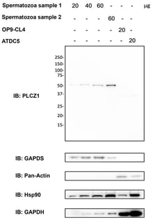
Background/Objectives: Phospholipase C zeta (PLCZ1; PLCζ) is a sperm-specific enzyme responsible for the Ca2+ oscillations required for oocyte activation, and altered PLCζ expression has been associated with fertilization failure in assisted reproductive technologies, particularly intracytoplasmic sperm injection (ICSI). This study aimed to develop and analytically validate a flow cytometry–based protocol for PLCζ quantification in human spermatozoa. Methods: The assay was established using normozoospermic samples and included validated positive and negative technical controls. Antibody specificity was confirmed by Western blot analysis. A defined gating strategy was used to assess linearity between fluorescence intensity and PLCζ expression. Analytical performance was evaluated for precision, reproducibility, stability, and sensitivity, including applicability to low sperm concentrations. Results: A linear relationship between fluorescence intensity and PLCζ expression was demonstrated. The assay showed high precision, reproducibility, and stability, with consistent results in samples stored up to 24 h at room temperature or up to one week post-fixation at 4 °C. Sensitivity testing confirmed suitability for low sperm concentrations. Conclusions: This work provides a standardized and analytically validated framework for PLCζ quantification using flow cytometry. Although the assay measures protein expression rather than functional competence or subcellular localization, it establishes a solid analytical basis for future studies to define clinically relevant PLCζ thresholds and assess its value as a biomarker of fertilization capacity.

9 February 2026

Primary antibody specificity control. Western blot analysis of PLCζ1 expression. Total proteins were extracted from two human samples, OP9-DL4 and ATDC5 cell lines (from left to right). Western blotting was performed with 20–60 μg of proteins. PLCζ1 (top panel), GAPDS (second panel), Pan-Actin (third panel), Hsp90 (fourth panel), and GAPDH (last panel) were revealed as described in the Section 2. (The original Western blot membranes corresponding to Figure 1 are provided in the Supplementary Materials (Supplementary Figures S1–S5)).

Diabetic retinopathy (DR) is a progressive retinal disorder and a leading cause of vision impairment worldwide affecting the livelihood of millions. Its pathogenesis is driven by chronic hyperglycemia-induced neuronal and microvascular injury, leading to capillary occlusion, increased vascular permeability, and the eventual formation of fragile neo vessels. These changes mark the progression from non-proliferative diabetic retinopathy (NPDR) to proliferative diabetic retinopathy (PDR). Diabetic macular edema (DME), characterized by blood–retinal barrier disruption and macular fluid accumulation, further contributes to vision loss. This review provides an integrative perspective on the cellular and molecular mechanisms of DR, highlighting both vascular and neuroglial contributions to retinal pathology. Current therapeutic approaches, including anti-VEGF agents and corticosteroids, offer symptomatic relief but are limited by the need for repeated administration and variability in patient response. Emerging evidence implicates the role of thioredoxin-interacting protein (TXNIP) as one of mediators of the disease progression. Strongly upregulated under hyperglycaemic stress, TXNIP induces oxidative damage, inflammation, and neuronal apoptosis, exacerbating neurovascular dysfunction. We explore potential therapeutic strategies such as gene therapy, TXNIP-targeted molecular interventions, and stem cell-based approaches aimed at achieving long-term modulation of disease mechanisms. This article thus attempts to address a comprehensive understanding of DR pathophysiology and innovative new strategies to improve long-term visual outcomes.

6 February 2026

Molecular mechanisms underlying diabetic retinopathy. Key processes including oxidative stress, inflammation, VEGF activation, blood–retinal barrier (BRB) breakdown, and TXNIP upregulation leading to retinal neurovascular dysfunction are illustrated. Color and arrow legend: Colored shapes represent distinct molecular pathways or signaling components involved in hyperglycemia-induced retinal damage. Yellow indicates cellular stress responses (e.g., ER stress). Purple denotes oxidative and nitrosative stress mediators (ROS/RNS). Green/teal represents antioxidant or redox-regulatory proteins (e.g., TRX). Blue and orange indicate metabolic and enzymatic pathways activated by hyperglycemia (e.g., PKC, hexokinase, NOS). Gray represents mitochondrial-related pathways (e.g., polyol pathway, mitochondrial oxidase). Red text highlights downstream pathological outcomes, including inflammatory factor activation and diabetic retinopathy. Solid arrows indicate activation, induction, or positive regulation of downstream pathways. Converging arrows illustrate the integration of multiple hyperglycemia-induced pathways leading to oxidative stress, inflammation, and retinal dysfunction.

Objectives: This study evaluated the impact of combining icariin with a bovine xenograft on the enhancement of early socket healing in Wistar rats. Methods: Male Wistar rats underwent incisive extraction and were randomized into three groups (n = 8/group): (1) control, (2) xenograft, and (3) icariin–xenograft. On days 7 and 14, the animals were decapitated and their mandibles were examined. Histological analysis was conducted to assess the collagen matrix and the expression of BMP-2 and HIF-1α. Results: The icariin–xenograft group exhibited superior outcomes compared to the xenograft-alone and control groups. Histological analysis showed an earlier arrangement of connective tissue and an improved collagen matrix outcome in the icariin-treated sockets. Immunohistochemistry revealed elevated BMP-2 and HIF-1α expression in the icariin–xenograft group, indicating enhanced osteogenic and angiogenic signaling. Conclusions: Icariin-enhanced xenografts speed up the repair of early extraction sockets by enhancing the development of the collagen matrix and increasing the activity of pathways that promote osteogenesis and angiogenesis in low-oxygen conditions. This bioactive grafting technology appears to be a cost-effective method for preserving sockets and performing regenerative therapy in dentistry.

4 February 2026

Diagram of the results for the collagen fibers (top of boxplot), HIF1-α (in the middle of boxplot) and BMP-2 (at the bottom of boxplot) on days 7 and 14.
  • Communication
  • Open Access

Diagnostic Value of Pax2 and PTEN Expression in Atypical Endometrial Hyperplasia in the Bulgarian Population

  • Angelina Mollova-Kyosebekirova,
  • Ekaterina Uchikova and
  • Nikoleta Parahuleva
  • + 3 authors

Background: Differentiating atypical endometrial hyperplasia, also known as endometrial intraepithelial neoplasia (EAH/EIN) from endometrial hyperplasia without atypia is crucial due to the higher risk of progression to endometrioid adenocarcinoma, associated with atypical lesions. Immunohistochemical markers such as PAX2 and PTEN have emerged as potential adjuncts to improve diagnostic accuracy in morphologically challenging cases. Objective: To evaluate the diagnostic value of PAX2 and PTEN expression in distinguishing atypical from endometrial hyperplasia without atypia in the Bulgarian population. Materials and Methods: A total of 96 endometrial hyperplasia cases (48 typical, 48 atypical) were included. Histopathological evaluation was performed on hematoxylin and eosin–stained sections, with two experienced pathologists confirming diagnoses according to the WHO criteria. Immunohistochemical analysis of PTEN and PAX2 was conducted on formalin-fixed, paraffin-embedded tissue sections. Results: PTEN expression loss was observed in 6% (3/48) of hyperplasia without atypia cases, compared with 81.3% (38/48) of EAH/EIN cases. For PAX2, strong nuclear staining was retained in 60% (29/48) of endometrial hyperplasia without atypia cases, with no complete loss of expression. In contrast, 64.6% (31/48) of EAH/EIN cases showed complete loss of PAX2 expression, while only 35.4% (17/48) preserved nuclear immunoreactivity. Together, these results highlight clear and consistent differences in PTEN and PAX2 expression between hyperplasia without atypia and EAH/EIN and may aid pathologists in distinguishing these two entities in routine diagnostic practice. Conclusions: Expression loss of PAX2 and PTEN is significantly associated with EAH/EIN. Immunohistochemical evaluation of these markers provides valuable adjunctive information for the diagnosis of morphologically ambiguous cases and may enhance diagnostic reproducibility and accuracy. Incorporating PAX2 and PTEN into routine assessment may guide appropriate clinical management and risk stratification of patients with endometrial hyperplasia.

2 February 2026

General view of areas with EAH/EIN, showing loss of PTEN expression, with the positive internal control of the stromal cells.

News & Conferences

Issues

Open for Submission

Editor's Choice

Get Alerted

Add your email address to receive forthcoming issues of this journal.

XFacebookLinkedIn
J. Mol. Pathol. - ISSN 2673-5261