Skip to Content
You are currently on the new version of our website. Access the old version .

Immuno

Immuno is an international, peer-reviewed, open access journal on immunological research and clinical applications published quarterly online by MDPI.

All Articles (221)

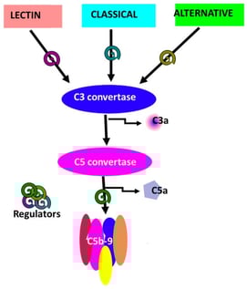
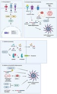
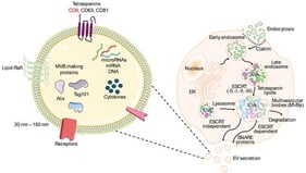
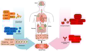
The complement system is a central component of innate immunity with established roles in host defense and emerging functions in neurodevelopment, synaptic remodeling, and neuroimmune communication within the central nervous system (CNS). In parallel, advances in nanotechnology have not only enabled targeted strategies for CNS drug delivery but have also revealed that many nanomaterials interact with and activate complement, influencing biodistribution, safety, and inflammatory responses. Opioid use disorder (OUD) is increasingly recognized as a condition associated with chronic neuroimmune dysregulation involving glial activation, altered cytokine signaling, and blood–brain barrier (BBB) disruption. However, direct experimental or clinical measurements of complement activation in OUD remain limited. Current evidence linking complement pathways to opioid exposure is derived largely from indirect observations, including transcriptomic alterations, glial phenotypes, and inflammatory signatures in preclinical and translational models, which collectively suggest, but do not yet definitively establish, complement involvement in opioid-induced neuroimmune signaling. This review synthesizes current knowledge at the intersection of complement biology, nanomedicine, and opioid-associated neuroimmune changes. It distinguishes well-established mechanisms of complement activation by nanomaterials from emerging and inferential evidence linking complement signaling to opioid exposure. This hypothesis-generating framework integrates complement signaling with opioid receptor and TLR4 pathways in glial and endothelial compartments, examining their potential protective and pathological CNS roles while outlining the translational promise and current evidence gaps of complement-aware nanotechnologies for addiction neuroscience.

13 February 2026

Schematic diagram of the complement system.

Research has shown that diet significantly influences the chance of developing chronic inflammatory diseases including inflammatory bowel disease, cardiovascular disease, obesity, type 2 diabetes and several types of cancer. Dietary components modulate the immune system by either promoting or mitigating inflammatory pathways. One such pathway is the activation of the NLRP3 inflammasome—a multiprotein complex that is involved in the innate immune response. The NLRP3 inflammasome is triggered by various stimuli including ionic flux, mitochondrial dysfunction, lysosomal damage and ROS. Upon activation through a two-signal process, an immune response is initiated that protects the body against pathogens and cellular stress. In a healthy body, this pathway is closely regulated to maintain homeostasis and prevent excessive inflammation that can result in tissue damage or chronic inflammatory diseases. Several components present in a human diet can activate or inhibit the NLRP3 inflammasome. To support a balanced diet, organizations like the WHO have developed dietary recommendations. These promote the consumption of fruits, vegetables, whole grains, lean proteins and healthy fats. These foods contain a variety of nutrients and bioactive compounds, including saturated fatty acids, cholesterol, omega-6 fatty acids and natural sugars, which are pro-inflammatory. At the same time, they also supply anti-inflammatory compounds such as monounsaturated fatty acids, antioxidants and probiotics. While current literature highlights the NLRP3 inflammasome as a critical regulator of inflammation, it lacks detailed insights into how the specific dietary components of a healthy diet influence its modulation. Therefore, this literature review elucidates the various mechanisms through which these dietary compounds modulate the NLRP3 inflammasome. The significance of maintaining a balance between pro- and anti-inflammatory components in the diet is highlighted by its role as a regulator of inflammatory diseases, for example, through mechanisms such as epigenetic pathways.

5 February 2026

Canonical activation and regulation of the NLRP3 inflammasome: (A) priming, (B) assembly, (C) cytokine processing, and (D) inhibitory mechanisms. Created with BioRender.com.

Extracellular vesicles (EVs) constitute a heterogeneous group of membrane-derived particles generated through distinct biogenesis pathways, each regulated by precise molecular mechanisms. They carry a diverse array of cargo that reflects the physiological or pathological state of their parent cells. Their classification continues to evolve, as advances in isolation and characterization techniques have revealed novel vesicle subpopulations beyond the traditional categories of microvesicles, and apoptotic bodies, further highlighting the complexity of the EV landscape. Within the central nervous system (CNS), neurons, microglia, astrocytes, oligodendrocytes, and endothelial cells actively release EVs that contribute to intercellular communication. Growing evidence demonstrates that these vesicles play critical roles in neuroinflammation and neurodegeneration by transporting bioactive molecules that influence disease pathways. Their ability to cross the blood–brain barrier allows CNS-derived EVs to be detected in peripheral fluids, making them promising candidates for noninvasive biomarkers. Moreover, EVs are increasingly being explored as therapeutic tools due to their stability, biocompatibility, and capacity to deliver targeted molecular cargo. In this review, we provide a comprehensive overview of EV biogenesis and release mechanisms in CNS cell types, discuss their emerging functions in neuroinflammatory and neurodegenerative disorders, and summarize current advances in EV-based diagnostics and therapeutic approaches, including ongoing clinical trials.

3 February 2026

Exosomes/sEVs (left) are nano-vesicles enriched in tetraspanins (e.g., CD9, CD63, CD81) and canonical markers such as ALIX and TSG101, and they carry proteins, cytokines, and nucleic acids (miRNAs, mRNAs, DNA). In cells (right), inward budding of the endosomal membrane generates intraluminal vesicles (ILVs) within early and late endosomes, forming multivesicular bodies (MVBs). ILVs arise through ESCRT-dependent mechanisms (ESCRT-0/-I/-II/-III with ALIX, TSG101, VPS4) or ESCRT-independent pathways organized by tetraspanin-enriched microdomains and lipid rafts. MVBs are either delivered to lysosomes for degradation or, aided by ER–endosome contact sites and small Rab GTPases (RAB27a/b, RAB11, RAB7, RAB35), trafficked to the plasma membrane. Fusion of MVBs with the plasma membrane and subsequent exosome release requires SNARE proteins (VAMP7, YKT6) and additional regulators, including syntenin, syndecan, PLD2, and ARF6.

Stem-cell differentiation technologies have traditionally relied on recombinant growth factors, cytokines, and morphogens to initiate and guide lineage specification toward clinically relevant cell types. These approaches have enabled substantial progress in regenerative medicine, as exemplified by recent advances in cell-replacement therapies for Parkinson’s disease, type 1 diabetes, and retinal degeneration. However, protein-based ligands and soluble factors are often limited by short half-lives, pleiotropic signaling, condition-dependent effects, and challenges in achieving precise spatial and temporal control in scalable systems. In this review, we survey differentiation strategies driven by administered substances, organizing the field into five material-centric modules: recombinant growth factors, cytokines, morphogens, exogenous ligands, and agonist antibodies. For each module, we summarize mechanistic principles, representative studies, controllable variables, and translational considerations. While growth factors, cytokines, morphogens, and exogenous ligands remain central tools for directing lineage commitment and maturation, recent studies indicate that agonist antibodies offer an additional and distinct means of controlling differentiation outcomes. These antibodies can function as receptor agonists while also imparting tissue-selective effects, enabling lineage specification with coordinated spatial targeting. By focusing on differentiation methods driven by administered molecules and excluding direct physical stimulation or complex 3D constructs, this review provides a framework that is particularly relevant to immunology and translational practice. We highlight agonist antibody-based induction as an emerging strategy that complements established ligand-based approaches and may broaden the design space for clinically applicable stem-cell differentiation.

31 January 2026

Molecular strategies for programming stem-cell differentiation through administered signals. Schematic overview of differentiation strategies driven by five major sections: (1) recombinant growth factors; (2) cytokines; (3) morphogens; (4) exogenous ligands; and (5) agonist antibody-based induction. Conventional approaches rely on recombinant growth factors and cytokines to promote lineage commitment and expansion, while morphogens provide concentration-dependent patterning cues during fate specification. Exogenous ligands, including hormonal and nuclear-receptor ligands, GPCR ligands, and ECM-mimetic peptides, enable chemically defined and temporally confined modulation of intracellular signaling to stabilize differentiation and promote maturation. In contrast, agonist antibodies function as drug-like surrogate ligands that directly engage cell-surface receptors, encoding receptor clustering geometry, signaling strength, and tissue tropism within a single molecule. Antibody-mediated induction uniquely couples fate specification with organ-specific homing, enabling differentiated cells to migrate to injured tissues and contribute to functional restoration. Together, these sections illustrate a continuum from traditional soluble factors to programmable antibody-based signaling platforms for stem-cell differentiation and regenerative repair.

News & Conferences

Issues

Open for Submission

Editor's Choice

Get Alerted

Add your email address to receive forthcoming issues of this journal.

XFacebookLinkedIn
Immuno - ISSN 2673-5601