Skip to Content

Hemato

Hemato - formerly Bloods - is an international, peer-reviewed, open access journal on hematology, published quarterly online by MDPI.
The Spanish Society of Hematology and Hemotherapy (SEHH) and the Nuclear Medicine Discovery (Nu.Me.D.) are affiliated with Hemato and their members receive discounts on the article processing charges.

Get Alerted

Add your email address to receive forthcoming issues of this journal.

All Articles (232)

  • Case Report
  • Open Access

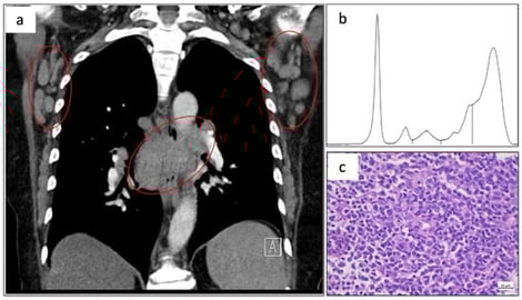
Angioimmunoblastic T-cell lymphoma (AITL) is a rare subtype of peripheral T-cell lymphoma (PTCL) and is frequently associated with autoimmune phenomena. Clinically, AITL shows an aggressive disease course and poor prognosis with currently available treatment strategies. We here report the case of a 64-year-old female patient who was diagnosed with AITL and showed a complicated clinical course due to concurrent immune-mediated thrombotic thrombocytopenic purpura (iTTP). To our knowledge, the presented case highlights a previously unreported association of both conditions. Treatment, including chemotherapy and iTTP-directed treatments, resulted in rapid clinical improvement and sustained remission of both the AITL and the concurrent iTTP. In AITL, transformed T-follicular helper cells (TFHs) are particularly thought to mediate hypersecretion of cytokines and excessive autoantibody production. Immunological disturbances to large parts mediated through these transformed TFHs are thought to trigger autoimmune conditions, as seen with iTTP in this patient. At 36 months post-treatment, the patient remains in complete remission for both AITL and iTTP. This case highlights the complex immunopathological relationship between AITL and autoimmune disorders possibly impeding diagnosis and treatment in a timely manner.

2 March 2026

(a) Computed tomography scan showing mediastinal mass and axillary lymphadenopathy (red circles). (b) Serum protein electrophoresis showing broad γ-region elevation favoring polyclonal hypergammaglobulinemia. (c) Representative hematoxylin and eosin-stained lymph node section (40× magnification) demonstrating a diffuse lymphoid infiltrate of pleomorphic, small-to-medium-sized atypical cells with irregular nuclei and prominent vascular network.

Background: Sickle cell disease (SCD) is a hereditary blood disorder marked by the production of abnormally shaped, rigid red blood cells that obstruct blood flow, resulting in pain, organ damage, and increased infection risk. SCD poses a significant public health challenge in Nigeria, which has the highest global burden, with about 150,000 affected children born annually. The high prevalence is exacerbated by limited healthcare infrastructure, low public awareness, and socio-economic barriers, making effective disease management difficult. Understanding the knowledge of home-based caregivers is essential to identify gaps that may impact care quality. This study was performed within the African Research and Innovative Initiative for Sickle Cell Education (ARISE, EC GA No 824021) project to develop best practice in the clinical management of SCD. Aim: This study explores the knowledge, experiences, and educational needs of home-based caregivers of children with SCD attending the Paediatric Haematology Clinic, ABUTH, Zaria. Methods: A qualitative case study design was used, involving in-depth interviews with ten purposively selected caregivers. Interviews were conducted in Hausa, transcribed, and translated into English. Thematic analysis was performed. Results: Four themes emerged: 1. Understanding of SCD aetiology 2. Knowledge of symptoms 3. Awareness of complications and 4. Knowledge of SCD type. Conclusions: Home-based caregivers had limited knowledge of the genetic basis of the disease, but possess some knowledge of SCD key symptoms, enabling basic disease management and healthcare seeking. However, there is a need to enhance caregiver education to improve care quality and health-seeking behaviour for children with SCD.

28 February 2026

Objective: This study aimed to investigate erythrocyte morphological alterations in hematological malignancies, with particular emphasis on structural differences among leukemia subtypes and anemia. Materials and Methods: Peripheral blood samples were obtained from 60 patients, including individuals with anemia (n = 10), acute lymphoblastic leukemia (ALL, n = 15), acute myeloid leukemia (AML, n = 15), chronic lymphocytic leukemia (CLL, n = 15), and chronic myeloid leukemia (CML, n = 5), as well as 10 healthy controls. Erythrocyte morphology was evaluated using light microscopy and scanning electron microscopy. Morphological abnormalities, including loss of biconcavity, poikilocytosis, echinocyte transformation, burr cells, and stomatocytes, were assessed in accordance with International Council for Standardization in Haematology (ICSH)-based morphological definitions. Results: Distinct erythrocyte morphological alterations were observed across disease groups. AML cases demonstrated pronounced central depression-like or perforation-like structures and hypochromasia. Lymphoid malignancies, particularly ALL and CLL, exhibited increased echinocyte formation, whereas chronic leukemias showed a higher prevalence of stomatocytes and cup-shaped cells. Quantitative scoring indicated that loss of biconcavity was most prominent in anemia, followed by AML, CML, ALL, and CLL. Poikilocytosis was most frequent in anemia, followed by ALL, CLL, AML, and CML. Conclusions: The findings indicate that erythrocyte shape alterations are more heterogeneous and prominent in lymphoid leukemias, whereas myeloid leukemias exhibit distinct ultrastructural membrane abnormalities. Although studies focusing on erythrocyte morphology in leukemia remain limited, the present results provide a foundational morphological reference dataset that may support the development and validation of artificial intelligence-based diagnostic approaches. Further studies involving larger cohorts and expanded imaging analyses are warranted to improve diagnostic accuracy and translational applicability.

26 February 2026

  • Case Report
  • Open Access

Primary Small Bowel MALT Lymphoma with Concurrent Pulmonary Dissemination: A Case Report

  • María José Robledo Rivera,
  • Adriana Margarita Trejos Tenorio and
  • Manuel Sureda González
  • + 2 authors

Marginal zone lymphomas (MZLs) are indolent mature B-cell neoplasms. Approximately 3.4% of gastrointestinal mucosa-associated lymphoid tissue (MALT) lymphomas involve the small bowel. Pseudoaneurysmal dilation has been reported in up to 35% of patients with small bowel MALT lymphoma. We report the case of a 73-year-old woman with ulcerative colitis in remission who presented with hematuria, constitutional symptoms, and progressive respiratory distress. Imaging incidentally revealed pseudoaneurysmal dilation of the small bowel and thoracic findings suggestive of pulmonary lymphangitic dissemination. A PET scan showed lung, nodal, and small bowel infiltration. Histopathological and flow cytometry examinations confirmed small bowel MZL, and bone marrow biopsy excluded marrow involvement. The patient was treated with R-CHOP chemotherapy and then R-Bendamustine, achieving complete clinical and radiological remission. This case illustrates a rare presentation of intestinal MALT lymphoma and emphasizes the diagnostic significance of correlating imaging and clinical findings in identifying pseudoaneurysmal dilatation and distinguishing it from other potential causes.

11 February 2026

News & Conferences

Issues

Open for Submission

Editor's Choice

XFacebookLinkedIn
Hemato - ISSN 2673-6357