
- Article

Effects of Microbial Biomass and Mineral Premixes on Growth Performance and Nutrient Utilisation in Penaeus monodon Fed Low Fishmeal Diets
- Ha H. Truong,
- Matthew R. P. Briggs and
- Cedric J. Simon
- + 3 authors
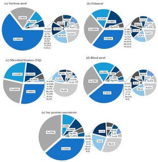
The growth performance of Penaeus monodon is often reduced when fishmeal is extensively replaced with terrestrial ingredients. This study evaluated the efficacy of a marine microbial biomass, NovaqPro™ (NQ), and inorganic mineral premixes in improving the performance of low fishmeal diets. Diets containing soybean meal, soy protein concentrate, and bloodmeal were formulated with fishmeal limited to 6%. Treatments included 10% NQ, an experimental inorganic mineral premix, a commercial mineral premix, and their combinations added to the low fishmeal control. A high fishmeal diet was also assessed as a benchmark of performance. NQ supplementation significantly improved shrimp growth, increasing weight gain by 78.7% compared with the low fishmeal control (2.77 vs. 1.55 g shrimp−1) and numerically improved by 25.3% compared with the high fishmeal diet (2.21 g shrimp−1). Similar responses were observed for FCR where NQ diets (1.47–1.68), as well as the high fishmeal diet (1.59), were superior to that of the control diet (2.02). Growth improvements were associated with increased feed intake and higher retention of protein and gross energy. In contrast, mineral premix supplementation did not improve growth, and weight gain was numerically reduced relative to the low fishmeal control. The NQ diet showed higher apparent digestibility of calcium, phosphorus, and magnesium compared with the high fishmeal diet. These results demonstrate that NQ is an effective mitigation strategy to reduce growth limitations associated with low fishmeal diets in P. monodon, without the need for additional inorganic mineral supplementation.
26 February 2026
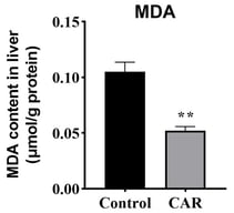
![Histopathological and ultrastructural alterations in the gallbladder of Cyprinus carpio under B[a]P exposure. (Top row) Light microscopy images (H&E staining) displaying the tissue architecture. Blue arrows indicate vacuolar degeneration of mucosal epithelial cells. In the high-magnification inset, Red arrows indicate nuclear deformities, characterized by irregular shape and disordered arrangement. (Bottom row) Transmission electron microscopy (TEM) images showing ultrastructural pathology. Green arrows point to numerous intracytoplasmic vesicular structures containing granular electron-dense material (likely incompletely degraded organelles or autophagic vacuoles). The blue boxes highlight the mitochondrial ultrastructure, displaying normal mitochondria with intact cristae in the control group (DC), contrasted with mitochondrial vacuolar degeneration in the treatment groups (DL and DH). The area within the red circle demonstrates submucosal edema, characterized by loosened connective tissue and widened interstitial spaces due to inflammatory exudation. Abbreviations: DC, control group (0 μg/L); DL, low-dose group (2.5 μg/L); DH, high-dose group (25 μg/L). N, nucleus.](https://mdpi-res.com/cdn-cgi/image/w=281,h=192/https://mdpi-res.com/fishes/fishes-11-00140/article_deploy/html/images/fishes-11-00140-ag-550.jpg)