Skip to Content
You are currently on the new version of our website. Access the old version .

Biosensors

Biosensors is an international, peer-reviewed, open access journal on the technology and science of biosensors, published monthly online by MDPI.

Indexed in PubMed | Quartile Ranking JCR - Q1 (Instruments and Instrumentation | Chemistry, Analytical)

All Articles (5,098)

Hybrid Oxygen-Sensing Bio-Scaffolds for 3D Micro-Tissue Models

  • Liang Li,
  • Alexander V. Zhdanov and
  • Dmitri B. Papkovsky

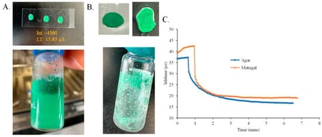
Culturing cells and micro-tissue samples in 3D bio-scaffolding structures is gaining popularity; however, precise control of tissue micro-environment in such systems remains challenging. We describe a family of new hybrid bio-scaffolds with 3D O2-sensing ability, produced by simple means from readily available bio-scaffolding and O2-sensing materials. Three different types of phosphorescent O2-sensing materials—polymeric microparticles (MPs), supramolecular probe MitoXpress and nanoparticulate probes NanO2 and Nano-IR (NPs)—were integrated in Matrigel and agarose scaffolding materials and evaluated. Key working characteristics of such hybrid scaffolds, including heterogeneity, stability, cytotoxicity, optical signals and O2-sensing properties, ease of fabrication and use, were compared. The results show superiority of the Matrigel hybrids with NanO2 and Nano-IR probes. Demonstration experiments were conducted with HCT116 cells and individual spheroids derived from these cells, culturing them in the Matrigel–NP hybrid scaffolds and monitoring oxygenation and local O2 gradients on a time-resolved fluorescence plate reader and by phosphorescence lifetime imaging microscopy (PLIM).

14 February 2026

Photographs of the agarose (A) and Matrigel (B) based precursor cocktails and spotted scaffold coatings doped with sensor MPs. (C) Magnitude and time of the response to changes in O2 concentration from 2.0 kPa to 15.0 kPa for the two types of coatings. Lifetime profiles were recorded using FirestingO2 reader, at 20 °C.

Severe Dengue fever can cause Dengue Hemorrhagic Fever (DHF), a life-threatening condition characterized by plasma leakage and hemoconcentration. A hematocrit (Hct) rise of ≥20% is a key indicator for medical intervention, but current monitoring is invasive and intermittent. This study aims to determine the optimal design parameters for a non-invasive optical sensor to continuously monitor hemoconcentration. We developed a high-fidelity Monte Carlo model of light transport in a multi-layered skin model, with the epidermis set to a 5% melanin volume fraction (Fitzpatrick type II/III). To ensure signal reliability, simulations were conducted with a high photon count (1×10⁸ photons), yielding a stochastic (Monte Carlo) signal-to-noise ratio of approximately 36 dB. We simulated diffuse reflectance at four characteristic wavelengths (577 nm, 660 nm, 800 nm—the isosbestic point—, and 940 nm) over source-detector separations of 0.5–8.0 mm. Sensor sensitivity was quantified as the reflectance change for a +25% relative Hct rise (e.g., 42% to 52.5%), mimicking severe hemoconcentration, and its dependence on baseline dermal blood volume fraction (BVF) was investigated. Sensor sensitivity showed a non-linear dependence on BVF, showing a direct correlation with perfusion level, reaching an optimal 6.41% for a robust 5% BVF at 8.0 mm. A dedicated sweep showed that even under low-perfusion shock conditions (1% BVF), the sensor maintains a highly significant sensitivity of 5.71% (also at 8.0 mm), indicating that sensitivity remains high across a physiologically relevant perfusion range. In the analysis, at a robust 5% BVF, the 800 nm wavelength demonstrated superior reliability, with peak sensitivity at 6.41% at 8.0 mm. Visible wavelengths (577 nm and 660 nm) exhibited high theoretical sensitivity, while 940 nm was compromised by water absorption. Based on these findings, a non-invasive optical sensor for hemoconcentration is most effective operating at 800 nm, within the evaluated spectral set, with a source-detector separation of ≥6.0 mm, targeting the deep dermis while minimizing superficial interference. This design provides an optimal balance of tissue penetration, robust sensitivity to Hct changes, and reduced sensitivity to oxygenation-related variability while maintaining signal stability. This work enables the design of a device for continuous monitoring, supporting continuous monitoring of hemoconcentration trends relevant to plasma leakage progression.

13 February 2026

Schematic of the Multi-Layered Skin Phantom and Optode Geometry. The 2D cross-section illustrates the three–layer skin model, including the epidermis (0.1 mm), dermis (1.5 mm), and subcutaneous fat (8.4 mm). The vertical axis is visually enhanced to clearly depict the thin superficial layers and is not to scale, as indicated by the dimension lines showing true physical thicknesses. The simulation geometry includes a single pencil–beam source and a linear array of 16 detectors on the surface, spanning source-detector separations (SDS) from 0.5 mm to 8.0 mm. Illustrative photon paths (dashed lines) show that light detected at longer SDS has, on average, interrogated deeper tissue volumes.

Traditional sample preparation for flow cytometry is often labor-intensive, operator-dependent, and reagent-consuming, limiting its suitability for automated and point-of-care biosensing applications. To address these challenges, this study presents a functional modular microfluidic system integrating immunomagnetic beads (IMBs) to enable automated intracellular immunofluorescence (IF) staining. The modular microfluidic platform is enabled by a dynamically actuated three-dimensional magnetic field that couples with IMBs within a microfluidic reaction chamber, requiring only one-dimensional magnet translation to induce effective three-dimensional bead motion. This magnetic–chip cooperative strategy significantly enhances microscale mixing and cell capture, facilitating automated immunostaining of the radiation biomarker in CD4+ cells. Finite element simulations were employed to guide magnetic field design by analyzing magnetic force distributions and identifying key parameters, including magnet material, size, spatial arrangement, and chip–magnet distance. Experimental validation using CD4+ cell capture confirmed the effectiveness of the magnetic mixing strategy, revealing ∇B·B as the critical design parameter. An N52 NdFeB magnet (6 mm diameter, 10 mm height) positioned within 2.2 mm of the chamber centerline stably retained IMBs at flow rates below 200 µL/min. Under optimized conditions (magnet translation speed of 8 mm/s and a 15 min mixing duration), a maximum cell capture efficiency of 86% was achieved. Subsequent automated γH2AX IF staining demonstrated a strong linear dose–response relationship (R2 > 0.9) in mean fluorescence intensity. This study demonstrates a robust and scalable strategy for automating complex IF staining workflows, highlighting the potential of magnetic-field-assisted microfluidic platforms for biosensing applications requiring reliable intracellular biomarker detection.

13 February 2026

Schematic diagram of microfluidic chip. (a) Overall display of the chip. (b) Layered chip display. (The red circles represent the perforations, indicating the inlet and outlet positions for the solution).

Deep Convolutional Neural Networks for Autofocus Control on a C. elegans Tracking System

  • Santiago Escobar-Benavides,
  • Jose-Julio Peñaranda-Jara and
  • Antonio-José Sánchez-Salmerón
  • + 1 author

Correct focal positioning is essential for microscopy imaging of live moving subjects such as Caenorhabditis elegans. However, many methods can be too slow to perform real-time control to keep the subject in focus. In this work, we propose a convolutional neural network-based method to perform one-shot prediction of the optimal focusing distance, without the need to scan iteratively the optical axis to find the optimal position. A new data augmentation technique is proposed, and its effectiveness is validated through statistical analysis. This technique is shown to improve results without the need for additional data collection. Several architectures are trained in z-stacks of images, using the proposed data augmentation technique, and compared on a validation set. Through this comparison, we find that the ConvNext V2, a novel architecture in this context, outperforms other models proposed in previous works. Furthermore, the impact of the Field of View used for the model’s prediction is studied, with the aim of further understanding the influence of spatial resolution and spatial compression on the performance of the model.

12 February 2026

Example of labeled stack of images using Vollath F5 index.

News & Conferences

Issues

Open for Submission

Editor's Choice

Reprints of Collections

Photonics for Bioapplications
Reprint

Photonics for Bioapplications

Sensors and Technology
Editors: Nélia Jordão Alberto, Maria de Fátima Domingues, Nunzio Cennamo, Adriana Borriello

Get Alerted

Add your email address to receive forthcoming issues of this journal.

XFacebookLinkedIn
Biosensors - ISSN 2079-6374