- Article

Proteasome Inhibition Amplifies Endoplasmic Reticulum (ER) Stress Responses: Comparative Proteomics of Chinese Hamster Ovary Cell Lines
- Christiana-Kondylo Sideri,
- David Ryan and
- Paula Meleady
- + 2 authors
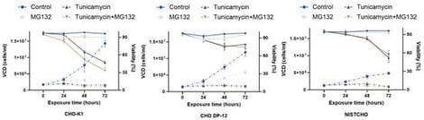
Chinese hamster ovary (CHO) cells are widely utilised in the biopharmaceutical industry to produce therapeutic proteins. Understanding the mechanisms of endoplasmic reticulum (ER) stress and its interplay with protein degradation pathways remains pivotal for improving production efficiency and product quality. In this study, we investigated the proteomic responses of CHO-K1 (non-producer), CHO DP-12 (IgG-producer), and NISTCHO (IgG-producer) cell lines under ER stress induced by a combination of the proteasome inhibitor MG132 and the glycosylation inhibitor tunicamycin. Viability, cell growth, and IgG titre were measured for 24 h, 48 h, and 72 h of treatment and the 48 h timepoint was used for the comparative analysis of the proteomic data across the three cell lines. Proteasome inhibition with MG132 intensified ER stress and altered ER-associated protein degradation (ERAD). Combined tunicamycin + MG132 treatment was associated with cell line-specific proteomic changes: NISTCHO upregulated ER translocation and glycoprotein quality control proteins (SSR4, SEC24C, UGGT1), CHO DP-12 activated redox/disulfide regulators (DNAJC10, CAPN1), while CHO-K1 showed broad proteome shifts, suggesting differences in baseline stress handling. These findings provide mechanistic insights into ER stress and protein quality control in CHO cells, offering a foundation for strategies to enhance cell line robustness and optimise biopharmaceutical production.
10 February 2026