Skip Content
You are currently on the new version of our website. Access the old version .

Bioengineering

Bioengineering is an international, peer-reviewed, open access journal on the science and technology of bioengineering, published monthly online by MDPI. 

Indexed in PubMed | Quartile Ranking JCR - Q2 (Engineering, Biomedical)

All Articles (5,918)

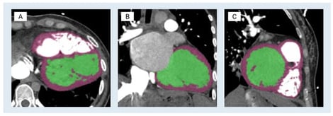
Three-dimensional (3D) anatomic modeling derived from high-resolution medical imaging, such as computed tomography (CT) and magnetic resonance imaging (MRI), has been increasingly adopted in preclinical testing and device development. This white paper describes a cardiac-specific workflow that integrates 3D printing and silicone molding for support device development and procedural simulation. Patient-derived computed tomography angiography data were segmented using FDA-cleared medical modeling software to isolate the left ventricular anatomy and were further processed in computer-aided design (CAD) to ensure accurate physiological wall thickness and structural fidelity. Material jetting 3D printing was performed on a Stratasys J750 using material distributions designed to mimic the mechanical properties of myocardium, thereby approximating myocardial compliance. In parallel, stereolithography apparatus molds were designed from the left ventricle CAD model to cast transparent, pliable left ventricular models in Sorta-Clear™ 18 silicone. The 3D-printed models preserved intricate morphological detail and were suitable for mechanical manipulation and device deployment studies, whereas silicone models offered tunable mechanical properties, transparency for visualization, and durability for repeated use. Together, these complementary modalities provided rapid manufacturing capability and application-relevant physical representation. Case-specific parameters, strengths, and limitations of both models in enhancing patient care and device testing are highlighted, with relevance to heart failure applications. Current knowledge gaps, workflow and integration challenges, and future opportunities are identified, positioning this work as a reference framework for continued innovation in anatomic modeling. Within the collaborative framework of Mayo Clinic’s Anatomic Modeling Unit and Simulation Center, this integrated modeling workflow demonstrates the value of multidisciplinary collaboration between engineers and clinicians. Clinically, these patient-specific left ventricular models may enable pre-procedural device sizing and positioning and may support simulation of mechanical circulatory support (MCS) deployment while identifying possible anatomic constraints prior to intervention. This workflow has direct applicability in advanced heart failure patients undergoing MCS support, such as the Impella axillary MCS device or the durable LVAD, with potential to reduce procedural uncertainty while reducing complications and improving peri-procedural outcomes. Additionally, these models also serve as high-accuracy educational tools, enabling trainees and multidisciplinary care teams to visualize and possibly rehearse procedural steps while gaining hands-on experience in a risk-free environment.

8 February 2026

Segmentation process of modeling the left ventricle. The blood pools of the left ventricle and the myocardium are shown in green and purple, respectively. (A) Axial view—a short-axis slice through the left ventricle (classic round/oval LV cavity). (B) Coronal view—a long-axis view showing the LV from anterior to posterior. (C) Sagittal view—the orthogonal long-axis view, slicing left–right through the LV.

The anatomy and mechanical strength of aortic valve leaflets are critical determinants of their biomechanical behavior and long-term structural integrity. The embryonic developmental period, when valves are forming, is critical to establish baseline leaflet properties. However, fetal stages of valve development, when valve leaflets are still forming and remodeling, are not well understood. The goal of this study is to investigate the biomechanical stress and deformation modes of developing valve leaflets during systole, and how leaflet biomechanics are affected by anatomy and material properties. To this end, the study employs a parametric approach to model the leaflet anatomy of an HH40 chick embryo, used here as a model of fetal cardiac development. To perform biomechanical analysis, a pressure profile derived from in ovo Doppler ultrasound measurements was applied, and an Ogden hyperelastic material model was employed following a sensitivity analysis. To determine the effect of valve anatomy on leaflet tissue deformation and stresses, we changed the leaflet midline curve (belly curve) from its native curvature to a linear profile and quantified biomechanical responses. Our analysis revealed a strong decrease in average leaflet effective stress as the belly curvature was shifted towards a linear profile. However, this reduction in average stress was at the expense of a biomechanical trade-off. The shift induced a progressive localization of stress concentration at the leaflet tips and commissures, and a distinct bending deformation mode at the tip under peak load. Our findings demonstrate that while the belly curve of the leaflet modulates tissue stress during valve opening, a low-stress anatomy does not align with hemodynamic performance. This work characterizes competing leaflet biomechanical responses (stress reduction versus failure modes) that shape valve leaflet formation, providing fundamental insights into developmental valve biomechanics.

6 February 2026

Doppler ultrasound-based acquisition and processing of aortic velocity over the cardiac cycle from HH40 (Day 14) chick embryo. (A) Imaging setup and location of the Doppler sample volume (gate) positioned distal to the aortic valve, with an insonation angle of about 35 degrees. (B) Pulsed-Wave Doppler velocity waveform. (C) Aortic blood flow velocity from a single cardiac cycle extracted from the Doppler waveform (left) and its corresponding pressure–time profile obtained using the simplified Bernoulli equation (right), used as a load boundary condition in FEA.

A Novel, Low-Cost, 3D-Printed Motorized Injector for Retinal Sheet Transplantation

  • Jerald Lim,
  • Francis Ung and
  • Andrew W. Browne
  • + 6 authors

Retinal transplantation offers promise for restoring vision in advanced retinal degeneration. However, manual delivery of retinal sheets is often hindered by imprecise placement and collateral tissue damage resulting from instrument instability. We introduce a novel, 3D-printed, motorized retinal sheet injector designed to enhance placement accuracy and minimize tissue injury. The motorized injector features an Arduino-controlled foot pedal with three discrete actuator positions (“Min”, “Mid”, “Max”). When compared via frame-by-frame motion analysis, the motorized system reduced tip variance by approximately threefold over manual methods. In addition, in in vitro gelatin trials, the motorized injector achieved significantly higher placement accuracy versus the manual injector, which suffered from occasional complete misplacements. The novel motorized retinal sheet injector markedly improves stability and placement accuracy relative to manual methods, potentially reducing complications associated with subretinal delivery. Safer subretinal delivery can pave the way for innovative research and advanced treatment for retinal disease.

6 February 2026

Manual injector (A,B) operating principles for transplanting sheets of retinal tissue into the subretinal space.

Immune checkpoint inhibitors (ICIs) targeting PD-1/PD-L1 and CTLA-4 are widely used in the treatment of several cancers and have significantly improved survival outcomes in responsive patients. However, a substantial proportion of patients fail to benefit from these therapies, underscoring the urgent need for accurate prediction of ICI response. We propose a deep learning framework, ICIsc, to accurately predict ICI response by integrating single-cell RNA sequencing (scRNA-seq) data with protein large language models. Specifically, patient representations are constructed using transcriptomic profiles and immune-related gene set scores as latent embedding features, while drug representations are derived from amino acid sequences of ICI encoded by the Evolutionary Scale Modeling 2 (ESM2). For bulk data, ICIsc employs a bilinear attention module to fuse patient and drug embeddings for response prediction. For scRNA-seq data, ICIsc infers cell–cell interactions using a single-sample network (SSN) approach and applies GATv2 to model immune microenvironment heterogeneity at the single-cell level. Benchmark evaluations and independent validation demonstrate that ICIsc consistently outperforms baseline models and exhibits robust generalization performance. SHAP-based interpretability analysis further identifies key genes (e.g., GAPDH) associated with immunotherapy response and patient prognosis. Overall, ICIsc provides an accurate and interpretable framework for predicting immunotherapy outcomes and elucidating underlying mechanisms.

6 February 2026

Overview of ICIsc framework. The model comprises three components. A patient feature encoder based on multi-layer perceptrons 


        M
        L
        P
      
      
        P


 learns latent patient representations, while an ICI feature encoder employs the protein language model ESM2 to encode ICI drugs. In the bulk model, patient and ICI representations are integrated via a bilinear attention module to generate fused embeddings for response prediction using 


        M
        L
        P
      
      
        B


. In the single-cell model, cell subtype embeddings, cell–cell interactions inferred by a single-sample network, and cell subtype proportions are incorporated into a GATv2 to derive patient representations, which are jointly used with ICI representations for immunotherapy response prediction using 


        M
        L
        P
      
      
        S


.

News & Conferences

Issues

Open for Submission

Editor's Choice

Get Alerted

Add your email address to receive forthcoming issues of this journal.

XFacebookLinkedIn
Bioengineering - ISSN 2306-5354