


Abstract
The careful planning of a sinus lift procedure is the key to avoiding surgical complications. In this clinical case, a female patient, 59 years old and totally edentulous in the maxilla, was referred to Egas Moniz’s Dental Clinic Implantology consultation with indication for bilateral external sinus lift of the maxillary sinuses prior to implant placement. Both orthopantomography and cone-beam computed tomography were used to show the anatomy of the maxillary sinuses, which presented multiple sinus septa. A multiple anterolateral window approach was applied in order to avoid perforation of the Schneiderian membrane while accessing it.
1. Introduction
An external maxillary sinus lift is a delicate surgical procedure that is performed when there is not enough bone available to allow implant placement in the posterior maxilla due to bone resorption of the alveolar process and pneumatization of the maxillary sinuses [1]. In order not to perforate the Schneiderian membrane, it is necessary to have full knowledge of the individual’s maxillary sinus anatomy [2]. The literature shows the relationship between the presence of sinus septa and perforation of the Schneiderian membrane during surgery for external elevation of the maxillary sinus [2]. The location of the access varies according to the morphology and anatomical location of the septa [2].
2. Materials and Methods
A female patient, 59 years old and totally edentulous in the maxilla, was referred for consultation at the Egas Moniz’s Dental Clinic Implantology with indication for bilateral external elevation of the maxillary sinuses. Orthopantomography and a CBCT (cone-beam computed tomography) scan revealed the presence of bilateral sinus septa in the anterolateral wall, resulting in its compartmentalization. Multiple accesses were performed for each sinus according to the anatomical position of each septum.
3. Results and Discussion
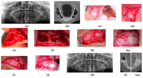
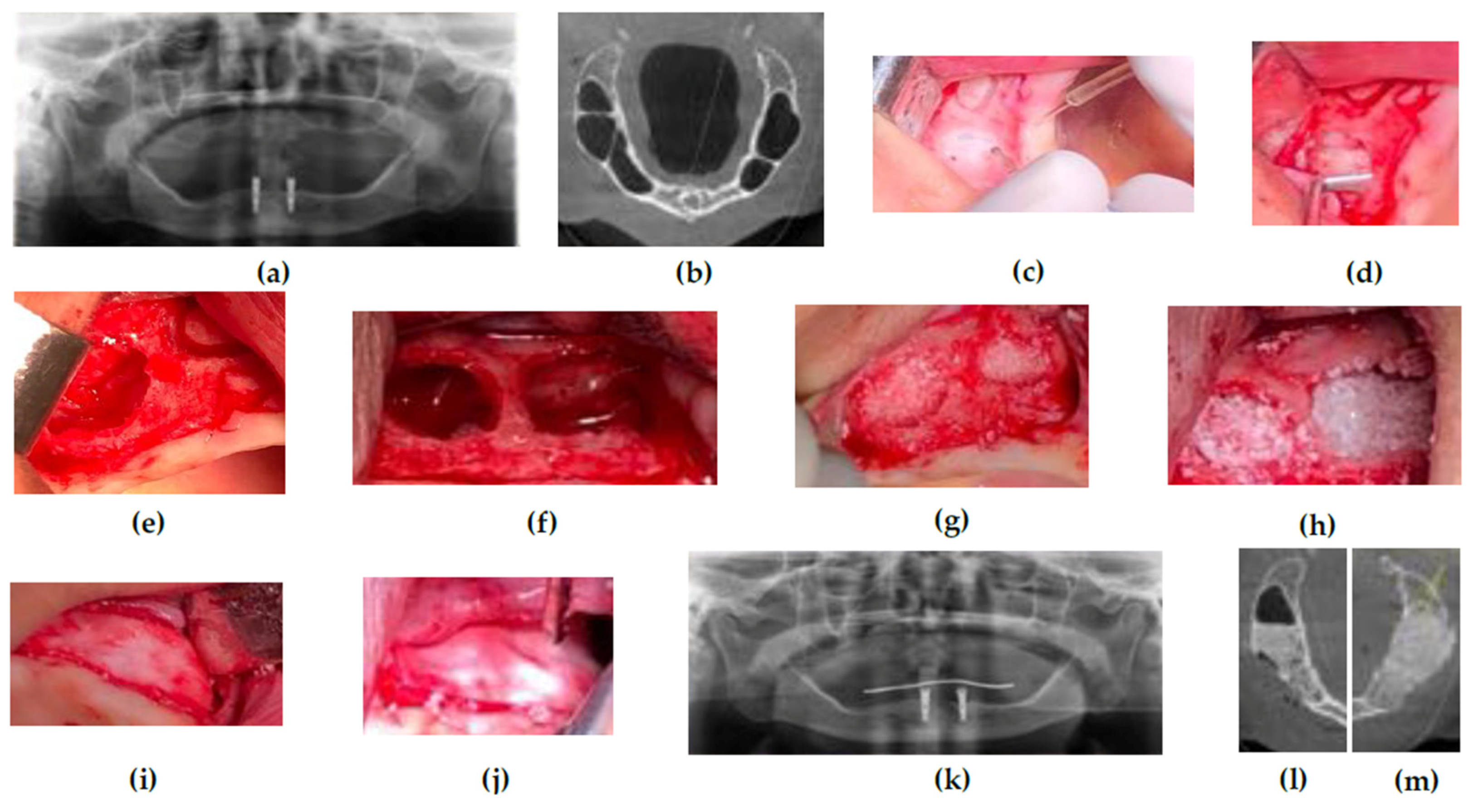
The same surgical approach was applied in both right and left maxillary sinuses. A full-thickness mucoperiosteal flap was executed at first, followed by an osteotomy using a round handpiece burr to gain sinus membrane access, creating two windows. Both access windows were elevated and the Schneiderian membrane was then detached. Both spaces were filled with xenograft Bio-Oss® and then covered with a Bio-Guide® collagen membrane. Orthopantomography and CBCT were performed after this procedure to check its final outcome. Multiple accesses allowed elevation of the maxillary sinus membranes with less risk of perforation (Figure 1).
Figure 1.
Surgical procedure: (a) initial orthopantomography; (b) initial CBCT scan; (c) osteotomy with round handpiece burr; (d) elevation of maxillary sinus access windows; (e) detached Schneiderian membrane on the right side; (f) detached Schneiderian membrane on the left side; (g) xenograft filling maxillary sinus access on the right side; (h) xenograft filling maxillary sinus access on the left side; (i) collagen membrane covering the graft on the right side; (j) collagen membrane covering the graft on the left side; (k) final orthopantomography; (l) final right-side CBCT scan; (m) final left-side CBCT scan.
In conclusion, septa should not be considered a contraindication to sinus lift surgery. Presurgical planning is crucial to reducing the risk of perforation of the Schneiderian membrane and enabling greater predictability of treatment success, thus reducing patient morbidity.
Institutional Review Board Statement
Not applicable.
Informed Consent Statement
Informed consent was obtained from all subjects involved in the study.
Conflicts of Interest
The authors declare no conflict of interest.
References
- Tassos Irinakis, T.I.; Valentin Dabuleanu, V.D.; Salwa Aldahlawi, S.A. Complications during Maxillary Sinus Augmentation Associated with Interfering Septa: A New Classification of Septa. Open Dent. J. 2017, 11, 140–150. [Google Scholar] [CrossRef] [Scilit] [PubMed]
- Magdalena Malec, M.M.; Tomasz Smektała, T.S.; Grzegorz Trybek, G.T.; Katarzyna Sporniak-Tutak, K.S. Maxillary sinus septa: Prevalence, morphology, diagnostics and implantological implications. Systematic review. Folia Morphol. 2014, 73, 259–266. [Google Scholar] [CrossRef] [Scilit]
Publisher’s Note: MDPI stays neutral with regard to jurisdictional claims in published maps and institutional affiliations. |
© 2021 by the authors. Licensee MDPI, Basel, Switzerland. This article is an open access article distributed under the terms and conditions of the Creative Commons Attribution (CC BY) license (https://creativecommons.org/licenses/by/4.0/).