Review

Thoracentesis for the Diagnosis and Management of Pleural Effusions: The Current State of a Centuries-Old Procedure

by
Michael J. Nicholson
1,*,
Christopher Manley
2 and
Danish Ahmad
2
1
Department of Thoracic Medicine and Surgery, Temple University Hospital, Philadelphia, PA 19140, USA
2
Division of Pulmonary and Critical Care, Fox Chase Cancer Center, Temple University Health System, Philadelphia, PA 19140, USA
*
Author to whom correspondence should be addressed.
J. Respir. 2023, 3(4), 208-222; https://doi.org/10.3390/jor3040020
Submission received: 3 November 2023 / Revised: 26 November 2023 / Accepted: 28 November 2023 / Published: 8 December 2023

Abstract

:
Thoracentesis is a fundamental procedure in interventional pulmonology, providing both diagnostic and therapeutic value. This review article offers a comprehensive analysis of thoracentesis, delving into pleural anatomy, procedural techniques, indications, and recent advancements. The article details the evolution of thoracentesis, including the crucial role of ultrasound guidance and emerging approaches that enhance precision and minimize complications. It addresses the wide range of indications for thoracentesis in diverse clinical scenarios, from the diagnosis of pleural effusions to therapeutic drainage of pleural collections. Furthermore, this review explores the management of coagulopathy and anticoagulation pertaining to thoracentesis. It will also provide strategies for preventing and managing complications, ensuring that thoracentesis remains a well-tolerated procedure with minimal risks. This article concludes by examining future directions in thoracentesis, including potential innovations and trends that will shape the landscape of interventional pulmonary medicine. This review serves as an essential resource for pulmonologists, interventional radiologists, and healthcare professionals, offering a comprehensive update on thoracentesis.

1. Introduction

Pleural effusion refers to the accumulation of fluid in the pleural space, which can occur due to various underlying medical conditions. It is a relatively common finding, with approximately 1.5 million cases reported annually in the United States [1]. Common causes include congestive heart failure, bacterial pneumonia, and malignancy [2]. Thoracentesis, also known as needle thoracostomy or pleural tap, was first described by American physician Henry Ingersoll Bowditch in 1852 and involves the removal of excess pleural fluid for diagnostic and therapeutic purposes. It is often the preferred initial procedure for diagnosis and management of pleural effusions, with approximately 178,000 thoracenteses performed in the United States annually [3]. Percutaneous pleural drainage is the third most commonly performed procedure in the intensive care unit [1]. Modern technology along with increased understanding of pleural physiology have led to the development of safe and effective thoracentesis techniques. This paper will explore the pathophysiology, diagnosis, and classification of pleural effusions while also providing a comprehensive overview of thoracentesis procedural techniques. By understanding the principles and best practices of thoracentesis, healthcare providers can ensure optimal outcomes and safety during this essential procedure.

2. Pleural Effusions

2.1. Pleural Space Anatomy

The pleural space refers to the anatomical cavity between the lungs and the chest wall. It is lined by two distinct membranes referred to as the visceral pleura and the parietal pleura. The visceral pleura is a thin, serous membrane composed of mesothelial cells and a connective tissue layer which covers the surface of the lungs. The parietal pleura lines the inner surface of the thoracic cavity and is divided into several regions based on location. The costal pleura lines the ribs, the diaphragmatic pleura lines the superior diaphragm, the mediastinal pleura lines the lateral mediastinum, and the cervical pleura extends into the neck region. The parietal pleura is more sensitive than the visceral pleura due to a richer innervation [4]. The pleura is supplied by branches of the intercostal, internal thoracic, and bronchial arteries. Understanding the neurovascular supply to the pleura and intracostal space is crucial to achieving effective local anesthesia and avoiding hemorrhagic complications. Pleural fluid is vital to parietal and visceral pleura function. It is produced by the mesothelial cells lining the pleura and reabsorbed by pleural lymphatics. It serves as a lubricant between the two layers, reducing friction during the respiratory cycle. Typically, the pleural space of a healthy individual contains approximately 0.3 mL/kg of pleural fluid [5].

2.2. Pleural Fluid Accumulation

Pleural fluid production occurs at an approximate rate of 0.2 mL/kg/h. Clearance of pleural effusion happens through the parietal pleura and can remove up to 0.3–3 mL/kg/h [5]. Any disruption in this delicate equilibrium can result in the development of pleural effusion. This imbalance arises when the rate of production surpasses reabsorption or when reabsorption mechanisms are compromised. Heightened fluid production may stem from hyperactive pleural mesothelial cells, often linked to inflammatory conditions such as infections, malignancies, or autoimmune diseases. Conversely, diminished fluid absorption can arise from direct lymphatic drainage obstruction or elevated hydrostatic pleural capillary pressures. The former has been observed in malignancies, while the latter is typical in hypervolemic conditions such as congestive heart failure [1].

2.3. Diagnostic Imaging

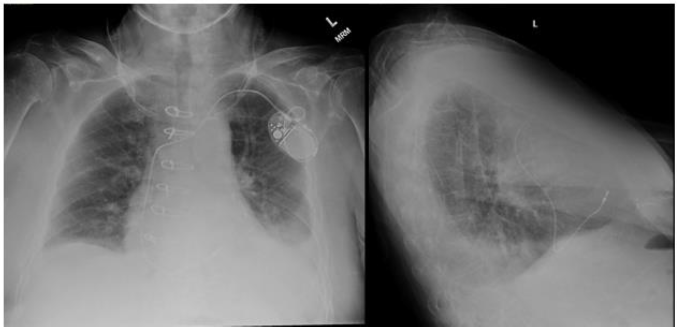
2.3.1. Chest Radiography

Standard posteroanterior and lateral chest radiography is often the initial modality employed to detect pleural effusion. On posteroanterior films, effusions are appreciated when the volume approaches 200 mL. Conversely, lateral views can visualize volumes as low as 50 mL [6]. In the intensive care setting, anteroposterior X-ray imaging is common, usually with the patient in a supine position. This positioning can cause pleural fluid to accumulate in the posterior thorax, making it less conspicuous on anteroposterior chest radiographs. In an upright patient, a pleural effusion typically presents on a chest X-ray as a uniform opacity in the lower lung field with blunting of the affected costophrenic angle (Figure 1). This effusion may display an upper border which curves up laterally, a radiological illusion created by a partially aerated lung between the anterior and posterior fluid layers known as the Ellis curve or meniscus sign [6]. Notably, a mediastinal shift away from the effusion is crucial for distinguishing pleural effusions from atelectasis. Utilizing lateral decubitus positioning to assess the layering of fluid is the most sensitive approach for detecting pleural effusions on chest radiographs. This positioning is capable of detecting effusions as small as 50 mL [7]. While chest radiography proves valuable in identifying pleural effusions, computed tomography and thoracic ultrasound offer greater sensitivity and detailed characterization of pleural fluid.

2.3.2. Computed Tomography

On CT scans, pleural effusions manifest as sickle-shaped opacities in the dependent regions of the thorax (Figure 2). Specific CT features, such as the presence of air in pleural fluid, pleural thickening, and pleural enhancement, can provide valuable insights into underlying pathologies such as empyema and malignancy [6]. Nevertheless, when it comes to detailing intrinsic pleural fluid attributes, ultrasonography has higher sensitivity. Despite its limitations, contrast-enhanced CT remains a widely utilized diagnostic tool in the investigation of pleural effusions, offering valuable information, especially in cases where a comprehensive assessment of pleural and lung pathology is warranted.

2.3.3. Ultrasound

Thoracic ultrasonography has emerged as an invaluable diagnostic tool for assessing pleural effusions. Offering portability, noninvasiveness, and radiation-free imaging, it facilitates convenient and safe bedside evaluation (Figure 3). The accuracy of thoracic ultrasonography in detecting pleural effusions has been confirmed through numerous studies [8,9,10]. Notably, a 2011 study by Xirouchaki et al. reported 100% sensitivity, specificity, and diagnostic accuracy for thoracic ultrasound in pleural effusion diagnosis [9]. The International Consensus Conference on Lung Ultrasound supports this, asserting that “for effusion detection, lung ultrasound is more accurate than supine radiography and equally accurate to CT” [7,11]. Thoracic ultrasonography facilitates the assessment of fluid homogeneity, septation, and echogenicity. Several studies highlight ultrasound’s superiority over CT imaging in detecting intrinsic pleural effusion characteristics. Depending on the effusion’s etiology and complexity, four main sonographic patterns can be identified: anechoic (purely black), complex non-septated (black with white strands), complex septated (black with white septa), and homogenously echogenic (purely white) (Table 1) [7,12]. Complex appearances are typically associated with exudative effusions, while anechoic effusions can be either transudative or exudative. Echogenic effusions require drainage to explore for underlying etiology, such as empyema or hemothorax. Patient positioning significantly influences thoracic ultrasound evaluation [12]. Pleural fluid accumulates dependently within the thorax, making the seated upright position ideal as it encourages fluid movement to the lower thorax. In cases where patients face challenges or contraindications to upright positioning, such as mechanically ventilated critically ill patients, the supine position with the ipsilateral arm adducted across the chest toward the contralateral side is preferred [13]. Accurately identifying the diaphragm’s position relative to the pleural effusion is crucial to avoid diaphragmatic injury during drainage. Further discussion on the procedural role of thoracic ultrasonography will be provided in the subsequent sections.

2.4. Pleural Fluid Analysis

2.4.1. Classification

The classic method for classifying pleural effusions is Light’s criteria. Modified from its original 1972 version, these criteria now comprise a pleural-fluid-to-serum-total-protein ratio greater than 0.5, a pleural-fluid-to-serum-lactate-dehydrogenase (LDH) ratio greater than 0.6, and pleural fluid LDH exceeding two-thirds the upper limit of normal levels (Table 2). The fulfillment of any of these criteria designates the effusion as an exudate. Light’s criteria exhibit high sensitivity, boasting a 94.7% accuracy rate in predicting exudative effusions [14]. The high sensitivity aims to prioritize the diagnosis of exudates. Conversely, Light’s criteria have a lower specificity, and 15–30% of transudates can be misdiagnosed as exudates [14]. Furthermore, there is also the known phenomenon of pseudo-exudates, which occur when transudative pleural effusion is classified as an exudate in the setting of diuresis [15]. An alternative approach to consider is the pleural fluid-only three-test combination. This method relies on the following pleural fluid parameters: total protein exceeding 3 g/dL, cholesterol surpassing 55 mg/dL, and pleural fluid LDH exceeding two-thirds the upper limit of normal levels. When any of these criteria are met, the effusion is classified as an exudate [16]. This approach offers distinct advantages. Firstly, it circumvents the significant reliance on LDH. Secondly, the criteria exclusively involve pleural fluid analysis, eliminating the need for serum samples. Multiple meta-analyses have consistently demonstrated similar accuracy between this approach and Light’s criteria. Ultimately, both methods hold significance in the classification of pleural effusions, catering to diverse clinical scenarios and diagnostic considerations.
Transudative effusions stem from imbalances in fluid and protein distribution within the systemic circulation. These effusions lack a specific pleural pathology. The most prevalent causes of transudates are conditions marked by fluid overload, such as congestive heart failure (CHF), cirrhosis, and nephrotic syndrome. CHF elevates pleural capillary hydrostatic pressure, nephrotic syndrome reduces pleural capillary oncotic pressure, and cirrhosis can have an effect on both [1]. Consequently, fluid disproportionately accumulates in the pleural space in the absence of pleural pathology. Management of transudates primarily involves addressing the underlying condition. However, in cases characterized by severe symptoms or compromised respiratory mechanics, therapeutic thoracentesis may be appropriate.
In contrast, exudative effusions result from direct involvement of the pleura. Impaired lymphatic drainage and increased capillary permeability in diseased pleura lead to the buildup of pleural effusions. The primary etiologies behind exudative effusions are infections and malignancies [1]. Occasionally, the underlying cause of an exudate remains unclear even after multiple thoracenteses. In such instances, more advanced sampling methods like pleural biopsy become necessary [17]. The differential diagnosis for exudative effusions is extensive, and delving into a detailed list of differentials falls outside the scope of this review.

2.4.2. Common Pleural Fluid Tests

In this section, we will delve into the standard biochemical, microbiological, and cytological tests routinely conducted for pleural effusions of unknown etiology.

2.5. Cell Count

Cell count analysis encompasses both the red blood cell (RBC) count and white blood cell (WBC) count, along with a differential assessment. This combination permits the enumeration of various cell types within the pleural space, offering insights into the etiology of the pleural effusion. Elevated RBC counts may arise from traumatic thoracentesis or hemothorax. For the diagnosis of hemothorax, a pleural fluid-to-blood hematocrit ratio is calculated, and a ratio exceeding 0.5 is deemed indicative of hemothorax [14].
Though the normal pleural fluid cell count varies among patients, a WBC count surpassing 1000 WBCs/mm4 is generally considered elevated. WBC counts are further categorized based on their differential. Effusions are classified as polymorphonuclear (PMN)-predominant, lymphocyte-predominant, or eosinophilic pleural effusions [7,14]. While these categories do not establish specific diagnoses, they narrow the differential and lend support to diagnoses indicated by subsequent analysis.
PMN-predominant effusions are typically associated with acute inflammation in settings such as acute infections or pulmonary embolism. Lymphocyte-predominant effusions indicate chronic processes such as malignancy or tuberculous pleurisy [14]. Eosinophilic pleural effusions are characterized by pleural eosinophilia exceeding 10% of the total nucleated cells. They are linked to pleural irritation and most commonly caused by the presence of air or blood within the pleural space [18]. However, a broad range of less common differential diagnoses also exist for eosinophilic effusions.

2.6. Total Protein

Pleural fluid total protein plays a crucial role in both Light’s criteria and the pleural fluid-only three-test combination. Elevated levels are indicative of exudative effusions. However, as previously mentioned, diuretic therapy can lead to artificially elevated levels due to pleural fluid concentration [15]. Consequently, this can result in the misclassification of transudates as exudates.

2.7. Lactate Dehydrogenase

Pleural fluid LDH also holds significance in the classification process using Light’s criteria or the pleural fluid-only three-test combination. Lactate dehydrogenase (LDH) is an enzyme distributed in all body tissues. Its elevation suggests an exudative pleural process. Although LDH levels generally do not correlate with specific diagnoses, markedly increased pleural fluid LDH levels (exceeding 1000 IU/L) have been linked to conditions like empyema and rheumatoid pleurisy [14].

2.8. Glucose

Under normal circumstances, glucose freely traverses the pleural membrane, resulting in pleural fluid glucose levels closely resembling the serum glucose levels. A decline in pleural fluid glucose is present in infectious and inflammatory contexts, where heightened glucose consumption and pleural abnormalities disrupt regular diffusion. Pleural fluid glucose levels below 60 or a pleural-to-serum glucose ratio under 0.5 suggest rheumatoid pleurisy, complicated parapneumonic effusions, empyema, and tuberculous pleurisy, among other possibilities [14].

2.9. Cholesterol

The presence of cholesterol in the pleural space signifies degenerative cell damage and heightened permeability. Elevated cholesterol levels imply an exudative effusion, serving as a positive criterion in the pleural fluid-only three-test combination. A notably high pleural cholesterol level (exceeding 250 mg/dL) signals a cholesterol effusion, also termed pseudochylothorax. Cholesterol effusions develop as a consequence of chronic pleural space inflammation, which is frequently observed in cases of longstanding tuberculous and rheumatoid pleuritis [7,14].

2.10. pH

The pleural fluid pH holds significant diagnostic and prognostic implications, with a distinctive role in guiding management decisions. Decreased pH levels are typically observed in exudative effusions. Pleural fluid acidosis is a result of heightened lactic acid and carbon dioxide production. These are increased due to elevated local metabolic activity, coupled with diminished hydrogen ion transport across diseased pleural membranes. Traditionally, a pH threshold below 7.20 has been employed to differentiate a complicated parapneumonic effusion from an empyema. Most guidelines advocate for thoracostomy tube insertion when the pleural fluid pH falls below 7.20. Moreover, lower pH levels have been associated with prolonged hospital stays and increased mortality rates [19].
Several factors related to thoracentesis can introduce confounding variables into pH results. These include contamination with lidocaine or heparin within the collection syringe, exposure to air, or delays in processing exceeding 4 h [11]. To ensure accurate results, pleural fluid intended for pH analysis should be collected using arterial blood gas syringes and promptly tested with a blood gas analyzer upon collection [19].

2.11. Adenosine Deaminase

Adenosine deaminase (ADA) is used in the diagnosis of tuberculous effusion. A pleural fluid ADA level less than 40 U/L essentially rules out tuberculous effusion with a negative predictive value of 98.7%. Values greater than 40 U/L also have a high positive predictive value of approximately 88% [14].

2.12. Amylase

Amylase is useful for ruling out rare etiologies of pleural effusion. A pleural fluid amylase level greater than the upper limit of what is normal for serum amylase or a pleural-fluid-to-serum-amylase ratio greater than two suggests pancreatitis or an esophageal rupture [7,14].

2.13. Triglycerides

Triglycerides are used in the diagnosis of chylothorax. Levels greater than 110 mg/dL are essentially diagnostic, whereas levels less than 50 mg/dL exclude the diagnosis [14]. If the values are intermediate, and the concern for chylothorax is high, then lipoprotein analysis of the pleural fluid for chylomicrons may be performed [7].

2.14. Albumin

The serum-to-pleural fluid albumin ratio serves as a valuable tool for effusion classification, particularly in the differentiation of pseudo-exudates. Acute diuresis can concentrate the pleural fluid, potentially yielding falsely elevated levels of individual protein components [15]. However, the use of a serum-to-fluid albumin ratio mitigates this effect. Ratios exceeding 1.2 indicate a transudative effusion.

2.15. Gram Stain and Culture

A positive gram stain or positive bacterial cultures from pleural fluid are indicative of a complicated parapneumonic effusion. Nevertheless, the diagnostic yield of a pleural fluid gram stain and culture is low, with only 60% of complicated parapneumonic effusions yielding causative organisms [7]. This yield can be enhanced by employing standard blood culture bottles for pleural fluid collection [20]. It is important to note that negative gram staining and culture growth do not definitively exclude a parapneumonic effusion. While a positive gram stain and culture can guide antimicrobial therapy, they are not obligatory for diagnosing a pleural infection.

2.16. Cytology

Previous data quoted the sensitivity of pleural fluid cytology for malignant cells at 60% on the initial thoracentesis, with the yield increasing by 27% for the second thoracentesis and an additional 5% for the third sampling [21]. Further study has revealed a more complex picture. These percentages can fluctuate based on factors such as sample preparation, the origin of the primary cancer (e.g., lung, breast, or ovarian), and the histologic classification of malignancy (e.g., adenocarcinoma, mesothelioma, or small cell carcinoma) [22]. Following two samples, the diagnostic yield from further sampling diminishes [22]. In cases where the diagnosis remains unclear, more invasive techniques such as pleural biopsy should be pursued [17]. In cases of known solid tumors and a high pretest probability of secondary malignant pleural effusion, the sensitivity of thoracentesis for that specific tumor type should be considered. Bypassing thoracentesis and proceeding directly to pleural biopsy should be considered in tumors with low thoracentesis sensitivity. These include sarcomas, head and neck cancers, and renal cell carcinoma [23].
The optimal volume for cytologic analysis remains uncertain. Earlier studies indicated heightened sensitivity with 60 mL compared to 10 mL samples. However, subsequent investigations did not demonstrate increased sensitivity with volumes exceeding 50 mL. The British Thoracic Society recommends 25–50 mL of pleural fluid for cytologic testing during diagnostic thoracentesis [11]. Utilizing cell blocks in conjunction with cytologic smears can enhance the yield for detecting malignancy [22]. If cytology will not be conducted immediately, then refrigerating the sample at 4 degrees Celsius can preserve its integrity for up to 14 days. Collecting samples in plain containers facilitates cellular separation, isolating malignant cells from the pleural fluid and enhancing the yield [11].

3. Thoracentesis

3.1. Indications

Thoracentesis plays a significant role in both diagnosing and treating pleural effusions. In cases where the etiology of a pleural effusion is uncertain, diagnostic thoracentesis is recommended. While specific guidelines are lacking, it is advisable to ensure at least 10 mm of fluid depth for thoracic ultrasound to mitigate complications associated with thoracentesis [11]. For most scenarios, unilateral pleural effusions should be sampled to evaluate for an exudate [24]. Bilateral pleural effusions linked to congestive heart failure or other hypervolemic conditions generally do not necessitate sampling. On the other hand, therapeutic thoracentesis is performed to relieve respiratory symptoms and enhance lung mechanics. The drainage of large pleural effusions may be able to enhance oxygenation by re-expanding poorly ventilated alveoli and restoring a normal ventilation-perfusion ratio. Pleural effusion aspiration also leads to improvements in transpulmonary pressure, plateau pressure, and compliance [11].

3.2. Contraindications

The only absolute contraindication to performing a thoracentesis is patient refusal. However, several relative contraindications warrant consideration. The primary relative contraindication is a bleeding diathesis. A comprehensive discussion of coagulation disorder assessment and management preceding thoracentesis will follow. Additionally, the presence of localized cutaneous conditions, such as herpes zoster, at the proposed puncture site constitutes another relative contraindication. Lastly, if thoracic ultrasound fails to reveal a safe site for fluid aspiration due to factors such as minimal fluid volume or risk to surrounding structures, then it is advisable to abort the procedure [11].

3.3. Preparation

3.3.1. Informed Consent

Before conducting a thoracentesis, obtaining informed consent from the patient is imperative and should be thoroughly documented. The consent details should be communicated in a language comprehensible to the patient. Many institutions offer pre-drafted consent forms for thoracentesis, outlining the acknowledged risks, benefits, and available alternatives to the procedure. We will address potential complications of thoracentesis in subsequent discussions. Patients must also be informed of their right to withdraw consent at any point before the procedure [11].

3.3.2. Anticoagulation and Antiplatelet Therapy

Urgent and emergent thoracentesis should not be deferred due to antithrombotic medication use, provided that the procedure’s benefits outweigh potential bleeding risks [11]. While every attempt should be made to address coagulation abnormalities resulting from these medications, the intervention should not be delayed. While no consensus guidelines exist for adjusting anticoagulation or antiplatelet therapy before elective thoracentesis, several small studies indicate safe performance in patients actively on clopidogrel, therapeutic unfractionated heparin, and warfarin without increased incidence of bleeding [25,26]. Nonetheless, clinicians remain cautious about conducting thoracentesis without stopping antithrombotic therapy. Exceptions often apply to aspirin and prophylactic heparin, which can be safely continued through thoracentesis [26]. Post-procedure day 1 is a suitable time to safely reinstate antithrombotic medications, assuming no immediate bleeding complications arise [26]. A comprehensive evaluation of procedural delay risk, bleeding risk, and thrombotic risk in the absence of antithrombotic medications should guide the development of an individualized, patient-centric strategy.

3.3.3. Pre-Procedural Laboratory Data

Before performing a thoracentesis, many centers require routine blood tests, including but not limited to a full blood count, coagulation studies, and electrolyte and liver function tests. However, the British Thoracic Society recommends that a coagulation profile is unnecessary if the patient lacks a history of coagulopathy and is not taking anticoagulants [11,27]. The Journal of Vascular and Interventional Radiology classifies thoracentesis as a procedure with low bleeding risk and does not routinely advise platelet counts or coagulation profiles before elective thoracentesis [25]. While no formal guidelines exist, many clinicians employ thresholds such as an INR below 2.0 and a platelet count above 50,000/µL. Numerous studies demonstrate the safety of performing thoracentesis with less stringent coagulation parameters, and no evidence suggests benefits in transfusing blood products to meet these arbitrary limits [27]. As with the management of antithrombotic therapy, an all-encompassing analysis of risks versus benefits should direct a personalized approach to patient selection.

3.3.4. Equipment

Multiple commercial thoracentesis kits are available which contain the necessary supplies to perform needle drainage. Most kits are based on the catheter-over-needle design, which will be described later. It is important to know the essential equipment in order to identify the limitations in one’s commercial kit and gather any missing supplies. If a commercial kit is not available, then the following list outlines the basic materials required to perform a safe and successful thoracentesis [28]:
  • A sterile tray, sterile drapes, skin antiseptic solution (e.g., iodine or chlorhexidine), sterile 4 × 4 gauze, sterile gown, sterile gloves, eye protection, mask, medical cap, sterile ultrasound probe cover, sterile ultrasound gel, and sterile dressing;
  • Ultrasound with both a low-frequency and high-frequency transducer;
  • Local anesthetic, preferably 5–10 mL of lidocaine 1% (10 mg/mL) without epinephrine;
  • A Luer lock syringe (10–20 mL), 18-gauge needle, and 22-gauge or 25-gauge needle for local anesthetic infiltration;
  • A 60 mL Luer lock syringe, 20-gauge or 22-gauge needle, over-the-needle catheter (6–8 Fr catheter with a 16–20-gauge needle), 3-way stop cock, intravenous tubing, scalpel, and collection chamber such as a 1000 mL suction cannister for needle insertion and pleural fluid drainage;
  • Iced blood gas syringe, aerobic and anaerobic blood culture bottles, a 50 mL clear collection cup, and a plain collection tube for sample collection and storage.

3.4. Procedure

3.4.1. Ultrasound Guidance

The integration of ultrasound guidance has significantly enhanced the safety and efficacy of thoracentesis [10]. For evaluating the puncture site, the preferred ultrasound probe is the low-frequency transducer, such as a convex or phased array probe. The probe should be placed transversely between two ribs. To minimize complications, it is important to identify the diaphragm and select the region that is a maximum distance between the visceral and parietal pleura for puncture [29].
Two primary ultrasound-guided thoracentesis techniques have been described. The first is the direct needle guidance approach, which involves real-time visualization of the needle as it enters the pleural space. After determining the aspiration site using the previously outlined techniques, the low-frequency transducer is exchanged for a high-frequency linear probe. This linear probe is positioned transversely between two ribs to visualize needle entry into the pleural space. It is crucial to visualize the entire trajectory of the needle from skin puncture to pleural space entry. This technique is more complicated due to the simultaneous operation of both the ultrasound probe and needle, along with the close positioning of the ultrasound probe to the needle path. The second technique is referred to as site marking. Here, the high-frequency transducer is used to identify the site for pleural aspiration, which is then marked for needle insertion. In this approach, preventing patient repositioning after marking the site is vital to prevent shifts in pleural fluid [29].
Numerous studies have compared blind pleural aspiration to ultrasound-guided procedures. They have consistently demonstrated superior results with the latter [10,11,13]. For instance, one study revealed a drop in pneumothorax occurrence from 18% in blind thoracentesis to 3% in ultrasound-guided thoracentesis. The diagnostic yield also improves with ultrasound guidance, as evidenced by a decrease from 33% to 0% in “dry taps” in a small, randomized controlled trial [13]. These findings are accentuated in studies specifically focusing on smaller pleural effusions. While ultrasound guidance has long been recognized for its reduction in pneumothorax risk, its impact on bleeding risk has not been previously emphasized.
The application of Doppler with thoracic ultrasound enables visualization of intercostal arteries, potentially allowing proceduralists to avoid vessel damage. Additionally, thoracic ultrasound is useful for assessing the presence of lung sliding prior to thoracentesis. This evaluation helps clinicians rule out post-procedure pneumothorax if lung sliding is noted both before and after the procedure [29].

3.4.2. Local Anesthesia

Starting with the injection of local anesthesia, the entire procedure should adhere to the aseptic technique. First, 1% lidocaine (10 mg/mL) without epinephrine should be drawn into a 10 mL Luer lock syringe with the use of an 18-gauge needle. A maximum of 3 mg/kg of lidocaine should be prepared, as higher doses can result in toxicity. The, the 18-gauge needle should be removed and replaced with a finer needle, such as a 22-gauge or 25-gauge. Local anesthetic is then infiltrated into the skin and subcutaneous tissues surrounding the ultrasound-guided puncture site. The needle is then advanced through the subcutaneous tissue layers toward the parietal pleura while injecting small amounts of local anesthetic along the tract. It is important to apply negative pressure to the syringe prior to injection to ensure lidocaine will not be injected into a blood vessel. Once the pleural space is accessed, as confirmed by a flash of pleural fluid within the syringe or directly visualized via ultrasound guidance, the remainder of the local anesthetic should be injected over the parietal pleura. Particular attention should be paid to the skin, periosteum, and pleura, which are the most highly innervated tissue. Opting for 1% lidocaine is preferred, as larger volumes of anesthetic increase the effective anesthetic area. Due to its increased concentration, a solution of 2% lidocaine would need to be given in smaller volumes to avoid toxicity. This diminishes the effective anesthetic area, potentially leading to increased patient discomfort [28]. The procedure may also be performed without the use of local anesthetic in certain clinical scenarios.

3.4.3. Accessing Pleural Space

Diagnostic thoracentesis can be performed with a 60 mL Luer lock syringe and a 20- or 22-gauge needle. The needle is advanced through the tract formed during local anesthetic infiltration. If using the site marking technique, then negative pressure is applied to the syringe throughout advancement through the tract. Upon entry into the pleural space, pleural fluid will fill the syringe. If using direct visualization, then there is no need to apply negative pressure until the needle is visualized entering the pleural space. After obtaining a 50–60 mL sample, the needle can be withdrawn and a sterile dressing applied. Therapeutic thoracentesis is most often performed using the catheter-over-needle technique. A small incision should be made at the marked site where the local anesthetic needle was inserted. The incision should accommodate insertion of the needle-over-catheter system, which classically features a 6–8 Fr catheter over a 16–20-gauge needle. The needle and catheter system are then advanced through the tract previously anesthetized. Upon entry into the pleural space, the catheter is advanced over the needle. Most catheters are marked with a black line which must be advanced until it is subcutaneous to ensure the proper position and function of the catheter within the pleural space. Once the catheter is advanced into the pleural space, the needle may be withdrawn and discarded appropriately [11,28].

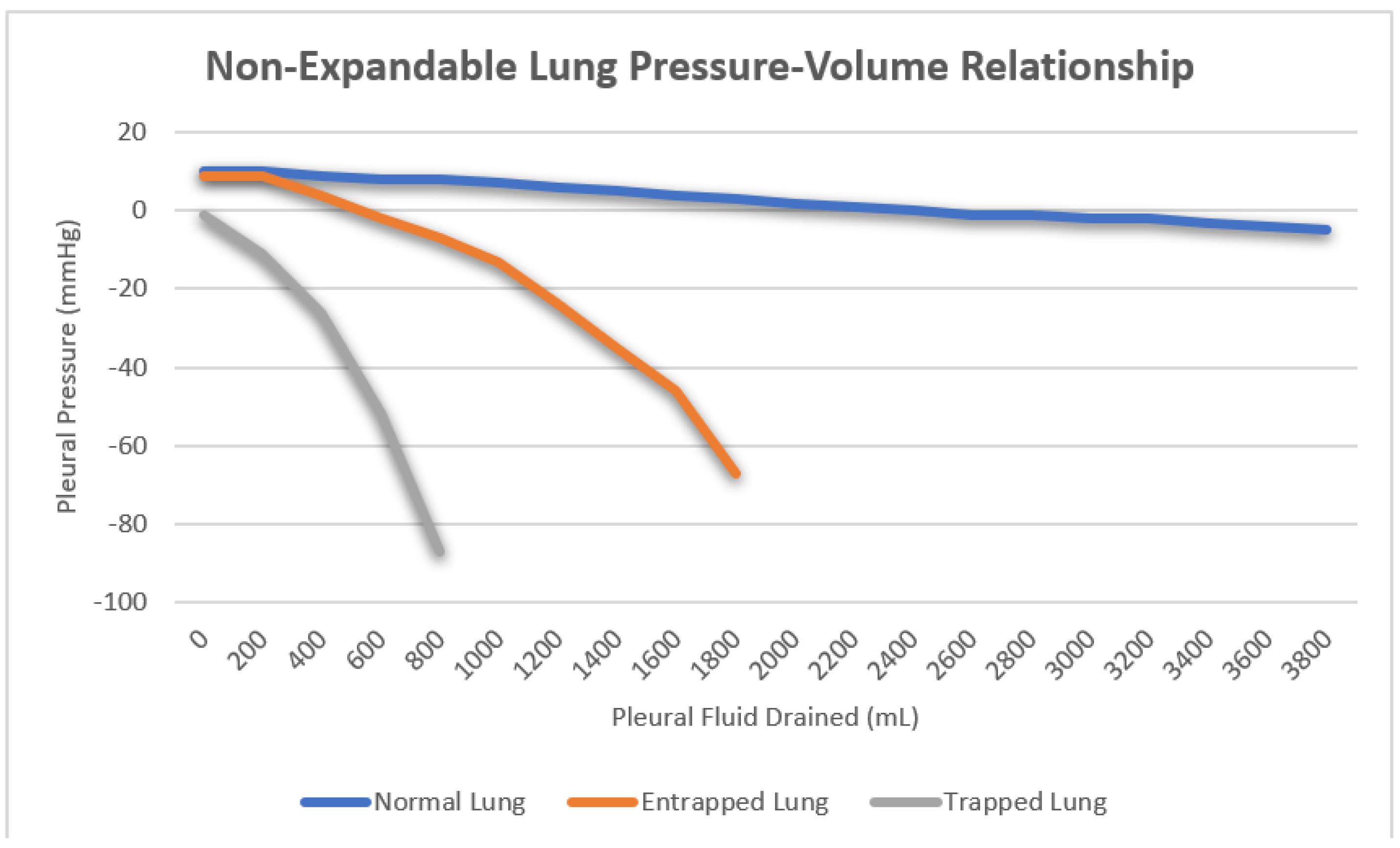
3.4.4. Pleural Manometry

Once the catheter is appropriately positioned within the pleural space, pleural manometry can be conducted using one of three methods. The original U-tube water manometry technique has fallen out of favor due to the improved ease and accuracy of more modern approaches. Hemodynamic electronic transducers and digital manometers have replaced U-tube water manometry, with hemodynamic electronic transducers emerging as the preferred method. All three methods offer pleural pressure measurements throughout the drainage process, which prove valuable for evaluating a non-expandable lung [30]. Non-expandable lung can be further categorized into lung entrapment and trapped lungs. In a healthy lung, the initial pleural pressure is positive, exhibiting a minimal decrease during fluid aspiration as the lung re-expands to occupy the space previously occupied by pleural effusion. Lung entrapment arises from visceral restriction due to an active pleural inflammatory process. Initially, the pleural pressure is positive and experiences a slight decrease as the lung re-expands, mirroring normal lung behavior. However, the progressive visceral restriction eventually prevents full lung re-expansion, causing a sharp drop in pleural pressure after fluid removal surpasses the point of maximum lung expansion. This phenomenon typically manifests after draining larger volumes in the range of one or more liters. Conversely, a trapped lung results from visceral thickening due to impaired pleural space healing resulting from a remote pleural process. Here, lung re-expansion is severely limited, creating a vacuum within the pleural space. Consequently, the initial pleural pressure is negative, and its sharp decline occurs rapidly with minimal fluid removal, often as little as 50–100 mL of fluid (Figure 4). Pleural manometry has also been proposed as a method to reduce the risk of re-expansion pulmonary edema following large volume thoracentesis. There are currently no data supporting the use of pleural manometry to affect any patient-related outcomes, and its use is strictly academic [30].

3.4.5. Sample Collection

As pleural fluid is withdrawn for analysis, its appearance should be documented. Terms such as serous, blood-tinged, serosanguinous, frank blood, purulent, frank pus, turbid, and milky can provide preliminary insights into the underlying cause even before formal analysis. It is advisable to collect three distinct samples for microbiological, biochemical, and cytological examinations. In cases of diagnostic thoracentesis, where the aspirated fluid is limited, judiciously distribute the fluid among these samples. Following British Thoracic Society recommendations, microbiological analysis necessitates only 5–10 mL, biochemical analysis requires 2–5 mL, and the remaining portion should be allocated for cytological analysis [11]. To enhance the yield, specific containers should be used for certain samples, as elaborated in the pleural fluid analysis section. Briefly, microbiological samples should be collected in both aerobic and anaerobic blood culture bottles [20]. For pH analysis, fluid should be collected in arterial blood gas syringes on ice and analyzed with a blood gas analyzer [19]. Cytology samples are best preserved in clear, plain containers, while biochemical samples can be collected in plain containers or serum blood collection tubes [11].

3.4.6. Fluid Drainage

When performing therapeutic thoracentesis to drain large-volume pleural effusions, the drainage speed and volume should be approached with caution. A slower removal rate is theoretically preferred to promote gradual lung re-expansion and mitigate the risk of re-expansion pulmonary edema. Multiple options for drainage include gravity drainage, hand syringe drainage via a three way stop-cock, and wall suction and vacuum bottle drainage [28]. There are no consensus recommendations for one method over the other, and much controversy exists regarding the preferred method. The maximum allowable drainage volume remains a topic of debate as well. Historically, a recommended upper limit of 1.5 L was advised for removal in a single thoracentesis. Larger drainage volumes were linked to increased incidence of re-expansion pulmonary edema and post-procedure pneumothorax. There is considerable mortality risk associated with re-expansion pulmonary edema and the robust evidence supporting minimal complication rates when draining less than 1.5 L [11,31]. Yet, studies have indicated reasonable safety even with larger drainage volumes, reaching up to 6.5 L in certain instances [30]. Irrespective of the drainage volume chosen, the thoracentesis should be promptly stopped if the patient develops chest pain, tightness, or breathlessness, as these symptoms might herald impending re-expansion pulmonary edema. Additionally, the procedure should be concluded once no more fluid can be effectively aspirated.

3.4.7. Needle Removal and Dressing

After the completion of fluid aspiration, the catheter can be safely removed. To prevent air entry into the pleural space, the patient should be guided to perform a maneuver that raises intrathoracic pressure as the catheter is withdrawn. This can be achieved by instructing the patient to hum continuously while the catheter is being removed. As the catheter is withdrawn, the humming assists in expelling any air present in the pleural space. Once the catheter has been successfully removed, a sterile dressing should be applied to the puncture site for proper wound care [28].

3.5. Post-Procedural Care

Routine post-procedural chest radiography is not necessary following an uncomplicated thoracentesis [11]. However, it is advisable if the patient continues to experience respiratory symptoms after the procedure, if multiple attempts were made during the aspiration, or if the operator perceives any complications. Interestingly, studies have shown that clinicians’ predictions of pneumothorax occurrence post-thoracentesis are quite accurate [32]. Despite this, many clinicians still opt for routine post-procedural imaging. Exceptions exist, such as in determining the next steps for malignant effusions, but these nuances are beyond the scope of this review [33]. An alternative approach which is gaining traction involves using thoracic ultrasound both before and after the procedure to confirm the presence of lung sliding, effectively ruling out a post-procedural pneumothorax. After the procedure, it is recommended to monitor vital signs and observe the patient for a brief period. Although there is no specific guideline for the duration of monitoring, patients without complications or symptoms typically do not require extended observation.

3.6. Complications

The three primary complications associated with thoracentesis are pneumothorax, hemothorax, and re-expansion pulmonary edema [32]. Both operator experience and ultrasound guidance have been shown to reduce the incidence of these complications [11].
Pneumothorax is the most frequent complication, with reported incidences varying across studies. Historically, the rate was around 18%, but with ultrasound guidance, this has decreased to approximately 3%. Recent estimates suggest an incidence of 0–6%, although pinpointing the exact incidence is challenging due to factors like a non-expandable lung and inadvertent air introduction. Management of post-thoracentesis pneumothorax ranges from chest tube placement (34.1% in one review) to conservative medical approaches, depending on the case. Overall, less than 2% of all thoracenteses require tube thoracostomy due to a complication [32].
Hemothorax, a rare but serious complication, occurs in less than 1% of cases. The risk can be minimized by selecting the puncture site at the superior rib margin. While Doppler ultrasound aids in identifying and preventing intercostal artery laceration, its impact on reducing bleeding complications remains uncertain.
Re-expansion pulmonary edema, marked by new alveolar infiltrates and hypoxemia within 24 h of the procedure, is an infrequent occurrence (less than 1%). Though asymptomatic cases are more common, symptomatic instances carry a substantial mortality rate of around 20% [31]. Several treatments, including continuous positive airway pressure, diuretics, and steroids, are used for symptomatic re-expansion pulmonary edema, but their efficacy is not strongly supported by data.

4. Discussion

Pleural effusions continue to be a prevalent medical condition encountered across inpatient and outpatient settings. Thoracentesis remains a pivotal procedure for both diagnosing and managing pleural effusions. Staying well informed about current guidelines and recommendations for thoracentesis equips physicians to conduct procedures that are effective and safe. The literature highlights a significant decrease in complications when practitioners adhere to proper anatomical positioning and employ contemporary equipment. Notably, thoracic ultrasonography has emerged as a reliable tool for assessing pleural effusions and guiding drainage procedures. Extensive evidence supports the use of ultrasound guidance in thoracentesis, positioning it as the new gold standard. The availability of thoracic ultrasonography should deter the practice of blind thoracentesis. Adhering to the correct protocol for preparation, pleural space aspiration, sample collection, and post-procedural care are crucial in continuing to improve patient outcomes and diagnostic yields.

5. Future Direction

While recent advancements have enhanced our understanding of thoracentesis, certain aspects of this procedure remain contentious. Specifically, there is ongoing debate regarding the preferred drainage method and the optimal volume of fluid to be removed. Currently, there is a dearth of large-scale clinical trials and comprehensive meta-analyses that offer definitive guidance on these matters. Given that both the chosen drainage method and the volume extracted can significantly impact the risk of complications, further investigation into these areas is imperative.
This review highlights compelling evidence supporting the use of ultrasound-guided thoracentesis. As additional data supporting this approach continue to accumulate, it is essential to prioritize the education of physicians in this technique. To maximize patient outcomes, it is advisable for ultrasound-guided thoracentesis, following the established protocols, to become the standard procedure for any physician performing pleural drainage.
Finally, improving adherence to guideline-directed care will improve outcomes. Veering from established practices leads to higher rates of complications, the need for repeat procedures, inconclusive results, and increased healthcare spending [34]. Nationwide efforts to educate and reinforce the principles covered in this review are crucial to achieve optimal benefits from performing thoracentesis.

Author Contributions

Conceptualization, M.J.N. and D.A.; methodology, M.J.N. and D.A.; resources, M.J.N., D.A. and C.M.; writing—original draft preparation, M.J.N.; writing—review and editing, D.A. and C.M.; supervision, D.A. and C.M. All authors have read and agreed to the published version of the manuscript.

Funding

This research received no external funding.

Institutional Review Board Statement

Not applicable.

Informed Consent Statement

Not applicable.

Data Availability Statement

Not applicable.

Conflicts of Interest

The authors declare no conflict of interest.

References

  1. Jany, B.; Welte, T. Pleural Effusion in Adults-Etiology, Diagnosis, and Treatment. Dtsch. Aerzteblatt Int. 2019, 116, 377–386. [Google Scholar] [CrossRef] [PubMed]
  2. Krishna, R.; Antoine, M.H.; Rudrappa, M. Pleural Effusion. In StatPearls; StatPearls Publishing: Treasure Island, FL, USA, 2023. Available online: https://www.ncbi.nlm.nih.gov/books/NBK448189/ (accessed on 6 September 2023).
  3. Karkhanis, V.S.; Joshi, J.M. Pleural effusion: Diagnosis, treatment, and management. Open Access Emerg. Med. 2012, 4, 31–52. [Google Scholar] [CrossRef] [PubMed]
  4. Charalampidis, C.; Youroukou, A.; Lazaridis, G.; Baka, S.; Mpoukovinas, I.; Karavasilis, V.; Kioumis, I.; Pitsiou, G.; Papaiwannou, A.; Karavergou, A.; et al. Pleura space anatomy. J. Thorac. Dis. 2015, 7 (Suppl. S1), S27–S32. [Google Scholar] [CrossRef] [PubMed]
  5. Steven, A.S. The Pathophysiology of Pleural Effusions. Annu. Rev. Med. 1990, 41, 7–13. [Google Scholar]
  6. Hooper, C.; Lee, Y.C.G.; Maskell, N. Investigation of a unilateral pleural effusion in adults: British Thoracic Society pleural disease guideline 2010. Thorax 2010, 65, ii4–ii17. [Google Scholar] [CrossRef] [PubMed]
  7. Roberts, M.E.; Rahman, N.M.; Maskell, N.A.; Bibby, A.C.; Blyth, K.G.; Corcoran, J.P.; Edey, A.; Evison, M.; de Fonseka, D.; Hallifax, R.; et al. British Thoracic Society Guideline for pleural disease. Thorax 2023, 78 (Suppl. S3), 1–42. [Google Scholar] [CrossRef] [PubMed]
  8. Prina, E.; Torres, A.; Carvalho, C.R. Lung ultrasound in the evaluation of pleural effusion. J. Bras. Pneumol. 2014, 40, 1–5. [Google Scholar] [CrossRef]
  9. Xirouchaki, N.; Magkanas, E.; Vaporidi, K.; Kondili, E.; Plataki, M.; Patrianakos, A.; Akoumianaki, E.; Georgopoulos, D. Lung ultrasound in critically ill patients: Comparison with bedside chest radiography. Intensive Care Med. 2011, 37, 1488–1493. [Google Scholar] [CrossRef]
  10. Vetrugno, L.; Guadagnin, G.M.; Orso, D.; Boero, E.; Bignami, E.; Bove, T. An easier and safe affair, pleural drainage with ultrasound in critical patient: A technical note. Crit. Ultrasound J. 2018, 10, 18. [Google Scholar] [CrossRef]
  11. Asciak, R.; Bedawi, E.O.; Bhatnagar, R.; Clive, A.O.; Hassan, M.; Lloyd, H.; Reddy, R.; Roberts, H.; Rahman, N.M. British Thoracic Society Clinical Statement on pleural procedures. Thorax 2023, 78 (Suppl. S3), s43–s68. [Google Scholar] [CrossRef]
  12. Yang, P.C.; Luh, K.T.; Chang, D.B.; Wu, H.D.; Yu, C.J.; Kuo, S.H. Value of sonography in determining the nature of pleural effusion: Analysis of 320 cases. AJR Am. J. Roentgenol. 1992, 159, 29–33. [Google Scholar] [CrossRef] [PubMed]
  13. Brogi, E.; Gargani, L.; Bignami, E.; Barbariol, F.; Marra, A.; Forfori, F.; Vetrugno, L. Thoracic ultrasound for pleural effusion in the intensive care unit: A narrative review from diagnosis to treatment. Crit. Care 2017, 21, 325. [Google Scholar] [CrossRef] [PubMed]
  14. Mercer, R.M.; Corcoran, J.P.; Porcel, J.M.; Rahman, N.M.; Psallidas, I. Interpreting pleural fluid results. Clin. Med. 2019, 19, 213–217. [Google Scholar] [CrossRef] [PubMed]
  15. Romero-Candeira, S.; Fernández, C.; Martín, C.; Sánchez-Paya, J.; Hernández, L. Influence of diuretics on the concentration of proteins and other components of pleural transudates in patients with heart failure. Am. J. Med. 2001, 110, 681–686. [Google Scholar] [CrossRef] [PubMed]
  16. Beaudoin, S.; Gonzalez, A.V. Evaluation of the patient with pleural effusion. Can. Med Assoc. J. 2018, 190, E291–E295. [Google Scholar] [CrossRef]
  17. Na, M.J. Diagnostic tools of pleural effusion. Tuberc. Respir. Dis. 2014, 76, 199–210. [Google Scholar] [CrossRef]
  18. Krenke, R.; Nasilowski, J.; Korczynski, P.; Gorska, K.; Przybylowski, T.; Chazan, R.; Light, R.W. Incidence and aetiology of eosinophilic pleural effusion. Eur. Respir. J. 2009, 34, 1111–1117. [Google Scholar] [CrossRef]
  19. Mark, C. Houston, Pleural fluid pH: Diagnostic, therapeutic, and prognostic value. Am. J. Surg. 1987, 154, 333–337. [Google Scholar] [CrossRef]
  20. Menzies, S.M.; Rahman, N.M.; Wrightson, J.M.; Davies, H.E.; Shorten, R.; Gillespie, S.H.; Davies, C.W.; Maskell, N.A.; Jeffrey, A.A.; Lee, Y.C.; et al. Blood culture bottle culture of pleural fluid in pleural infection. Thorax 2011, 66, 658–662. [Google Scholar] [CrossRef]
  21. Garcia, L.W.; Ducatman, B.S.; Wang, H.H. The value of multiple fluid specimens in the cytological diagnosis of malignancy. Mod. Pathol. 1994, 7, 665–668. [Google Scholar]
  22. Desai, N.R.; Lee, H.J. Diagnosis and management of malignant pleural effusions: State of the art in 2017. J. Thorac. Dis. 2017, 9 (Suppl. S10), S1111–S1122. [Google Scholar] [CrossRef] [PubMed]
  23. Grosu, H.B.; Kazzaz, F.; Vakil, E.; Molina, S.; Ost, D. Sensitivity of Initial Thoracentesis for Malignant Pleural Effusion Stratified by Tumor Type in Patients with Strong Evidence of Metastatic Disease. Respiration 2018, 96, 363–369. [Google Scholar] [CrossRef] [PubMed]
  24. Li, D.; Ajmal, S.; Tufail, M.; Panchal, R.K. Modern day management of a unilateral pleural effusion. Clin. Med. 2021, 21, e561–e566. [Google Scholar] [CrossRef] [PubMed]
  25. Patel, I.J.; Davidson, J.C.; Nikolic, B.; Salazar, G.M.; Schwartzberg, M.S.; Walker, T.G.; Saad, W.A. Standards of Practice Committee, with Cardiovascular and Interventional Radiological Society of Europe (CIRSE) Endorsement. Consensus guidelines for periprocedural management of coagulation status and hemostasis risk in percutaneous image-guided interventions. J. Vasc. Interv. Radiol. 2012, 23, 727–736. [Google Scholar] [CrossRef] [PubMed]
  26. Vikas Pathak JErin Allender Mollie, W.G. Management of anticoagulant and antiplatelet therapy in patients undergoing interventional pulmonary procedures. Eur. Respir. Rev. 2017, 26, 170020. [Google Scholar] [CrossRef]
  27. Segal, J.B.; Dzik, W.H. Paucity of studies to support that abnormal coagulation test results predict bleeding in the setting of invasive procedures: An evidence-based review. Transfusion 2005, 45, 1413–1425. [Google Scholar] [CrossRef] [PubMed]
  28. LeVasseur, T. Chapter 76 Thoracentesis. In The Mont Reid Surgical Handbook, 6th ed.; Textbook; W.B. Saunders: Philadelphia, PA, USA, 2008; pp. 835–838. [Google Scholar]
  29. Liang, S.J.; Tu, C.Y.; Chen, H.J.; Chen, C.H.; Chen, W.; Shih, C.M.; Hsu, W.H. Application of ultrasound-guided pigtail catheter for drainage of pleural effusions in the ICU. Intensive Care Med. 2009, 35, 350–354. [Google Scholar] [CrossRef]
  30. Hu, K.; Chopra, A.; Huggins, J.T.; Nanchal, R. Pleural manometry: Techniques, applications, and pitfalls. J. Thorac. Dis. 2020, 12, 2759–2770. [Google Scholar] [CrossRef]
  31. Havelock, T.; Teoh, R.; Laws, D.; Gleeson, F. Pleural procedures and thoracic ultrasound: British Thoracic Society pleural disease guideline 2010. Thorax 2010, 65, i61–i76. [Google Scholar] [CrossRef]
  32. Cantey, E.P.; Walter, J.M.; Corbridge, T.; Barsuk, J.H. Complications of thoracentesis: Incidence, risk factors, and strategies for prevention. Curr. Opin. Pulm. Med. 2016, 22, 378–385. [Google Scholar] [CrossRef]
  33. Feller-Kopman, D.J.; Reddy, C.B.; De Camp, M.M.; Diekemper, R.L.; Gould, M.K.; Henry, T.; Iyer, N.P.; Lee, Y.C.G.; Lewis, S.Z.; Maskell, N.A.; et al. Management of Malignant Pleural Effusions. An Official ATS/STS/STR Clinical Practice Guideline. Am. J. Respir. Crit. Care Med. 2018, 198, 839–849. [Google Scholar] [CrossRef] [PubMed]
  34. Ost, D.E.; Niu, J.; Zhao, H.; Grosu, H.B.; Giordano, S.H. Quality Gaps and Comparative Effectiveness of Management Strategies for Recurrent Malignant Pleural Effusions. Chest 2018, 153, 438–452. [Google Scholar] [CrossRef] [PubMed]
Figure 1. Posteroanterior (PA) and lateral chest radiography demonstrating a left sided pleural effusion. Image courtesy of Abdelkader Mallouk, Radiopaedia.org, rID: 71570.
Jor 03 00020 g001
Figure 2. Computed tomography (CT) of the chest demonstrating bilateral pleural effusions. Image courtesy of Paul Leong, Radiopaedia.org, rID: 26474.
Jor 03 00020 g002
Figure 3. Thoracic ultrasonography demonstrating an anechoic pleural effusion with surrounding structures. Image (left) courtesy of Mohammad Osama Hussein Yonso, Radiopaedia.org, rID: 22793. Image (right) courtesy of Zhan Lim, Radiopaedia.org, rID: 94919.
Jor 03 00020 g003
Figure 4. Three distinct pressure/volume curves representing the mean pleural pressure as volume is removed from the pleural space in normal lung, lung entrapment and trapped lung respectively.
Jor 03 00020 g004
Table 1. Demonstrating the most likely etiologies of pleural effusions based on their ultrasonographic appearance.
Effusion TypeAnechoicComplex Non-SeptatedComplex SeptatedEchogenic
TransudativeXX
ExudativeXXXX
Hemorrhagic X
Table 2. Different diagnostic criteria use for Light’s criteria and the pleural fluid only three test combination.
Light’s CriteriaPleural Fluid Only Three Test Combination
Pleural fluid to serum total protein > 0.5Total protein > 3g/dL
Pleural fluid to serum lactate dehydrogenase (LDH) ratio > 0.6Cholesterol > 55mg/dL
Pleural fluid LDH > two-thirds upper limit of normalLDH > two-thirds the upper limit of normal
Disclaimer/Publisher’s Note: The statements, opinions and data contained in all publications are solely those of the individual author(s) and contributor(s) and not of MDPI and/or the editor(s). MDPI and/or the editor(s) disclaim responsibility for any injury to people or property resulting from any ideas, methods, instructions or products referred to in the content.

