
Phage Therapy for Cardiac Implantable Electronic Devices and Vascular Grafts: A Targeted Literature Review


Abstract
1. Introduction
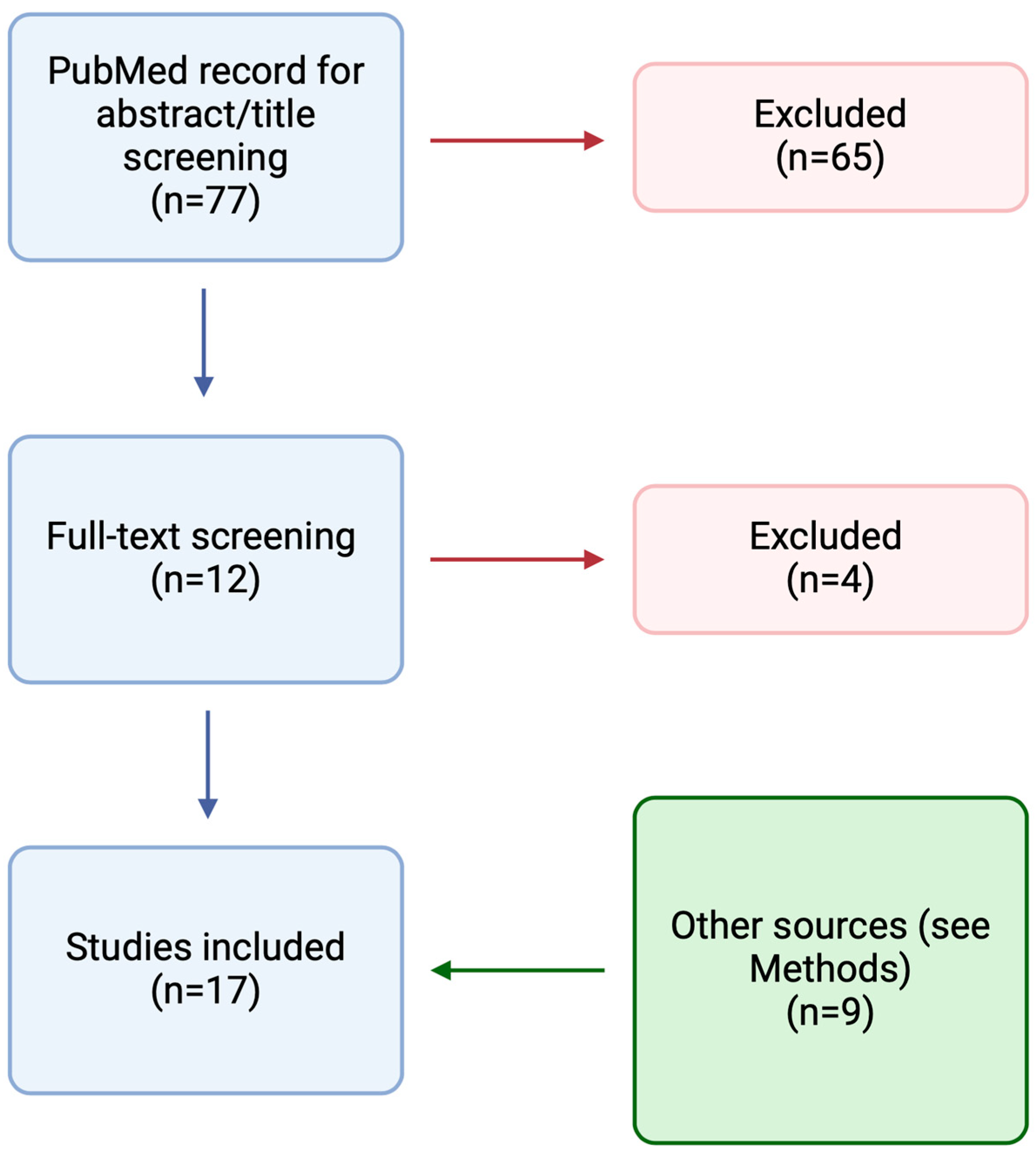
2. Methods
3. Results
3.1. Demographic, Clinical, and Microbiological Characteristics
3.2. Phage Therapy and Concomitant Treatment
3.3. Efficacy and Safety
4. Discussion
4.1. Which Patients with CIED and Vascular Graft Infections Were Treated?
4.2. How Were Phages Administered?
4.3. What Is the Role of Concomitant Antibiotics?
4.4. What Is the Role of Surgery?
4.5. Is There a Pattern between Management and Outcomes?
4.6. Is Phage Therapy Safe?
5. Conclusions
Author Contributions
Funding
Institutional Review Board Statement
Informed Consent Statement
Data Availability Statement
Conflicts of Interest
References
- Zinoviev, R.; Lippincott, C.K.; Keller, S.C.; Gilotra, N.A. In Full Flow: Left Ventricular Assist Device Infections in the Modern Era. Open Forum Infect. Dis. 2020, 7, ofaa124. [Google Scholar] [CrossRef] [PubMed]
- Glikson, M.; Nielsen, J.C.; Kronborg, M.B.; Michowitz, Y.; Auricchio, A.; Barbash, I.M.; Barrabés, J.A.; Boriani, G.; Braunschweig, F.; Brignole, M.; et al. 2021 ESC Guidelines on cardiac pacing and cardiac resynchronization therapy. Eur. Heart J. 2021, 42, 3427–3520. [Google Scholar] [CrossRef] [PubMed]
- Pibarot, P.; Dumesnil, J.G. Prosthetic Heart Valves. Circulation 2009, 119, 1034–1048. [Google Scholar] [CrossRef]
- Pennel, T.; Zilla, P. Clinical Applications and Limitations of Vascular Grafts. In Tissue-Engineered Vascular Grafts; Springer International Publishing: Cham, Switzerland, 2020; pp. 3–34. [Google Scholar]
- Mostafavi, E.; Dubey, A.K.; Walkowiak, B.; Kaushik, A.; Ramakrishna, S.; Teodori, L. Antimicrobial surfaces for implantable cardiovascular devices. Curr. Opin. Biomed. Eng. 2022, 23, 100406. [Google Scholar] [CrossRef]
- Caldara, M.; Belgiovine, C.; Secchi, E.; Rusconi, R. Environmental, Microbiological, and Immunological Features of Bacterial Biofilms Associated with Implanted Medical Devices. Clin. Microbiol. Rev. 2022, 35, e0022120. [Google Scholar] [CrossRef]
- Blomströ M-Lundqvist, C.; Traykov, V.; Erba, P.A.; Burri, H.; Nielsen, J.C.; Bongiorni, M.G.; Poole, J.; Boriani, G.; Costa, R.; Deharo, J.C.; et al. European Heart Rhythm Association (EHRA) international consensus document on how to prevent, diagnose, and treat cardiac implantable electronic device infections-endorsed of Clinical Microbiology and Infectious Diseases (ESCMID) in collaboration with the European Association for Cardio-Thoracic Surgery (EACTS). Europace 2020, 22, 515–549. [Google Scholar]
- Totten, K.M.C.; Patel, R. Phage Activity against Planktonic and Biofilm Staphylococcus aureus Periprosthetic Joint Infection Isolates. Antimicrob. Agents Chemother. 2022, 66, e0187921. [Google Scholar] [CrossRef]
- Chegini, Z.; Khoshbayan, A.; Moghadam, M.T.; Farahani, I.; Jazireian, P.; Shariati, A. Bacteriophage therapy against Pseudomonas aeruginosa biofilms: A review. Ann. Clin. Microbiol. Antimicrob. 2020, 19, 45. [Google Scholar] [CrossRef] [PubMed]
- Rubalskii, E.; Ruemke, S.; Salmoukas, C.; Boyle, E.C.; Warnecke, G.; Tudorache, I.; Shrestha, M.; Schmitto, J.D.; Martens, A.; Rojas, S.V.; et al. Bacteriophage therapy for critical infections related to cardiothoracic surgery. Antibiotics 2020, 9, 232. [Google Scholar] [CrossRef]
- Aslam, S.; Lampley, E.; Wooten, D.; Karris, M.; Benson, C.; Strathdee, S.; Schooley, R.T. Lessons Learned from the First 10 Consecutive Cases of Intravenous Bacteriophage Therapy to Treat Multidrug-Resistant Bacterial Infections at a Single Center in the United States. Open Forum Infect. Dis. 2020, 7, ofaa389. [Google Scholar] [CrossRef]
- Petrovic Fabijan, A.; Lin, R.C.; Ho, J.; Maddocks, S.; Ben Zakour, N.L.; Iredell, J.R. Safety of bacteriophage therapy in severe Staphylococcus aureus infection. Nat. Microbiol. 2020, 5, 465–472. [Google Scholar] [CrossRef] [PubMed]
- Tkhilaishvili, T.; Potapov, E.; Starck, C.; Mulzer, J.; Falk, V.; Trampuz, A.; Schoenrath, F. Bacteriophage therapy as a treatment option for complex cardiovascular implant infection: The German Heart Center Berlin experience. J. Hear. Lung Transplant. 2022, 41, 551–555. [Google Scholar] [CrossRef] [PubMed]
- Onallah, H.; Hazan, R.; Nir-Paz, R. Compassionate Use of Bacteriophages for Failed Persistent Infections During the First 5 Years of the Israeli Phage Therapy Center. Open Forum Infect. Dis. 2023, 10, ofad221. [Google Scholar] [CrossRef] [PubMed]
- Green, S.I.; Clark, J.R.; Santos, H.H.; Weesner, K.E.; Salazar, K.C.; Aslam, S.; Campbell, J.W.; Doernberg, S.B.; Blodget, E.; Morris, M.I.; et al. A Retrospective, Observational Study of 12 Cases of Expanded-Access Customized Phage Therapy: Production, Characteristics, and Clinical Outcomes. Clin. Infect. Dis. 2023, 77, 1079–1091. [Google Scholar] [CrossRef] [PubMed]
- Aslam, S.; Roach, D.; Nikolich, M.P.; Biswas, B.; Schooley, R.T.; Lilly-Bishop, K.A.; Rice, G.K.; Cer, R.Z.; Hamilton, T.; Henry, M.; et al. Pseudomonas aeruginosa ventricular assist device infections: Findings from ineffective phage therapies in five cases. Antimicrob. Agents Chemother. 2024, 68, e0172823. [Google Scholar] [CrossRef] [PubMed]
- Pirnay, J.P.; Djebara, S.; Steurs, G.; Griselain, J.; Cochez, C.; De Soir, S.; Glonti, T.; Spiessens, A.; Vanden Berghe, E.; Green, S.; et al. Retrospective, observational analysis of the first one hundred consecutive cases of personalized bacteriophage therapy of difficult-to-treat infections facilitated by a Belgian consortium. medRxiv 2023. [Google Scholar] [CrossRef]
- Chan, B.K.; Turner, P.E.; Kim, S.; Mojibian, H.R.; Elefteriades, J.A.; Narayan, D. Phage treatment of an aortic graft infected with Pseudomonas aeruginosa. Evol. Med. Public Heal. 2018, 2018, 60–66. [Google Scholar] [CrossRef] [PubMed]
- Duplessis, C.; Biswas, B.; Hanisch, B.; Perkins, M.; Henry, M.; Quinones, J.; Wolfe, D.; Estrella, L.; Hamilton, T. Refractory Pseudomonas Bacteremia in a 2-Year-Old Sterilized by Bacteriophage Therapy. J. Pediatric Infect. Dis. Soc. 2018, 7, 253–256. [Google Scholar] [CrossRef]
- Mulzer, J.; Trampuz, A.; Trampuz, A.; Potapov, E.V.; Potapov, E.V. Treatment of chronic left ventricular assist device infection with local application of bacteriophages. Eur. J. Cardio-Thorac. Surg. 2020, 57, 1003–1004. [Google Scholar] [CrossRef]
- Gilbey, T.; Ho, J.; Cooley, L.A.; Fabijan, A.P.; Iredell, J.R. Adjunctive bacteriophage therapy for prosthetic valve endocarditis due to Staphylococcus aureus. Med. J. Aust. 2019, 211, 142–143.e1. [Google Scholar] [CrossRef]
- Püschel, A.; Skusa, R.; Bollensdorf, A.; Gross, J. Local Treatment of Driveline Infection with Bacteriophages. Antibiotics 2022, 11, 1310. [Google Scholar] [CrossRef]
- Grambow, E.; Junghans, S.; Kröger, J.C.; Reisinger, E.C.; Krause, B.J.; Groß, J. Treatment of an Infected TEVAR with Extra- and Endovascular Bacteriophage Application. EJVES Vasc. Forum 2022, 56, 20–23. [Google Scholar] [CrossRef] [PubMed]
- Rojas, S.V.; Junghans, S.; Fox, H.; Lazouski, K.; Schramm, R.; Morshuis, M.; Gummert, J.F.; Gross, J. Bacteriophage-Enriched Galenic for Intrapericardial Ventricular Assist Device Infection. Antibiotics 2022, 11, 602. [Google Scholar] [CrossRef] [PubMed]
- Blasco, L.; López-Hernández, I.; Rodríguez-Fernández, M.; Pérez-Florido, J.; Casimiro-Soriguer, C.S.; Djebara, S.; Merabishvili, M.; Pirnay, J.P.; Rodríguez-Baño, J.; Tomás, M.; et al. Case report: Analysis of phage therapy failure in a patient with a Pseudomonas aeruginosa prosthetic vascular graft infection. Front. Med. 2023, 10, 1199657. [Google Scholar] [CrossRef] [PubMed]
- Racenis, K.; Lacis, J.; Rezevska, D.; Mukane, L.; Vilde, A.; Putnins, I.; Djebara, S.; Merabishvili, M.; Pirnay, J.P.; Kalnina, M.; et al. Successful Bacteriophage-Antibiotic Combination Therapy against Multidrug-Resistant Pseudomonas aeruginosa Left Ventricular Assist Device Driveline Infection. Viruses 2023, 15, 1210. [Google Scholar] [CrossRef] [PubMed]
- Simpson, E.A.; MacLeod, C.S.; Stacey, H.J.; Nagy, J.; Jones, J.D. The Safety and Efficacy of Phage Therapy for Infections in Cardiac and Peripheral Vascular Surgery: A Systematic Review. Antibiotics 2023, 12, 1684. [Google Scholar] [CrossRef]
- Slopek, S.; Weber-Dabrowska, B.; Dabrowski, M.; Kucharewicz-Krukowska, A. Results of Bacteriophage Treatment of Supparative Bacterial Infections in the Years 1981–1986. Arch. Immunol. Ther. Exp. 1987, 35, 569–583. [Google Scholar]
- Hannan, M.M.; Xie, R.; Cowger, J.; Schueler, S.; de By, T.; Dipchand, A.I.; Chu, V.H.; Cantor, R.S.; Koval, C.E.; Krabatsch, T.; et al. Epidemiology of infection in mechanical circulatory support: A global analysis from the ISHLT Mechanically Assisted Circulatory Support Registry. J. Heart Lung Transplant. 2019, 38, 364–373. [Google Scholar] [CrossRef]
- Greenspon, A.J.; Eby, E.L.; Petrilla, A.A.; Sohail, M.R. Treatment patterns, costs, and mortality among Medicare beneficiaries with CIED infection. Pacing Clin. Electrophysiol. 2018, 41, 495–503. [Google Scholar] [CrossRef]
- Chakfé, N.; Diener, H.; Lejay, A.; Assadian, O.; Berard, X.; Caillon, J.; Fourneau, I.; Glaudemans, A.W.; Koncar, I.; Lindholt, J.; et al. Editor’s Choice-European Society for Vascular Surgery (ESVS) 2020 Clinical Practice Guidelines on the Management of Vascular Graft and Endograft Infections. Eur. J. Vasc. Endovasc. Surg. 2020, 59, 339–384. [Google Scholar] [CrossRef]
- Peng, Q.; Tang, X.; Dong, W.; Sun, N.; Yuan, W. A Review of Biofilm Formation of Staphylococcus aureus and Its Regulation Mechanism. Antibiotics 2022, 12, 12. [Google Scholar] [CrossRef] [PubMed]
- Kamali, E.; Jamali, A.; Ardebili, A.; Ezadi, F.; Mohebbi, A. Evaluation of antimicrobial resistance, biofilm forming potential, and the presence of biofilm-related genes among clinical isolates of Pseudomonas aeruginosa. BMC Res. Notes 2020, 13, 27. [Google Scholar] [CrossRef] [PubMed]
- Hernando-Amado, S.; Martínez, J.L. Special Issue: ‘Antimicrobial Resistance in Pseudomonas aeruginosa’. Microorganisms 2023, 11, 744. [Google Scholar] [CrossRef] [PubMed]
- Yerushalmy, O.; Khalifa, L.; Gold, N.; Rakov, C.; Alkalay-Oren, S.; Adler, K.; Ben-Porat, S.; Kraitman, R.; Gronovich, N.; Shulamit Ginat, K.; et al. The Israeli Phage Bank (IPB). Antibiotics 2020, 9, 269. [Google Scholar] [CrossRef] [PubMed]
- Rosner, D.; Clark, J. Formulations for Bacteriophage Therapy and the Potential Uses of Immobilization. Pharmaceuticals 2021, 14, 359. [Google Scholar] [CrossRef] [PubMed]
- Nang, S.C.; Lin, Y.W.; Fabijan, A.P.; Chang, R.Y.; Rao, G.G.; Iredell, J.; Chan, H.K.; Li, J. Pharmacokinetics/pharmacodynamics of phage therapy: A major hurdle to clinical translation. Clin. Microbiol. Infect. 2023, 29, 702–709. [Google Scholar] [CrossRef] [PubMed]
- Łusiak-Szelachowska, M.; Międzybrodzki, R.; Drulis-Kawa, Z.; Cater, K.; Knežević, P.; Winogradow, C.; Amaro, K.; Jończyk-Matysiak, E.; Weber-Dąbrowska, B.; Rękas, J.; et al. Bacteriophages and antibiotic interactions in clinical practice: What we have learned so far. J. Biomed. Sci. 2022, 29, 23. [Google Scholar] [CrossRef]
- Gu Liu, C.; Green, S.I.; Min, L.; Clark, J.R.; Salazar, K.C.; Terwilliger, A.L.; Kaplan, H.B.; Trautner, B.W.; Ramig, R.F.; Maresso, A.W. Phage-Antibiotic Synergy Is Driven by a Unique Combination of Antibacterial Mechanism of Action and Stoichiometry. MBio 2020, 11, 10–1128. [Google Scholar] [CrossRef]

| Patient | Article Reference | Year | Country | Age | Sex | Pathogen | Type Infection | Number/Type of Phage(s) | Route of Phage Administration | Highest Dose of Phage Administered (PFU/mL) | Duration |
|---|---|---|---|---|---|---|---|---|---|---|---|
| 1 | Chan, BK [18] | 2018 | US | 76 | M | P. aeruginosa | Vascular graft infection | One (OMKO1) | In situ through a CT-guided needle | 1 × 10⁷ | Once |
| 2 | Duplessis, C [19] | 2018 | US | 2 | M | P. aeruginosa | bacteremia after the ASD/VSD closures | Two (not specified) | iv | 3.5 × 10⁵ | q6h for six doses, resumed after 11 days |
| 3 | Rubalskij, E (1) [10] | 2020 | Germany | 52 | M | S. aureus E. faecium P. aeruginosa | Vascular graft infection | Four (CH1, Enf1, PA5, PA10) | Topically + iv + intraoperative | 1 × 10⁸ | q24h for 2 separate days |
| 4 | Rubalskij, E (2) [10] | 2020 | Germany | 59 | M | S. aureus | Vascular graft infection | One (CH1) | In situ through a chest tube | 1 × 10⁹ | q12h for 2 days |
| 5 | Rubalskij, E (3) [10] | 2020 | Germany | 62 | M | S. aureus | Pleural empyema after LVAD implantation | One (CH1) | In situ through a drainage | 1 × 10⁹ | q12h (14 doses) |
| 6 | Rubalskij, E (4) [10] | 2020 | Germany | 51 | M | S. aureus | LVAD | Four (Sa30, CH1, SCH1, SCH111) | In situ through a catheter + intranasal + per os | 1 × 10⁹ | 9 + 6 days |
| 7 | Rubalskij, E (5) [10] | 2020 | Germany | 45 | M | S. aureus | Implantable infusion pump | One (Sa30) | Intraoperative (new pump covered) | 4 × 10¹⁰ | Once |
| 8 | Rubalskij, E (6) [10] | 2020 | Germany | 66 | M | E. coli | Sternal wall healing disorder after mitral valve replacement and AoCo bypass surgery | Two (ECD7, V18) | Intraoperative | 5 × 10¹⁰ | Once |
| 9 | Aslam, S (1) [11] | 2020 | US | 65 | M | S. aureus | LVAD | Three (AB-SA01) | iv | 3 × 10⁹ | q12 for 28 days |
| 10 | Aslam, S (2) [11] | 2020 | US | 64 | M | P. aeruginosa | Vascular graft infection | Three (PPM2) | iv | 2.6 × 10⁶ | q12h for 6 weeks |
| 11 | Mulzer, J [20] | 2020 | Germany | 67 | M | S. aureus | LVAD | Two (PYO, Sb-1) | In situ through a catheter | 1 × 10⁷ | q8h for 10 days |
| 12 | Gilbey, T [21] and Petrovic Fabijan, A (1) [12] | 2019 and 2020 | Australia | 65 | M | S. aureus | Prosthetic valve endocarditis | Three (AB-SA01) | iv | 1 × 10⁹ | q12h for 14 days |
| 13 | Petrovic Fabijan, A (2) [12] | 2020 | Australia | 69 | F | S. aureus | Prosthetic valve endocarditis | Three (AB-SA01) | iv | 1 × 10⁹ | q12h for 14 days |
| 14 | Petrovic Fabijan, A (3) [12] | 2020 | Australia | 21 | M | S. aureus | Prosthetic valve endocarditis | Three (AB-SA01) | iv | 1 × 10⁹ | q12h for 28 days |
| 15 | Petrovic Fabijan, A (4) [12] | 2020 | Australia | 81 | M | S. aureus | Prosthetic valve endocarditis | Three (AB-SA01) | iv | 1 × 10⁹ | q12h for 14 days |
| 16 | Puschel, A [22] | 2022 | Germany | 57 | M | P.mirabilis S. aureus | LVAD | Cocktail (SniPha 360) | In situ in the interface between driveline and coating | 1 × 10⁷ | Once |
| 17 | Grambow, E [23] | 2022 | Germany | 67 | F | S. aureus | Vascular graft infection | Cocktail (SniPha 360) | Intraoperative + in situ through VAC system + hydrogel at the second stage of the surgery + stent graft covered with hydrogel | NA | 6 days |
| 18 | Rojas, SV [24] | 2022 | Germany | 49 | M | S. aureus | LVAD | Cocktail (SniPha 360) | Intraoperative | 1 × 10⁷ | Once |
| 19 | Tkhilaishvili, T (1) [13] | 2022 | Germany | 41 | M | S. aureus C.acnes | LVAD + vascular graft infection | Two (PYO, Sb-1) | In situ through a drainage + intraoperative | 1 × 10⁷ | q8h for 14 days |
| 20 | Tkhilaishvili, T (2) [13] | 2022 | Germany | 67 | M | S. aureus | Abscess at the thoracotomy scar site with connection to the LVAD pump | Two (Sb-1, Pyo) | In situ through a drainage + intraoperative | 1 × 10⁷ | q8h, 10 days |
| 21 | Tkhilaishvili, T (3) [13] | 2022 | Germany | 68 | M | P. aeruginosa | LVAD | Two (Autophage [personalized phage], PYO) | Topically | 1 × 10⁸ | q12h for 12 days (Autophage), q12h for 5 days (PYO) |
| 22 | Tkhilaishvili, T (4) [13] | 2022 | Germany | 53 | M | P. aeruginosa | LVAD | Three (PNM, 14/1, PT07) | Topically + iv + intraoperative | 1 × 10⁸ | q12h for 5 days topical + once iv |
| 23 | Onallah, H (1) [14] | 2023 | NA | 29 | M | P. aeruginosa | LVAD | Three (PASA16, SaWIQ0488Phi, PaWRA02Phi87) | Topically + iv + intraoperative | 4.7 × 10¹⁰ | q24h for 2 weeks |
| 24 | Blasco, L [25] | 2023 | Spain | 50 | M | P. aeruginosa | Vascular graft infection | Three (PNM, 14/1, PT07) | iv | 2.1 × 10⁹ | q24h for 7 days |
| 25 | Racenis, K [26] | 2023 | Latvia | 50 | M | P. aeruginosa | LVAD | Two (PNM, PT07) | In situ through a catheter + iv | 1 × 10⁷ | q24h for 8 days (iv) + q24h for 4 days (topical) |
| 26 | Green, SI [15] | 2023 | US | 67 | M | S. aureus | LVAD | Two (SA4, DUD2) | iv + intraoperative | 3 × 10¹⁰ | q12h for 6 weeks |
| 27 | Green, SI [15] | 2023 | US | 73 | M | P. aeruginosa | LVAD | Two (6917, 6959) | iv + in situ | 1 × 10¹¹ | q12h for 6 weeks |
| 28 | Green, SI [15] | 2023 | NA | NA | NA | S. aureus | LVAD | One (SA4) | iv | 1 × 10⁹ | q12h for 6 weeks |
| 29 | Aslam, S (3) [11] and Aslam, S (1) [16] | 2020 and 2024 | US | 60 | M | P. aeruginosa | LVAD | Three (GD-1) | iv | 1.9 × 10⁷ | q8h for 6 weeks |
| 30 | Aslam, S (4) [11] and Aslam, S (2) [16] | 2020 and 2024 | US | 82 | M | P. aeruginosa | LVAD | First treatment (i) initially two (PAK_P1, E127) (ii) then one (PAK_1) (iii) then two (PAK_P1, PAK_P5) Second treatment (iv)four (PPM3) | First treatment topically + iv Second treatment iv | 4 × 10¹⁰ | First treatment (i) q8h for 2 weeks (ii) q4h for 11 weeks (iii) q12h for 3 weeks Second treatment (iv) q12h for 6 weeks |
| 31 | Onallah, H (2) [14], Aslam, S (3) [16] | 2023 and 2024 | Israel | 10 | F | P. aeruginosa | LVAD | One (PASA16) | iv | 1.72 × 10¹¹ | q12h for 49 days |
| 32 | Aslam, S (4) [16] | 2024 | Israel | 52 | M | P. aeruginosa | LVAD | One (PASA16) | iv | 5 × 10¹⁰ | q24h for 2 weeks |
| 33 | Pirnay, JP (1) [17] | pre-print | Belgium | NA | NA | P. aeruginosa | LVAD | Three (14-1, PNM, PT07) | Intraoperative + iv + topical through a dressing | 1 × 10⁸ | q24h for 10 days (iv) + q12h for 5 days (topical) |
| 34 | Pirnay, JP (2) [17] | pre-print | Belgium | NA | NA | P. aeruginosa | LVAD | Two (PNM, PT07) | iv + intralesional | 1 × 10⁷ | q24h for 8 days (iv) + q24 for 5 days (intralesional) |
| Patient | Article Reference | Concomitant Surgery | Concomitant Antibiotics | Adverse Events (Type) | Clinical Improvement | Eradication of the Targeted Bacteria | Follow-Up | On Antimicrobial Suppression at Follow-Up |
|---|---|---|---|---|---|---|---|---|
| 1 | Chan, BK [18] | No | Yes | Yes (aortic perforation due to the CT-guided procedure) | No | Yes | 18 months | No |
| 2 | Duplessis, C [19] | No | Yes | Yes (heart failure) | No | Yes | Patient died during treatment course | NA |
| 3 | Rubalskij, E (1) [10] | Yes | Yes | No | No | Yes | 2 months, patient died due to a new infection | NA |
| 4 | Rubalskij, E (2) [10] | No | Yes | No | Yes | Yes | 7 months after bacteriophage therapy, no signs of infection at PET/CT | NA |
| 5 | Rubalskij, E (3) [10] | Yes | Yes | No | Yes | Yes | 20 months. Patient died after heart transplant due to transplant failure | NA |
| 6 | Rubalskij, E (4) [10] | No | Yes | No | No | No | Patient died 1.5 months after beginning phage therapy due to S. aureus sepsis | NA |
| 7 | Rubalskij, E (5) [10] | Yes | Yes | No | Yes | Yes | 1.5 months after phage therapy, no signs of infection were observed | NA |
| 8 | Rubalskij, E (6) [10] | Yes | Yes | No | Yes | Yes | Alive at last follow-up | NA |
| 9 | Aslam, S (1) [11] | No | Yes | No | Yes | No | 7 months | No |
| 10 | Aslam, S (2) [11] | No | Yes | No | Yes | Yes | 3 months | NA |
| 11 | Mulzer, J [20] | Yes | Yes | No | Yes | Yes | 9 months after discharge, no local signs of infection | NA |
| 12 | Gilbey, T [21] and Petrovic Fabijan, A (1) [12] | No | Yes | No | No | Yes | 3 months | No |
| 13 | Petrovic Fabijan, A (2) [12] | No | Yes | No | Yes | Yes | 3 months (additional surgery day 50, death day 90) | NA |
| 14 | Petrovic Fabijan, A (3) [12] | Yes | Yes | No | No | No | 3 months (additional surgery day 15 followed by a subsequent phage cycle) | NA |
| 15 | Petrovic Fabijan, A (4) [12] | No | Yes | No | No | NA | Patient died day 27 of respiratory failure | NA |
| 16 | Puschel, A [22] | Yes | Yes | No | Yes | No | 2 months after discharge, patient readmitted for mild local infection at the driveline exit by S. aureus only | No |
| 17 | Grambow, E [23] | Yes | Yes | No | Yes | Yes | 12 months | No |
| 18 | Rojas, SV [24] | Yes | Yes | No | Yes | No | 3 months, PET/CT with significant decrease of the outflow graft infection. Patient readmitted 6 months later with local infection of the driveline exit with S. aureus without interrelation to the former focus | No |
| 19 | Tkhilaishvili, T (1) [13] | Yes | Yes | No | Yes | Yes | 12 months | No |
| 20 | Tkhilaishvili, T (2) [13] | Yes | Yes | No | Yes | Yes | 30 months | No |
| 21 | Tkhilaishvili, T (3) [13] | Yes | Yes | No | Yes | Yes | Relapsed | NA |
| 22 | Tkhilaishvili, T (4) [13] | Yes | Yes | Yes (slight increase in GGT and direct bilirubin, mild nausea) | Yes | Yes | Patient died 4 months after bacteriophage therapy due to LVAD pump thrombosis, no signs of infection | No |
| 23 | Onallah, H (1) [14] | No | Yes | Yes (elevated ALK) | Yes | Yes | 3 months | No |
| 24 | Blasco, L [25] | No | Yes | No | No | No | 10 months | No |
| 25 | Racenis, K [26] | Yes | Yes | No | Yes | Yes | 6 weeks after driveline repositioning, slight residual metabolic activity at PET/CT; no signs of inflammation at the wound. PET/CT negative at 34 months. No clinical recurrence at 21 months | No |
| 26 | Green, SI (1) [15] | Yes | Yes | No | Yes | Yes | 32 months | No |
| 27 | Green, SI (2) [15] | Yes | Yes | No | Yes | Yes | 27 months | No |
| 28 | Green, SI (3) [15] | NA | Yes | No | No | No | Bacteremia recurred after the end of phage and antibiotic therapy | Yes |
| 29 | Aslam, S (3) [11] and Aslam, S (1) [16] | No | Yes | No | No | No | 7 months (when it was performed a heart transplant with surgical cultures positive for P. aeruginosa) | Yes |
| 30 | Aslam, S (4) [11] and Aslam, S (2) [16] | Yes | Yes | Yes (fever, wheezing, and shortness of breath) | No | No | 4.5 months (when the patient died due to a septic event) | Yes |
| 31 | Onallah, H (2) [14] and Aslam, S (3) [16] | No | Yes | Yes (elevated LFT 2xULN, high fever) | No | No | 2 months (when the patient died) | No |
| 32 | Aslam, S (4) [16] | No | Yes | No | No | No | 0.5 months | Yes |
| 33 | Pirnay, JP (1) [17] | Yes | Yes | No | Yes | Yes | NA | NA |
| 34 | Pirnay, JP (2) [17] | No | Yes | No | Yes | Yes | NA | NA |
Disclaimer/Publisher’s Note: The statements, opinions and data contained in all publications are solely those of the individual author(s) and contributor(s) and not of MDPI and/or the editor(s). MDPI and/or the editor(s) disclaim responsibility for any injury to people or property resulting from any ideas, methods, instructions or products referred to in the content. |
© 2024 by the authors. Licensee MDPI, Basel, Switzerland. This article is an open access article distributed under the terms and conditions of the Creative Commons Attribution (CC BY) license (https://creativecommons.org/licenses/by/4.0/).
Share and Cite
Passerini, M.; Petri, F.; Suh, G.A. Phage Therapy for Cardiac Implantable Electronic Devices and Vascular Grafts: A Targeted Literature Review. Pathogens 2024, 13, 424. https://doi.org/10.3390/pathogens13050424
Passerini M, Petri F, Suh GA. Phage Therapy for Cardiac Implantable Electronic Devices and Vascular Grafts: A Targeted Literature Review. Pathogens. 2024; 13(5):424. https://doi.org/10.3390/pathogens13050424
Chicago/Turabian StylePasserini, Matteo, Francesco Petri, and Gina A. Suh. 2024. "Phage Therapy for Cardiac Implantable Electronic Devices and Vascular Grafts: A Targeted Literature Review" Pathogens 13, no. 5: 424. https://doi.org/10.3390/pathogens13050424
APA StylePasserini, M., Petri, F., & Suh, G. A. (2024). Phage Therapy for Cardiac Implantable Electronic Devices and Vascular Grafts: A Targeted Literature Review. Pathogens, 13(5), 424. https://doi.org/10.3390/pathogens13050424